poly(a) elongation during xenopus oocyte maturation is...
TRANSCRIPT

Poly(A) elongation during Xenopus oocyte maturation is required .for . translational recruitment and is mediated by a short sequence element L. Lynn McGrew, 1 Eva Dworkin-Rast l , 2 Mark B. D w o r k i n , 2 and Joel D. Richter 1
1Worcester Foundation for Experimental Biology, Shrewsbury, Massachusetts 01545 USA; 2Ernst-Boehringer-Institut, A-1121 Vienna, Austria
Xenopus oocytes contain several mRNAs that are mobilized into polysomes only at the completion of meiosis (maturation) or at specific times following fertilization. To investigate the mechanisms that control translation during early development, we have focused on an mRNA, termed G10, that is recruited for translation during oocyte maturation. Coincident with its translation, the poly(A) tail of this message is elongated from - 9 0 to 200 adenylate residues. To identify the cis sequence that is required for this cytoplasmic adenylation and recruitment, we have synthesized wild-type and deletion mutant G10 mRNAs with SP6 polymerase. When injected into oocytes that subsequently were induced to mature with progesterone, wild-type G10 mRNA, but not mutant transcripts lacking a 50-base sequence in the 3'-untranslated region, was polyadenylated and recruited for translation. The 50-base sequence was sufficient to confer polyadenylation and translation when fused to globin mRNA, which does not normally undergo these processes during oocyte maturation. Further mutational analysis of this region revealed that a U-rich sequence 5' to the AAUAAA hexanucleotide nuclear polyadenylation signal, as well as the hexanucleotide itself, were both required for polyadenylation and translation. The 50-base cis element directs polyadenylation, but not translation per se, as a transcript that terminates with 3'-deoxyadenosine (cordycepin) is not recruited for translation. The available data suggest that the dynamic process of polyadenylation, and not the length of the poly(A) tail, is required for translational recruitment during oocyte maturation.
[Key Words: Xenopus oocyte; poly(A); meiosis; oogenesis]
Received January 4, 1989; revised version accepted April 3, 1989.
Early embryonic development in several animals is pro- grammed by mRNAs that are present in the egg at the time of fertilization (Smith and Richter 1985; Davidson 1986). Many of these maternal mRNAs are synthesized throughout oogenesis, but are not translated until oo- cytes resume meiosis (maturation) or are subsequently fertilized. In Xenopus, for example, the transcripts en- coding the core histones (Adamson and Woodland 1977) and the proto-oncogene c-mos (Sagata et al. 1988) among others (Smith et al. 1988a; for review, of. Richter 1987) all enter polysomes from a nontranslating compartment during oocyte maturation. Other mRNAs, such as those encoding histone H1 (Woodland et al. 1979; Flynn and Woodland 1980), fibronectin (Lee et al. 1984), and lamin (Stick and Hausen 1985), remain quiescent during these periods, and enter polysomes later in embryogenesis. A third class of maternal transcripts, exemplified by ribo- somal protein L1, is translated in oocytes but is re- pressed during maturation (Baum et al. 1988; Hyman and Wormington 1988). These observations indicate that several mechanisms control the expression of maternal mRNA.
One of these modes of translational control appears to
be the availability of a factor(s) that limits the amount of message that can be assembled into polysomes. This was inferred from experiments that show that although mRNA injected into Xenopus oocytes or into sea urchin eggs is translated, it is accompanied by a reciprocal de- crease in the translation of endogenous mRNAs (Laskey et al. 1977; Asselbergs et al. 1979; Richter and Smith 1981; Collin et al. 1987). Candidates for the factor that limits overall translation include eukaryotic initiation factor 4A (eIF-4A) in Xenopus oocytes (Audet et al. 1987) and eIF-4F in sea urchin eggs (Huang et al. 1987). It seems unlikely, however, that these factors could dis- criminate between different mRNAs to such an extent that they could account for all of the differential mRNA translation that takes place in early development. Therefore, other mechanisms must control the transla- tion of specific mRNAs, one of which appears to be re- pressor proteins. In this case, it is envisioned that mRNAs are bound by proteins that inhibit translation, which degrade or dissociate from the transcripts when they are to enter polysomes (Spirin 1969; Jenkins et al. 1978; Richter and Smith 1984; Crawford and Richter 1987; Grainger and Winkler 1987; Kick et al. 1987; Swi-
GENES & DEVELOPMENT 3:803-815 �9 1989 by Cold Spring Harbor Laboratory Press ISSN 0890-9369/89 $1.00 803
Cold Spring Harbor Laboratory Press on July 8, 2018 - Published by genesdev.cshlp.orgDownloaded from

McGrew et al.
Figure 1. Gel blot analysis of G10 RNA during early develop- ment. (A) Stage-6 (control) and progesterone-matured {mature) oocytes were homogenized, centrifuged through 35% sucrose, and the RNA extracted from the polysomal pellet (P) and the postpolysomal supematant (S). The RNA was then analyzed by a gel blot probed with nick-translated G10 cDNA. A group of mature oocytes was also homogenized and centrifuged in solu- tions that contained 20 mM EDTA. The RNA was processed as described above. (B) SP6-derived G10 mRNA was translated in a reticulocyte lysate supplemented with [aSS]methionine (IVT). Newly synthesized G10 protein was then examined without further purification (-Ab), or following immunoprecipitation (+ Ab). G10 protein was also immunoprecipitated from control and mature oocytes that were injected with [aSS]methionine. All proteins were resolved on an SDS-15% polyacrylmide gel followed by autoradiography. {C) One microgram of poly{A) + RNA from oocytes and embryos of several developmental stages was probed on a gel blot with nick-translated G10 cDNA. These same RNA samples were also probed with a cDNA encoding Xenopus actin.
derski and Richter 1988; for review, see Richter 1987; Rosenthal and Wilt 1987).
Changes in the primary structure of maternal m R N A may also be important for translational regulation during early development. Following oocyte maturat ion in Xenopus and fertilization in several marine inverte- brates, a specific subset of maternal poly(A) + m R N A un- dergoes a size increase that is due to polyadenylation (Colot and Rosbash 1982; Rosenthal et al. 1983; Dworkin and Dworkin-Rastl 1985; Rosenthal and Wilt 1986; for review, see Rosenthal and Wilt 1987). Al- though many of these messages also enter polysomes at these times, it is unclear whether translation requires adenylation or whether these two processes merely co- incide temporally but are functionally unrelated. In ad- dition, the cis elements that are important for adenyla- tion and/or translation of these mRNAs have not been identified. In our investigations, we have defined a 50- nucleotide cis element at the 3' end of a Xenopus oocyte maternal m R N A that is necessary and sufficient for both polyadenylation and translation during oocyte matura- tion. With in this e lement reside two sequences, U U U U U U A U and AAUAAA, which are both required
for these processes. We also show that polyadenylation and translation are blocked if the message 3' terminus is modified to contain cordycepin. These data suggest that the process of polyadenylation is required for m R N A re- crui tment during oocyte maturation.
R e s u l t s
Characterization of maternal GI O mRNA
Oocyte maturat ion in Xenopus is accompanied by the recrui tment of specific maternal mRNAs into poly- somes. An example of one such m R N A that is mobil ized at this t ime is G10, which was cloned from an oocyte cDNA library (Dworkin et al. 1985). To study G10 m R N A translation, its distribution between polysomal and nonpolysomal fractions was analyzed in fully grown but immature stage-6 oocytes, and stage-6 oocytes in- duced to mature in vitro wi th progesterone. Oocytes were homogenized, centrifuged through 35% sucrose, and the RNA was extracted from the pellet, which con- tains polysomes, and the supernatant, which contains nontranslat ing mRNAs. A Northern gel blot probed wi th nick-translated G10 cDNA is shown in Figure 1A.
804 GENES & DEVELOPMENT
Cold Spring Harbor Laboratory Press on July 8, 2018 - Published by genesdev.cshlp.orgDownloaded from

In stage-6 oocytes, -16% of G10 mRNA was found in the pellet fraction whereas in mature oocytes, this amount increased to >60%. This demonstrated that G10 mRNA enters polysomes from a nontranslating compartment in response to progesterone. As a control, homogenates from mature oocytes were also centrifuged through sucrose that contained EDTA, which disso- ciates the ribosomes from the mRNA. In this case, no G10 sequences were found in the pellet. It is also evident from Figure 1A that G10 mRNA increases in size fol- lowing maturation, which indicates that some struc- tural modification accompanies its assembly into poly- somes.
We have confirmed that polysomal recruitment of G10 mRNA during maturation does indeed lead to the production of G10 protein. Control and mature oocytes were injected with [aSS]methionine followed by immun- oprecipitation of G10 protein using a rabbit polyclonal antibody directed against a G10-~-galactosidase fusion protein. For comparison, G10 protein synthesized in a reticulocyte lysate from an SP6-derived G10 mRNA (see below) was also immunoprecipitated. Figure 1B shows that G10 synthesis in stage-6 (control) oocytes was vir- tually undetectable. Following maturation, the syn- thesis of G10 protein was observed clearly. Therefore, these data agree with the polysome isolation experi- ments which demonstrate that G10 mRNA is translated predominantly during oocyte maturation.
The steady-state levels of G10 mRNA were examined during early development. Figure 1C shows a gel blot of poly(A) + RNA isolated from stage-5 and stage-6 oocytes, shed eggs, blastulae (7 hr postfertilization), gastrulae (12 hr postfertilization), and neurulae (19 hr postfertiliza- tion) probed with labeled G10 cDNA. Although the mass of G10 mRNA stays relatively constant through- out early development, it is clear that this transcript is modified in the egg, as seen by a slower electrophoretic
Figure 2. Assessment of poly{A) tail length of G10 mRNA. One microgram of poly(A) + RNA from total ovary and shed eggs was hybridized to oligo(dT) and then incubated in the ab- sence (-) or presence ( + ) of RNase H. The products were then analyzed by a gel blot probed with radiolabeled G10 cDNA. Ri- bosomal RNA (18S and 28S) markers are noted.
Maternal mRNA expression
migration rate. G10 mRNA is further modified in the early embryo, but in this case, its migration rate in- creases rather than decreases. For comparison, the same RNA probed with actin cDNA demonstrates that these transcripts do not have an altered electrophoretic mo- bility.
Several investigators have shown that there is consid- erable processing of poly(A) during the early develop- ment of several species (for review, see Rosenthal and Wilt 1987). To investigate whether the size increase of G10 mRNA that occurs during maturation was due to adenylation, we compared the length of the poly(A) tail of G10 mRNA from ovarian material (oocytes) and eggs. Poly(A) + RNA from these cells was hybridized to oligo(dT); the R N A - D N A hybrids were then digested with RNase H and analyzed on a Northern gel blot (Fig. 2). Following RNase H treatment of ovarian RNA, the size of G10 mRNA decreased from about 850 nucleo- tides to about 760 nucleotides, indicating a poly(A) tail length of nearly 90 residues. RNase H digestion of egg RNA caused G10 mRNA to decrease in size from about 960 nucleotides (mean length, range 850-1070 nucleo- tides) to about 760 nucleotides, a difference of -200 res- idues. This shows that during oocyte maturation, G10 mRNA becomes hyperadenylated, which is concomitant with its mobilization into polysomes.
The complete sequence of G10 RNA, and the putative amino acid sequence of the protein it encodes, is shown in Figure 3. The predicted composition of 144 amino acids, corresponding to the longest open reading frame, agrees with the in vitro translation product of this RNA, which is 18.4 kD (Swiderski and Richter 1988). Up- stream of the putative initiation codon are two addi- tional AUG sequences (nucleotides 30-32 and 82-84), both of which are followed by termination codons (nu- cleotides 36-38 and 97-99, respectively). Based on con- text, the AUG most likely to be used in vivo is the third AUG codon (Kozak 1986). Although a comparison of the amino acid sequence of G10 protein with those of other proteins compiled in the National Biomedical Resource Foundation-Protein Identification Resource Data Base (Release 12.0) did not reveal any significant homologies, two features of the G10 protein should be mentioned. First, a hydrophilic region encompassing amino acid res- idues 2 -10 (corresponding to nucleotide residues 200-226) is reminiscent of a nuclear targeting sequence (Dingwall and Laskey 1986), in which positively charged residues tend to be flanked with the helix-breaking res- idues glycine or proline (Burglin and DeRobertis 1987). Second, amnio acid residues 102-119 (corresponding to nucleotide residues 500-553) fit exactly a consensus zinc-binding finger (Payre and Vincent 1988), which might indicate that G10 interacts with DNA. At present, however, we do not know the in vivo function of these regions in the G10 protein.
Injection of GIO mRNA into oocytes
Our next goal was to identify the sequence(s) of G10 mRNA that regulates polyadenylation and/or transla-
GENES & DEVELOPMENT 805
Cold Spring Harbor Laboratory Press on July 8, 2018 - Published by genesdev.cshlp.orgDownloaded from

McGrew et al.
10 20 30 40 50 60 �9 I �9 I | I
T TTG TTA GTA TTA CGG TAA AAT CCG ACA TAT GAC GTA GTT AAG CGT CGC CTT GAA ATT GAA
70 80 90 100 110 120
AAG ACC GTG TGG COC GTG GGA TGA TTA GCA TAC AGT GAC TGG GCG CGT ACA CAG GAA GCG
130 140 150 160 170 180 �9 �9 �9 �9 | �9
GAA ATA ACC TCT GTA AAA GGA AGC GAG AGA GAA GC, C TAC AGT GCG TAG TAC AGA CAA GTA
190 200 210 220 230 2~0
CAA TCA CTA ACG AAC ATG[CCA AAA GTA AAG CGG AGC AGO AAG CCTJCCA CCA OAT GGA TGG Met Pro Lys Val Lys ArE Set Arg Lys Pro Pro Pro Asp GIy Trp
250 260 270 280 290 300
GAG CTC ATT GAG CCA ACC TTA GAT GAA CTT GAC CAG AAA ATG AGA GAG GCG GAA ACT OAT GIu Leu Ile Glu Pro Thr Leu Asp Glu Leu Asp Gln Lys Met ArE Glu Ala Glu Thr Asp
310 320 330 3~0 350 360
CCC CAT GAG GGA AAG AGA AAA GTA GAG TCC TTG TGG CCG ATC TTC AGO ATT CAT CAT CAG Pro His Glu GIy Lys ArE Lys Val Glu Set Leu Trp Pro Ile Phe Arg Ile His His Gln
370 380 390 400 410 420 �9 �9 | �9 �9 �9
AAA ACT CGC TAT ATC TTT GAC CTA TTT TAC AAG AGA AAA GCC ATC AGC AGA GAG CTA TAC Lys Thr ArE Tyr lle Phe Asp Leu Phe Tyr Lys Arg Lys Ala Ile Set Arg Glu Leu Tyr
430 4~0 ~50 460 ~70 ~80 I �9 �9 I | �9
GAC TAC TGC ATC AGO GAA GGA TAT GCT OAT AAG AAC CTC ATT GCA AAA TGG AAA AAA CAG Asp Tyr Cys Ile Arg Glu Gly Tyr Ala Asp Lys Asn Leu Ile Ala Lys Trp Lys Lys Gln
~90 500 510 520 530 540
OOC TAT GAA AAC CTG TGT TGC CTG CGC TGT ATA CAG ACC AGA GAC ACC AAC TTT GGG ACC Gly Tyr GIu Asn Leu Cys Cys Leu Arg Cys Ile Gln Thr ArE Asp Thr Asn Phe Gly Thr
550 560 570 580 590 600
AAT TGT ATT TGC AOG GTC CCA AAA ACC AAA CTG GAA GTG GGG AGG ATC ATT GAG TGC ACA ASh Cys Ile Cys ArE Val Pro Lys Thr Lys Leu Glu Val Gly Arg Ile Ile Glu Cys Thr
610 620 630 640 650 660
CAC TOT GGC TGC AGA GGA TGC TCT GGA TAA AGA ATG GGT CCG ATT CTA TTT TTG ACT TCT His Cys Gly Cys ArE GIy Cys Set Gly ---
670 680 690 700 710 720
GGA GTA TTT TAA GC-C CGG CGA CTG AAA TTC, TGT TTT GAA AGT TTA TCT ATA ACG TCA CGT
730 740 750 760 770 780 �9 �9 | �9 ,II �9
CAT TTT AAT ATT ATT TGT GTT TTT TAT AAA GGT GTA ATA AAC ATG ACA TTT CAT GGA AAA i
790
AAA AAA AAA AAA AAA A
Figure 3. DNA sequence and derived amino acid sequence of G10 cDNA. Two upstream AUG codons are indicated by the single underlines. A putative nuclear targeting signal, encompassing amino acid residues 2 - 1 0 is boxed. A consensus zinc-binding finger is denoted by a double line. The consensus nuclear polyadenylation hexamer is denoted by a bold line.
806 GENES & DEVELOPMENT
Cold Spring Harbor Laboratory Press on July 8, 2018 - Published by genesdev.cshlp.orgDownloaded from

Maternal mRNA expression
tion during oocyte maturation. To do this, G10 cDNA, which had been cloned into the SP6 promoter-con- taining vector pMW005 (Dworkin and Dworkin-Rastl 1985) by oligo(dG) : oligo(dC) tailing, was used as a tem- plate for in vitro RNA synthesis. The a2P-labeled run-off transcripts from pG10 linearized at the HindIII site {Fig. 4A) were injected into oocytes, which were then induced to mature with progesterone. Upon maturation, the RNA was extracted and analyzed on urea-polyacryl- amide gels followed by autoradiography. In these experi- ments, SP6-derived G10 mRNA was not adenylated (data not shown}, nor was it mobilized into polysomes (Swiderski and Richter 1988}. Because the injected G10 mRNA might be refractory to endogenous regulatory controls due to extraneous vector and cloning se- quences, the same pG10 run-off transcripts were incu- bated with oligo(dT) and RNase H, which removed se- quences downstream of the 3'-untranslated region in- cluding most, if not all, of the poly(A) tail. When injected into oocytes this RNA, G10/dT, increased in size from 850 nucleotides to about 945 nucleotides {mean length; range 890-1000 nucleotides) in mature oocytes (Fig. 4B). As these added nucleotides were sensi- tive to treatment with oligo(dT) and RNase H (Fig. 4B), we infer that injected G10/dT mRNA was adenylated
with nearly 100 residues during maturation. To assess whether this polyadenylation was specific for G10/dT mRNA, we performed an identical experiment with SP6- derived Xenopus B-globin mRNA, which was also dea- denylated prior to injection (globin/dT). This transcript was not adenylated during oocyte maturation {Fig. 4B), which demonstrates that this reaction occurs only on specific mRNA substrates.
To define the sequence in G10 mRNA mediating this maturation-specific polyadenylation, we linearized pG10 at either the NaeI or SspI sites (Fig. 4A), synthe- sized radiolabeled run-off transcripts, and injected them into oocytes that were then incubated with proges- terone. Neither deletion mutant was adenylated (Fig. 4B), suggesting that the polyadenylation signal resides in the 3' terminal 50 nucleotides of G10 mRNA (Fig. 4A).
Because the injected G10/dT transcripts were polyade- nylated during maturation, we wished to know whether they were also assembled into polysomes at this time. Therefore, 32P-labeled G10/dT mRNA was injected into oocytes that were subsequently treated with proges- terone, followed by preparation of polysome and super- natant fractions as described in Figure 1. The distribu- tion of G10/dT mRNA between these two fractions was analyzed by gel electrophoresis and autoradiography.
Figure 4. Size and polysomal distribution of in- jected G10 mRNA. (A) Diagram of the SP6 plasmid containing the entire G10 cDNA. (B) SP6-derived, radiolabeled deadenylated G10 RNA {G10/dT), dea- denylated globin RNA (globin/dT), G10 RNA syn- thesized from DNA linearized at the NaeI site (GlO/NaeI), and G10 RNA synthesized from DNA linearized at the SspI site (GlO/SspI) were injected into stage-6 oocytes that were subsequently incu- bated in the absence or presence of progesterone. Following maturation, the RNA was extracted from all oocytes and analyzed on a polyacrylamide-urea gel followed by autoradiography. The RNA from oo- cytes injected with G10/dT mRNA also was hybrid- ized to oligo(dT) and treated with RNase H prior to electrophoresis. Size markers from h DNA digested with HindlII and EcoRI are noted. (C) G10/dT and GlO/SspI RNAs, synthesized as described in B, were injected into oocytes, some of which were subse- quently treated with progesterone. Following matu- ration, the oocytes were homogenized and centri- fuged through 35% sucrose as described in Fig. 1. The injected RNA sedimenting with the polysomal pellet (P) or postpolysomal supematant {S) was visu- alized by autoradiography.
GENES & DEVELOPMENT 807
Cold Spring Harbor Laboratory Press on July 8, 2018 - Published by genesdev.cshlp.orgDownloaded from

McGrew et al.
Densi tometr ic scanning of the autoradiograph shown in Figure 4C demonstrated that this transcript was 18% polysomal in control oocytes and nearly 70% polysomal in mature oocytes. This is consistent wi th the poly- somal distribution of endogenous G10 m R N A (Fig. 1A). We also assessed whether GlO/SspI mRNA, which was not polyadenylated during maturation, still retained the capacity to assemble into polysomes. This message was not translated in either control or mature oocytes (Fig. 4C), which indicates that a sequence in the 3' terminal 50 nucleotides is necessary for both polyadenylation and translation of G10 mRNA.
A 50-base cis element of GIO rnRNA is sufficient for polyadenylation and translation
We have investigated whether the 50-base cis sequence of G10 m R N A is sufficient to mediate maturation-spe- cific polyadenylation and translation of another m R N A that does not normal ly undergo these processes. The SspI-EcoRI fragment of pG10 (Fig. 4A), was fused to a Xenopus [3-globin cDNA that was truncated at the
BstEII site (pXBG10, Fig. 5A). This cDNA was linearized at the EcoRI site (Fig. 5A); a radiolabeled run-off tran- script was generated, deadenylated wi th oligo(dT] and RNase H, and injected into oocytes that were induced to mature with progesterone. This RNA increased in size from 790 nucleotides to 1005 nucleotides during matu- ration (Fig. 5B). Because the increase in size was sensi- tive to t reatment wi th oligo(dT) and RNase H (Fig. 5B), we infer that this chimeric RNA was adenylated wi th about 215 residues. We also should note that G10 mRNA, as well as chimeric RNAs that include the G10 3'-untranslated region migrate aberrantly slowly in our u rea-acry lamide gels; the reason for this is unclear, but could be due to secondary structure anomalies.
Polyadenylation of G10 m R N A at maturat ion nor- mal ly takes place on a transcript that already contains a poly(A) tail. Our injection experiments, however, have used RNAs that lack poly(A). To test whether an in- jected RNA can be polyadenylated if it contains an ex- isting poly(A) tail, we treated a run-off XBG10 RNA with oligo(dC) and RNase H, resulting in an m R N A con- taining 20 adenylate residues at its 3' terminus [the
Figure 5. Size and polysomal distribution of in- jected G10/globin chimeric RNA. (A) Diagram of pXBG10, in which the Xenopus [3-globin 5'-un- translated region (5' UTR) and coding sequence, terminated by a BstEII cleavage site, are joined to the the 3' UTR of G10, whose 5' boundary is an SspI site. (B) Radiolabeled XBG10 RNA, treated with either oligo(dT) and RNase H (XBG10/dT), or oligo(dC] and RNase H (XBG10/dC), was in- jected into oocytes, some of which were matured with progesterone. The RNA was then extracted and analyzed by electrophoresis and autoradiog- raphy. RNA from XBG10/dT mRNA injected oo- cytes was also treated with oligo(dT) and RNase H prior to electrophoresis. (C) Oocytes were in- jected with radiolabeled globin RNA synthesized from a template linearized at the BstEII site and XBG10/dT mRNA and their polysomal distribu- tions, before and after maturation, were deter- mined as described in Fig. 4.
808 GENES & DEVELOPMENT
Cold Spring Harbor Laboratory Press on July 8, 2018 - Published by genesdev.cshlp.orgDownloaded from

length of the poly(A) tail in XBG10]. Upon injection and subsequent maturation, this RNA was polyadenylated with an additional 245 residues (Fig. 5B), demonstrating that polyadenylated as well as nonpolyadenylated sub- strates are used efficiently in this reaction.
We determined whether the chimeric XBG10 mRNA, which is polyadenylated during oocyte maturation, also assembles into polysomes. First, however, the poly- somal distribution of the control mRNA, Xenopus ~- globin mRNA, truncated at the BstEII site, was exam- ined. This RNA was neither polyadenylated nor re- cruited into polysomes following oocyte maturation (Fig. 5C). In contrast, XBG10/dT mRNA was not only polyadenylated, but also assembled into polysomes at this time (17% polysomal in control oocytes versus 70% polysomal in mature oocytes). These data were con- firmed by experiments in which oocytes injected with XBG10/dT mRNA, which encodes globin, were also in- jected with pS]methionine. Newly synthesized protein that corresponds to the size of globin was observed in mature, but not Control oocytes (data not shown). There- fore, these data show that the 50-base element of G10 mRNA is not only necessary, but also sufficient for po- lyadenylation and translation during maturation.
Two sequences within the 50-base element are required for polyadenylation
A comparison of the 3'-untranslated region of G10 mRNA with those of several other transcripts that un- dergo poly(A) elongation during early development re- veals two conserved regions (Fig. 6). The first is the hex- anucleotide AAUAAA, a nuclear pre-mRNA cleavage and polyadenylation signal that is present in virtually all polyadenylated mRNAs. The second region consists of a U-rich sequence followed by an AU dinucleotide 4-13 bases upstream of the hexanucleotide. To assess whether these sequences, or others, are important for polyadenylation and translation, three additional radio- labeled mutant SP6-derived RNAs were constructed.
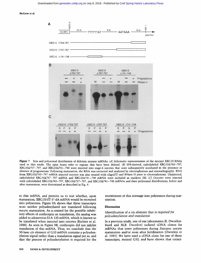
The first transcript, XBG10&763-797, consists of the 3'-untranslated region of G10 mRNA truncated immedi- ately after the AAUAAA hexanucleotide fused to Xeno- pus B-globin coding sequences (Fig. 7A; mutants are numbered according to the nucleotide sequence shown
Maternal mRNA expression
in Fig. 3). This transcript was both polyadenylated (Fig. 7B) and recruited into polysomes (Fig. 7C) during matu- ration. Therefore, sequences downstream of the hexanu- cleotide are not necessary for these processess. The mu- tant mRNA XBG10&757-797 lacks the hexanucleotide and all downstream G10 sequences (Fig. 7A). This tran- script was neither polyadenylated (Fig. 7B) nor recruited for translation (Fig. 7C), which demonstrates the re- quirement of this sequence for these responses to matu- ration. The final transcript, XBG10A741-748, lacks only the UUUUUUAU octanucleotide (Fig. 7A). The deletion of this sequence also inhibited polyadenylation and translation following maturation (Fig. 7B, C). These data therefore show that two sequences within the G10 mRNA 3'-untranslated region, UUUUUUAU and AAUAAA, are both required for polyadenylation and subsequent translation.
Polyadenylation is required for translation of G10 mRNA
The data presented thus far show that the poly(A) tail of endogenous G10 mRNA increases from 90 to 200 res- idues in response to progesterone, that of G10/dT mRNA from 0 to -95 residues, and that of XBG10/dT mRNA from 0 to -215 residues. This might suggest that a poly(A) tail size of >90 residues is sufficient for G10 mRNA translation in oocytes. To determine whether this is the case, we have added a long poly(A) tail to XBG10 (mean length of 210 residues; range 100-325 res- idues), and have examined its translation in oocytes. Figure 8 shows that <10% of these molecules were translated in oocytes, and demonstrates that a long poly(A) tail is insufficient for translation. Following oo- cyte maturation, these molecules were not only further adenylated (with an additional 175 residues), but also were recruited into polysomes (by 30%). Therefore, G10 mRNA translation is not controlled by the number of adenylate residues, but could be regulated by the dy- namic process of polyadenylation.
To examine the relationship between polyadenylation and translation we attempted to uncouple these two re- sponses to maturation by adding 3'-deoxyadenosine (cordycepin) to the 3' terminus of XBG10/dT mRNA. This prevents the polymerization of adenylate residues
Reference
X. laevis G10 UUUUUUAU 8 b AAUAAA ~ A n This study
X. laevis B4 UUUUUAAU ~ 13 b ~ AAUAAA ~ A n Smith et al., 1988b
X. laevis D7 CUUUUUAU 4 b AAUAUA ~ A n Smith et al., 1988a
clam cyclin GCUUUUAU ~ 5 b m AAUAAA ~ A n Swenson et al., 1986
mouse HPRT UUUUAAAU ~ 7b ~ AAUAAA ~ A n Koneckietal., 1982
Figure 6. Sequence comparison of a portion of the 3'-untranslated region of several mRNAs that undergo cytoplasmic polyadenyla- tion during early development.
GENES & DEVELOPMENT 809
Cold Spring Harbor Laboratory Press on July 8, 2018 - Published by genesdev.cshlp.orgDownloaded from
McGrew et al.
Figure 7. Size and polysomal distribution of deletion mutant mRNAs. (A) Schematic representation of the mutant XBG10 RNAs used in this study. The open boxes refer to regions that have been deleted. (B) SP6-derived, radiolabeled XBG10A763-797, XBG10A757-797, and XBG10A741-748 were injected into stage-6 oocytes that were subsequently incubated in the presence or absence of progesterone. Following maturation, the RNA was extracted and analyzed by electrophoresis and autoradiography. RNA from XBG10A763-797 mRNA injected oocytes was also treated with oligo(dT) and RNase H prior to electrophoresis. Uninjected, radiolabeled XBG10A757-797 mRNA and XBGlOA741-748 mRNA were included as markers (M). (C) Oocytes were injected with radiolabeled XBG10A763-797, XBG10A757-797, and XBG10A741-748 mRNAs and their polysomal distributions, before and after maturation, were determined as described in Fig. 4.
to this mRNA, and permits us to test whether, upon maturat ion, XBG 10/dT-3'-dA m R N A would be recruited into polysomes. Figure 9A shows that these transcripts were neither polyadenylated nor translated following oocyte maturat ion. As a control for the possible inhibi- tory effects of cordycepin on translation, the analog was added to adenovirus E1A 13S mRNA, which is known to be translated when injected into oocytes (Richter et al. 1988). As seen in Figure 9B, cordycepin did not inhibit translation of this mRNA. Thus, we conclude that the 50-base cis element of G10 m R N A contains a polyaden- ylation signal rather than a translation signal per se, and that the process of polyadenylation is required for the
recrui tment of this message into polysomes during mat- uration.
D i s c u s s i o n
Identification of a cis dement that is required for polyadenylation and translation
In a previous study, one of our laboratories (E. Dworkin- Rastl and M.B. Dworkin) isolated cDNA clones for m R N A s that enter polysomes during Xenopus oocyte matura t ion and/or soon after fertilization (Dworkin et al. 1985). We have used a eDNA clone for one of these transcripts, termed G10, and have shown that coinci-
810 GENES & DEVELOPMENT
Cold Spring Harbor Laboratory Press on July 8, 2018 - Published by genesdev.cshlp.orgDownloaded from

Maternal mRNA expression
Figure 8. Polysomal distribution of XBG10 RNA that contains a long poly(A) tail. Radiolabeled XBG10/dT mRNA was incu- bated with poly{A) polymerase and ATP. The resulting RNA (XBG10-rAloo_a2s) , containing 100-325 adenylate residues, was injected into oocytes, some of which were induced to mature with progesterone. The polysomal distribution of this RNA was determined as described in Fig. 4.
dent with its translation is a lengthening of its poly(A) tail from - 9 0 residues to -200 residues. To identify the cis element that is required for maturation-specific po- lyadenylation and/or translation, we have synthesized wild-type and deletion mutant SP6-derived G10 mRNAs, injected them into oocytes, matured the oo- cytes in vitro by treatment with progesterone, and as- sessed whether they become polyadenylated and enter polysomes. Wild-type G10 mRNA, but not mutant tran- scripts lacking the 3' terminal 50 nucleotides, is polyad- enylated and recruited for translation at maturation. When this 50-base sequence was linked to a frog f~- globin mRNA lacking the 3'-untranslated region, the polyadenylation and polysome recruitment phenotypes of G10 mRNA were conferred to this transcript. Thus, a sequence within this 50-base region is necessary and sufficient for polyadenylation and translational recruit- ment during maturation.
This 50-base sequence includes the canonical AAUAAA motif that is important for positioning the cleavage site of nuclear pre-mRNAs that are subse- quently polyadenylated (for review, see Nevins 1983; Humphrey and Proudfoot 1988). Because this hexamer is
present on nearly all cytoplasmic poly(A) + mRNAs, it would clearly be insufficient to direct the specific cyto- plasmic polyadenylation that we report here [e.g., Figs. 1 and 4 show that actin and globin mRNAs, which con- tain the polyadenylation hexamer, are not adenylated at maturation). However, a nucleotide sequence compar- ison of the 50-base cis element of G10 with other mRNAs that undergo cytoplasmic polyIA) elongation during early development revealed the presence of a conserved octamer 5' of the AAUAAA hexanucleotide {Fig. 6). To assess whether the octamer, or another se- quence, was important for cytoplasmic polyIA) elonga- tion, three mutant mRNAs were constructed in which various regions of the G10 3'-untranslated region were deleted {Fig. 7). One mutant transcript (XBG10A763- 797) that contained both the po!yadenylation hexamer and the octamer described above but lacked 16 bases 3' of the hexamer, was polyadenylated and recruited in re- sponse to progesterone. A second mutant mRNA (XBG10A757-797), which lacked the hexamer as well as the following 16 bases, was neither polyadenylated nor recruited for translation. This demonstrates that the hexanucleotide is necessary for cytoplasmic poly(A) elongation. A third mutant (XBG10A741-748), which lacked only the UUUUUUAU octanucleotide, was not polyadenylated or translated during maturation. There- fore, within the 50-base 3'-untranslated region of G10 mRNA are two sequences that together are necessary for polyadenylation and translation.
Polyadenylation is required for translation
Several studies have shown a strong, although not abso- lute, correlation between polyadenylation and transla- tion during early development. For example, following fertilization in SpisuIa, several cytoplasmic RNAs be- come adenylated and are recruited for translation (Ro- senthal et al. 1983; Rosenthal and Ruderman 19871. Sim- ilar phenomena accompany fertilization in Urechis {Ro- senthal and Wilt 1986) and oocyte maturation in the mouse (Paynton et al. 19881. Conversely, other mRNAs that are deadenylated during maturation or fertilization are no longer translated IRosenthal et al. 1983; Ro- senthal and Ruderman 1987; Hyman and Wormington
Figure 9. (A) Polysomal distribution of RNAs that contain cordycepin. Radiolabeled XBG10/dT RNA was incubated with poly(A I polymerase and cordy- cepin (to yield XBG10/dT-3'-dAI and then injected into oocytes. The polysomal distribution of this RNA, before and after maturation, was determined as described in Fig. 4. (BI Radiolabeled SP6-derived adenovims E1A RNA was incubated in the absence (E1A) or presence of cordycepin (E1A-3'-dA) and then injected into oocytes. The potysomal distribu- tion of this RNA was determined as described in Fig. 4.
GENES & DEVELOPMENT 811
Cold Spring Harbor Laboratory Press on July 8, 2018 - Published by genesdev.cshlp.orgDownloaded from

McGrew et al.
1988). Finally, some mRNAs do not follow this general pattern in that they are deadenylated during oocyte mat- uration, but enter, rather than leave polysomes (Ru- derman et al. 1979; Ballantine and Woodland 1985). Taken together, these studies suggest a more than coin- cidental relationship between polyadenylation and translation, but whether it is one of cause and effect still remains unclear.
In our attempts to uncouple polyadenylation from translation, we added cordycepin to the 3' terminus of XBG10 mRNA, thus rendering it incapable of polyaden- ylation. This molecule was unable to be recruited into polysomes during maturation, and establishes that, in this case, polyadenylation is necessary for translation. We also added cordycepin to XBG10-rA10o_32s mRNA to assess whether this transcript also was unable to be re- cruited at maturation. Unfortunately, this message un- derwent some hydrolysis of the poly(A) tail, which re- sulted in a transcript devoid of the cordycepin. We also injected cordycepin directly into oocytes, but this, inter- estingly, inhibited maturation. Although one might sus- pect that a message with a poly(A) tail of -200 residues (the number present on endogenous G10 mRNA in ma- ture oocytes) might be required for translation, G10/dT mRNA contained only -90 residues when it was trans- lated at maturation. Therefore, these aggregate data strongly suggest that G10 mRNA translation requires the dynamic process of polyadenylation, and not merely a set number of adenylate residues. This was clearly demonstrated by the experiment in which the addition of up to 325 adenylate residues to XBG10 mRNA failed to stimulate its translation in oocytes; this message was translated, however, when further polyadenylated in re- sponse to progesterone. These results should be com- pared with those of other studies that show that the mere presence of a poly(A) tail can increase translational efficiency (Palatnik et al. 1984; Drummond et al. 1985; Gallili et al. 1988). These apparent discrepancies may suggest that recruitment of mRNA into polysomes (i.e., release from a block of translation) and increased trans- lational efficiency are fundamentally different responses to polyadenylation. In the former case, many oocyte mRNAs are sequestered from the translational appa- ratus as assessed by their slower rate of sedimentation relative to the 80S monosome. Following maturation, several of these messages become fully loaded with ribo- somes and have a mean sedimentation value of 300S (Richter et al. 1982). In the latter case, poly(A) might not induce mRNAs to enter polysomes, but rather could me- diate the number of ribosomes on messages that already are being translated (Palatnik et al. 1984). It should also be noted that message recruitment into polysomes, and not increased translational efficiency, is responsible for the twofold increase in protein synthesis observed during oocyte maturation (Richter et al. 1982).
We estimate that oocytes contain -30 pg of G10 mRNA, all of which is polyadenylated at maturation. However, only about 60% of this message assembles into polysomes. As such, one might speculate that a translation factor becomes rate limiting after polyadeny-
lation has occurred. However, we injected about 700- fold excess SP6-derived mRNA relative to endogenous G10 mRNA, yet all of this material was polyadenylated and about 70% was recruited into polysomes. Therefore, it is unlikely that a translational factor becomes rate limiting following polyadenylation. It is possible that this incomplete translational activation of G10 mRNA reflects the time at which the assays were performed. Although polyadenylation occurs immediately after nu- clear membrane dissolution, polysome assembly prob- ably continues over the next several hours. Indeed, by the 4-cell stage, G10 mRNA can be detected only in polysomes (Dworkin et al. 1985, and unpubl.).
In this study, we have shown that the recruitment of G10 mRNA into polysomes during oocyte maturation requires the process of polyadenylation. Poly(A) elonga- tion, in turn, is mediated by a 50-base sequence element that resides in the 3'-untranslated region. Contained within this 50-base sequence are two elements, UUU- UUUAU and AAUAAA, which are each required for po- lyadenylation and subsequent translation. The observa- tion that neither element can act independently of the other suggests two possibilities for factor interaction. First, one factor, such as a sequence-specific poly(A) polymerase, might require both elements for proper binding and/or activity. This factor could always be cy- toplasmic or could become cytoplasmic only after its re- lease from the nucleus following dissolution of the nu- clear envelope (germinal vesicle breakdown) at matura- tion. Alternatively, different factors could interact with each element; one factor that confers message speci- ficity by binding to the UUUUUUAU sequence, and an- other factor, possibly a general poly(A) polymerase, that interacts with the AAUAAA sequence. Presently, we are attempting to distinguish between these possibilities.
M a t e r i a l s a n d m e t h o d s
Extraction of RNA and gel blot analysis
RNA was extracted from Xenopus laevis stage-6 oocytes (Du- mont 1972), embryos (staged according to Nieuwkoop and Faber 1976), or whole ovary with p-aminosalicylic acid (PAS), SDS, and phenol/chloroform (Kirby 1965). Poly(A) + RNA was selected by four passages through an oligo{dT) column (type 7, P-L Biochemicals). RNAs were resolved by electrophoresis through 1.3% agarose gels containing 2.2% formaldehyde, blotted to nitrocellulose (Thomas 1980), and probed with nick- translated G 10 or actin cDNA clones. General procedures have been described in Maniatis et al. (1982).
Poly(A) § RNA was deadenylated in vitro by a modification of the procedure described by Colot and Rosbash (1982). Two mi- crograms of total ovary or egg RNA was incubated with 2 ~g of oligo(dTh2_is in a reaction containing 40 mM Tris-HC1 (pH 7.5), 10 ~ MgClw and 50 rnM KC1 for 15 rain at 37~ One unit of RNase H (Promega Biotec) was added to the reaction and the incubation continued for another 30 rain at 37~ The RNA was then extracted with phenol and chloroform, precipitated with ethanol, and collected by centrifugation. Approximately 1 ~g of RNA from each sample was analyzed on a gel blot.
812 GENES & DEVELOPMENT
Cold Spring Harbor Laboratory Press on July 8, 2018 - Published by genesdev.cshlp.orgDownloaded from

Polysome isolation
Oocytes were defolliculated manually and were induced to ma- ture by incubation in Barth's medium (Maniatis et al. 1982) containing 1 ixg/ml progesterone. Maturation was detected by the presence of a white spot at the animal pole 5-8 hr after the addition of hormone. These oocytes, and control oocytes, in pools of 10, were homogenized in 750 ~1 of 300 mM NAG1, 20 mM Tris-HC1 (pH 7.4), 10 mM MgC12, 0.5% Nonidet P-40, 20 ~g/ml polyvinyl sulfate, and 4 mM vanadyl ribonucleoside complex {Bethesda Research Laboratories) and centrifuged at 12,000g for 15 min at 4~ The supernatants were layered over a step gradient of 10% sucrose (2 ml) and 35% sucrose (2.5 ml), both in polysome buffer. Polysomes were centrifuged through the sucrose cushions at 35,000 rpm for 2 hr at 4~ in a Beckman SWS0.1 rotor. The RNA was extracted from the polysomal pellets by suspension in 0.5 ml 4 M guanidine isothiocyanate, 50 mbi Tris-HC1 (pH 7.6), 5 mM EDTA, 0.5% sodium lauryl sarkosinate, and 0.1 M [3-mercaptoethanol, followed by phenol/ chloroform extraction and ethanol precipitation. The collected ethanol precipitate was suspended in a solution of 10 mM Tris- HC1 (pH 7.6), 5 mM EDTA, 1% SDS followed by phenol/chloro- form extraction and ethanol precipitation. The RNA in the su- pernatants, which were devoid of polysomes, was precipitated with 2 volumes of ethanol and then extracted with guanidine and phenol/chloroform as described above. In some cases, the postmitochondrial supernatants were adjusted to 20 mM EDTA prior to ultracentrifugation through sucrose, which also con- tained 20 m_M EDTA. The purified RNA from pellets and super- natants was suspended in equal volumes (-2 ~1 per oocyte), and RNA yields were determined spectrophotometrically. Three oocyte equivalents were analyzed for each supematant and pellet fraction.
DNA sequencing and plasmid construction
DNA sequencing was performed on double-stranded plas- mid DNA by the dideoxy chain-termination method (Sanger 1972) using a Sequenase sequencing kit (U.S. Biochemical Corp.), as well as by the method of Maxam and Gilbert (1980).
The plasmid pG10 {Dworkin et al. 1985) isolated from a Xenopus oocyte cDNA library, contains a 797-bp insert cloned downstream of the SP6 promoter. To construct the XBG10 fu- sion plasmid, a Xenopus B-globin clone (pSP64XBM, Krieg and Melton 1984) was digested with BstEII and filled-in with DNA polymerase I (Klenow fragment). This clone was then digested with EcoRI and ligated to a SspI-EcoRI fragment from pG10.
To construct mutant plasmids that contain deletions within the G10 mRNA 50-base cis element, oligonucleotides specific for each mutant sequence were synthesized. Complementary oligonucleotides containing a 5' BstEII site and a 3' SalI site, were annealed at 50~ for 60 min in 0.1 M NaC1 and were li- gated into the BstEII and SalI sites of pSP64XBM. The plasmid pXBG10A763-797 contained the oligonucleotide sequence 5'- GTTACCACTAGAATATTATTTGTGTTTTTTATAAAGG_ TGTAATAAAG, annealed to its complement 5'-TCGACT- TTATTACACCTTTATAAAAAACACAAATAATATTCTA - GTG; plasmid pXBG10A757-797 contained the oligonucleotide sequence 5'-GTFACCACTAGAATATFATYFGTG i T V i T iAT- AGGTGTG, annealed to its complement 5'-TCGACAC- ACCTTTATAAAAAACACAAATAATATTCTAGTG; and the plasmid pXBG10A741-748 contained the oligonucleotide sequence 5'-GTTACCACTAGAATATTATTTGTGAAAGGT- GTAATAAACATGACATTTCATGGG, annealed to its com- plement 5'-TCGACCCATGAAATGTCATGTTTATTACAC- CTTTCAGAAATAATATrCTAGTG.
Maternal mRNA expression
In vitro transcription and oocyte injection
The following DNA templates were used for transcription in vitro: pG10 linearized at the HindIII, NaeI, or SspI sites; pSP64XBM linearized at the PstI (in the vector, Krieg and Melton 1984) or BstEII (in coding sequence, Patient et al. 1983) sites; pXBG10 linearized at the EcoRI site; pXBG10a763-797, pXBG10A757-797, and pXBG10a741-748 linearized at the SalI site (cf. Fig. 7); and pSP13S (containing the E1A 13S mRNA coding sequence, Richter et al. 1988) linearized at the HindIII site. Transcription by SP6 polymerase was carried out essen- tially as described by Krieg and Melton (1984), except that the reaction mixture contained 50 ~M GTP, 500 ~M GpppG, and 60 ~Ci of [a2P]dCTP (800 Ci/mmole Amersham). Following syn- thesis, the 3' termini of several of the RNAs were removed (G10/dT, globin/dT, XBG10/dT, XBG10/dC) by RNase H diges- tion in the presence of either oligo(dT) or oligo(dC) as described previously. The RNA was extracted with phenol/chloroform and precipitated with ethanol.
Manually defolliculated oocytes were injected with 20-40 ng of radiolabeled RNA (3 x 10⁶ cpm/}xg) and cultured in the ab- sence or presence of of progesterone. Within 1 hr after the ap- pearance of a white spot at the animal pole, the oocytes were homogenized and centrifuged through sucrose cushions as de- scribed earlier. Alternatively, the RNA was extracted directly from the injected oocytes and analyzed without further treat- ment or after deadenylation with oligo(dT) and RNase H.
To add cordycepin to mRNA, 5 ~g of radiolabeled XBG10/dT or E1A RNA was incubated in a 50-}xl reaction containing 200 ~M 3' dATP (cordycepin), 50 mM Tris-HCl (pH 8.0), 10 mM MgC12, 50 ~g/ml of BSA, 250 mM NaC1, 1 mM MnC12, 2 mM DTT, 1.5 U/~I of RNasin, and 3 units of poly(A) polymerase (Pharmacia) at 37~ for 20 rain. This reaction produced the RNAs XBG10-3'-dA and E1A-3'-dA. The 3' terminus of XBG10 mRNA was also modified to contain 100-325 adenylate res- idues {XBG10-rA10o_a2s) by incubation of 5 ~g of radiolabeled XBG10/dT RNA with 50 ~M ATP, 50 mM Tris-HC1 (pH 8.0), 10 mM MgC12, 50 ~g/ml of BSA, 250 mM NaC1, 1 mM MnC12, 2 mM DTT, 1.5 U/fzl of RNasin, and 3 units of poly(A) poly- merase for 5 min at 37~ All radioactive RNAs were resolved by electrophoresis in 5% polyacrylamide-8.3 Ivt urea gels, fol- lowed by autoradiography. Sizes of the transcripts were deter- mined by comparison with k DNA that was digested with Hin- dIII and EcoRI.
Fusion protein construction, antiserum generation, and immunoprecipitation
An -550-bp DdeI-HindIII fragment from pG10, filled-in at the DdeI site, was cloned into pUR290 which was cut with HpaI and HindIII (Riither and Miiller-Hill 1983). This resulted in the fusion of the amino-terminal 140 amino acids of ~-galactosi- dase with amimo acids 22-144 of G10. Induction and fusion protein isolation from Escherichia coli, as well as immuniza- tion protocols in rabbits have been described (Smith et al. 1988b).
Fifty control and mature stage-6 oocytes were injected with [aSS]methionine (8 x 10⁴ cpm/oocyte, 1200 Ci/mmole, Amer- sham), cultured for 2 hr, and homogenized in 500 ~1 of Barth's medium containing 0.1% Triton X-100. Following brief centrif- ugation to remove the yolk, the supernatant was adjusted with 1 x RIPA buffer [50 mM Tris-HC1 (pH 7.5), 150 mM NAG1, 1% deoxycholate, 1% Triton X- 100, 0.1% SDS] and incubated with G10 antiserum for 2 hr at room temperature. Protein A-Sepha- rose was added and incubation was continued for an additional 2 hr. The beads were then washed in 1 x RIPA buffer and the G10 protein was eluted by boiling in SDS gel loading buffer and
GENES & DEVELOPMENT 813
Cold Spring Harbor Laboratory Press on July 8, 2018 - Published by genesdev.cshlp.orgDownloaded from

McGrew et al.
was analyzed by SDS-15% polyacrylamide gel electrophoresis and autoradiography. In a similar experiment, [aSSJmethionine- labeled reticulocyte lysate synthesized G10 protein was im- munoprecipitated with G10 antiserum and analyzed as de- scribed above.
A c k n o w l e d g m e n t s
We thank A. Shrutkowski for technical assistance and I. Good- child for synthesizing oligonucleotides. This work was sup- ported by grants from the National Institutes of Health (CA40189), the National Science Foundation (DCB8719300), and the American Cancer Society, Massachusetts Division (Cancer Research Scholar Award) awarded to J.D.R.
R e f e r e n c e s
Adamson, E.D. and H.R. Woodland. 1977. Changes in the rate of histone synthesis during oocyte maturation and very early development of Xenopus laevis. Dev. Biol. 57: 136- 149.
Asselbergs, F.A.M., W.J. Van Venrooij, and H. Bloemendal. 1979. Messenger RNA competition in living Xenopus oo- cytes. Eur. J. Biochem. 94: 249-254.
Audet, R.G., J. Goodchild, and J.D. Richter. 1987. Eukaryotic initiation factor 4A stimulates translation in microinjected Xenopus oocytes. Dev. Biol. 121" 58-68.
Ballantine, J.E.M. and H.R. Woodland. 1985. Polyadenylation of histone mRNA in Xenopus oocytes and embryos. FEBS Lett. 180:224-228
Baum, E.Z., L.E. Hyman, and W.M. Wormington. 1988. Post- translational control of ribosomal protein L1 accumulation in Xenopus oocytes. Dev. Biol. 126: 141-149.
Burglin, T.R. and E.M. DeRobertis. 1987. The nuclear migra- tion signal of Xenopus laevis nucleoplasmin. EMBO I. 6: 2617-2625.
Colin, A.M., B.D. Brown, J.N. Dholakia, C.L. Woodley, A.J. Wahba, and M.B. Hille. 1987. Evidence for simultaneous derepression of messenger RNA and the guanine nucleotide exchange factor in fertilized sea urchin eggs. Dev. Biol. 123: 354-363.
Colot, H.V. and M. Rosbash. 1982. Behavior of individual ma- ternal pA + RNAs during embryogenesis of Xenopus laevis. Dev. Biol. 94: 79-86.
Crawford, D.R. and J.R. Richter. 1987. An RNA-binding protein from Xenopus oocytes is associated with specific message sequences. Development 101: 741-749.
Davidson, E.H. 1986. Gene activity in early development. Aca- demic Press, New York.
Dingwall, C. and R.A. Laskey. 1986. Protein import into the cell nucleus. Annu. Rev. Cell Biol. 2: 367-391.
Drummond, D.R., J. Armstrong, and A. Colman. 1985. The ef- fect of capping and polyadenylation on the stability, move- ment, and translation of synthetic messenger RNAs in Xenopus oocytes. Nucleic Acids Res. 13: 7375-7394.
Dumont, J.N. 1972. Oogenesis in Xenopus laevis (Daudin): I. Stages of oocyte development in laboratory maintained an- imals. J. Morphol. 136: 153-180.
Dworkin, M.B. and E. Dworkin-Rastl. 1985. Changes in RNA titers and polyadenylation during oogenesis and oocyte mat- uration in Xenopus laevis. Dev. Biol. 112: 451-457.
Dworkin, M.B., A. Shrutkowski, and E. Dworkin-Rastl. 1985. Mobilization of specific maternal RNA species into poly- somes after fertilization in Xenopus laevis. Proc. Natl. Acad. Sci. 82: 7636-7640.
Flynn, J.M. and H.R. Woodland. 1980. The synthesis of histone
H1 during early amphibian development. Dev. Biol. 75: 222-230.
Galili, G., E.E. Kawata, L.D. Smith, and B.A. Larkins. 1988. Role of the 3'-poly(A) sequence in translational regulation of mRNAs in Xenopus laevis oocytes. J. Biol. Chem. 263: 5764-5770.
Grainger, J.L. and M.M. Winkler. 1987. Fertilization triggers unmasking of maternal mRNAs in sea urchin eggs. Mol. Cell. Biol. 7: 3947-3954.
Huang, W.I., L.H. Hansen, W.C. Merrick, and R. Jagus. 1987. Inhibitor of eukaryotic initiation factor 4F activity in unfer- tilized sea urchin eggs. Proc. Natl. Acad. Sci. 84: 6359- 6363.
Huarte, J., D. Belin, A. Vassalli, S. Strickland, and J.-D. Vassalli. 1987. Meiotic maturation of mouse oocytes triggers the translation and polyadenylation of dormant tissue-type plasminogin activator mRNA. Genes Dev. 1: 1201-1211.
Humphrey, T. and N.J. Proudfoot. 1988. A beginning to the bio- chemistry of polyadenylation. Trends Genet. 4: 243-245.
Hyman, L.E. and W.M. Wormington. 1988. Translational inac- tivation of ribosomal protein mRNAs during Xenopus oo- cyte maturation. Genes Dev. 2: 598-605.
Jenkins, N.A., J.F. Kaumeyer, E.M. Young, and R.A. Raft. 1978. A test for masked message: The template activity of mes- senger ribonucleoprotein particles isolated from sea urchin eggs. Dev. Biol. 63: 279-298.
Kick, D., P. Barrett, A. Cummings, and J. Sommerville. 1987. Phosphorylation of a 60 kD polypeptide from Xenopus oo- cytes blocks messenger RNA translation. Nucleic Acids Res. 15: 4099-4109.
Kirby, K.S. 1965. Isolation and characterization of ribosomal ri- bonucleic acid. Biochem. J. 96: 266-269.
Konecki, D.S., J. Brennand, J.C. Fuscoe, C.T. Caskey, and A.C. Chinai. 1982. Hypoxanthine-guanine phosphoribosyltrans- ferase genes of mouse and Chinese hamster: Construction and sequence analysis of cDNA recombinants. Nucleic Acids Res. 10: 6763-6775.
Kozak, M. 1986. Point mutants define a sequence flanking the AUG initiator codon that modulates translation by eukary- otic ribosomes. Cell 44: 283-292.
Krieg, P.A. and D.A. Melton. 1984. Functional messenger RNAs are produced by SP6 in vitro transcription of cloned cDNAs. Nucleic Acids Res. 12: 7057-7070.
Laskey, R.A., A.D. Mills, J.B. Gurdon, and G.A. Partington. 1977. Protein synthesis in oocytes of Xenopus laevis is not regulated by the supply of messenger RNA. Cell 11: 345- 351.
Lee, G., R. Hynes, and M. Kirschner. 1984. Temporal and spa- tial regulation of fibronectin in early Xenopus development. Cell 36: 729-740.
Maniatis, T., E.F. Fritsch, and J. Sambrook. 1982. Molecular cloning: A laboratory manual. Cold Spring Harbor Labora- tory, Cold Spring Harbor, New York.
Maxam, A.M. and W. Gilbert. 1980. Sequencing endqabeled DNA with base-specific chemical cleavages. Methods En- zymoI. 65: 499-560.
Nevins, J.R. 1983. The pathway of eukaryotic mRNA forma- tion. Armu. Rev. Biochem. 52: 441-466.
Nieuwkoop, P.D. and J. Faber. 1956. Normal table of Xenopus laevis (Daudin). North Holland Publishing Company, Am- sterdam.
Palatnik, C.M., C. Wilkins, and A. Jacobson. 1984. Transla- tional control during early Dictyostelium development: Possible involvement of poly{A) sequences. Cell 36: 1017- 1025.
Patient, R.K., R. Harris, M.E. Walmsley, and J.G. Williams.
814 GENES & DEVELOPMENT
Cold Spring Harbor Laboratory Press on July 8, 2018 - Published by genesdev.cshlp.orgDownloaded from

1983. The complete nucleotide sequence of the major adult ~-globin gene of Xenopus laevis. 1. Biol. Chem. 258: 8521- 8523.
Paynton, B.V., R. Rempel, and R. Bachvarova. 1988. Changes in state of adenylation and time course of degradation of ma- ternal mRNAs during oocyte maturation and early embry- onic development in the mouse. Dev. Biol. 129: 304-314.
Payre, F. and A. Vincent. 1988. Finger proteins and DNA-spe- cffic recognition: Distinct patterns of conserved amino acids suggest different evolutionary modes. FEBS Lett. 234: 245- 250.
Richter, J.D. 1987. Molecular mechanisms of translational con- trol during the early development of Xenopus laevis. In Translational regulation of gene expression (ed. J. Ilan), pp. 111-139. Plenum Publishing Company, New York.
Richter, J.D. and L.D. Smith. 1981. Differential capacity for translation and lack of competition between mRNAs that segregate to free and membrane-bound polysomes. Cell 27: 183-191.
�9 1984. Reversible inhibition of translation by Xenopus oocyte-specific proteins. Nature 309: 378-380.
Richter, J.D., W.J. Wasserman, and L.D. Smith. 1982. The mechanism for increased protein synthesis during Xenopus oocyte maturation. Dev. Biol. 89: 159-167.
Richter, J.D., J.M. Slavicek, J.F. Schneider, and N.C. Jones. 1988. Heterogeneity of adenovirus type 5 E1A proteins: Multiple serine phosphorylations induce slow-migrating electrophoretic variants but do not affect E1A-induced tran- scriptional activation or transformation. J. Virol. 62: 1948- 1955.
Rickles, R.J., A.L. Darrow, and S. Strickland. 1988. Molecular cloning of complementary DNA to mouse tissue plasmin- ogen activator mRNA and its expression during F9 terato- carcinoma cell differentiation. J. Biol. Chem. 263: 1563- 1569.
Rosenthal, E.T. and F.H. Wilt. 1986. Patterns of maternal mes- senger RNA accumulation and adenylation during oogenesis in Urechis caupo. Dev. Biol. 117: 55-63.
�9 1987. Selective messenger RNA translation in marine invertebrate oocytes, eggs, and zygotes. In Translational reg- ulation of gene expression (ed. J. Ilan), pp. 87-110. Plenum Publishing Company, New York.
Rosenthal, E.T. and J.V. Ruderman. 1987. Widespread changes in the translation and adenylation of maternal messenger RNAs following fertilization of Spisula oocytes. Dev. Biol. 121: 237-246.
Rosenthal, E.T., T.R. Tansey, and J.V. Ruderman. 1983. Se- quence-specific adenylations and deadenylations accom- pany changes in the translation of maternal messenger RNA after fertilization of Spisula oocytes. J. Mol. Biol. 166: 309- 327.
Ruderman, J.V., H.R. Woodland, and E.A. Sturgess. 1979. Mod- ulations of histone messenger RNA during the early devel- opment of Xenopus laevis. Dev. Biol. 71:71-82.
Rfither, U. and B. MUller-Hill. 1983. Easy identification of cDNA clones. EMBO J. 2: 1791-1794.
Sagata, N., M. Oskarsson, T. Copeland, J. Brumbaugh, and G.F. Vande Woude. 1988. Function of c-mos proto-oncogene product in meiotic maturation in Xenopus oocytes. Nature 335: 519-525.
Sanger, F., S. Nicklen, and A.R. Coulson. 1977. DNA se- quencing with chain-terminating inhibitors. Proc. Natl. Acad. Sci. 74: 5463-5467.
Smith, L.D. and J.D. Richter. 1985. Synthesis, accumulation, and utilization of maternal macromolecules during oo- genesis and oocyte maturation. In Biology of fertilization.
Maternal mRNA expression
(eds. A. Metz and A. Monroy), pp. 141-188�9 Academic Press, New York.
Smith, R.C., M.B. Dworkin, and E. Dworkin-Rastl. 1988a. De- struction of a translationally controlled mRNA in Xenopus oocytes delays progesterone-induced maturation�9 Genes Dew. 2: 1296-1306.
Smith, R.C, E. Dworkin-Rastl, and M.B. Dworkin. 1988b. Ex- pression of a histone Hl-like protein is restricted to early Xenopus development. Genes Dev. 2: 1284-1295.
Spirin, A.S. 1969. Informosomes. Eur. J. Biochem. 10: 20-35. Stick, R. and P. Hausen. 1985. Changes in the nuclear lamina
composition during early development of Xenopus laevis. Cell 41: 191-200.
Swenson, K.I., K.M. Farrell, and J.V. Ruderman. 1986. The clam embryo protein cyclin A induces entry into M phase and resumption of meiosis in Xenopus oocytes. Cell 47: 861- 870.
Swiderski, R.E. and J.D. Richter. 1988. Photocrosslinking of proteins to maternal mRNA in Xenopus oocytes. Dev. Biol. 128: 349-358.
Thomas, P.S. 1980. Hybridization of denatured RNA and small DNA fragments transferred to nitrocellulose. Proc. Natl. Acad. Sci. 77: 5201-5205.
Woodland, H.R., J.M. Flynn, and A.J. Wyllie. 1979. Utilization of stored mRNA in Xenopus embryo and its replacement by newly synthesized transcripts: Histone H1 synthesis using interspecies hybrids. Cell 18: 165-171.
GENES & DEVELOPMENT 815
Cold Spring Harbor Laboratory Press on July 8, 2018 - Published by genesdev.cshlp.orgDownloaded from
10.1101/gad.3.6.803Access the most recent version at doi: 3:1989, Genes Dev.
L L McGrew, E Dworkin-Rastl, M B Dworkin, et al. element.translational recruitment and is mediated by a short sequence Poly(A) elongation during Xenopus oocyte maturation is required for
References
http://genesdev.cshlp.org/content/3/6/803.full.html#ref-list-1
This article cites 55 articles, 15 of which can be accessed free at:
License
ServiceEmail Alerting
click here.right corner of the article or
Receive free email alerts when new articles cite this article - sign up in the box at the top
Copyright © Cold Spring Harbor Laboratory Press
Cold Spring Harbor Laboratory Press on July 8, 2018 - Published by genesdev.cshlp.orgDownloaded from