pmod software release notesdoc.pmod.com/pdf/releasenotes.pdf · fixed issue with loading of rtss...
TRANSCRIPT
Printed on 19 April, 2018
PMOD Software Release Notes
Version 3.9
PMOD Technologies
2 PMOD Software Release Notes
Maintenance Builds of Release 3.9 Build 5 April 19, 2018
VOI statistics selection, new default: Only Averaged and SD are initially selected and the configuration dialog is displayed before first run.
VOI statistics of dynamic data: Bug fixed (introduced in Build 4).
Peak VOI for dynamic data: Peak calculation is now correctly done for each frame, and save/retrive of dynamic peak VOIs was fixed.
Iso-contouring VOI for dynamic data: Restricting VOI was not handled correctly.
FuseIt: Protocol behavior fixed; correctly retrieves data and transformation.
PNEURO: Batch mode could be blocked by a dialog window appearing due to a mapping problem.
Build 4 March 22, 2018
Main innovation: added features for assessing and documenting oncologic lesions. Please refer to the dedicated flyer in the News section on the PMOD website.
Global oncology configuration establishes the following defaults: SUV display units, show MIP image in inverted gray, show patient name and ID, use terms "MTV" and "TLG" in statistics.
Hot-key (Ctrl+Q) driven placement of a peak VOI in lesion.
Hot-key (Ctrl+L) generation of an iso-contour VOI of lesion based on %SUVmax, %SUVpeak, or absolute SUV.
Hot-key based assessment of lesions relative to liver: (Ctrl+Shift+U) places 3cm sphere as reference, (Ctrl+U) performs iso-contouring at minimal level of tumor uptake and calculates SUVpeak.
Generation of an iso-contour VOI of lesion based on %SUVmax, %SUVpeak, or absolute SUV.
Configuration of the hot-key VOIs and the overlay statistics in a new panel of the data inspector.
Overlay of relevant VOI statistics (average, peak, max, MTV) in image.
Sorting of VOI list by descending SUVpeak, SUVmax, SUVmean, MTV, diameter.
Statistics units synchronized with display units (SUV type).
Lesion documentation via screen capture (Ctrl+E).
Other improvements and changes:
Slowness on computer without internet connection due to update and documentation checking: improved, but it is recommended to install the documentation locally and switch off the update checking.
PNEURO: Saving both parametric maps and TACs in batch mode.
PXMOD: ssq map added to the SRTM2 model.
PKIN/Parametric mapping: Lack of synchronization between curve and parameter
PMOD Software Release Notes
PMOD Software Release Notes 3
update fixed.
Texture analysis: NaN support, applicable for dynamic series.
PGEM: Export of OpenFOAM's volume mesh to Fluent format.
VOI statistics: Fixed problem in sphericity (was always zero).
Build installation: existing start scripts are not overwritten.
Database: Compatibility with current mySQL established.
R Statistics: Bland-Altman works with NaN.
File saving: Prevent file system write error for file names longer than 255 chars.
Build 3 Jan. 15, 2018
Database of demo data extended by PCARDM and PGEM (CFD) examples.
PXMOD: Two models (Patlak, irreversibe 2-compartment model with basis function fitting) added for the parametric mapping of dynamic FDG whole-body data. They account for the slice-wise timing definitions in the image header.
Slice-wise timing for TAC transfer to PKIN: average TAC uses timing of the slice in the VOI center; pixel-wise TAC uses timing of individual slice.
PKIN: Reference curve was not shown when reference region was the first in the list.
Amyloid Cortical Composite atlas added. It contains the definition of the most commonly used composite region for analyzing amyloid data, as well as several reference regions.
Mouse atlas renamed to "Mouse (Ma-Benveniste-Mirrione)" to credit the work of Ma and Benveniste who did the extensive atlas groundwork.
PFUS/PNEURO: Normalization templates derived from ADNI data (FDG, AV45 for AD and controls) added to the list of predefined templates. Brain masking for AV45 templates improved.
Pig Brain Atlas (CH. Malbert) labels fixed: Left/right corrections, Superior_cerebellar_peduncle moved to the non-lateralized Posterior_fossa.
PNEURO: User defined MNI atlases automatically offer the standard normalization templates if MNI is mentioned in the atlas manifest.
PNEURO: Option for enabling/disabling CSF masking and CSF threshold slider added.
PNEURO: Transformation results of a PET/MR workflow are cleared before PET-only protocol execution.
PNEURO: Anatomical reference control added to batch mode panel.
PNEURO/Compare to Norm: Variance pooling estimation corrected. Missing statistics VOIs fixed when repeating analysis with modified parameters.
VOI/RTSS: Fixed problem with conversion to RTSS when contours were defined in different plane orientations. Fixed issue with loading of RTSS data. Fixed problem with wrong series instance UID in RTSS objects created by PMOD.
VOI: Problem of calculating fractal dimension for VOIs defined in X and Y directions fixed.
VOI: Texture analysis can be started from Calculate Statistics menu of VOI Tool.
PCARD: Segmentation parameters for mouse data has been adjusted (active models + valve plane fitting).
4 PMOD Software Release Notes
PCARD: Manually edited VOIs are now saved correctly to protocols. Protocols for data with changed frame times are now retrieved correctly.
PCARD: Specific protocol retrieval panels for cardiac MR applications.
Pipeline processing: Can be started from command line script and run with or without user interface. Fixed problem with updating the QC tool state.
Interpolation tool: Fixed reading of transformation when deformation field was previously loaded. Fixed saving of resliced image using deformation field transformation.
PGEM: Improved conversion of CFD results to images for comparison with tomographic data.
PGEM: Image data can be loaded while visualizing CFD results.
R Console: Aggregate filtering in the presence of NA values fixed; scatter plot fixes.
R Console: Linear Mixed Effect Models is now working with data containing NA values (which are excluded from the analysis).
Network license: More detailed information in license server dialog. If all licenses are occupied there is an information that no license is available and connection can terminate other Pmod instance.
Philips PAR/REC images: Now assumes miliseconds as trigger times units. Improved slice sorting and timing.
Build 2 Nov. 9, 2017
Dynamic Data with slice-dependent timing: Use of the VOI center location timing when transferring VOI TACs to PKIN. Automatic reformatting improved for handling of per-slice information.
VOI: Configuration of the statistics measures of interest introduced for minimizing calculation time.
VOI: Tumor volume segmentation method fixed and improved. Is now working for group of selected VOIs, and a VOI called "Bck" is automatically selected as background VOI.
PXMOD/Resting state model: Fix to avoid masking of correlation matrix.
PXMOD/VOIs tab: fraction mode is new default for interactively outlined VOIs.
PCARDP/Gated: Segmentation method limited to EPI/ENDO Active Model.
PCARDP/Perfusion: Problem in sector TAC calculation fixed.
PCARDM: Segmentation parameters units unified to mm.
PCARD protocols: VOI saving in protocols fixed.
Texture analysis: Information in the tab titles improved.
PGEM: DTI report problem fixed when patient information was missing.
Fusion: Parameters of motion correction transformation can be presented as a graph.
Build 1 Oct. 30, 2017
Initial upload of 3.9 version.

PMOD Software Release Notes 5
6 PMOD Software Release Notes
Release Notes 3.9
New Features and Revisions
General Improved handling of the data path history when selecting from the file system rather than the database.
SUV dialog window and inspector revised. Support for SUVlbm by Janmahastian method. Use of UCUM units (e.g. g/ml{SUVlbm(Janma)} when saving SUV images to enhanced DICOM objects, otherwise the DICOM SUV Type definition (0054,1006).
Texture analysis within VOIs.
Command line execution of pipeline definitions which include an input file list (e.g. ./java/jre/bin/java -Xmx1200M -jar pmod.jar PIPELINE[noGUI] pipe_definition_file.pipeProc).
Pipeline processing: Restriction that a file can only appear once on the input list was removed.
DICOM server starting a pipeline processing with the received data: all non-image data are also saved.
External tool for calculation z-score values relative to the average and the standard deviation within a specified VOI (frame-wise operation for dynamic data). Useful for transforming the data as input to the supervised clustering method).
New option to keep triangulation at the same 3D coordinate when switching between images in a viewport.
Layout panel: "Go to coordinate origin" and "Go to x/y/z coordinate" buttons to support well-defined triangulation.
Extended list of isotopes and radiopharmaceuticals.
Proper units are set for images generated in situations beyond parametric mapping.
Skeletonization added to "Segmentation" external tool.
New automatic VOI generation approach resulting in the hottest connected voxels within a VOI.
External tool for calculation of texture features within VOIs.
PMOD Software Release Notes 7
PNEURO Template, Atlas and Parcellation resources converted to compressed images for saving space
White matter parcellation added, resulting in a white matter VOI for each atlas VOI.
Masking of grey matter regions by CSF improved for the maximum probability method.
Revision of the tree organization for better organizing the additional VOIs.
Batch mode improved in several ways: support for mapping added; option to create and save a quality control image showing the generated VOIs on top of the images; new interface item element for specifying the averaging range of dynamic series; saving of TACs, parametric maps, transformations and protocols.
Option to select the dynamic PET instead of the average PET after the VOIs have been outlined.
Support for user-defined atlases in parcellation improved. Supported also in batch mode.
Warning added when a loaded transformation doesn’t match the space of input data.
PKIN
Grid fitting: Start iterative fitting with initial parameter combinations which cover the whole physiologic parameter space.
Monte Carlo: Randomized fitting supported.
Monte Carlo: Noise can be added to tissue TAC instead of model curve. This allows studying situations where the model underlying the data differs from the fitted model.
Monte Carlo: The generated data and the fits can be added to the fitting history for inspection and export.
Monte Carlo: Noise generation using bootstrap method.
Parameter explorer: The listed results correspond either to the current model parameters, or to the latest successful fit. Previously non-fitted regions were omitted.
Fitting history included in parameter explorer to browse histories across regions more easily.
Parameter sensitivity to acquisition duration is now also supported for coupled fits. Creates history entries with the same revision number.
Number of fits required for convergence is also added to the fit details list.
Batch fitting with specified model configuration: Only the tissue model is replaced, the blood configuration now remains unchanged.
Fitting can be stopped after calling "Fit all".
Negative values allowed in blood data.
Blood curve resampling improved. Didn't work properly in all configurations.
"non-blood" model filter introduced which is default until blood data is loaded.
Averaging window introduced in "Plasma Ratio" model for use with bolus/infusion data, where the ratio equals Vt.
Standard error supported in the aggregation of kinetic modeling results.

8 PMOD Software Release Notes
Support for TACs with different timing across regions. This is particularly relevant for dynamic whole-body and dosimetry studies.
Dosimetry model: Output of cumulated activities for the IDAC2.1 dosimetry program (www.idac-dose.org).
Massive speed improvements for the transfer of pixel-wise TACs.
PXMOD Revised SRTM2 model, allowing to calculate k2' as the median for the SRTM results in a region and restricted to a physiological range.
Model for DWI MR images. Supports different calculations of the ADC maps including fit of mono exponential.
Model for PCASL MR images which are organized in two dynamic series (control, label), and a proton density image. Supports calculation of average perfusion.
Model for the seed-based correlation analysis of resting state fMRI data. A matrix showing the correlation between the signals of multiple VOIs is shown as image and can be explored with the data inspector probe.
Individual timing is supported for each pixel. This feature is particularly relevant for dynamic data acquired with continuous bed motion.
PCARD Gated PET: Improved volume and wall-thickening accuracy based on a new interpolated data set (called VOLUME).
Gated PET: More precise valve plane fitting.
Cardiac PET: Acceptance test revised.
Perfusion PET: Transmural gradient for flow areas calculated from the ratio of the endo and epi averages of the relevant segments.
Contours are smoothed.
Optional correction of myocardium TACs for activity remaining from previous scan.
Cardiac MR: Active model segmentation added; can be initialized by markers.
PSEG Clustering method when using external TACs replaced: the supervised clustering algorithm developed by Turkheimer et al. (2007) for PK11195 is now used instead of k-means.
Brain extraction added to the masking step.
PFUS Color table lt/ut propagation in absolute values supported.
P3D
Functionality useful for vessel rendering and analysis: Skeletonization of a segment and creation of a vectorized path; creation of planes which can be moved along the path and always are orthogonal to; cutting of object by this plane.
Cutting of SR objects by use of a circle.
Texture display on oblique planes.
PGEM CFD: Integration with new CFD calculation engine OpenFoam 5.0.
CFD: Use of 4D-Flow derived flow boundary conditions.
CFD: Improvements in case creation GUI, case status detection, case downloading, log display and error detection.
CFD: Cutting interface shows loaded image data.
Added possibility of loading 4D flow data to a scene presenting CFD simulations
PMOD Software Release Notes 9
results.
Added possibility of generating and using skeletons of geometric structures, and cutting those structures by planes perpendicular to the skeletons.
Loaded image data available on 3D page for all processing types.
Heart model updated.
Model with brain regions added.
R Statistics Console
NaN are not removed when statistics are transferred to R Console.
Master table only includes mutual VOIs included in all aggregates
New function "pm.copyColumns()" for copying columns between aggregates.
GUI of Linear Models is not freezing any more during execution.
Improved NA handling when filtering variable is equal NA.
Version control introduced for required packages.
Data Formats Handling of wrongly formatted injection date in some versions of Albira microPET files.
Matlab image loader extended to read 4D matrices as dynamic images.
Improved handling of reversed image indices in the PET DICOM object.
Fix for handling of DICOMDIR files with icon images.
Initial reading support for the evolving quantitative PET extension of the BIDS data standard (bids.neuroimaging.io), including encoding of ancillary information in the JSON format.
Zürich, October 30, 2017
10 PMOD Software Release Notes
Maintenance Builds of Release 3.8 Build 7 Oct. 2, 2017
MacOSX: Improvements for popup panels which were sometimes hidden, depending on system configuration.
PXMOD: Fixed problem with using the interactively defined VOI in later stage of the workflow.
PBAS: Transfer of TACs to PKIN sets units to kBq/cc when "Global Scale factor to [kBq/cc]" is switched on.
PFUS/General: Cubic spline interpolation optimized to avoid long waiting time for dynamic data.
PCARD/Gated analysis of small animal data: Volume calculation fixed; segmentation improved; mass calculation fixed.
PGEM: Fix related to 4D flow protocol execution; OpenFOAM version number included in acceptance test.
Build 6 July 24, 2017
PXMOD/DWI model: zero gradients are now accepted (as opposed to the DTI model).
DICOM Server (when started from ToolBox): Pipeline applied to received images now saves also VOIs, statistics and captures.
PNEURO: Saving template atlas from outlined VOIs preserves hierarchical structure.
PNEURO: Hybrid, matched PET/MR data which was acquired in different orientations don’t require the matching step any more.
PCARD: MBF polar plots had wrong titles.
DICOM: Adjustments to RTSS contour generation for improving MIM compatibility.
DICOM: Improvements in reading dynamic MR series (GE private elements; velocity encoding parameter; ASL data sorting).
Support for reading gzipped Nifti images.
Merging of multiple static series into a single dynamic series: fixed time sorting.
R Linear Models and Linear Discrimination: Bug fix related to the "relative to" option.
R: Improvements in restricted master table creation including proper handling of the selected part.
PGEM: Tracking improvements; CFD case creation streamlined; STL generation fixed.
PGEM/4DFlow: Data management improved; protocol support for Philips data; fixes to report.
Build 5 May 15, 2017
PNEURO: Ability to save partial-volume corrected images during batch mode and from the "Save all" facility.
PXMOD: Edited image acquisition times are now maintained. They were overwritten before because the images were loaded again before calculation.
The window/level mode of changing the color table thresholds was improved.
Problem fixed in top-hat edge-enhancement procedure.
PMOD Software Release Notes 11
PGEM/4DFlow: Velocity formats with 3 files (e.g. Philips PAR/REC) are now supported by protocol.
Build 4 March 6, 2017
VOI: Erasing was not working properly for VOIs created by "Outline Maps".
PNEURO Batch: Option to save TACs as processing output.
PNEURO Parametric Mapping: Mapping configuration saved in protocol; maps saved in batch mode; maps available in the "Save all" dialog window.
PKIN: Procedure for explicit input curve resampling revised. Instead of using the fraction data timing, the denser whole-blood timing is used.
PKIN: Randomized fitting adds more variation to parameters with very small values.
PCARD: For EPI/ENDO segmentation an approximation of the myocardial mass is included as a result in the MBF workflow.
Image saving: Problem fixed when saving PET images with inconsistent decay correction parameters (times, factors). This information will be omitted.
PGEM: Acceptance test fixed.
PGEM, 4D Flow: Fusion image added as input image on the 3D tab; report improved.
PGEM, DTI: Bug fixed in the report and content improved.
PGEM, CFD: Interface for cutting surface mesh debugged and new feature added (appending cuts).
PGEM: VOIs are verified before adding them to the model (pixel size, origins).
Build 3 Jan. 4, 2017
PNEURO: Batch mode now supports saving a protocol of the processing, as well as saving the transformations.
PKIN: Metabolites data were not saved when a reference tissue model was selected.
PKIN: Inclusion of the standard error in the aggregation. Usable for statistics in the R console.
PKIN: When fitting the parameters of the tissue and the blood models together, the shape parameters of whole blood were not adjusted.
VOI: Converter into DICOM RTSTRUCT improved.
VOI: Plugin added for VOI masking in pipeline.
Manual matching of individual frames of dynamic data improved to retain original orientation.
Aggregation: Improved handling of non-standard units (unit names enclosed in square brackets).
Memory usage of the matrix reduction option during loading improved.
Build 2 Nov. 16, 2016
PKIN: Revised options for saving from the fitting history and the parameter overview.
PCARD: Tracer retention analysis added (called HED in 3.7). Polar plots corrected and VOI selection adjusted for transmural analysis. Retrieval of gated protocols fixed.
PGEM: Fixed problem with handling of the VOI tracking properties in protocol retrieval. Name extension of VOI in 3D scene now corresponds to tracking

12 PMOD Software Release Notes
function. Improvements in the 3D scene control and movie creation.
P3D: 3D protocols with multi planes scene improved. Fixed track coloring in velocity mode with frame selection.
Improved reading of velocity encoding (VENC) from DICOM and Philips PAR/REC.
Build 1 Oct. 27, 2016
Initial upload of 3.8 version.
PMOD Software Release Notes 13
Product Release 3.8 Considerable effort was spent to optimize PMOD for the ever increasing data sizes, and improvements and extensions were achieved in the various tools.
New Features and Revisions
General Support for data sets with very high resolution. Previously there was a limit of 29 voxels per volume, which prevented the loading of some animal CT series.
User interface scales better for ultra-high display resolution. Larger fonts and scale factor for icon size introduced in the Appearance configuration.
Multiple network licenses can be handled by a single server.
System for tracking the processing progress of a study in the Audit Trail license type.
Administration tool for starting, stopping and monitoring server processes.
Sound can be disabled.
Installers for R added to distribution (Windows, Mac only).
Curve export proposes curve name as file name default.
User-specific default database when using a tailored database set.
Pipeline: Button added to set all intermediate plugin to results save mode.
Segmentation tool extended by histogram display, minimal cluster size, morphological closing and maximal number of clusters.
The patient age is shown in the image info dialog if it is available in the data or can be calculated from the birth and scanning date.
VOI
VOI atlas of the domestic pig brain added (Courtesy: C-H. Malbert).
VOI-based image cropping simplified.
Interpolation between ROIs defined by B-splines on distant slices.
Copy/paste of multiple selected contours.
Change of the plane orientation in which ROIs are defined.
Tool to reduce the density of polygon vertices.
Additional statistics values: NumberOfNaNs - the number of VOI voxels with NaN value; VolumeWithoutNaN - VOI volume minus the volume of NaN voxels.
Morphological VOI tool parameters serialized.
Support for VOI generation from segmentation results.
In pipeline, the VOI Statistics plugin uses all VOIs associated to the image series.
14 PMOD Software Release Notes
PNEURO Multi-channel segmentation using two MR images for improving segmentation.
User-defined atlases and the 6 probability maps segmentation can also be used for the parcellation method.
All 6 segments calculated by the 6 probability maps segmentation are now saved.
Muller-Gartner-type partial volume correction variants added, including the region-based voxelwise method (RBV; Thomas et al, EJNMMI 2010).
Further harmonization of the parcellation and the maximum probability approaches: Brain splitting which is relevant for the left/right separation of white matter and cerebellum can be inspected, recalculated and switched off if it causes problems; when protocols are loaded and use of the saved VOIs enabled, the segmentation step is skipped if the result space corresponds to the VOIs.
Parametric mapping configuration is also stored in the protocol.
Batch mode: Full access to the masking details; brain split control added; new organization of PVC controls; type of protocol indicated.
Improvements related to normal databases: Support for pixel-wise age regression, allowing for the prediction of the image pattern at a certain age; normalization presets for non-human data; reorientation to standard orientation supported.
PKIN
Parametric mapping tool for the analysis of pixel-wise TACs: the TACs of VOI pixels can be transferred to PKIN, analyzed with any model, and parametric maps generated from the results which are in the space of the input image. This tool is suitable for studying tissue heterogeneity, for example in tumors.
Data editor which allows for easily editing loaded data and importing new data via the clipboard. In this way TACs may also be appended to an existing data set.
Visualization of multiple model curves at the same time for comparing the results of different models visually.
Spectral analysis with subtraction of a fixed blood contribution.
Implementation of the SAIF spectral analysis variant (Veronese et al, 2010) which is applicable for irreversible tracers.
Extension of the model with vascular compartment to allow for reversible binding, not just trapping.
Addition of a reference tissue model based on the partial saturation approach for estimation of Bavail and appKD using a single injection protocol with raclopride (Wimberley et al, 2014).
Curve multiplication with exponential.
Bolus/Infusion model returns the percentage of the syringe content to inject as the bolus.
Display of the reference TAC when using reference tissue models.
Facility to plot all regional residuals at once.
Save KM parameters: uses last fit result rather than currently displayed model configuration; standard error added.
Parameter export from fitting history: all fitting results with common revision.
PMOD Software Release Notes 15
PCARD Gated PET: Improved segmentation; wall thickening polar plot added.
A new method was introduced for analyzing the transmural perfusion gradient using parametric flow mapping and separation of endo- and epicardial muscle (Sciagrà et al, 2015).
Smoothing parameter added for the MBF parametric map calculation, which has an impact on the transmural analysis.
In Stress/Rest comparison difference and reversibility added.
Dynamic polar plots can be saved as a dynamic image series so that parametric mapping may be applied.
Several options can be reconfigured when loading protocols (sampling, MBF mapping, transmural analysis).
Masking as part of the cropping procedure was retired.
PXMOD Model for resting-state fMRI data analysis. It performs the required preprocessing steps like de-trending, filtering, slice timing correction, motion correction, and calculates the correlation coefficients.
k2’ map added to SRTM model.
Better masking support: K-means clustering in the 2-tissue compartment ridge regression model uses the mask; thresholding is not considered if an explicit mask is defined.
Implementation of the spectral analysis with iterative filter (SAIF) model which is applicable for irreversible tracers (Veronese et al, 2010).
Check added that the sizes of the image and the mask agree.
P3D
Distance calculation between two markers.
Introduction of oblique planes which can cut 3D objects, optionally with restriction in a circle. Can be applied for multiple cuts.
Reading and visualization of OpenFOAM polyMesh data representing calculation results such as pressure or velocity on a 3D mesh.
Optional inclusion of wire frame for solid objects.
16 PMOD Software Release Notes
R Statistics Console
Linear discriminant analysis (LDA) added. The functionality covers the determination of the discriminant coefficients from a training set, is validation using a validation set, and the application for classifying an individual case.
Linear Mixed Effects model number of covariates increased;
ANOVA analysis extended to 5 groups.
The scripts using a mastertable can generate relative statistics for the analysis, and save the potentially restricted working mastertable in the workspace.
Facility added to remove packages; recommended when installing a new R version.
Several plots were improved regarding output and serialization.
Variable edit supports outlier removal using <, >, =, Range from to, and Automatic Quartiles Detection (Boxplot) method.
Export of tables and simple R variables to csv format for import in Excel, SAS, SPSS.
Vector element names visible as column headers.
Data Formats TIFF: Extended to support save of multiframe files and monochrome images including support for ModelPixelScaleTag (hex 0x830E) to preserve pixel size.
Previous origin values were used when no origins were specified in the file: BrainVisa ROI, Minc, Analyze avw, Matlab matrixes, Interfile, Hidac PET, Varian, GE Advance, Anatomist, nifti (when qform and sform are not defined). Now the origin is set to zero.
Interfile: Unsigned short value representation added for saving.
PSAMPLE Branching ratio introduced for conversion of the radioactive counts to activity concentration.
Event buffer introduced for recording the times of external events in the PSAMPLE file.
Zürich, October 27, 2016
PMOD Software Release Notes 17
Maintenance Builds of Release 3.7 Build 11 Jan. 2, 2017
PKIN: Metabolites data were not saved when a reference tissue model was selected.
PKIN: When fitting the parameters of the tissue and the blood models together, the shape parameters of whole blood were not adjusted.
VOI: STD and STDV were wrong after recalculations triggered by user input in the statistics user interface.
Build 10 Oct. 14, 2016
MacOSX Sierra caused very slow database access and image loading due to inefficient IPv6 implementation. A workaround was implemented to avoid the issues.
The '-upgrade' switch for updating a database when starting a transaction server was improved.
PKIN: The tracer retention model used wrong plasma integral, because only half of the acquisition duration was integrated.
PKIN: Viewing of .kinpar files improved to handle the case of different models in different regions.
DICOM: Improved reading of several study types (molecubes SPECT, perfusion CT, fMRI, dynamic PET).
DICOM: Fixed problem with adding RTSS objects to Dicomdir. Solves DB export for series with RTSS.
Problem fixed with timing when only a subset of the frames was loaded.
Origins can't change any more when applying operations during loading.
Build 9 August 22, 2016
PNEURO: Optional background VOI added for use in partial-volume correction.
PNEURO: Selection of the model for mapping fixed when retrieving protocol.
PSEG: Bug fixed in the LMA PVC when using the Clustering method.
PKIN: t* in MRTM0 was not correctly handled.
PCARDP: Different internal handling of the interpolation, because the RAM demand was excessive for small animal data.
Database loading: Fixed issues (selected series, timing) when loading from a transaction server with selection.
Build 8 July 23, 2016
Absolute values for MIP projections supported.
Wistar Rat Atlas (Tohoku) added.
PSEG: Cluster analysis extended to use a set of external TACs as clustering centroids.
PSEG: Background removal revised.
PKIN: Display of the weighted residuals changed from r/sd^2 to r/sd. Doesn't have an impact on the calculations.
Fuse It: Modality dependent display configuration added.
Improved loading of multiframe TIFF images.
Installation from command line without GUI: java -jar setup.jar -quiet "D:/"
18 PMOD Software Release Notes
Build 7 April 29, 2016
VOI: Volume of hypoxic voxels added to the statistics measures.
Calculation of TAC timing for scanners with continuous acquisition mode updated.
PCARDP: Polar sampling improved for the case of myocardium definition by endo/epi outlines.
Interpolation tool: Explicit options (other than the default mirroring behavior) to handle extrapolation outside the volume boundaries.
SUV display switch: The SUV conversion was applied even in the case of values already in SUV.
Facility to generate an R server starting script from the Config panel.
R: Assigning condition to a variable by the "Set" function improved.
Build 6 April 7, 2016
PNEURO: Parcellation can be deactivated.
PXMOD: Cardiac perfusion model for ammonia has a new masking option. The hardwired (vLV+vRV)>0.75 criterion is replaced by an explicit threshold.
PCARDP: The MBF parameter image is now also masked when applying Epi/Endo segmentation.
PCARDP: contour detection in the gated images improved.
VOI: Improvement of a slight problem in pixel classification when interactively defined vertices were exactly at voxel corners.
Build 5 March 8, 2016
DB structure adjusted for better support of multiple parallel clients on a single computer (eg. ATL version on a single server). Verification of DB versions added to automatic check. Note: Existing databases may need upgrading via the DATABASES tab on the "Config" tool after installation of Build 5.
New server manager functionality. The manager user interface can be started by adding the "-admin" argument in the RunPmod script. It allows starting/stopping/monitoring the server processes (DICOM, database, license servers), viewing logs and outputs, and freeing ATL licenses. The server manager doesn't consume a license.
There was a problem with WIBU key access when exiting sleep mode. Improved to avoid PMOD shutdown.
Distribution: Java VM updated to 1.8.0_73.
VOI: Interpolation between B-spline ROIs results in B-splines.
VOI: Two options for calculating the Peak VOI results: using the full sphere, or only the intersection of the sphere with the original VOI.
R: Improved handling of NaN values in the basic statistics scripts.
R: Functionality to edit aggregate values and apply arithmetic operations to them added.
R: Extraction of external covariates introduced.
PKIN: User interface for a second input function (metabolites) removed, which was not used.
PKIN: AUC of the "Residence times" model was wrong if integration from time 0 was enabled. Additional slight improvements.
PKIN: Fix for km files created from PCARDP.

PMOD Software Release Notes 19
PNEURO: "Save all" option extended to include protocol saving.
The same default parameters for tissue probability maps normalization are used in all tools (same as in PNEURO).
The replace tool now supports NaN as the replacement value.
PCARDP: RV region is now outlined closer to the LV. Cropping avoids truncation of any defined VOI.
PGEM: PICO probabilistic tracking method added. Requires tensor image calculated in PXMOD with LUT option and saved in database or as DICOM.
DICOM: The "Accumulated counts" information is preserved when saving images as DICOM NM objects.
Patient information can be edited when importing DICOM data into a database, and when copying data between databases.
Build 4 Dec. 21, 2015
VOI: The mirror operation can now be combined with cloning of the original VOIs.
VOI: Transformation with the deformation field from the 6 probability maps normalization is now also supported.
VOI: Improvement of RTSS export. Exclusions are handled in a DICOM-compliant by introducing bridges.
PKIN: Residence times model could fail when the exponential integration didn't start from time 0.
PKIN: Vt was added to the 3-Tissue Compartment Model. A model variant with K1/k2 as fit parameter is added.
PXMOD: The LSRTM model returns NaN instead of 0 for outliers.
PCARD: The gated analysis now includes 17-segment results.
Segmentation: Option added to adjust the standard brain mask to the data (using normalization of a brain template to the input image).
Font size extended to 32 for supporting displays with very high resolution.
Configuration: Alphabetical sorting supported for configuration lists, like models.
Build 3 Nov. 20, 2015
VOI: Change of the hypoxia index calculation in fraction mode. Now only pixels with at least 50% VOI inclusion are used.
PNEURO brain norm editor: Automatic cropping added.
PNEURO Batch mode: Hammers 1mm was not supported properly. Warning message added indicating that only the 4 standard atlases are supported.
PXMOD, P3D, PVIEW: Brain extraction now also working on DWI MR data.
PKIN: LSRTM model had not been included in model list.
Gated PET: Automatic valve plane fitting improved. Use of an explicitly defined maximum value in polar plot switched off per default.
Philips par/rec data: Loading improved for 4D flow data.
Transaction server: Loading speed improved for multi-file images.
Transaction server: Issue solved when saving to a 3.7 database from a pre-3.7 installation.
Build 2 Nov. 6, 2015
Transaction server: backward compatibility and stability improved.
PNEURO: Affine initialization for 3 probability maps segmentation by overlap
20 PMOD Software Release Notes
method.
PNEURO: Transfer of multiple parametric maps to Fuse It.
PXMOD: handling of RGB maps and failures in "Save all".
VOI: Choice for the plane orientation when outlining atlases.
VOI: Fix of the "Group" statistics results in the presence of contour and atlas VOIs.
VOI: Fixes in the calculation of statistics for probabilistic VOIs.
PGEM: Protocols for the tracking applications improved.
PCARDM: Fix in linearity correction and protocol saving/retrieving.
Fusion display: tooltips for the tabs indicating which images are mixed.
Build 1 Oct. 26, 2015
Initial upload of 3.7 version.
PMOD Software Release Notes 21
Product Release 3.7 Along with many general improvements, the 3.7 product release extends PMOD to new application domains. A new PXMOD model calculates parametric diffusion maps and the diffusion tensors from DWI MR images. Further processing of the tensors and tractography is supported by the PGEM tool. In a similar manner, PGEM is able to calculate and visualize streamlines derived from MR 4D flow images. The cardiac tools are extended by functions for the analysis of gated data, both from humans and animals. Following up on requests from scientists developing new tracers, support for the analysis of dosimetry image data has been added and arranged in a smooth workflow.
Features
General Thorough use of NaN values ("not a number") to indicate results and pixel values which are not valid (PKIN, PXMOD, PNEURO, Fuse It). Formats supporting float values preserve NaNs. In DICOM objects NaNs are stored as padding values. NaNs are disregarded in statistics.
Merging of static images into a dynamic series now supports decay correction. This convenience allows correcting separately acquired statics as occurs in dosimetry studies to the same time point.
For PET data, the display units can be switched between kBq/cc and SUV.
Support of predefined window settings for the configured color tables.
Revised communication between transaction servers resulting in more reliable and faster connections.
External tool added for reading DWI MRI gradient information.
Curve units handling improved.
Image capture into a DICOM series now possible in all directions, not only z-plane.
Revised handling of the origin when the pixel size is changed: The location can be set to the center, stay at the same anatomical location, or the origin values can be retained.
Simplified addition of an existing database to the list of data sources.
VOI
Automatic PET Tumor volume segmentation method implements which is independent of the signal-to-background ratio. (Jentzen W: JNMT 2015).
The fractal VOI shape dimension can be calculated.
In addition to transferring the VOI average TAC to PKIN, the individual pixel TACs can be transferred.
A timing offset can be added to the TACs from data with multiple table positions, depending on the VOI position in the volume.
Support for probabilistic atlases which specify the VOI probability per pixel instead of a clear delineation.
An option was added to automatically back up VOIs at regular intervals.
Organ lists for dosimetry purposes added with assigned volume and mass.
Basic support of Osirix csv VOIs. The files have to be renamed to *.voi.
Option to triangulate the planes in the VOI center instead of the first slice when
22 PMOD Software Release Notes
double-clicking into the VOI list.
SUV unit is serialized in the statistics viewer.
Histogram display of multiple VOIs improved.
Histogram of pixel values can be displayed when iso-contouring in a VOI.
PGEM Complete revision of the tool and extension by new functionality.
Loading of diffusion tensor images and fiber tracking with various methods and restrictions.
Loading of MR 4D flow images and calculation of streamlines.
3D visualization of fiber tracks or streamlines.
Saving of fiber tracks or streamlines as VTK files which can be used for visualization in other programs (e.g. ParaView).
Generation of dynamic PET phantom images based on a VOI atlas and corresponding TACs.
PNEURO 6-probability maps normalization added (corresponds to the segmentation-based normalization in SPM12).
Volume modulation option added when generating the normalized MR in the atlas space. The results can be used for brain morphometry.
Mapping part extended by adding the VOI & Fusion tab as in PXMOD.
Transfer of dynamic input data and resulting parametric maps to Fuse It for comparison.
Norm editor supports segmentation and volume modulation.
Species support for the Maximum Probability approach.
Parcellation now generates the same cortical areas as Maximum Probability. A different normalization has been used before.
Skull stripping added as a new option for rigid matching.
A warning icon is shown when non-standard definition of the grey/white matter segments are used.
Addition of the DTI model for calculating diffusion maps.
PMOD Software Release Notes 23
PFUSEIT
6-probability maps normalization added. Resulting deformation maps can be saved.
Masks supported for probability maps normalizations.
Triple fusion page added.
"Primate" added as an additional species for settings configuration.
External tools enabled for Reference and Input data.
Batch improvements: Saving of job lists; use of pairing to simplify configuration.
Volume modulation (preserve total amount) option introduced for reslicing, in case an elastic transformation is applied. Required for morphometry.
Fusion balance buttons activate the corresponding image tabs.
Scientific capture added.
Support for NaNs in image algebra.
Interpolation settings of reslicing are serialized between sessions.
Landmark setting problem fixed when the image was zoomed.
PKIN
An optional blood volume correction was added to non-compartment models (Logan, Patlak, MA1, MA2, Bolus/Infusion, Plasma Ratio, Tracer Retention). For blood volume fractions >0 the whole blood contribution (frame average) is subtracted before the actual analysis. Note that data saved with these modes is not compatible with prior versions.
Three irreversible models added. Two of them support the Flux as a fitting parameter, which can be initialized from the Patlak plot. The parameter lambda*k3 is also calculated.
Revision of the "Residence times" model, supporting standard organ volumes. Function to export the results as an OLINDA/EXM case file.
Introduction of a taskbar containing the most useful fitting options as well as model filter presets. A "?" supports tab-sensitive helps.
When the conversion flag is enabled during model switching, the vB, t* and Flux parameters are also copied among the relevant models.
Loaded blood measurements can be replaced by the interpolation function values. A new plasma activity curve can be generated from the resampled input curve.
A deconvolution methodology for dispersed blood data acquired with online sampling systems has been implemented (Munk et al, Med Phys 2008).
It is not any more required that all regional TACs have the same timing. For instance, in dynamic whole-body acquisitions there is a time offset between different parts of the body.
The TACs of individual VOI pixels can directly be transferred to PKIN. Up to 10'000 TACs are supported.
The result of the Runs Test is returned as a p-value rather than a binary.
Monte Carlo simulation support added for non-compartment methods with TAC as target (MLAIR, MRTM, MRTM2, LLSQ) as well as non-TAC targets (Logan, Patlak, Logan Ref, Patlak Ref). For the latter, noise is added to the measured TAC.
Composite file format extended for incorporating demographic information.
Blood data can also be modified using the "Edit data" window.
24 PMOD Software Release Notes
Direct transfer of kinetic parameter sets (.kinPar) to R without the need of aggregating them first.
Use of parameter names instead of the parameter index in .km files. This means that 3.7 PKIN files are not compatible with older installations.
The initialization, conversion and random fits options are also supported in batch mode.
Better handling of curve units for non-radioactivity data.
PCARD Workflow for the analysis of gated series added (PET and MR).
CT perfusion workflow added.
Cropping changed from cube to ellipsoid.
P3D
Reduction percentage of the number of surface triangles introduced. Speed can be improved and the object size decreased without notable surface degradation.
Visualization of tracks such as DTI fiber tracks and 4D Flow streamlines.
Visualization of vector fields such as the deformable matching deformation field.
Morphological closing option added to the segmentation process.
R Statistics Console
Better support for covariates in the LME script.
Automatic parsing of VOI name suffixes (_l, _r) and generation of a condition vector Left, Right, Other.
Polar plot visualization added.
Script added for the analysis of MR relaxometry data.
Loading of curves added.
Support for saving of tables as result files and their aggregation.
Report now includes patient information.
PXMOD
A model was added for the analysis of DTI MRI images. It calculates various parametric diffusion maps as well as the diffusion image tensors, which can be used for fiber tracking. To accommodate data from different sources a facility was added for loading gradient information from text files.
Unused tabs are hidden.
Opening of the input data settings window is much faster.
Parallel processing changed so that one processor remains free.
NaNs are returned for masked pixels, when parametric mapping fails, or when the result value is beyond the restriction range.
Data Formats DICOM: Added support to handle and save padding values (values outside actual imaged area). NOTE: this affects the precision of scaling floating point data to 16-bit short representation and may change region statistics when short data representation is used during processing or as a final result.
DICOM: Support for reading and saving DWI MRI gradient information.
DICOM: Special cases extended by protocol name.
Reader of Philips PAR/REC research file format implemented for 4D flow images.
JPEG: Added support to read image resolution (JFIF APP0 x, y density and units) for defining the image pixel size.
New loader "Generated" which allows creating empty, chessboard and fractal

PMOD Software Release Notes 25
image series. Such synthetic data may be useful for testing.
Zürich, October 26, 2015
26 PMOD Software Release Notes
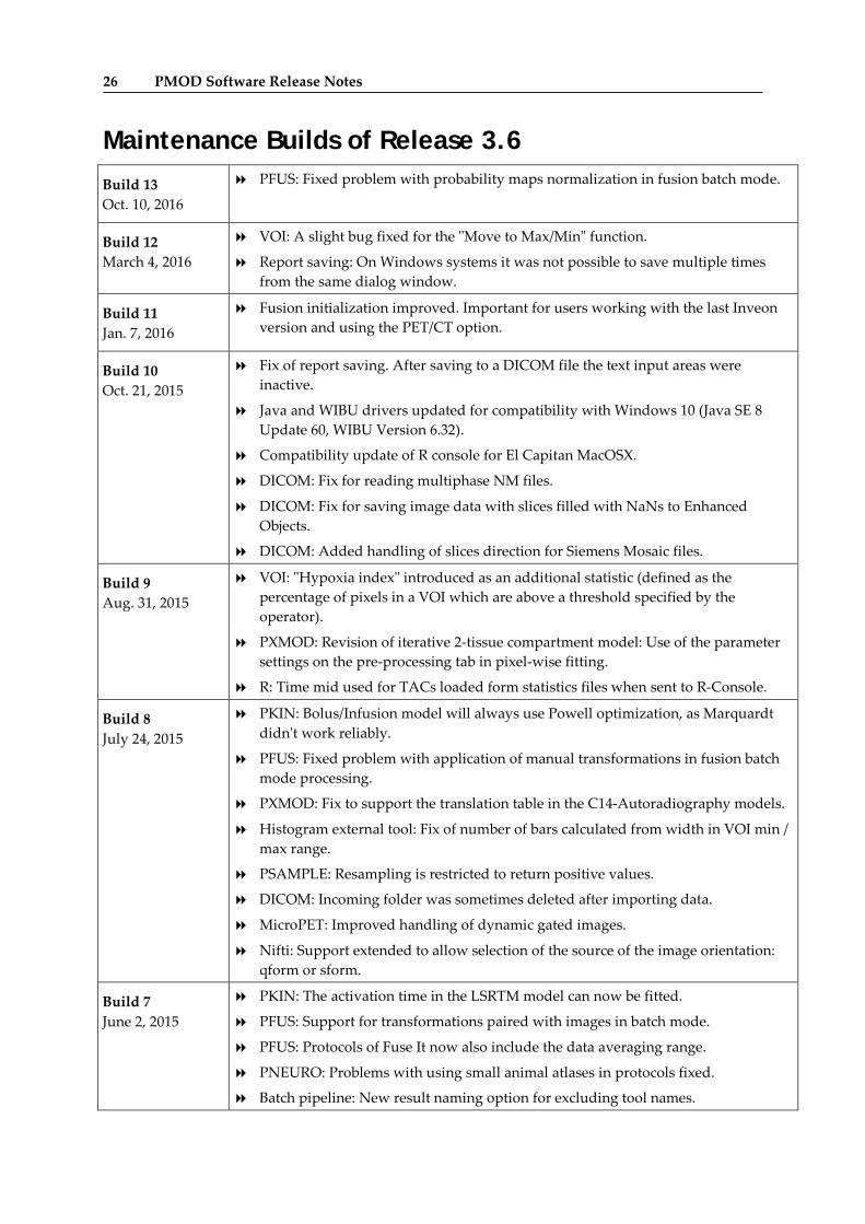
Maintenance Builds of Release 3.6 Build 13 Oct. 10, 2016
PFUS: Fixed problem with probability maps normalization in fusion batch mode.
Build 12 March 4, 2016
VOI: A slight bug fixed for the "Move to Max/Min" function.
Report saving: On Windows systems it was not possible to save multiple times from the same dialog window.
Build 11 Jan. 7, 2016
Fusion initialization improved. Important for users working with the last Inveon version and using the PET/CT option.
Build 10 Oct. 21, 2015
Fix of report saving. After saving to a DICOM file the text input areas were inactive.
Java and WIBU drivers updated for compatibility with Windows 10 (Java SE 8 Update 60, WIBU Version 6.32).
Compatibility update of R console for El Capitan MacOSX.
DICOM: Fix for reading multiphase NM files.
DICOM: Fix for saving image data with slices filled with NaNs to Enhanced Objects.
DICOM: Added handling of slices direction for Siemens Mosaic files.
Build 9 Aug. 31, 2015
VOI: "Hypoxia index" introduced as an additional statistic (defined as the percentage of pixels in a VOI which are above a threshold specified by the operator).
PXMOD: Revision of iterative 2-tissue compartment model: Use of the parameter settings on the pre-processing tab in pixel-wise fitting.
R: Time mid used for TACs loaded form statistics files when sent to R-Console.
Build 8 July 24, 2015
PKIN: Bolus/Infusion model will always use Powell optimization, as Marquardt didn't work reliably.
PFUS: Fixed problem with application of manual transformations in fusion batch mode processing.
PXMOD: Fix to support the translation table in the C14-Autoradiography models.
Histogram external tool: Fix of number of bars calculated from width in VOI min / max range.
PSAMPLE: Resampling is restricted to return positive values.
DICOM: Incoming folder was sometimes deleted after importing data.
MicroPET: Improved handling of dynamic gated images.
Nifti: Support extended to allow selection of the source of the image orientation: qform or sform.
Build 7 June 2, 2015
PKIN: The activation time in the LSRTM model can now be fitted.
PFUS: Support for transformations paired with images in batch mode.
PFUS: Protocols of Fuse It now also include the data averaging range.
PNEURO: Problems with using small animal atlases in protocols fixed.
Batch pipeline: New result naming option for excluding tool names.
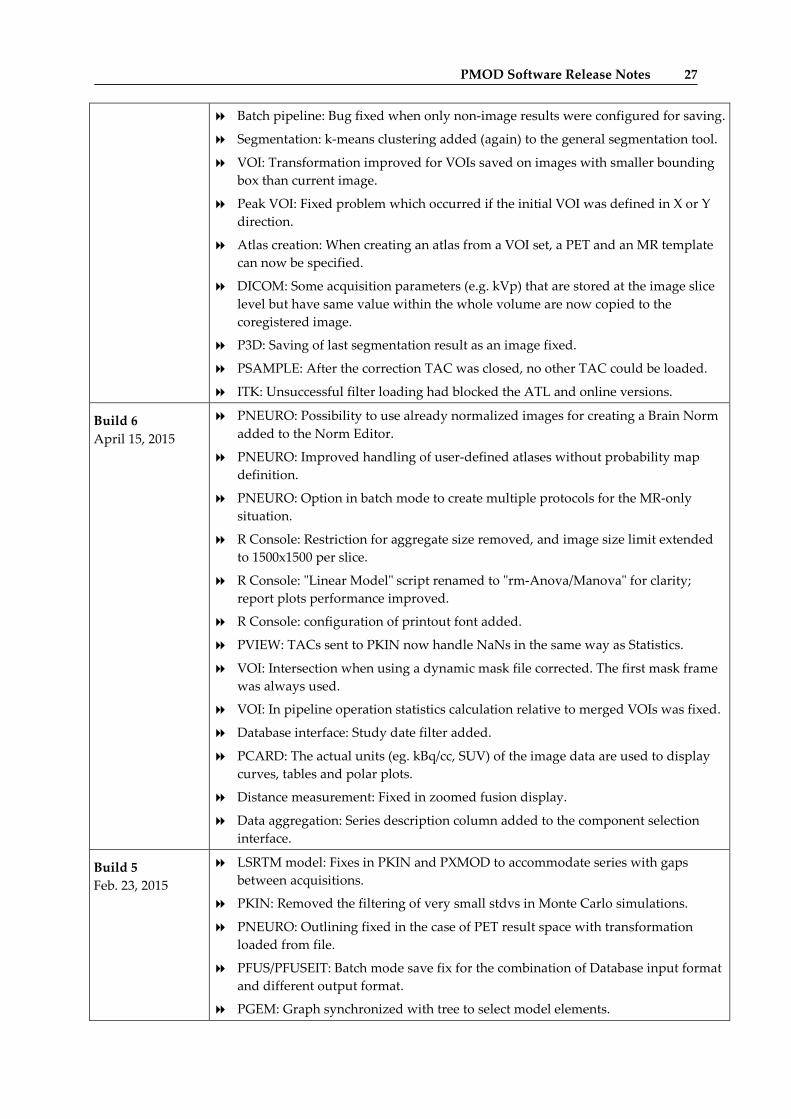
PMOD Software Release Notes 27
Batch pipeline: Bug fixed when only non-image results were configured for saving.
Segmentation: k-means clustering added (again) to the general segmentation tool.
VOI: Transformation improved for VOIs saved on images with smaller bounding box than current image.
Peak VOI: Fixed problem which occurred if the initial VOI was defined in X or Y direction.
Atlas creation: When creating an atlas from a VOI set, a PET and an MR template can now be specified.
DICOM: Some acquisition parameters (e.g. kVp) that are stored at the image slice level but have same value within the whole volume are now copied to the coregistered image.
P3D: Saving of last segmentation result as an image fixed.
PSAMPLE: After the correction TAC was closed, no other TAC could be loaded.
ITK: Unsuccessful filter loading had blocked the ATL and online versions.
Build 6 April 15, 2015
PNEURO: Possibility to use already normalized images for creating a Brain Norm added to the Norm Editor.
PNEURO: Improved handling of user-defined atlases without probability map definition.
PNEURO: Option in batch mode to create multiple protocols for the MR-only situation.
R Console: Restriction for aggregate size removed, and image size limit extended to 1500x1500 per slice.
R Console: "Linear Model" script renamed to "rm-Anova/Manova" for clarity; report plots performance improved.
R Console: configuration of printout font added.
PVIEW: TACs sent to PKIN now handle NaNs in the same way as Statistics.
VOI: Intersection when using a dynamic mask file corrected. The first mask frame was always used.
VOI: In pipeline operation statistics calculation relative to merged VOIs was fixed.
Database interface: Study date filter added.
PCARD: The actual units (eg. kBq/cc, SUV) of the image data are used to display curves, tables and polar plots.
Distance measurement: Fixed in zoomed fusion display.
Data aggregation: Series description column added to the component selection interface.
Build 5 Feb. 23, 2015
LSRTM model: Fixes in PKIN and PXMOD to accommodate series with gaps between acquisitions.
PKIN: Removed the filtering of very small stdvs in Monte Carlo simulations.
PNEURO: Outlining fixed in the case of PET result space with transformation loaded from file.
PFUS/PFUSEIT: Batch mode save fix for the combination of Database input format and different output format.
PGEM: Graph synchronized with tree to select model elements.
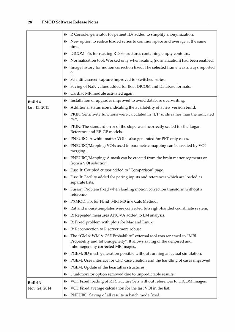
28 PMOD Software Release Notes
R Console: generator for patient IDs added to simplify anonymization.
New option to reslice loaded series to common space and average at the same time.
DICOM: Fix for reading RTSS structures containing empty contours.
Normalization tool: Worked only when scaling (normalization) had been enabled.
Image history for motion correction fixed. The selected frame was always reported 0.
Scientific screen capture improved for switched series.
Saving of NaN values added for float DICOM and Database formats.
Cardiac MR module activated again.
Build 4 Jan. 13, 2015
Installation of upgrades improved to avoid database overwriting.
Additional status icon indicating the availability of a new version build.
PKIN: Sensitivity functions were calculated in "1/1" units rather than the indicated "%".
PKIN: The standard error of the slope was incorrectly scaled for the Logan Reference and RE-GP models.
PNEURO: A white-matter VOI is also generated for PET-only cases.
PNEURO/Mapping: VOIs used in parametric mapping can be created by VOI merging.
PNEURO/Mapping: A mask can be created from the brain matter segments or from a VOI selection.
Fuse It: Coupled cursor added to "Comparison" page.
Fuse It: Facility added for paring inputs and references which are loaded as separate lists.
Fusion: Problem fixed when loading motion correction transform without a reference.
PXMOD: Fix for PBnd_MRTM0 in 6 Calc Method.
Rat and mouse templates were converted to a right-handed coordinate system.
R: Repeated measures ANOVA added to LM analysis.
R: Fixed problem with plots for Mac and Linux.
R: Reconnection to R server more robust.
The “GM & WM & CSF Probability” external tool was renamed to “MRI Probability and Inhomogeneity". It allows saving of the denoised and inhomogeneity corrected MR images.
PGEM: 3D mesh generation possible without running an actual simulation.
PGEM: User interface for CFD case creation and the handling of cases improved.
PGEM: Update of the heartatlas structures.
Dual-monitor option removed due to unpredictable results.
Build 3 Nov. 24, 2014
VOI: Fixed loading of RT Structure Sets without references to DICOM images.
VOI: Fixed average calculation for the last VOI in the list.
PNEURO: Saving of all results in batch mode fixed.
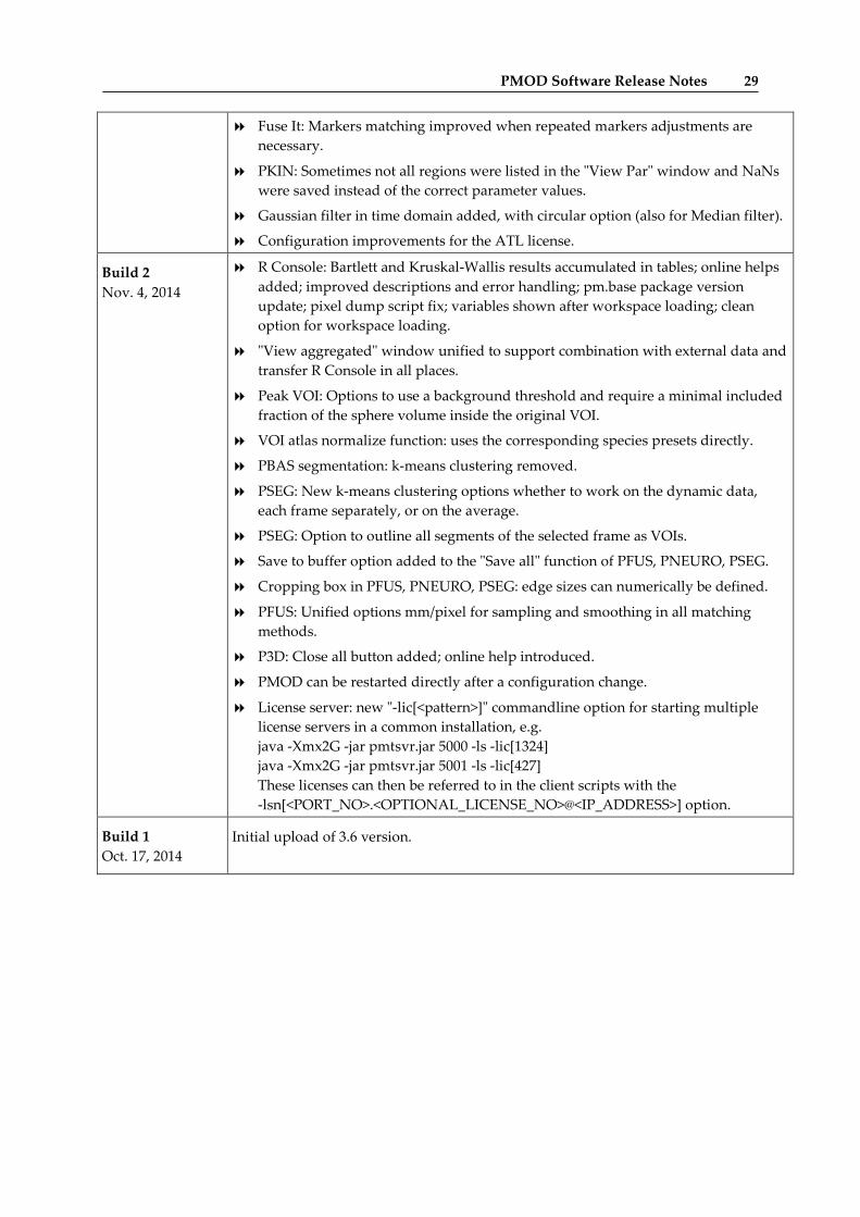
PMOD Software Release Notes 29
Fuse It: Markers matching improved when repeated markers adjustments are necessary.
PKIN: Sometimes not all regions were listed in the "View Par" window and NaNs were saved instead of the correct parameter values.
Gaussian filter in time domain added, with circular option (also for Median filter).
Configuration improvements for the ATL license.
Build 2 Nov. 4, 2014
R Console: Bartlett and Kruskal-Wallis results accumulated in tables; online helps added; improved descriptions and error handling; pm.base package version update; pixel dump script fix; variables shown after workspace loading; clean option for workspace loading.
"View aggregated" window unified to support combination with external data and transfer R Console in all places.
Peak VOI: Options to use a background threshold and require a minimal included fraction of the sphere volume inside the original VOI.
VOI atlas normalize function: uses the corresponding species presets directly.
PBAS segmentation: k-means clustering removed.
PSEG: New k-means clustering options whether to work on the dynamic data, each frame separately, or on the average.
PSEG: Option to outline all segments of the selected frame as VOIs.
Save to buffer option added to the "Save all" function of PFUS, PNEURO, PSEG.
Cropping box in PFUS, PNEURO, PSEG: edge sizes can numerically be defined.
PFUS: Unified options mm/pixel for sampling and smoothing in all matching methods.
P3D: Close all button added; online help introduced.
PMOD can be restarted directly after a configuration change.
License server: new "-lic[<pattern>]" commandline option for starting multiple license servers in a common installation, e.g. java -Xmx2G -jar pmtsvr.jar 5000 -ls -lic[1324] java -Xmx2G -jar pmtsvr.jar 5001 -ls -lic[427] These licenses can then be referred to in the client scripts with the -lsn[<PORT_NO>.<OPTIONAL_LICENSE_NO>@<IP_ADDRESS>] option.
Build 1 Oct. 17, 2014
Initial upload of 3.6 version.
30 PMOD Software Release Notes
Product Release 3.6 The 3.6 product release brings major functional improvements for pipeline processing which allows constructing comprehensive analysis procedures, a completely novel population statistics via the R console, and an additional tool (PFUSEIT) for image fusion with redesigned workflow and new features. Also added is a new tool (PGEM) for the development of geometric models. Additionally, many improvements were implemented for the various tools and the platform, whereby the list below only highlights the major points.
Features
General Batch Pipeline Processing: Substantial extensions of functionality such as
Brain VOIs can be generated on anatomic images and applied to functional images for TAC generation.
Generated TACs can be submitted for kinetic modeling.
Quality-control options added: image viewing, VOI editing, JPEG image saving.
Results can directly be aggregated for population statistics.
Various external tools optimized for use with batch pipeline: CoRegistration and motion correction with interpolation choice; and VOI statistics with spatial transformation, morphological operations, and relative to reference region; VOI based PVC simplified; Interpolation tool can apply transformation; Background removal tool; Segmentation tool supports VOI outlining.
VOIs:
New inclusion criteria for contour VOIs: 1) All pixels with a fractional part covered by the VOI are included, and the average is weighted according to the included pixel area. 2) Using pixels with 50% or 100% area included, with unweighted averaging.
SUVpeak: Generation of explicit sphere VOIs within an enclosing VOI. The positioning options are centering on the maximal pixel, or to maximize the average enclosed in the sphere.
Localization of a number of hottest pixels within a VOI.
Morphological operations (erosion, dilation etc.) of a whole group of VOIs, for instance to shrink automatically generated VOIs by 1 pixel.
Calculations of VOI pixel value histograms, which can be analyzed in R (skewness, kurtosis).
Use of predefined VOI name lists.
Tool for reducing the number of contour vertices.
Eraser working on all active VOIs when the Group tab is selected.
Miscellaneous:
Java Version 8 is bundled with the distribution, now also for Mac.
Aggregation improved, particularly for group analysis. Group name and condition can be specified.
Animated GIF option for saving movies added.
PMOD Software Release Notes 31
Yearly filter added to improve components listing speed. Important for large databases.
DB components loading speed optimization from large databases. Particularly relevant for PXMOD and PNEURO.
Automatic crop box calculation added based on subject end modality (PFUS, PALZ, PNEURO, PSEG).
Spline interpolation introduced for reslicing (PFUS, PALZ).
Changed:
The Automatic Brain VOIs external tool is based on image matching technology and is therefore now bundled with the PFUS option.
PFUSEIT The new "Fuse It" tool guides the user step-by-step from the input images to the registered and post-processed results. Common tasks can be achieved in only a few straightforward steps, whereas complications are handled separately by additional steps. The new tool is launched as a companion tool to the 3.6 version of the proven Fusion tool, for allowing users familiar with the old interface to continue their work as-is. This coexistence will be supported for some future versions, but new developments will mainly be included in the "Fuse It" tool.
Novelties in "Fuse It":
generation of rotating MIP images fused from up to three sources, including animation over time;
explicit use of species information (HUMAN, RAT, MOUSE) for proper matching presets;
parallel viewing of up to 6 fused renderings;
whole-body layout for improved display of non-cubic data;
reference can be reoriented before the matching;
a "Run all" operation mode which applies the configured methods to data selected in the database;
support of overlapping criteria (Dice, Jaccard, volume difference, signed volume difference, specificity, Sensitivity)
transformation of markers after matching;
support for the normalization of CT brain images using a conversion of the HU values and template images from an elderly population (Clinical Toolbox, Chris Rorden).

32 PMOD Software Release Notes
R statistics Console
Population analysis implemented based methodology developed by Prof. K. Herholz
Statistical analysis of study data with numerous regional outcome values, which raises the problem of multiple comparisons.
Streamlined MANOVA and Linear Mixed Effects (LME) analysis workflows.
Scenarios covered include the comparison of groups, the interaction with regional effects, and the handling of covariates, such as age and gender.
Additional tests: Kruskal-Wallis, Variance analysis using the Bartlett test.
Import of data from SPSS, SAS, Systat, Stata, CSV and conversion to aggregates.
Improvements for pixel-dump analysis.
Own operating-system independent package pm.base.tar.gz added containing the scripts.
Multiple user interface improvements.
PKIN
New:
Fitting mode which sequentially shortens the data segment used.
The model history supports multiple new options. The change of a parameter across different fits (eg. shortening) can be visualized as a plot.
The configuration of all models (tissue, blood) can be saved in a single file.
The Sigmoid, Watabe and Hill functions can also be used as plasma fraction functions.
Sigmoid parent fraction function added which was described for [11C]-PBR28 by Owen et al.
Two-tissue compartment model developed by Rizzo et al. for the analysis of [11C]-PBR28 uptake in the human brain.
Alpert’s Linearized Simplified Reference Tissue Model (LSRTM) for noninvasive detection of neuromodulatory changes in specific neurotransmitter systems added.
Improved:
Handling of fitting failures: Regions are not shown in the parameter overview and not saved in the kmPar file. Failures in the reference region are not reported.
Monte Carlo simulations now use an independent seed for each data sample.
Changed:
Runs test: The restriction of >8 runs and >16 samples was removed, and a correction for small sample sizes (N<50) was introduced.
Inclusion of the residual weighting scheme into the tissue model configuration file (.kmModel).
PMOD Software Release Notes 33
PNEURO Animal brain atlases (mouse, rat, monkey) also supported.
Completely revised batch mode which allows defining a processing scheme for a whole list of input data. In this way processing variants can easily be applied to large data samples.
Automatic image cropping mode added so that the entire processing can run without user interaction.
Alternative normalization of MR images which uses the template images directly, rather than the tissue probability maps.
A Hammers atlas variant with 1mm resolution is available which provides more accurate VOIs in the MR space.
VOIs resulting from parcellation can also be obtained in the atlas space.
Parametric mapping can directly be performed in PNEURO, if the PXMOD tool is licensed. The VOIs can be applied to the maps, and the maps can also be used in norm comparisons.
PSEG Automatic species recognition added based on the image volume.
The anatomic images (MR, CT) are directly resampled to the functional images.
K-means clustering added as an alternative method for generating cluster maps.
P3D JOGL is used as 3D engine allowing 3D on Mac with Java 1.8.
New page for the generation of high-quality rotating MIPs. Up to 3 images may be fused. For dynamic scans, the temporal evolution of the images can be included in the rotations.
A scene consisting of surface objects can be saved to an STL file. The object tree is reconstructed after loading the STL
Support for the more compact binary STL format.
3D properties can be set on the Segment page before rendering.
PXMOD Alpert’s Linearized Simplified Reference Tissue Model (LSRTM) for noninvasive detection of neuromodulatory changes in specific neurotransmitter systems
Image definition and loading are now done in a single step.
Several user interface optimizations.
PGEM This new tool leverages the PMOD platform for supporting various types of simulations. A model builder allows for the construction of anatomic structures from VOIs, resulting in a geometric model. Such models can be enriched by multimedia annotations for creating educational 3D-scenes, and complemented by tissue properties. Based on the model construct, phantom images can easily be generated which represent a well-defined input for external simulators of image acquisition instruments or physiology, e.g., OpenFOAM® or Fluent®. An alternative output constitutes atlases which can be employed for automatic VOI generation.
Data Formats Improved handling of floating point image data with NaN values (interpolation and smoothing).
Fixed problem with reading patient and acquisition information from de-identified files with no de-identification method element (0012,0063) included.
Information on image de-identification can be shown in the series info dialog, if present in the DICOM file.

34 PMOD Software Release Notes
Corrected encoding of HU for DICOM enhanced images.
DICOM Incoming folder support for MicroPET and Interfile. Device specific folders with an initial transformation.
Zürich, October 17, 2014
PMOD Copyright Notice 35
Copyright © 1996-2018 PMOD Technologies LLC. All rights reserved.
The PMOD software contains proprietary information of PMOD Technologies LLC; it is provided under a license agreement containing restrictions on use and disclosure and is also protected by copyright law. Reverse engineering of the software is prohibited.
Due to continued product development the program may change and no longer exactly correspond to this document. The information and intellectual property contained herein is confidential between PMOD Technologies LLC and the client and remains the exclusive property of PMOD Technologies LLC. If you find any problems in the document, please report them to us in writing. PMOD Technologies LLC does not warrant that this document is error-free.
No part of this publication may be reproduced, stored in a retrieval system, or transmitted in any form or by any means, electronic, mechanical, photocopying, recording or otherwise without the prior written permission of PMOD Technologies LLC.
PMOD Technologies LLC Sumatrastrasse 25 8006 Zürich Switzerland +41 (44) 350 46 00 [email protected] http://www.pmod.com
PMOD Copyright Notice