pmod release notes2 pmod release notes maintenance builds of release 3.1 build 7 aug. 15, 2010 pkin:...
TRANSCRIPT

Printed on 7 August, 2010
PMOD Release Notes
Version 3.1

2 PMOD Release Notes
Maintenance Builds of Release 3.1
Build 7
Aug. 15, 2010
PKIN: Fixes for inconsistencies in the fitted start time of the MA1 and MA2 models.
MySQL: Fix for compatibility with the latest MySQL 5.1.41.
Merging of files into a dynamic study: At the end of the merging process the Edit
Times dialog window is shown so that the user can verify the timing and correct of
necessary.
Interfile: Added the times for static tomographic images, not only dynamic ones.
PXMOD: With some DICOM objects only one parametric map was saved due to a
naming problem.
DICOM: Fixed reading of DICOMDIR with references to non image objects. The
problem caused repeated entries for the same image on the selection list.
DICOM: Improvements for Philips enhanced MR DICOM objects to support DTI
images.
DICOM: Fixed special case "Image start time not at zero". It resulted in a wrong
timing and a message about missing time in the file.
DICOM loading window: Fixed a problem with de-selecting files by double-clicking.
Buil.d 6
May 30, 2010
PKIN: Fix of a bug in the B/I optimization model which occurred with coupled
fitting, and when optimizing a tissue TAC together with the plasma curve.
PKIN: Bug fixed in MP4A RLS Model which caused an exception during fitting.
PKIN: Dual Ligand, Cold model did not consider the cold tracer from the second
injection at times after the third injection.
PXMOD: Batch mode loading error messages were not added to the final message.
PFUS: For some combinations of pixel size and bounding box a wrong bounding box
was obtained after normalization.
Anatomical orientation: Fix of a problem with missing orientations after the initial
loading operations AVERAGE_VOLUMES, VOLUMES_SUM and
INTEGRATE_VOLUMES.
Anatomical orientation: The reorientation tool in combination with certain data
types could result in obviously wrong value scaling.
DICOM: Fix of a problem with with UN elements (unknown value representation).
DICOM: Loading procedure modified to correctly sort retired NM objects with
wrong image orientation (old GE data).
PALZ: New normalization setting "Compensate Origins displacement (Difference
between origins of Template and Normalized)" added to improve normalization
robustness.
A setup was tested for Solaris on SPARC systems and has been found running, with
the exception of license key support and the native ITK libraries. In such a
configuration a network license must be employed which is served by one of the
Release Notes of PMOD Software

Release Notes of PMOD Software 3
officially supported system.
Build 5
March 29, 2010
SUV values: There was a SUV calculation problem for GE and PMOD data if a non-
zero value for the post-injection activity had been entered. This problem was in
Builds 1-4 of version 3.1, but not in version 3.0.
Image loading: When only a subset of the frames of a dynamic data set was loaded
the timing could be incorrect.
Philips ImageIO: The image data were loaded with a scale factor 1/1 and the units set
to kBq/cc, even if the specified units were not kBq/cc. This could introduce a scaling
error of the data.
DICOM: Images are now loaded even if the FMI header length in the file is incorrect
(GE Signa Excite).
IRW DICOM: Fix of a slice scaling bug which occasionally occurred and was obvious
in the loaded images. Version 3.0 did not suffer from this problem.
DICOM Sever: Starting of the server with the provided script failed.
PKIN: Retrieving of default settings for the metabolite correction did not work
properly.
PKIN: The Print Report function generated a plot that did not correctly display the
metabolite data.
VOI: When sending curves to PKIN the specified curve operations such as adding an
offset were not applied.
VOI: VOIs with different definitions for different frames were not correctly saved.
If the distance was measured on two synchronized data sets, minor distance
differences could occur.
Pipe processing: The definition files were changed to be smaller and faster
interpreted. Saved Pipe processing definitions may require recreation.
Report printing of RGB images: The proportional scaling of the image was change to
better exploit the image area.
The zoom factor in the zoom list was not adjusted after zoom manipulations.
Build 4
Feb. 2, 2010
Dramatic speed improvement of database operations by a factor of 100 and more.
Taking advantage of the improvement requires updating the database structure as
follows:
After installation of Build 4 select the Config button on the ToolBox. In the Users
configuration window select the DATABASE tab, and then the local data source.
First perform a backup of the database tables with the Save SQL Backup button.
Then start database upgrading with the Update button. Note that remote
Transaction Server databases need to be upgraded on the machine hosting the
database.
PMOD Workbook added to the distribution: It is provides step-by-step tutorials for
the major tools using the example data available after PMOD installation. The
workbook can also be downloaded after the login into the Support area of the PMOD
website.
PKIN: Loading multi-column text files with more column headers than data columns
caused a program failure.
PFUS: Motion correction with the "previous" or "following" frame as reference did
not work properly.
4 PMOD Release Notes
VOI: The contour names had not been saved with the VOI definition.
Pipe processing: Adding the specified prefix to the file name did not always work.
"Interpolation" external tool: support for manual transformations had been missing.
Ecat saving: Activity dose changed from MBq to Bq. Patient height changed from m
to cm.
Analyze, NiFTi, Interfile, microPET, Afni image loading: importing the acquisition
times from a file reset the scaling factor, so that wrong values were loaded.
Analyze, Afni, Neurostat, Graphic image loading: the times from the preceding
loading operation was not remembered.
WIBU USB key: Drivers for Debian/Ubuntu Linux systems added to the distribution.
Build 3
Dec. 1, 2009
VOI: Propagation on the ROI level caused contours to change from "-" to "+".
VOI: The time of VOIs created with the iso-contour tool and "Apply to all frames" in
dynamic series were all set to zero.
Edit times: The change of acquisition times acquisition start not from zero were not
correctly saved.
Saving DICOM: the saving as NM DICOM objects caused a program error in certain
conditions.
Rat template (W.Schiffer): the template included some distorted regions.
AAL-VOIs template: The image origin was adjusted to that of the other templates in
the MNI space.
NifTI: Fix for the 2 files mode to store frame times.
Build 2
Nov. 11, 2009
VOI: Compatibility fix required to compensate changes in the treatment of origins
and pixel sizes.
SUV dialog window: Synchronization of the list and the entered half-life.
Edit frame time: Improvement of time generation by step and duration.
Build 1
Oct. 26, 2009
Initial upload of 3.1 version.
Product Release 3.1
The 3.1 release incorporates more than 130 improvements with main highlights in VOI and kinetic modeling
functionalities.
CAUTION: There are important improvements in kinetic modeling which may change the fitting results,
please see below.
General New:
During the installation of the new version the configuration of the previous version
can be imported.
A new button on the ToolBox allows for checking whether the installation is up to
date.
Release Notes of PMOD Software 5
New color tables added.
The list menu of the color tables now includes icons representing the coloring of the
different tables.
New interface to the ITK (Insight Toolkit). It is exploited by a new external tool
which allows to apply 28 image processing filters, and in the segmentation tool
which offers ITK-based segmentations.
A pipe tool was introduced for multistudy processing.
For example: Loading [scaling | smoothing | replacing ] saving.
A new external tool was added for starting scripts of the operating system.
Dual-screen support: Menus are correctly shown, and dialog windows are shown on
the secondary screen.
Printing of the help pages is supported.
Changed:
When merging loaded series into a dynamic study the user can define which series
are used and in which order.
When merging loaded series which cover parts of an object (table positions) into a
single study the user can define which series are used and in which order.
The logging was improved.
Database New:
Free text search in the study and series descriptions.Viewer for loading JPEG and
TIFF images which are saved in the database.
Free text comments which are related to certain image series can be saved in the DB.
New "Flat" selection mode with all series in a single list.
Ability to switch off list updating after every change of a filter element.
Transaction Server: A facility was added which generates an operating-system
specific starting script for the transaction server.
Arrow buttons added for stepping through the patients list.
It is now possible to filter for more than one modality by an OR combination.
Fixed:
Sometimes entries remained in the Projects list although all related data had been
removed.
Sometimes could disappear from a patient, although the user had chosen not to
delete the attached components when deleting images.
VOIs New:
Regular geometric VOIs were added which are not consisting of ROIs, so that they
can be scaled in all directions. With this function an enclosing VOI can much quicker
be defined.
Single pixels can be added or removed from a VOI.
Extended operations on the contour level. Contours can be additive (adding the
included pixels) or subtractive (removing the including pixels) for the VOI.
6 PMOD Release Notes
Facility for applying operations at the contour level.
Extended operations on the VOI level. VOIs can be merged to form new VOIs.
During the merge, VOIs can be additive or subtractive, as the contours. This allows
to easily create VOIs with hollow inclusions.
AND operation with a mask (eg gray matter). Only VOI pixels are considered in the
statistics which are also in the mask.
Definition of a new VOI which consists of the pixels along a line.
New solution for the case when the data origin has changed which causes the VOIs
to be shifted. The VOI origin can be adjusted to the new data origin.
Splitting of a VOI into a set of VOIs on the ROI or contour level.
The VOI ordering in the list can be changed.
Insertion of vertices into a closed contour at any position is now possible.
An eraser of configurable size and shape was added for quickly removing ranges of
vertices.
The hammer tool now has a configurable size.Default sizes for the regular objects
can be saved.
Drawing contours and Iso-Contours are now also possible in NxM layouts.
PKIN CAUTION:
Significant Change related to Compartment Models: In prior versions the whole-blood
contribution in the operational equation was calculated by interpolation of the whole-
blood activity at frame midpoint and multiplication with the frame duration. The
calculation has now been changed to the integration of whole-blood activity during the
frame duration using the interpolation model of the whole-blood curve. This calculation
represents a better approximation of the imaging physics and tends to result in a better
modeling of the initial part of the TACs.
As a result of this change, the model curves calculated with version 3.1 differ from that
of prior versions, and correspondingly fitting results will also differ. Particularly, the
default value of vB may have to be revised.
Change of the Lumped constant for the FDG models. The old default value of 0.437
was replaced by a new value of 0.89. This value had been determined by Graham et al (J
Nucl Med 2002; 43:1157–1166) using 11C Glucose and FDG in 2002. Their results: Normal
brain: 0.89+/-0.08; cerebellum: 0.78+/-0.11.
New:
Support for Randomized fitting. Random sets of initial parameters are generated
and the data fitted. The parameter set among the results with has minimal chi
squared is returned as the fitting estimate. Hereby the potential of getting stuck in a
local minimum is reduced.
Option for Automatic parameter initialization of compartment models using linear
least squares estimates before starting the fit.
Facility for establishing User-defined initial parameters per model.
Improvement of the History functionality. It is now a list to which the user can add
manually, and from which he can delete entries. The list shows the model

Release Notes of PMOD Software 7
configuration with the estimated parameters and fit criteria. Therefore it can also be
used for quickly comparing the results with different model configurations.
Support for Data cloning into an additional window which can be used for a side-
by-side comparison of models.
Comments can be added to a data set, and later edited.
Noise can be added to TACs in the Edit Data dialog.
Whole-blood and plasma data can be loaded from a single file.
New model: linear least squares method for 1- and 2-tissue compartment models
with blood spillover.
The definition of the valid samples can now be copied to all regions.
PXMOD New:
New model: Iterative fitting of a 2-tissue compartment model with ridge regression.
This model allows controlling the degree how much the individual parameters may
be adjusted from their initial values.
Option for separating the blood curves and the tissue TACs into two separate curve
displays to improve visibility on small displays.
Changed:
2-tissue compartment model: was extended by additional output parameters (Vt, Vs,
k3/k4, flux, ssq).
Partial volume correction method 2: the gray matter threshold is now applied to the
smoothed segment, not the original. This reduces the overcorrection problem along
the mask boundaries.
CAUTION: The lumped constant for the FDG models was corrected from the old default
value of 0.437 to a new value of 0.89.
PFUS New:
Brain Normalization II: Default configurations which sets appropriate parameters
for human or small animal applications. (Note: Brain Normalization I should NOT
be used for small animal applications).
Templates of the rat brain added for normalization, matching and VOI statistics
(courtesy of Wynne Schiffer).
Added the functionality to save all registered images at once.
Optional identity line added to the 2D scatter plots of VOI pixel values.
Changed:
VOIs can now also be enabled in the Scientific Output of PFUS.
P3D New:
Selection of objects directly in the scene by double-clicking.
When rendering VOIs, their color is used for surface shading.
Support for more than one set of orthogonal planes.
By using thresholds, the pixels below a certain value can be set to transparent in the
8 PMOD Release Notes
planes.
Plane definitions are also saved in protocols.
Support for more than one marker.
Choice to enable automatic refreshing after changing the VR properties.
Various ITK segmentations available.
Changed:
Revision of the user interface of the control section.
Planes are now also objects in the tree.
Organization of the color profiles for volume rendering improved.
Navigator window is no more hidden in certain situations.
PCARD New:
Save protocol function for easily saving all information related to a data processing
(data, transformations, VOIs). Allows to exactly reproduce a data analysis.
New default for setting the start time of the first frame to zero.
New default for rotating the loaded images to anatomical orientation. This makes the
automatic reorientation procedure more robust.
Different default reorientation for Humans and Animals.
Changed:
Scaling in the reorientation function was disabled.
More significant numbers are shown in the data investigator of the polar plot.
RV and LV VOIs are no more required for the processing of static scans.
Data Formats New:
Native support for JPEG compressed DICOM loader which avoids the use of the JAI
ImageIO library which was not available on Mac.
DICOM Advanced settings: Default button added.
DICOM Server: A facility was added which generates an operating-system specific
starting script for the DICOM server.
Fixed:
DICOMDIR: Under certain conditions (records including sequences) the system
hanged after deleting a file based on the DICOMDIR listing.
DICOM: When scanning a read protected folder an error occurred (mainly MAC,
Linux)
Java Runtime
Environment
(JRE)
On Windows and Linux systems a dedicated JRE is installed together with PMOD
and used for the execution of the program. Therefore, automatic upgrading of Java
by the operating system should not affect the PMOD installation.
On Mac OS X systems the internal JRE is used and therefore the PMOD installation
might be more vulnerable to system updates.
PMOD 3.1 has been tested on Snow Leopard (10.6) and
Java(TM) SE Runtime Environment (build 1.6.0_15-b03-219)
Java HotSpot(TM) 64-Bit Server VM (build 14.1-b02-90, mixed mode)
Release Notes of PMOD Software 9
Zürich, Oct. 26, 2009
Maintenance Builds of Release 3.0
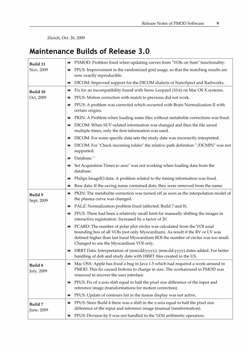
Build 11
Nov, 2009
PXMOD: Problem fixed when updating curves from "VOIs on Sum" functionality.
PFUS: Improvement in the randomized grid usage, so that the matching results are
now exactly reproducible.
DICOM: Improved support for the DICOM dialects of NanoSpect and Radworks.
Build 10
Oct, 2009
Fix for an incompatibility found with Snow Leopard (10.6) on Mac OS X systems.
PFUS: Motion correction with match to previous did not work.
PFUS: A problem was corrected which occurred with Brain Normalization II with
certain origins.
PKIN: A Problem when loading some files without metabolite corrections was fixed.
DICOM: When SUV-related information was changed and then the file saved
multiple times, only the first information was used.
DICOM: For some specific data sets the study date was incorrectly interpreted.
DICOM: For "Check incoming folder" the relative path definition "./DCMIN" was not
supported.
Database: "
Set Acquisition Times to zero" was not working when loading data from the
database.
Philips ImageIO data: A problem related to the timing information was fixed.
Raw data: If the saving name contained dots, they were removed from the name.
Build 9
Sept, 2009
PKIN: The metabolite correction was turned off as soon as the interpolation model of
the plasma curve was changed.
PALZ: Normalization problem fixed (affected: Build 7 and 8).
PFUS: There had been a relatively small limit for manually shifting the images in
interactive registration. Increased by a factor of 20.
PCARD: The number of polar plot circles was calculated from the VOI axial
bounding box of all VOIs (not only Myocardium). As result if the RV or LV was
defined higher than last basal Myocardium ROI the number of circles was too small.
Changed to use the Myocardium VOI only.
HRRT Data: Interpretation of (mm/dd/yyyy), (mm:dd:yyyy) dates added. For better
handling of dob and study date with HRRT files created in the US.
Build 8
July, 2009
Mac OSX: Apple has fixed a bug in Java 1.5 which had required a work-around in
PMOD. This fix caused buttons to change in size. The workaround in PMOD was
removed to recover the user interface.
PFUS: Fix of z-axis shift equal to half the pixel size difference of the input and
reference image (transformations for motion correction).
PFUS: Update of contours list in the fusion display was not active.
Build 7
June, 2009
PFUS: Since Build 4 there was a shift in the z-axis equal to half the pixel size
difference of the input and reference image (manual transformation).
PFUS: Division by 0 was not handled in the %Dif arithmetic operation.

10 PMOD Release Notes
PXMOD: Problem fixed which occurred when updating TACs from VOIs after
switching between models.
PXMOD: The behavior when using VOI templates for updating the TACs was not
correct.
PXMOD: When sending data to PKIN form the data inspector only the input curve
was transferred, not both the input curve and the whole-blood curve.
PXMOD: An error was not handled when all model parameters were disabled from
fittting.
DICOM data: When saving dynamic data as NM DICOM objects the timing was not
correctly saved if there were gaps between successive acquisitions.
AFNI data: The origin was interpreted differently when loading the images directly
than when showing the loading dialog. This caused VOI problems.
NEUROSTAT data: The non-standard encoding as floating-point is now supported.
NIFTI data: Images created by PMOD were confusing to SPM5. The sform matrix
was corrected.
Fixed a slight problem with the context menu of the color bar for setting the window
to the upper third of the value range.
Build 6
May, 2009
PXMOD: Selection of the mask file caused replacement of the original input data by
the mask data.
PXMOD: When "Interactive loading" was initially selected, changing of the input
data was not possible thereafter.
VOI: In fusion mode the iso-contour VOI was always calculated on the "A" image.
VOI: In zoom mode, edition of the vertices with the "Hammer" tool resulted in
unpredictable shifts.
VOI: In the case of zoom and image shift "Unfinish" resulted in a wrong starting
point.
DICOM: On Windows platform a problem with reading JPEG lossless compressed
DICOM data was fixed.
DICOM: The limitation of file names to eight capital characters was dropped. This
behavior caused the "Save all" feature in PXMOD to sometimes generate only a
single file.
DICOM: The regular scanning of an input directory by the DICOM server was not
working.
TIFF: Fix for saving results in tiff format.
Matlab data loading: There was a misleading scaling factor from previous loading
which is now set to 1.
Set Pixel Size: After changing the pixel size the display was not been updated
properly.
Build 5
April, 2009 IMPORTANT: JRE 1.6 update 12 introduced an incompatibility which causes all prior
PMOD versions to stop working. This incompatibility has been fixed in the current Build
5. For prior PMOD version please use a JRE below 1.6 update 12.
PXMOD: Fix for the BP calculation in the MRTM2 model. A wrong k2' had been
applied for the BP calculation in the earlier builds of version 3.0.
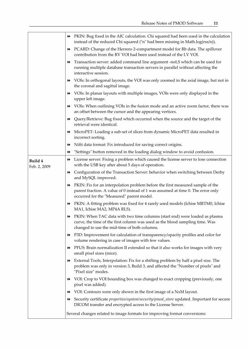
Release Notes of PMOD Software 11
PKIN: Bug fixed in the AIC calculation. Chi squared had been used in the calculation
instead of the reduced Chi squared ("n" had been missing in Math.log(rss/n)).
PCARD: Change of the Herrero 2-compartment model for Rb data. The spillover
contribution from the RV VOI had been used instead of the LV VOI.
Transaction server: added command line argument -noLS which can be used for
running multiple database transaction servers in parallel without affecting the
interactive session.
VOIs: In orthogonal layouts, the VOI was only zoomed in the axial image, but not in
the coronal and sagittal image.
VOIs: In planar layouts with multiple images, VOIs were only displayed in the
upper left image.
VOIs: When outlining VOIs in the fusion mode and an active zoom factor, there was
an offset between the cursor and the appearing vertices.
Query/Retrieve: Bug fixed which occurred when the source and the target of the
retrieval were identical.
MicroPET: Loading a sub-set of slices from dynamic MicroPET data resulted in
incorrect sorting.
Nifti data format: Fix introduced for saving correct origins.
"Settings" button removed in the loading dialog window to avoid confusion.
Build 4
Feb. 2, 2009
License server: Fixing a problem which caused the license server to lose connection
with the USB key after about 5 days of operation.
Configuration of the Transaction Server: behavior when switching between Derby
and MySQL improved.
PKIN: Fix for an interpolation problem before the first measured sample of the
parent fraction. A value of 0 instead of 1 was assumed at time 0. The error only
occurred for the "Measured" parent model.
PKIN: A fitting problem was fixed for 4 rarely used models (Ichise MRTM0, Ichise
MA1, Ichise MA2, MP4A RLS).
PKIN: When TAC data with two time columns (start end) were loaded as plasma
curve, the time of the first column was used as the blood sampling time. Was
changed to use the mid-time of both columns.
P3D: Improvement for calculation of transparency/opacity profiles and color for
volume rendering in case of images with few values.
PFUS: Brain normalization II extended so that it also works for images with very
small pixel sizes (mice).
External Tools, Interpolation: Fix for a shifting problem by half a pixel size. The
problem was only in version 3, Build 3, and affected the "Number of pixels" and
"Pixel size" modes.
VOI: Crop to VOI bounding box was changed to exact cropping (previously, one
pixel was added).
VOI: Contours were only shown in the first image of a NxM layout.
Security certificate properties/system/security/pmod_store updated. Important for secure
DICOM transfer and encrypted access to the License Server.
Several changes related to image formats for improving format conversions:

12 PMOD Release Notes
Conversion Explore Vista Interfile -> DICOM: more data fields are interpreted.
DICOM special cases are not applied for images created by PMOD itself.
DICOM special case "Time start not at zero (move to zero) "added: To make sure the
acquisition times start from time 0.
PMOD now reads and writes energy windows (DICOM NM, DICOM PET, Ecat).
PMOD reads/writes decay factor and relative to what time it must be applied
(DICOM).
PMOD reads/writes decay correction factor (ECAT).
PMOD preserves the original series date/time if the image data is not modified.
Note: series date/time is stored only in DICOM PET objects.
Build 3
Dec. 9, 2008
Installer for the Linux 64-Bit USB-key drivers replaced by WIBU pre-release (WkRt-
Lin64-5.21.500-1.x86_64.rpm).
Correction of handling the image origins while rotating and mirroring images
during loading.
Improved origin support for NIfTI image files.
AAL VOI template image (ROI_MNI_V4.nii) replaced by a new file with origins
compatible with SPM templates.
P3D: Fix for the 3D rendering of VOIs. It was only functional for VOIs defined in the
z-plane.
PKIN: Fix of a bug which could result in endless looping when fits encounter a
certain error condition.
PKIN: Default metabolite correction set to Fixed, not Measured.
PKIN Fix: The model on the Metabolites tab was not reset when loading new data.
VOIs: Fix for ROI propagation with "ROI Follow Max". It was only functional for
VOIs defined in the z-plane.
"New Contour" added to the context menu of VOI definition.
PFUS: Fix fixed problem of the rotation center when combining a normalization
transformation with other transformations.
PFUS: Support for the combination of rigid transformations with rotation centers not
in image center (calculated with initial parameters from principal axis or center of
mass methods).
PFUS: Fix for using VOIs defined as part of VOI templates in scatter plots.
PFUS: Menu entry for user-defined templates which must be prepared in the
resources/templates/usertemplates subdirectory as NIfTI files.
PCARD: Correction of the calculation of the segmental TACS from the polar plot
samples. The resulting TACs are smaller in magnitude (the division was by the
number of samples minus one before) and composed of slightly different polar
samples (boundary samples were considered in more than one segment).
Note: This change produces different results as compared to the prior versions by up
to 10% in individual segments, particularly in apical segments. The effect is smaller
for territories and global values.
PCARD: Fix for a problem of VOI positioning after manual VOI adjustments.
PCARD: Fix for static series. They were smoothed even if the box for the automatic
application was not checked.
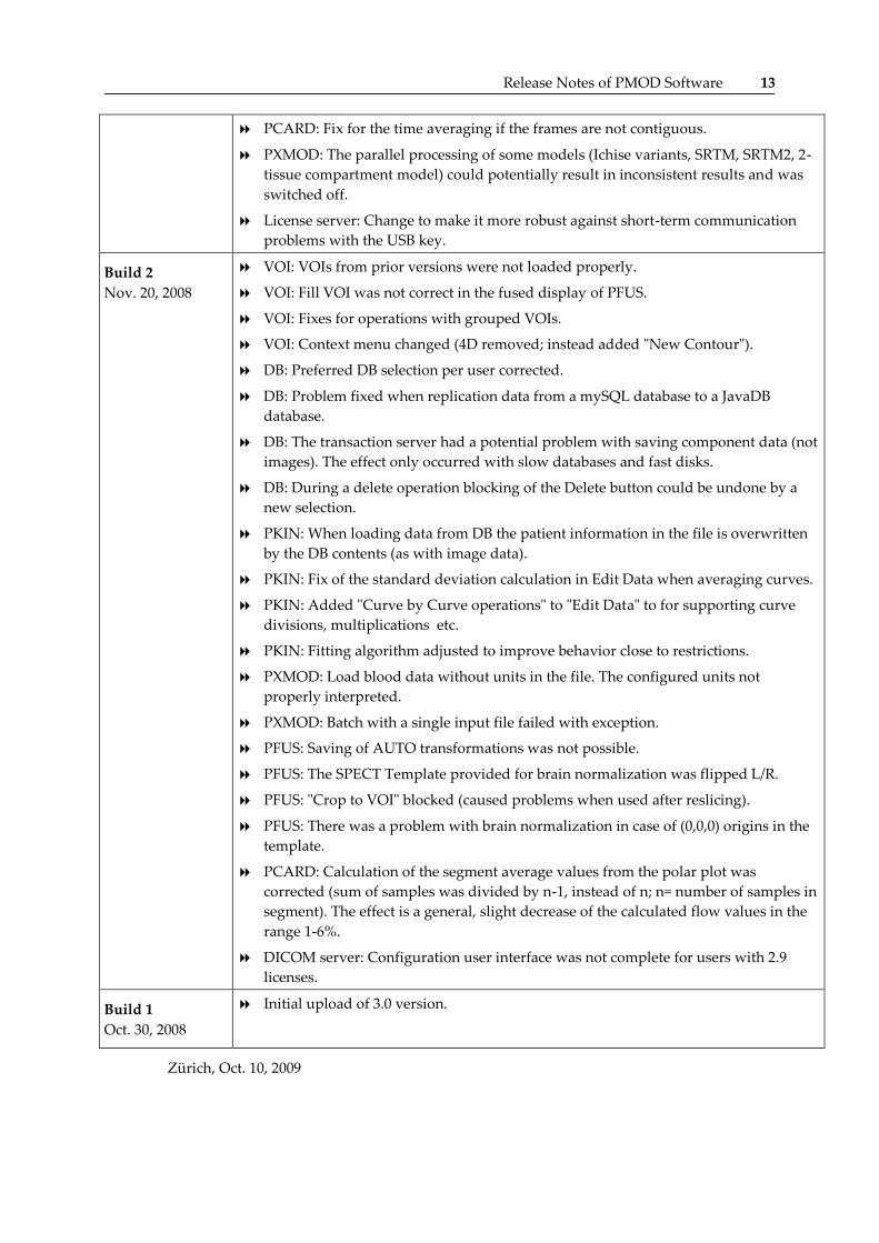
Release Notes of PMOD Software 13
PCARD: Fix for the time averaging if the frames are not contiguous.
PXMOD: The parallel processing of some models (Ichise variants, SRTM, SRTM2, 2-
tissue compartment model) could potentially result in inconsistent results and was
switched off.
License server: Change to make it more robust against short-term communication
problems with the USB key.
Build 2
Nov. 20, 2008
VOI: VOIs from prior versions were not loaded properly.
VOI: Fill VOI was not correct in the fused display of PFUS.
VOI: Fixes for operations with grouped VOIs.
VOI: Context menu changed (4D removed; instead added "New Contour").
DB: Preferred DB selection per user corrected.
DB: Problem fixed when replication data from a mySQL database to a JavaDB
database.
DB: The transaction server had a potential problem with saving component data (not
images). The effect only occurred with slow databases and fast disks.
DB: During a delete operation blocking of the Delete button could be undone by a
new selection.
PKIN: When loading data from DB the patient information in the file is overwritten
by the DB contents (as with image data).
PKIN: Fix of the standard deviation calculation in Edit Data when averaging curves.
PKIN: Added "Curve by Curve operations" to "Edit Data" to for supporting curve
divisions, multiplications etc.
PKIN: Fitting algorithm adjusted to improve behavior close to restrictions.
PXMOD: Load blood data without units in the file. The configured units not
properly interpreted.
PXMOD: Batch with a single input file failed with exception.
PFUS: Saving of AUTO transformations was not possible.
PFUS: The SPECT Template provided for brain normalization was flipped L/R.
PFUS: "Crop to VOI" blocked (caused problems when used after reslicing).
PFUS: There was a problem with brain normalization in case of (0,0,0) origins in the
template.
PCARD: Calculation of the segment average values from the polar plot was
corrected (sum of samples was divided by n-1, instead of n; n= number of samples in
segment). The effect is a general, slight decrease of the calculated flow values in the
range 1-6%.
DICOM server: Configuration user interface was not complete for users with 2.9
licenses.
Build 1
Oct. 30, 2008
Initial upload of 3.0 version.
Zürich, Oct. 10, 2009
14 PMOD Release Notes
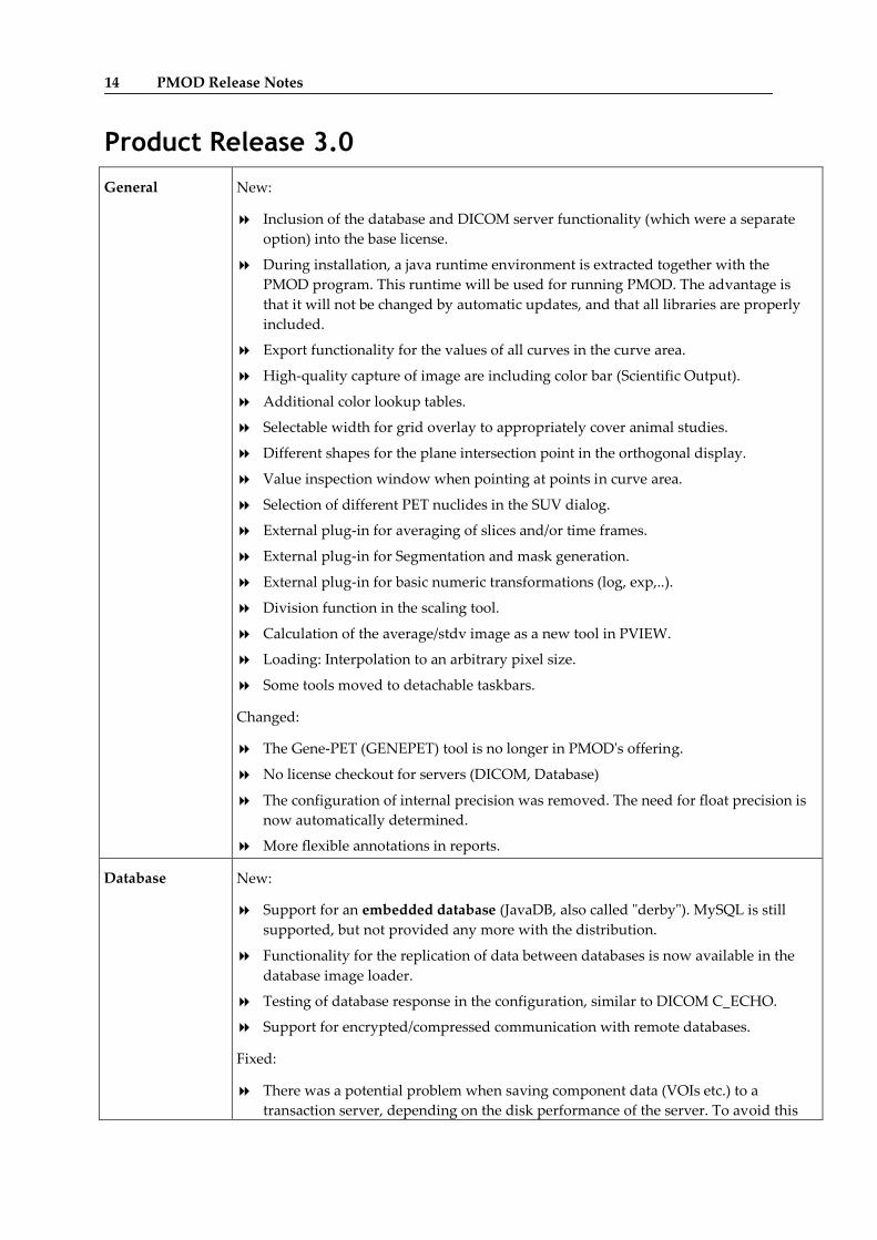
Product Release 3.0
General New:
Inclusion of the database and DICOM server functionality (which were a separate
option) into the base license.
During installation, a java runtime environment is extracted together with the
PMOD program. This runtime will be used for running PMOD. The advantage is
that it will not be changed by automatic updates, and that all libraries are properly
included.
Export functionality for the values of all curves in the curve area.
High-quality capture of image are including color bar (Scientific Output).
Additional color lookup tables.
Selectable width for grid overlay to appropriately cover animal studies.
Different shapes for the plane intersection point in the orthogonal display.
Value inspection window when pointing at points in curve area.
Selection of different PET nuclides in the SUV dialog.
External plug-in for averaging of slices and/or time frames.
External plug-in for Segmentation and mask generation.
External plug-in for basic numeric transformations (log, exp,..).
Division function in the scaling tool.
Calculation of the average/stdv image as a new tool in PVIEW.
Loading: Interpolation to an arbitrary pixel size.
Some tools moved to detachable taskbars.
Changed:
The Gene-PET (GENEPET) tool is no longer in PMOD's offering.
No license checkout for servers (DICOM, Database)
The configuration of internal precision was removed. The need for float precision is
now automatically determined.
More flexible annotations in reports.
Database New:
Support for an embedded database (JavaDB, also called "derby"). MySQL is still
supported, but not provided any more with the distribution.
Functionality for the replication of data between databases is now available in the
database image loader.
Testing of database response in the configuration, similar to DICOM C_ECHO.
Support for encrypted/compressed communication with remote databases.
Fixed:
There was a potential problem when saving component data (VOIs etc.) to a
transaction server, depending on the disk performance of the server. To avoid this
Release Notes of PMOD Software 15
problem both the transaction server and the clients should use version 3.0
VOIs New:
Mask by VOI number for generating template images.
Contours can now also be outlined in the coronal or sagittal orientations.
Undo function for contour VOIs.
Cropping the image volume to VOI bounding boxes.
Single-button creation of an atlas from an image and the outlined contours.
Changed
Revised user interface to enlarge the available image space.
Calculation of TACs: up to 5 times faster.
Creation of VOI masks from a number of hottest pixels (external tool).
Creation of VOI masks using segmentation techniques (external tool)
PKIN New:
Functionality for metabolite correction which includes fitting of parent fractions
with different models and the usage of population correction curves.
Ability to directly copy of the parameters from the overview panel to the clipboard.
Can be pasted into Excel.
"Details" tab with new fit quality parameters (coefficient of determination, residuals
root mean square) and criteria for model comparison (Schwartz criterion, Runs test).
Changed:
Menu reorganized and moved to the lower left corner.
When loading .km files from a database, patient/study information is overwritten by
the contents in the database.
Fixes:
Bolus/Infusion Optimization had a problem when measured weights were used
during fitting and simultaneous fitting of the plasma parameters was enabled.
The calculation of the sensitivity function used parameter changes which were too
big (100% instead of 1%).
Blood delay fitting sometimes failed due to a problem in resampling with negative
times.
Fix for calculation of standard deviations in curves created in the Edit Data tool.
16 PMOD Release Notes
PXMOD New:
SRTM2 model for BPnd calculation with fixed k2'.
1-tissue compartment model with ridge regression (Zhou GRRSC).
Threshold parameter in the model pre-processing section to allow for background
masking.
Possibility to outline VOIs and save them as TAC definitions for the PXMOD
models.
Mask files can now be selected for masking the processing.
Changed:
The MA1 model was removed as a standalone model; it is included in the model
which calculates the distribution volume by 3 different methods.
It is no longer possible to directly return a corrected input curve from PKIN to
PXMOD.
The whole blood activity must be defined in the blood dialog, not any more in the
pre-processing dialog (2-tissue compartments model, 1-tissue compartment models).
When a model processing is interrupted, the calculated pixels are displayed.
Fixed:
Some problems with batch mode were solved.
BPnd values calculated with SRTM method were not correct in early builds of
version 2.95.
The configuration of value units was not properly applied when loading certain
blood files.
Batches with only a single batch operation failed.
PFUS New:
Automatic matching and normalization can be started for all loaded series, not only
one-by-one.
Support for independent manual transformations of the frames of a dynamic study.
Toolbar with shortcut buttons for the most important tasks.
Changed:
Brain normalization II now includes a resampling grid randomization to avoid local
minima. Consequently, the result may vary slightly between runs.
Adjusted order of the rotation buttons for a more intuitive behavior.
Transformations are always applied when data is loaded ("Auto" check box
removed).
Fixed:
Due to an interpolation problem there had been a slight shift of the resliced images
by 1/2 of the original pixel size.
The combination of set landmark and manual matching are now correctly
saved/replayed in protocols.

Release Notes of PMOD Software 17
SPECT template replaced (left and right were mirrored before).
P3D New:
Synchronization of two movies with the real time scale.
Ghost objects to configure rendering options beforehand.
Interpolation to isotropic pixel size during loading to reduce the flipping effects of
VR objects.
Control over smoothing during segmentation.
Segmentation of a definable number of hottest pixels.
Annotation in the 3D scene by adding a Note
Changed:
VR controls are now also in the properties area, not in a separate dialog window.
More control over opacity function
Some detachable taskbars.
Data Formats Memory usage optimized for large DICOM objects
Added: Loader for NeuroStat files
Philips format: support for SUV-related information.
Philips format: Support for Syntegra CT data.
Zürich, Oct. 30, 2008

18 PMOD Release Notes
Maintenance Release 2.95
General Most Important - Compatibility with the "Leopard" OS on Mac:
The Leopard (10.5) release of the Mac OSX turned out to be incompatible with all
PMOD versions. The reason was the missing support for some of the user interface
widgets used by PMOD.
The whole PMOD user interface was revised to avoid the affected elements. As a
result, PMOD has now a platform-specific look on Mac systems. On Linux and Vista
systems, both the cross-platform look (used traditionally by PMOD) and the system
look are now available. The selection is available as a configuration switch in the
"Config" functionality.
New plug-in "Basic Operations" in the external tools with some basic pixel-wise
operations (ln, exp, 1/x, etc).
Bug fixed which occurred when loading the acquisition timing from a file, if the
number of frames did not match the number of file entries.
The database "Save Query" now also remembers the last used database.
PKIN Fix of "Batch Mode" which was trapped in a never ending loop.
Fix of a problem occurring during Drag & Drop of a data file.
Fix of a saving problem with "Create Synthetic KM File" if a new, dense sampling
was defined.
Fix of a problem with "Edit Data" which occurred when deleting TACs in studies
without blood data.
PXMOD Pre-processing stage: Derived parameters (which have no effect on the way how the
calculations are performed) are indicated more clearly.
Fixed problem with "VOIs on Sum": After creating VOIs, the "Update PMOD
Curves" did not produce valid time-activity curves.
Saving of the parametric maps using the file "File" menu did not work correctly. The
input data were saved rather than the results.
PFUS Separate color table for the fusion overlay.
Support for dynamic studies in "Image algebra".
New menu entries as shortcuts for Fusion MIPs, Image Algebra, and Scatter Plots.
PVIEW
VOIs
Fixes for different problems occurring with iso-contour VOIs. The functionality was
reduced and corrected to work more robustly.
Fix for a problem with splitting a dynamic sequence into static frames.
Fix of a protocol saving with transformations calculated by automatic methods.
Release Notes of PMOD Software 19
P3D Surface cutting of SR objects is now also supported in addition to the existing cutting
of VR objects.
Unrelated elements in the objects can be selected (with CTRL+Click) and their
properties modified at once.
Cutting parameters are now also saved in protocol files.
New facility for enlarging the image area during the segmentation definition.
Selective rendering of VOIs (as stripes or surfaces) is supported without loading
images.
Fix in loading protocols: the data format was not always correctly detected.
Fix in the texturing of small objects.
Optimization of memory usage for VR rendering.
Support for textures sizes which are not a power of two.
Data Formats MicroPET: extended so that the CT images from Inveon systems can also be loaded.
Adliswil, February 26, 2008
Product Release 2.9
PCARD The perfusion difference can be calculated as an alternative to the perfusion reserve
to avoid artifacts by the division of very low perfusion values.
Visualization of the myocardium sampling points is now available for quality
control. They are represented as circles in the short axis images. If the P3D option is
available, they can more accurately be rendered as spheres in a 3D scene together
with the left ventricle shape.
A polar plot of the Chi squared criterion of the kinetic model fit was added as an
option.
A new sampling mode "Radial Max, Averaged" was added. This mode adds
immediate neighbor pixels of the samples at the radial maximum to reduce noise
effects.
Water model: The TAC results of a first analysis can be used for a subsequent new
Factor Analysis.
DeGrado cardiac NH3 model only considers the first 4 minutes for fitting and
disables later measurements which are not supported by the model. Problems are
avoided by this behavior if a user loads longer-lasting acquisitions.
Rb 1-compartment model: The default parameters used for the flow-dependent
extraction function according to the paper Lortie et al. Quantification of myocardial
blood flow with 82Rb dynamic PET imaging. Eur J Nucl Med Mol Imaging. 2007 (in
press).
A bug was corrected in the calculation of the apical TACs. Some pixels from the apex
had also included in their calculation.
The division into basal, mid-ventricular and apical slices in the short-axis images
was revised to more closely approach three equal thirds.
20 PMOD Release Notes
PKIN A history of fitted models is maintained for each region, through which one can
browse quickly.
Data Editor to rename, rearrange, average, scale, and delete regional TACs from the
data set.
Points of the input curve which are switched off will not be used for the linear
interpolation of the blood data.
The applicability of sensitivity functions was extended to more non-compartment
models.
Fix for a conversion problem when loading vector data in Ci/cc.
Fix for a problem when generating synthetic PXMOD data (occurring only with
ny=1).
PXMOD Parametric maps are differently organized, not any more as the frames of a dynamic
series. Dynamic series resulting from PVE and PVE2 processing are also available in
the parametric maps area.
Optimization of batch operation.
PFUS The user interface was slightly adjusted to make the image algebra clearer.
A new non-linear matching method was added which is SPM5 compatible.
PVIEW
VOIs
New function to split a VOI into single-slice VOIs.
Iso-contour Auto-VOI: real-time outline on all slices and other improvements
Direct 3D visualization of VOIs (requires P3D option).
VOI Templates: Automatic contouring of the different labels was added to create an
outline version of the template.
P3D Selection of colors for surface rendered objects from a palette is now possible.
The properties of an entire branch in the object tree can be manipulated, for instance
be set to invisible.
Significant improvements in RAM consumption.
Data Formats A new loader format called "Autodetect" was added: It allows selecting data in
different image formats and recognizes the format type automatically from the file
contents.
Nifti: Support for writing was added.
MicroPET: A flag was added in the loading dialog to take into account the branching
factor to the data values (division by the fraction). This is only a compatibility flag
and not needed with new data, which has the branching ratio already applied.
Philips Mosaic SUV data: The unit section of a loading dialog was extended with a
choice for units defined within the file. That choice is only available when PMOD
recognizes more than one unit defined in the file.
DICOM Native support for Lossless Compression was added. The JAI installation is only
needed for data with lossy compression.
Support for compressed connections.
General Rounding of x-axis tick numbers improved to avoid identical ticks.
External tool for image interpolation: The user can now also specify the pixel size of
the image volume which is calculated from the original data by image interpolation.
Users button in toolbar renamed to Config.
Release Notes of PMOD Software 21
Data Inspector bug fix: when interrogating in the z-planes, values from the
neighboring plane (slice number decremented by 1) were reported.
Adliswil, October 2007
Maintenance Release 2.85
NEW: Network
Licensing
Implementation of a network licensing scheme:
A license server manages a number of purchased licenses. When starting PMOD on
client machines within a network, the license server is contacted to request a free license.
If one is available, it is checked out from the license pool, and PMOD starts. Otherwise,
the user is informed that currently there is no license available. Since only the server
requires the USB key, sharing of PMOD licenses is much easier.
Installation
Running
The RAM usage has been improved, so that the memory needs during continuous
processing are markedly reduced.
The blocking disclaimer dialog window at start-up has been removed.
If only one user is configured, an explicit log in after start-up is no longer required.
Improvements for starting individual tools from the command line.
In some installation the generation of Quicktime movies fails because of library
problems. It is therefore recommended to install the Java Media Framework (JMF)
which can be downloaded from:
http://java.sun.com/products/java-media/jmf/2.1.1/download.html
In some installation the reading of compressed DICOM data fails because of library
problems. It is therefore recommended to install the Java Advanced Image I/O
Tools (jre) which can be downloaded from:
https://jai-imageio.dev.java.net/binary-builds.html
General Better support for NaN (Not a Number) values in Ecat, Matlab and Analyze files.
More filtering options instead of only Gaussian smoothing are now available in the
tools section.
PCARD The MBF values are now reported in [ml/min/g], assuming a cardiac tissue density of
1.04 g/ml.
Streamlining of the Coronary Flow Reserve report which now also includes a bar
representation of the segmental flow reserves.
Changes in the layout so that the images of the horizontal long axis view have now
the apex on top, and the annotations are in cardiology style.
If the automatic short-axis reorientation procedure fails, the user can define a marker
in the left ventricle cavity. With this information, reorientation works more robustly.
A new shortcut has been introduced to quickly apply average rotation parameters,
bringing the images roughly to short-axis reorientation. This can serve as a starting
point for manual reorientation.
PKIN A new reference model suitable for the non-invasive quantification of the
acetylcholinesterase activity with the MP4A tracer was added.
A new variant of the dual-input curve model (with metabolites entering tissue) was
added which includes a transfer coefficient linking the exchangeable compartments
in tissue.
22 PMOD Release Notes
A reference model has been implemented in which the target tissue is modeled by a
1-tissue model, whereas the reference tissue is described by a 2-tissue model (Watabe et
al, ).
The Logan plot model now additionally calculates the slope using the perpendicular
distances from the regression line. The resulting distribution volume should be less
biased in the presence of a high noise level.
Unit change of kinetic parameters: "ml tissue" was replaced by ccm (cm3). ml is now
only used for fluid volumes.
Name changes of composed parameters. The new notations are:
Vt = total distribution volume
Vs = distribution volume of specific binding
Vnd = distribution volume of non-displaceable uptake (K1/k2)
Vns = Distribution volume of non-specific binding (k5/k6)
Coupled Fit: a bug in the %COV of the derived parameters was fixed (they
represented values from the single fits).
PXMOD A new reference model suitable for the non-invasive quantification of the
acetylcholinesterase activity with the MP4A tracer was added.
An new model was implemented which calculates the distribution volume with
three methods at once: the standard Logan plot, the Logan plot using the
perpendicular distances, and Ichise's MA1 method. In this order, the methods have
less bias, but increasing variance. The advantage is that three Vt-maps are available
in a single run.
PFUS Improved batch mode to allow arbitrary Reference/Reslice pairs instead of a global
reference only.
Simplified user interface for creating rotating MIPs of fused images.
PVIEW
VOIs
The isocontour auto-VOI can now work on all frames of a dynamic series, for
instance to measure the volume changes of a vessel in dynamic microscopy images.
The isocontour auto-VOI now supports multiple contours per slice for outlining
disconnected structure components.
Database A new backup procedure was developed which saves the database tables together
with the actual data files into a zip archive. The data from such archives can be
restored into an existing database at later times.
Data Formats Reading and writing of Ecat data was further improved.
A reader was added to load blood data directly from the ABSS blood sampler file
(http://www.allogg.se; corrected singles).
DICOM Query/Retrieve improvements: With some systems (eg. GE Advantage Windows)
Q/R from PMOD failed because some unsupported elements were requested. By
reducing the number of requested elements Q/R from PMOD now works more
widely.
Adliswil, April 2007
Product Release 2.8
Licensing The use of the single-user PMOD license is more strictly controlled. PMOD may now only
be used by a single person at a time who may run up to two concurrent sessions (for

Release Notes of PMOD Software 23
side-by-side comparisons).
Caution: When PMOD is started a third time, the first session is stopped.
Installation
Running
The 2.8 version requires the current Java version JRE 1.5.
The individual PMOD tools can also be started directly from the command line (and
therefore from other programs). Files to be loaded are specified as command line
arguments. For instance, the fusion tool could be started from a different application
with a reference and a reslice study loaded.
General Configuration of plug-ins such as models or loaders is easier because model name
and explanation is available.
The zoom factor can be entered numerically.
PCARD The cardiac modeling tool is undergoing a complete revision. The improvements in the
current version include
Improved documentation
Stress/Rest side-by-side processing.
General pre-processing for anatomical blood volume and myocardium images.
Automatic procedure to transform the data into standard short axis orientation.
Automatic outlining of the left ventricle and right ventricle VOIs, as well as the
myocardium centerline.
New polar resampling for more flexibility in the segment model definition.
AHA 17-segment model.
Calculation of the Coronary Flow Reserve from the rest and stress results.
The revision will continue and is scheduled for completion in the 2.85 version.
PXMOD The pixel-wise modeling tool has also be revised and significantly improved:
Tabs added to allow for the parallel processing of multiple data sets.
Improved documentation.
Button to pop up a short model explanation.
The TACs from single pixels can directly be transferred to the PKIN tool together
with the blood data.
Parametric maps can directly be sent to the PFUS tool for a comparison.
Menu newly structured.
New Patlak Reference Model for calculating Kocc for FDOPA studies.
The file names can not be edited any more. To redefine a file the ... button must be
used.
After activating the x button processing will start from scratch including all data
loading.
PKIN New button to pop up a short model explanation.
Undo/redo button to switch back and forth between models which have already
been fitted.
New model for cardiac perfusion with NH3 model: 2 compartments (Hutchins) with
exponential metabolite correction (van den Hoff).
24 PMOD Release Notes
New model for cardiac perfusion with 11C acetate model (van den Hoff) with
corrections for metabolite buildup and flow-dependent extraction fraction.
The interpolation of blood values before the first sample was changed. In earlier
versions, all blood values before the first sample were considered as 0. Now, the
blood values are linearly interpolated assuming a blood value of 0 at time 0. To
avoid unexpected results the user should define a blood value of 0 at a suitable time.
"Fit blood param" check box has been moved to the first tab for more ease of use.
P3D Improved documentation.
Loading of VRML data sets improved to integrate Amira results in PMOD scenes.
PFUS Batch application of an existing transformation matrix to a set of studies.
Normalization templates switched from the Analyze to the Nifti format.
Direct transfer of matched studies to the 3D tool.
Support for user-defined templates with corresponding masks.
PVIEW
VOIs
Split to disk exception fixed.
Improved implementation of saving VOIs as DICOM RT Structure sets.
Database mySQL 5 is now required (supporting local character sets). If a mySQL 4.x server is
running, please first un-install, then install the new 5.x version. Finally copy the data
directory containing the database tables from the old to the new mysql version.
Migration between databases is much faster.
Backup of database tables to file system (automatic schedule or manual).
Data Formats New loader added for images in the Nifti format (http://nifti.nimh.nih.gov/nifti-1).
TIFF loader improved; data from AIDA systems can now be loaded.
DICOM Support for national character sets (Umlaute) added.
Fix for situations where character set is not encoded in the object (Xeleris). The new
Advanced DICOM configuration "Use selected character set if not present in object".
It is recommended to define "ISO-8859-1" as the default (Latin-1 character set for
Western Europe).
New Query loader allows to display images from a remote DICOM server without
first saving the data to disk.
Database import server: Specification of a directory which is scanned continuously
by the DICOM server. Found DICOM image series are imported into a database or
can be converted to another format.
Flag to suppress the saving of SUV-related information which is encoded in private
GE attributes (can cause problems on some older systems).
Studies with multiple acquisitions in one series can now be loaded. The different
acquisitions are shown as separate series.
Bug fixed with with NM Recon Tomo objects. Acquisition time was 1000 times to
big.
Adliswil, Oct 2006

Release Notes of PMOD Software 25
Maintenance Release 2.75
New: PGENE
Tool
A new tool has been developed in collaboration with the group of Prof. L. Strauss from
the German Cancer Research Center. It supports the statistical analysis of gene chip data
to detect genes which are over expressed in disease processes. They represent potential
targets for new radio pharmaceuticals, to be applied in tumor diagnostics and isotope
therapy. Additionally, the gene expressions can be correlated with quantitative PET
parameters using different regression relationships, resulting in a ranked list of
significant correlations.
PKIN Improved documentation.
New summary page showing the parameters of all regions at once.
Volume-weighted averaging of TACs added to the View Data Tool.
Monte Carlo simulation fixes.
PXMOD Ichise's MA1 model for the calculation of the total DV added.
The iterative 2-compartment model improved to allow for the spillover correction
with whole blood instead of using the input curve.
The Times/Origins shown are read from the selected files if they contain the
information.
Saving of the functional maps as DICOM was only possible once. Units are encoded
when saving to DICOM, and decoded when loading back. Parameter names are
appended to series description instead of overwriting.
PFUS Improved documentation.
Addition of 2D and 3D (with 3D option) scatter plots to visualize the pixel values of
VOIs.
Improvement of initial reslicing: not the whole volume is calculated, just the visible
slices.
Specification of a common point for the initial reslicing so that manual matching can
be started from an approximate match.
The interpolation method used in reslicing can now be selected by the user.
Combination of the current transformation with saved transformation to generate
combined transformation. For instance the stereotactic normalization of an
individual MRI to a template and the matching transform of a PET of the same
individual to that MRI can be combined into a single transform which directly
normalizes the PET to the template.
As a significant improvement, the resliced images are now always calculated from
the original images by combining the initial/automatic and the manual transform.
Bug fixed when applying motion correction to a sub-range of frames.
PVIEW - VOIs Improvement of the Iso-Contour ROI/VOI; bias removed; also working for cold
spots; inner and outer contour.
Mirroring of VOIs in the z-direction (across slices) added.
Improvements when saving VOIs as DICOM RT-Structure Sets.

26 PMOD Release Notes
Database Automatic backup of the mySQL tables with a time schedule.
Updates to the mySQL database structure can now be performed from the PMOD
database configuration interface, rather than using system-specific batch scripts.
Data Formats Mosaic Format (Philips animal and human PET scanners, GE Quest scanner) added.
Bug fixes in the MicroPET format: 1) The slice thickness was calculated wrongly for
older files (transaxial_crystal_pitch/2 instead of axial_crystal_pitch/2). 2) Values were
not always scaled correctly. 3)
Data Formats -
DICOM
C-Store is now possible for a subrange of the slices (with preview functionality).
SUV-related informations are saved with DICOM objects (NM and PT).
New tool to anonymize DICOM data with minimal modification of the data
attributes. The advantage is that all private information is maintained, and the
objects can be used for the analysis of DICOM data posing problems in PMOD. This
feature is only available as an export from the DICOM database.
Implementation of an improved strategy for reading Siemens Biograph PET data.
Because of a certain Siemens implementation detail the images appear with the
Head/Feet orientation mirrored. Selection of the "Anatomical Reorientation" button
on the loading dialog allows to fix this problem.
BrainDB Visualization of the brain area used for scaling when applying a percentile range
Installation Detects an existing 2.7 installation and allows to maintain the prior properties.
The Java native libraries for the USB key driver support are automatically installed.
Processing After resizing (External Tools/Resize), VOIs were not usable any more because of
origin changes. This behavior has been improved.
Release Notes of PMOD Software 27
P3D Improvements in transparency in combination with the new Java3D 1.4 version
(currently not available on Mac OSX). The slice images can also be set to transparent.
Improvements in the protocols.
Adliswil, May 2006
Product Release 2.7
New: Brain
Database Tool
The new PMOD Brain Database Tool is a general tool for establishing the normal
pattern of a certain type of brain images, and to compare patient images against it.
After a brain database has been establised, it can easily be exported and made
available to other users.
The database construction in principle consists of the following steps:
The acquisition of images from a set of normal controls with most preferably the
same acquisition and image reconstruction protocols the patients are studied
with.
The stereotactic normalization of the control images, so that the anatomy of the
normalized images is comparable within a certain accuracy.
The scaling of the pixel values in the normalized images relative to an internal
reference to allow for the pooling of the data.
The analysis of the values across the control collective in each pixel of the
stereotactic anatomy to establish the normal value and its deviation across the set
of normal data.
Patient images are normalized and scaled in the same way as the control images, and
the resulting pixel values compared with the normal values. This process results in a
map showing the differences between the patient images and the normal pattern,
expressed as a z-score values. The z- score map can be investigated in a multitude of
ways including fusion with the patient images and 3D rendering (separate option).
New:
PVIEW Tool
The functionality of the two base tools Image Gateway (PGATE) and VOI Tool (PVOI)
has been merged into a single tool (PVIEW). In this way the situation that a study was
loaded in the "wrong" tool is avoided.
VOI Analysis Iso-contour VOI tool. It looks for a closed iso-contour at a specified image value
within an enclosing VOI.
Automatic procedure for myocardium VOI delineation (Auto-VOI).
Support for loading standard VOI templates such as AAL and using the labeled
pixels for VOI statistics in stereotactically normalized studies.
Support for binary masks in VOI statistics.
Improved contour ROI tool.
Display Reports: The report quality (image resolution) has been improved and can now be
configured per user in the settings.
28 PMOD Release Notes
Reports and screen captures can now be saved as DICOM SC images, and to the
clipboard.
Movies can be saved as DICOM objects.
The access to the 3D tool is available wherever images have been loaded (requires
P3D option). This avoids the need to first save processed images, and loading
them in the 3D tool afterwards.
DICOM
DB Server
Images saved to the DICOM DB are now always saved as Recon Gated Tomo NM
DICOM objects which allow efficient loading of large dynamic data sets (see
DICOM Conformance Statement for details). This change is transparent to the
user, and the modality and the units are preserved.
Support for mySQL 4.1 including non empty root passwords.
Data Formats When loading DICOM series which contain different image types, an additional
selection layer is provided to load a subset of the data. Such series result from
dedicated analysis protocols.
Demographic data can be loaded from an Interfile header file into a loaded image
series. This functionality has been to facilititate conversion processes to DICOM.
The batch conversion now includes a section of study level attributes including
the accession ID. This makes it possible to convert jpeg images (which were
created as a study documentation to DICOM secondary capture images) for
archival in a PACS system.
3D Rendering Orientation box which rotates with the scene and shows patient's left, head
anterior.
Markers can be defined at arbitrary positions in the scene.
Segments based on region growing.
Visual feedback of the pixels included in the segment before starting of the
rendering.
Predefined volume renderings for angio CT with contrast agent.
Predefined volume renderings for angio CT with matched PET or SPECT
PXMOD Improved partial volume correction method (PVE2correct) which obviates the need
for outlining a white-matter VOI.
PFUS MNI templates have been prepared in the HFS orientation. Using them as a
reference in stereotactic normalization results in images in standard radiological
orientation.
Changed bounding box options for pruning the results of stereotactic
normalization. The selection includes the Talairach extent or the full template
extent.
PCARD Two-compartment model for FDG is available.
A dialog allows to select the kinetic model and set the parameter starting values
before transferring control to the kinetic modeling tool. This is particularly
relevant for the FDG models to provide the plasma glucose value.
A new button is available to start a fusion layout containing the two factor images
of water studies, myocardium and blood volume.
Automatic procedure for myocardium VOI delineation. A model-based approach
tries to detect the myocardium center line after the images have been resliced to
Release Notes of PMOD Software 29
short axis orientation.
PKIN The PKIN application now has several tabs to hold multiple data sets. Multiple
.km files can be loaded at once, and more data sets can be added incrementally.
The axis ranges (x and y) can now be set to arbitrary values, not only to values
within the range of measurements.
Random number generation for Monte Carlo simulations has been improved.
WIP:
Documentation
The revision of the PMOD documentation is in progress. The base functionality and
several tool descriptions are now available both in html and as printable PDF
documents.
Adliswil, October, 2005
Maintenance Release 2.65
Display Anatomical orientation: As a new feature, the anatomical orientations are displayed, if
sufficient patient positioning information is contained in the data.
Orthogonal planes view: The position of the blue lines indicating the planes location
has slightly changed. Now the position is in the slice center, not at the slice border as
before.
Mirroring: There are now several mirroring options available for data after loading.
Processing Improvements of the processing tools ("screw driver" tab):
A new checkbox is available to modify the processing mode: Either, the loaded original
data is modified (eg. by smoothing, scaling, etc), or the study is first copied and the
copy processed.
An "average" tab has been introduced to average over time after a dynamic series has
been loaded.
"smooth": A bug in Gaussian smoothing was fixed. Due to this bug the data had only
been smoothed in the x-direction.
"External/SUV": There is a new tool has added to the "External" section to transform
PET activity data into SUV units.
PVOI The ROIs can now also be edited while operating in the orthogonal planes view, not
only in the axial 1x1 layout.
To adjust imported VOIs to a new study they can also be shifted along the z-axis
(across slices) and scaled in x and y (orthogonal planes view).
To improve control of small VOIs, the "Fill" mode was revised to unequivocally show
all pixels belonging to a VOI. The user can select between a raster and a filled mode.
DICOM Anatomical annotations are derived from the DICOM positioning information and
shown in the images.
The user can define the patient orientation for data loaded from non-DICOM formats.
When subsequently saving the data, this information is encoded in DICOM attributes,
so that the correct anatomical annotations are shown when the data is loaded next
time.
When DICOM Data is loaded into PMOD and saved again, the DICOM attributes are

30 PMOD Release Notes
changed. To allow for transferring the original DICOM data to remote DICOM servers,
a new "DICOM send" facility has been added which sends the data directly off the disk
without introducing DICOM changes.
Tilt correction: To display data aquired with a tilted gantry correctly in the orthogonal
planes view, a slice offset must be corrected for. Such an (optional) tilt correction has
been added for DICOM data.
The format of the Dicom time attributes was corrected.
DICOM Q/R has been improved, and is working properly with GE Advantage
Windows and Xeleris in both directions.
Data Formats Ecat: Fix for reading gated image data as a dynamic series.
Analyze: Correction of an endian error affecting the scaling factor when saving short
integers in LE.
Gated PET is loaded as a dynamic series, not as a list of separate studies. This allows to
display movies more easily.
Database The database scheme was extended to allow for two new fields: a text field
"Diagnosis" and a date field "Last used". These fields can be used for grouping data
and to form more detailed queries. Additionally, more attributes were indexed for
faster responses.
These changes require that existing databases (2.6 and below) must be migrated to
be used with the 2.65 version!
Please refer to the Database upgrade guide for details.
JPEG data can now also be saved in the database.
All types of component data can be searched and exported from the database using the
"Aggregate Components" Dialog on the DB configuration page.
P3D Volume rendering of dynamic studies supported.
Optional texturing of individual objects.
Saving/retrieving of protocol definitions.
Cutting out quadrants in volume-rendered scenes.
PCARD Revision of the calculation of the segments from the center line and the septal angle. In
the VOI tool the myocardium pixels can now be inspected in the "Fill" mode.
The standard deviation shown in the report can be configured to be in % (default), or
in absolute units.
The width of the myocardium in the synthetic study showing the calculated parameter
can be defined. This feature is intended to be used with 3D rendering, so that
perfusion can be shown as a texture on the myocardium wall surface.
The Patlak kinetic model has been modified so that it can be used from within the
PCARD tool.
PKIN During saving operations (.km, .kinpar files), the parameter standard errors are saved
in absolute units rather than %COV. An error was fixed in this conversion which
affected parameters with units 1/min.
The units of the MRGlu was changed from [mg/min/100ml] to [umol/min/100g] (factor
from old to new: 5.336). Models affected are the Patlak and FDG 2compartment model.
Adliswil, May 3, 2005
Release Notes of PMOD Software 31
Product Release 2.6
Licensing
USB-hardware key licensing only: the IP-based licenses have been abandoned due to
practical difficulties in the nowadays mostly dynamic environment.
A free upgrade to version 2.6 is only available for customers who purchased a 2.5
version. All prior versions require an upgrade which can be ordered through the
support section on www.pmod.com using the login provided to you shortly.
General
Changes and new features
The principal components analysis of dynamic data has been implemented as an
external tool.
Some methods for wavelet denoising have been implemented as an external tool.
Denoising can be applied per slice, per slice over time, and per frame.
DICOM Server The DICOM server performance has been dramatically improved. Images are received
more than 10 times faster.
Data Formats
New internal data representation: In order to limit the amount of required RAM images
have traditionally been stored internally as a 2-byte integer per pixel plus a scale factor
and an offset per slice. Herby the precision of the calculation is somewhat limited with
large dynamic ranges. Truncation artifacts can occur, especially in operations such as
pixel-wise divisions which might produce big outlier numbers. To avoid such problems,
pixel values can now internally be stored as floating numbers (High precision mode). The
user has to select between the two modes by setting a configuration checkbox
appropriately (Users settings, Statistics panel). Please note that configuring the high-
precision mode will double the amount of RAM which PMOD requires for image data.
PXMOD
New models
Two-tissue compartment model with pixelwise iterative fitting.
Partial volume correction of brain studies based on white and gray matter segment
images.
Modified models
The accuracy of the flow calculations was improved in the Alpert model. The
difference with respect to the old implementation is typically in the range of 1%.
PFUS
Motion correction: the frames of a dynamic series can be aligned using the automatic
matching methods implemented in PFUS. There are different strategies available, eg.
to register to the average of several frames, or sequentially to the
preceeding/succeeding frame.
Batch mode for automatic registration: A number of studies can be selected for
subsequent matching to a single reference. For instance, a set of studies can be
scheduled for spatial normalization in a batch operation.
The performance of the spatial normalization has been significantly improved.
PCARD
Rubidium model (WIP): As a new model for use with the PCARD tool the 82Rb model as
described by Herrero et al. has been implemented in PKIN. This model can be applied,
but is to be regarded as a works-in-progress since it has not yet been validated against
water or ammonia in a prospective study.
32 PMOD Release Notes
PKIN
New models:
Logan's reference model (requiring an estimate of k2) which was only available in
PXMOD is now also implemented in PKIN.
Herrero's model for cardiac 82Rb PET studies.
Ichise's multilinear methods MA1 and MA2 for calculating the total DV with minimal
noise-induced bias.
Adliswil, October 21, 2004
Copyright © 1996-2010 PMOD Technologies Ltd.
All rights reserved.
The PMOD software contains proprietary information of PMOD Technologies Ltd; it is
provided under a license agreement containing restrictions on use and disclosure and is also
protected by copyright law. Reverse engineering of the software is prohibited.
Due to continued product development the program may change and no longer exactly
correspond to this document. The information and intellectual property contained herein is
confidential between PMOD Technologies Ltd and the client and remains the exclusive
property of PMOD Technologies Ltd. If you find any problems in the document, please
report them to us in writing. PMOD Technologies Ltd does not warrant that this document
is error-free.
No part of this publication may be reproduced, stored in a retrieval system, or transmitted in
any form or by any means, electronic, mechanical, photocopying, recording or otherwise
without the prior written permission of PMOD Technologies Ltd.
PMOD Technologies Ltd
Sumatrastrasse 25
8006 Zürich
Switzerland
+41 (44) 350 22 37
http://www.pmod.com