pineal region tumors - sno · pineal region tumors, and histologic documentation is a prerequisite...
TRANSCRIPT
193
7
Pineal Region Tumors
JEFFREY C. ALLEN, JEFFREY BRUCE, LARRY E. KUN, AND LAUREN LANGFORD
Tumors of the pineal region can be divided into threecategories (Rubinstein, 1970): (1) germ cell tumors,which range from surgically curable mature ter-atomas to the malignant germ cell tumors capable ofmetastasizing throughout the neuraxis; (2) pinealparenchymal tumors, such as the low-grade pineocy-toma and the malignant pineoblastoma; and (3) othertumors such as intra-axial astrocytomas, ependymo-mas and mixed gliomas, and rare tumors of sur-rounding structures (meningioma, dermoid, epider-moid).
GERM CELL TUMORS
Background and Pathology
Germ cell tumors are relatively uncommon, consti-tuting 3% to 5% of large institutional pediatric tumorseries and 0.5% to 1% of adult brain tumor series(Hoffman et al., 1983). Table 7–1 summarizes thefrequency of predominantly adult surgical pathologycases seen at the New York Neurologic Institute(Bruce and Stein, 1995). Germ cell tumor cases con-stituted 37% of their series of 160 cases. The originof these non-neuroectodermal, primary brain tumorsis unknown but may be related to an early period ofontogeny when the fetal germinal cells migrate widelythroughout the body, including the central nervoussystem (CNS). Normally, germinal cells not residingin tissues destined to form sex organs become apop-totic and die; presumably some may occasionally sur-vive and over many years transform into a neoplasm.
The sites of origin of germ cell tumors in the CNS areunique, that is, extra-axial locations in proximity tothe pineal gland and infundibulum. In unusual in-stances when germ cell tumors arise in the brain ofinfants or patients with Down’s syndrome, other lo-cations may predominate (Chik et al., 1999).
Germ cell tumors have several unique epidemio-logic features such as the age at onset, sites of ori-gin, and racial and sex predilections. The peak ageat onset lies within the second and third decades oflife, thereby including both the pediatric and adultpopulations. The primary sites of origin lie withinmidline extra-axial spaces such as the pineal region(45%), suprasellar region (35%), both regions(10%), and other locations (10%). Interestingly, theincidence of CNS germ cell tumors is extraordinarilyhigh in the Japanese population, constituting 15% to18% of primary CNS tumors in several large institu-tional series compared with 3% to 5% in North Amer-ican reports (Jennings et al., 1985).
More than 95% of CNS germ cell tumors are bio-logically malignant, that is, capable of rapid growth,invasion, and metastasis. Histologically, approxi-mately 65% of CNS germ cell tumors are pure ger-minomas, and most of the remaining ones are eitherpure nongerminomatous germ cell tumors (NGGCT)such as embryonal carcinoma, endodermal sinus oryolk sac tumor, choriocarcinoma, immature or ma-lignant teratoma, or mixed malignant tumors. Matureteratomas that are slow growing and noninvasive arethe least common variant (�5%). The pure germi-nomas are more commonly found in the pineal re-gion, whereas NGGCT occur more frequently in the
3601_e07_p193-207 2/15/02 4:34 PM Page 193
suprasellar region (Edwards et al., 1988; Malo-golowkin et al., 1990; Rueda-Pedraza et al., 1987).There exists a male predominance (3/1 [US] to 10/1[Japanese]) in germ cell tumor series in the pineallocation, but there is an equal sex distribution or fe-male predominance for suprasellar primary tumors(Jennings et al., 1985).
Because the management and prognosis of patientswith intracranial germ cell tumors is very dependenton histology, it is imperative to establish an unequiv-ocal diagnosis prior to the administration of radio-therapy (RT) and/or chemotherapy. For example,germinomas are readily curable with RT alone orcombinations of RT and chemotherapy; NGGCT are
potentially curable with maximal surgical debulkingand intensive chemotherapy and RT; and teratomasare curable with surgery alone (Jennings et al.,1985).
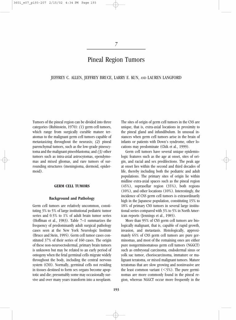
Primary intracranial germ cell neoplasms are his-tologically similar to gonadal germ cell tumors. Ger-minoma is histologically identical to the seminoma(testes) or dysgerminoma (ovary). Germinomas aretypically composed of two cell types: large, uniformpolyhedral cells with clear cytoplasm that resembleprimordial germ cells; and smaller lymphoid cells.The large cells contain abundant intracytoplasmicglycogen. Their round nuclei contain one or moreprominent nucleoli (Fig. 7–1). Although not neces-
194 PRIMARY CENTRAL NERVOUS SYSTEM TUMORS
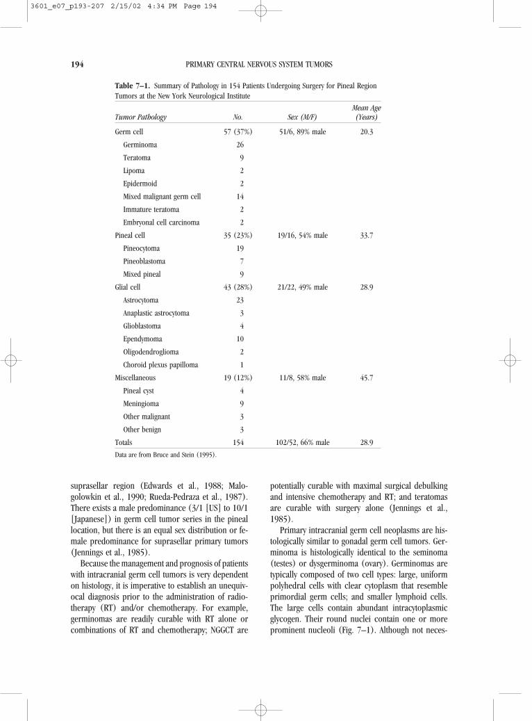
Table 7–1. Summary of Pathology in 154 Patients Undergoing Surgery for Pineal RegionTumors at the New York Neurological Institute
Mean AgeTumor Pathology No. Sex (M/F) (Years)
Germ cell 57 (37%) 51/6, 89% male 20.3
Germinoma 26
Teratoma 9
Lipoma 2
Epidermoid 2
Mixed malignant germ cell 14
Immature teratoma 2
Embryonal cell carcinoma 2
Pineal cell 35 (23%) 19/16, 54% male 33.7
Pineocytoma 19
Pineoblastoma 7
Mixed pineal 9
Glial cell 43 (28%) 21/22, 49% male 28.9
Astrocytoma 23
Anaplastic astrocytoma 3
Glioblastoma 4
Ependymoma 10
Oligodendroglioma 2
Choroid plexus papilloma 1
Miscellaneous 19 (12%) 11/8, 58% male 45.7
Pineal cyst 4
Meningioma 9
Other malignant 3
Other benign 3
Totals 154 102/52, 66% male 28.9
Data are from Bruce and Stein (1995).
3601_e07_p193-207 2/15/02 4:34 PM Page 194
sary for diagnosis, immunohistochemical studies canhelp in problem situations when only limited mater-ial is available. No single antigen, however, has beenidentified as “specific” for germinoma (Table 7–2).The lymphocytic component is immunopositive forantibodies to leukocyte common antigen, whereas thelarger epithelioid cells may react to antibodies forplacental alkaline phosphatase and cytokeratins (Fe-lix and Becker, 1990). Some germinomas may alsocontain syncytiotrophoblastic cells, which are im-munoreactive with antibodies to human �-chorionicgonadotropin.
From a treatment perspective, the malignant NGGCTs are the most challenging. Endodermal sinustumor and choriocarcinoma resemble extra-embry-onic tissues, and the embryonal carcinoma appearssimilar to fetal embryonal tissue. Endodermal sinustumors (yolk sac tumor) consist of glomeruloidstructures composed of a space lined by tumor cells
with an invaginated vascular pedicle covered by amonolayer of the same cells. This tumor is consid-ered to represent a neoplasm whose cells are par-tially differentiated into extra-embryonic structuresthat express yolk sac potential (Gonzalez-Crussi,1979). Embryonal cell carcinoma is considered themost histogenetically primitive of the germ cell tu-mors, with features of anaplastic columnar tocuboidal cells arranged in sheets and cords (Bjorns-son et al., 1985). This tumor shows a variable pat-tern of acinar, papillary tubular, or solid structures.A lymphocyte infiltrate may also be present but is notas abundant as in germinomas. Embryonal cell carcinoma may give rise to a multiplicity of tumor ad-mixtures, with the most advanced form being ter-atoma. Choriocarcinomas are examples of differ-entiation along extraembryonic pathways and arecomposed solely of cytotrophoblastic and syncy-tiotrophoblastic cells without true villous formation.The primary immunohistochemical marker of this tu-mor is human �-chorionic gonadotropin, which issecreted by the syncytiotrophoblast; however, positivestaining for human �-chorionic gonadotropin is notexclusively diagnostic of choriocarcinoma (Midgleyand Pierce, 1962). Primary intracranial choriocarci-nomas are rare (Bjornsson et al., 1986).
A relatively uncommon form of germ cell tumor isthe pure teratoma, a tumor that is composed of ma-ture tissues from all three germ cell layers (endo-derm, mesoderm, and ectoderm). Pineal teratomasare complex mixtures of tissues that occur most fre-quently in males, in contrast to sacrococcygeal ter-atomas, which occur more often in females. Pinealteratomas are largely well differentiated or mature,but immature teratomas with malignant features dooccur (Bjornsson et al., 1985). Any CNS teratoma,
Pineal Region Tumors 195
Figure 7–1. Germinomas contain large cells with clear cy-toplasm and scattered lymphocytes. Hematoxylin and eosin,�200.
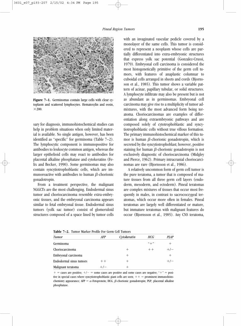
Table 7–2. Tumor Marker Profile For Germ Cell Tumors
Tumor AFP Cytokeratin HCG PLAP
Germinoma “�” �
Choriocarcinoma � �� �/�
Embryonal carcinoma � �
Endodermal sinus tumors �� � �/�
Malignant teratoma �/� �
� � cases are positive; �/� � some cases are positive and some cases are negative; “�” � posi-tive in special cases where syncytiotrophoblastic giant cells are seen; �� � prominent immunohisto-chemistry appearance; AFP � �-Fetoprotein; HCG, �-chorionic gonadotropin; PLP, placental alkalinephosphatase.
3601_e07_p193-207 2/15/02 4:34 PM Page 195

however, can harbor foci of a malignant germ cell tu-mor; therefore, adequate sampling is of the utmostimportance. If no other malignant elements are de-tectable, this variant can be managed with radical sur-gical resection alone. This strategy also pertains toother mature teratoid or embryonal tumors, such asdermoids and epidermoids.
Tumor Markers
The tumor markers �-fetoprotein (AFP), human �-chorionic gonadotropin (HCG), placental alkalinephosphatase, and lactic dehydrogenase isoenzymesare useful in the diagnosis and treatment monitoringof germ cell tumors. Elevations in levels of AFP alonein cerebrospinal fluid (CSF) and serum are found inpure endodermal sinus tumor. Elevated levels of bothHCG and AFP are found in embryonal carcinoma, andhigh levels of HCG alone are found in choriocarci-noma. The serum and CSF levels in cells of AFP maybe 10 to 100 times baseline. Serum HCG levels maybe 100 times baseline, and there may be a CSF/serumgradient, especially when lumbar CSF is assayed.Modest elevations of HCG may be found in germi-noma, usually in the presence of elevated placentalalkaline phosphatase and/or lactic dehydrogenaseisoenzymes (Allen, 1987). A serum or CSF HCG �50IU/L and/or an AFP �25 ng/ml in the presence of amidline CNS tumor is supportive of a diagnosis of NGGCT (Calaminus et al., 1997).
Clinical Presentation
The clinical presentation of germ cell tumors varieswith the site(s) of primary and/or metastatic disease.For suprasellar primary tumors, especially germino-mas, the prodrome may be long, that is, from monthsto several years. Typically, a child may present withsigns and symptoms of hypopituitarism (i.e., diabetesinsipidus, growth failure, or secondary hypothy-roidism) or precocious puberty. Initial neurodiag-nostic studies may be noninformative. Children withacquired hypopituitarism are prime candidates toharbor germ cell tumors and should be followed ex-pectantly with magnetic resonance imaging (MRI)scans at regular intervals. Visual field or acuity im-pairments and hydrocephalus occur late in the pro-drome, when the tumor is large or disseminated. AnMRI scan may disclose an extra-axial enhancing massin the suprasellar and/or pineal region or intraven-
tricular seeding in the third or lateral ventricles withhydrocephalus. A diagnostic surgical procedure istypically performed after a stress or therapeutic doseof corticosteroids is administered.
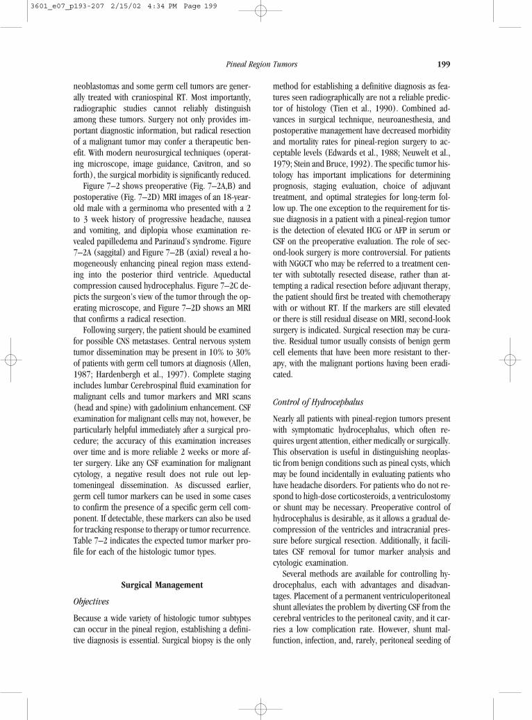
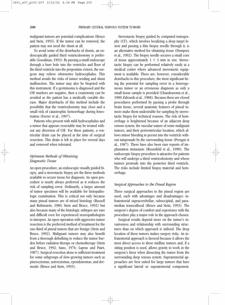
Patients with primary tumors arising in the pinealregion tend to have a much shorter prodrome (i.e.,weeks to several months). These patients usuallypresent with signs and symptoms of raised intracra-nial pressure, such as headache, diplopia, andlethargy due to aqueductal obstruction. Tectal com-pression can cause Parinaud’s syndrome (verticalgaze paresis, impaired pupillary light reflex, and con-vergence nystagmus). The MRI scan will reveal apineal region-enhancing mass, which may protrudeinto the posterior region of the third ventricle withacute hydrocephalus (Fig. 7–2). Following high-dosecorticosteroid therapy, the patient may need to be sta-bilized with ventriculostomy before definitive surgery.
General Management Considerations
The management of primary intracranial germ cell tu-mors is changing. Surgery is becoming an increas-ingly safer procedure for patients with suprasellar andpineal region tumors, and histologic documentationis a prerequisite for optimum therapy. The manage-ment of germ cell tumors is histology dependent anddifferent protocols are emerging for patients withpure germinomas and nongerminoma germ cell tu-mors. During a prior era when surgical approachesto the pineal region were associated with consider-able morbidity and mortality, the standard practicewas to administer a therapeutic/diagnostic course offocal radiation therapy to 20 Gy. If the tumor under-went a major regression, a presumptive diagnosis ofgerminoma was made and the patient then receiveda full course (55 Gy) of focal and, in some institu-tions, craniospinal radiation. If the tumor did not re-spond to the 20 Gy dose, a diagnostic surgical pro-cedure could be performed.
This approach is no longer favored for several rea-sons. Germinomas are not the most prevalent tumorsin the pineal region in either pediatric or adult se-ries, and the incidence of various tumor types is notreadily predictable. For example, the incidence ofgerm cell tumors in the pineal region reported in tworecent operative pediatric series from the Children’sHospital of Philadelphia (Packer et al., 1984) and theUniversity of California, San Francisco (Edwards etal., 1988) were 32% and 61%, respectively. The in-
196 PRIMARY CENTRAL NERVOUS SYSTEM TUMORS
3601_e07_p193-207 2/15/02 4:34 PM Page 196
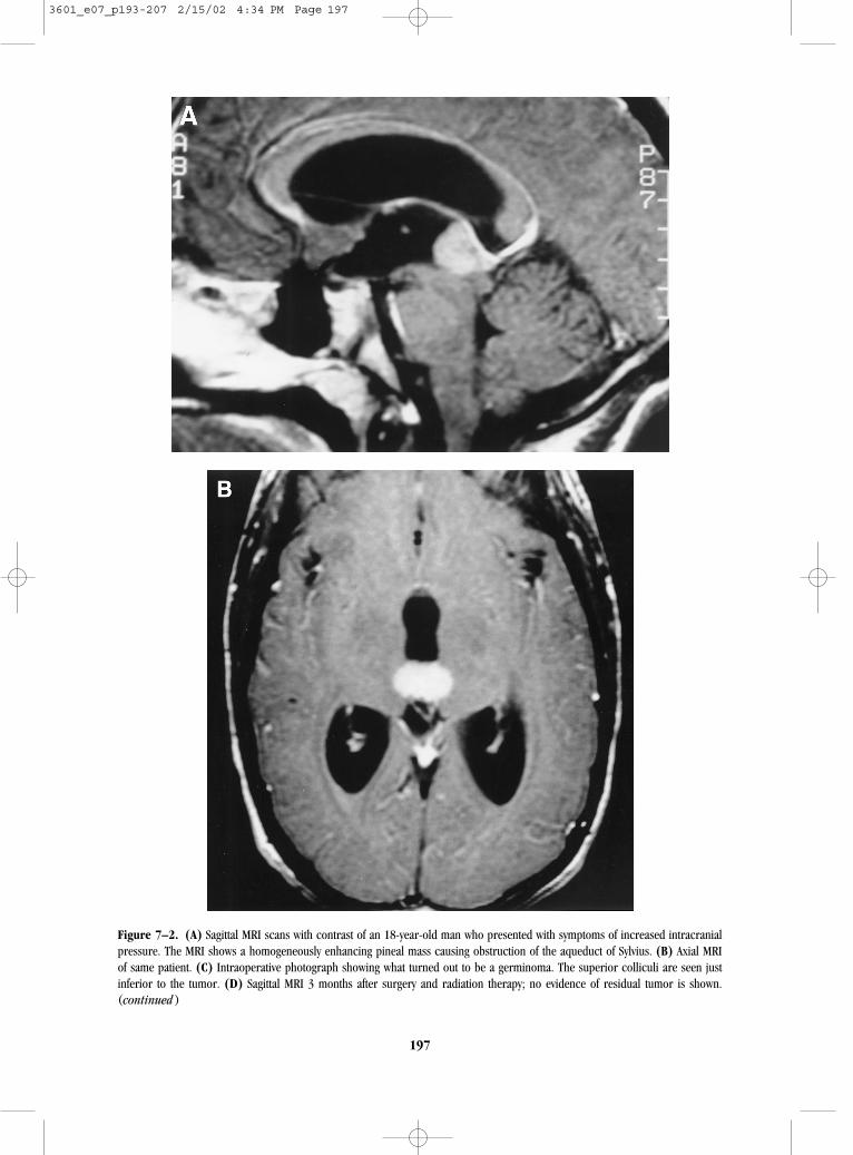
Figure 7–2. (A) Sagittal MRI scans with contrast of an 18-year-old man who presented with symptoms of increased intracranialpressure. The MRI shows a homogeneously enhancing pineal mass causing obstruction of the aqueduct of Sylvius. (B) Axial MRIof same patient. (C) Intraoperative photograph showing what turned out to be a germinoma. The superior colliculi are seen justinferior to the tumor. (D) Sagittal MRI 3 months after surgery and radiation therapy; no evidence of residual tumor is shown.(continued )
197
3601_e07_p193-207 2/15/02 4:34 PM Page 197

cidence in two adult series was 38% (Linggood andChapman, 1992; Bruce and Stein, 1995). The inci-dence of pure germinoma in the two pediatric serieswas 20% (7/35) and 31% (11/36), respectively.
Other malignant tumors (pineoblastoma and ma-lignant ependymoma) may respond to RT, but their
management is quite different from that for germi-noma. If a low-grade tumor such as an astrocytoma,pineocytoma, or teratoma exists, radical surgical re-section alone rather than RT may be the treatment ofchoice. For certain tumors, such as ependymoma andmalignant astrocytoma, focal RT is indicated; pi-
198 PRIMARY CENTRAL NERVOUS SYSTEM TUMORS
Figure 7–2. (Continued)
3601_e07_p193-207 2/15/02 4:34 PM Page 198
neoblastomas and some germ cell tumors are gener-ally treated with craniospinal RT. Most importantly,radiographic studies cannot reliably distinguishamong these tumors. Surgery not only provides im-portant diagnostic information, but radical resectionof a malignant tumor may confer a therapeutic ben-efit. With modern neurosurgical techniques (operat-ing microscope, image guidance, Cavitron, and soforth), the surgical morbidity is significantly reduced.
Figure 7–2 shows preoperative (Fig. 7–2A,B) andpostoperative (Fig. 7–2D) MRI images of an 18-year-old male with a germinoma who presented with a 2to 3 week history of progressive headache, nauseaand vomiting, and diplopia whose examination re-vealed papilledema and Parinaud’s syndrome. Figure7–2A (saggital) and Figure 7–2B (axial) reveal a ho-mogeneously enhancing pineal region mass extend-ing into the posterior third ventricle. Aqueductalcompression caused hydrocephalus. Figure 7–2C de-picts the surgeon’s view of the tumor through the op-erating microscope, and Figure 7–2D shows an MRIthat confirms a radical resection.
Following surgery, the patient should be examinedfor possible CNS metastases. Central nervous systemtumor dissemination may be present in 10% to 30%of patients with germ cell tumors at diagnosis (Allen,1987; Hardenbergh et al., 1997). Complete stagingincludes lumbar Cerebrospinal fluid examination formalignant cells and tumor markers and MRI scans(head and spine) with gadolinium enhancement. CSFexamination for malignant cells may not, however, beparticularly helpful immediately after a surgical pro-cedure; the accuracy of this examination increasesover time and is more reliable 2 weeks or more af-ter surgery. Like any CSF examination for malignantcytology, a negative result does not rule out lep-tomeningeal dissemination. As discussed earlier,germ cell tumor markers can be used in some casesto confirm the presence of a specific germ cell com-ponent. If detectable, these markers can also be usedfor tracking response to therapy or tumor recurrence.Table 7–2 indicates the expected tumor marker pro-file for each of the histologic tumor types.
Surgical Management
Objectives
Because a wide variety of histologic tumor subtypescan occur in the pineal region, establishing a defini-tive diagnosis is essential. Surgical biopsy is the only
method for establishing a definitive diagnosis as fea-tures seen radiographically are not a reliable predic-tor of histology (Tien et al., 1990). Combined ad-vances in surgical technique, neuroanesthesia, andpostoperative management have decreased morbidityand mortality rates for pineal-region surgery to ac-ceptable levels (Edwards et al., 1988; Neuwelt et al.,1979; Stein and Bruce, 1992). The specific tumor his-tology has important implications for determiningprognosis, staging evaluation, choice of adjuvanttreatment, and optimal strategies for long-term fol-low up. The one exception to the requirement for tis-sue diagnosis in a patient with a pineal-region tumoris the detection of elevated HCG or AFP in serum orCSF on the preoperative evaluation. The role of sec-ond-look surgery is more controversial. For patientswith NGGCT who may be referred to a treatment cen-ter with subtotally resected disease, rather than at-tempting a radical resection before adjuvant therapy,the patient should first be treated with chemotherapywith or without RT. If the markers are still elevatedor there is still residual disease on MRI, second-looksurgery is indicated. Surgical resection may be cura-tive. Residual tumor usually consists of benign germcell elements that have been more resistant to ther-apy, with the malignant portions having been eradi-cated.
Control of Hydrocephalus
Nearly all patients with pineal-region tumors presentwith symptomatic hydrocephalus, which often re-quires urgent attention, either medically or surgically.This observation is useful in distinguishing neoplas-tic from benign conditions such as pineal cysts, whichmay be found incidentally in evaluating patients whohave headache disorders. For patients who do not re-spond to high-dose corticosteroids, a ventriculostomyor shunt may be necessary. Preoperative control ofhydrocephalus is desirable, as it allows a gradual de-compression of the ventricles and intracranial pres-sure before surgical resection. Additionally, it facili-tates CSF removal for tumor marker analysis andcytologic examination.
Several methods are available for controlling hy-drocephalus, each with advantages and disadvan-tages. Placement of a permanent ventriculoperitonealshunt alleviates the problem by diverting CSF from thecerebral ventricles to the peritoneal cavity, and it car-ries a low complication rate. However, shunt mal-function, infection, and, rarely, peritoneal seeding of
Pineal Region Tumors 199
3601_e07_p193-207 2/15/02 4:34 PM Page 199
malignant tumors are potential complications (Bruceand Stein, 1993). If the tumor can be removed, thepatient may not need the shunt at all.
To avoid some of the drawbacks of shunts, an en-doscopically guided third ventriculostomy is prefer-able (Goodman, 1993). By passing a small endoscopethrough a burr hole into the ventricles and floor ofthe third ventricle into the prepontine cistern, the sur-geon may relieve obstructive hydrocephalus. Thismethod avoids the risks of tumor seeding and shuntmalfunction. The tumor may also be biopsied withthis instrument. If a germinoma is diagnosed and theCSF markers are negative, then a craniotomy can beavoided as the patient has a medically curable dis-ease. Major drawbacks of this method include thepossibility that the ventriculostomy may close and asmall risk of catastrophic hemorrhage during fenes-tration (Ferrer et al., 1997).
Patients who present with mild hydrocephalus anda tumor that appears resectable may be treated with-out any diversion of CSF. For these patients, a ven-tricular drain can be placed at the time of surgicalresection. This drain is left in place for several daysand removed when tolerated.
Optimum Methods of Obtaining Diagnostic Tissue
An open procedure, an endoscopic visually guided bi-opsy, and a stereotactic biopsy are the three methodsavailable to secure tissue for diagnosis. An open pro-cedure is nearly always preferred as it reduces therisk of sampling error. Ordinarily, a larger amountof tumor specimen will be available for histopatho-logic examination. This is critical not only becausemany pineal tumors are of mixed histology (Russelland Rubinstein, 1989; Stein and Bruce, 1992) butalso because many of the histologic subtypes are rareand difficult even for experienced neuropathologiststo interpret. An open operation with aggressive tumorresection is the preferred method of treatment for theone-third of pineal tumors that are benign (Stein andBruce, 1992). Malignant tumors may also benefitfrom a thorough debulking to reduce the tumor bur-den before radiation therapy or chemotherapy (Steinand Bruce, 1992; Sano, 1976; Lapras and Patet,1987). Surgical resection alone is sufficient treatmentfor some subgroups of slow-growing tumors such aspineocytomas, astrocytomas, ependymomas, and der-moids (Bruce and Stein, 1993).
Stereotactic biopsy guided by computed tomogra-phy (CT), which involves localizing a deep target le-sion and passing a thin biopsy needle through it, isan alternative method for obtaining tissue (Dempseyet al., 1992). The biopsy needle secures a small coreof tissue approximately 1 � 3 mm in size. Stereo-tactic biopsy can be performed relatively easily at amedical center where advanced stereotactic equip-ment is available. There are, however, considerabledrawbacks to this procedure, the most significant be-ing the potential for sampling error in a heteroge-neous tumor or an erroneous diagnosis as only asmall tissue sample is provided (Chandrasoma et al.,1989; Edwards et al., 1988). Because these are closedprocedures performed by passing a probe throughbrain tissue, several anatomic features of pineal tu-mors make them undesirable for sampling by stereo-tactic biopsy for technical reasons. The risk of hem-orrhage is heightened because of an adjacent deepvenous system, the vascular nature of some malignanttumors, and their periventricular location, which al-lows minor bleeding to persist into the ventricle with-out tamponade by the surrounding tissue (Peragut etal., 1987). There have also been rare reports of im-plantation metastasis (Rosenfeld et al., 1990). Theendoscopic biopsy procedure is attractive for patientswho will undergo a third ventriculostomy and whosetumors protrude into the posterior third ventricle.The risks include limited biopsy material and hem-orrhage.
Surgical Approaches to the Pineal Region
Three surgical approaches to the pineal region areused, each with advantages and disadvantages: in-fratentorial supracerebellar, suboccipital, and para-median transcallosal (Bruce and Stein, 1993). Thesurgeon’s degree of comfort and experience with theprocedure play a major role in the approach chosen.
Surgical results depend more on the tumor’s in-vasiveness and relationship with surrounding struc-tures than on which approach is utilized. The deeplocation of these tumors makes surgery risky. An in-fratentorial approach is favored because it allows themost direct access to these midline tumors and, if asitting position is used, allows gravity to work in thesurgeon’s favor when dissecting the tumor from thesurrounding deep venous system. Supratentorial ap-proaches are best suited for large tumors that have a significant lateral or supratentorial component.
200 PRIMARY CENTRAL NERVOUS SYSTEM TUMORS
3601_e07_p193-207 2/15/02 4:34 PM Page 200
Supratentorial approaches have the disadvantage ofrequiring brain retraction or sacrifice of bridgingveins, which can lead to focal neurologic deficits.
Surgical Results
Most large surgical series reported by experiencedneurosurgeons using microsurgical techniques re-port morbidity rates ranging from 0% to 12% andmortality rates ranging from 0% to 8% (Bruce andStein, 1993). The largest series cited a mortality rateof 4% and a major morbidity rate of 3% (Stein andBruce, 1992). Most series involving stereotactic bi-opsy demonstrated minimum mortality and morbid-ity; however, some errors in diagnosis occurred(Bruce and Stein, 1993). The most common opera-tive-related complications involve extraocular move-ment dysfunction related to tectal trauma (Bruce andStein, 1993). Altered mental status and ataxia can oc-cur, but generally these deficits are temporary. Manyof these problems are present preoperatively as a re-sult of tumor compression and hydrocephalus andthus make it difficult to distinguish preoperative frompostoperative morbidity. Ultimately, most complica-tions are transient and improve with time. Shunt mal-function is another frequent complication (Bruce andStein, 1993).
Multimodality Treatment Considerations
Much recent interest has been aroused concerningthe application of adjuvant RT and/or chemotherapyfollowing the diagnosis of a malignant germ cell tu-mor. Most clinical investigators use different treat-ment strategies for the management of germinomasand nongerminoma germ cell tumors such as endo-dermal sinus tumors, choriocarcinomas, embryonalcarcinomas, and immature teratomas. Germinomasare readily curable with high-dose RT alone or withcombinations of moderate-dose chemotherapy andRT, and the major concern is to minimize the late ef-fects of therapy. NGGCTs are less responsive to ther-apy, and the goal is to improve survival by intensifi-cation of treatment.
Radiotherapy
Radiation has been the primary curative treatment forgerminomas arising in the pineal and suprasellar re-
gions. Durable disease control rates in excess of 65%to 90% are well documented in the literature (Lin-stadt et al., 1988; Legido et al., 1989; Dearnaley etal., 1990; Jenkin et al., 1990; Fuller et al., 1994).However, this survival rate is achieved at a high priceas the late effects of RT on cognitive and neuroen-docrine function may be significant. In addition, manymedical centers also employ craniospinal RT (36 Gy)regardless of whether CNS metastases are present,and this therapy has additional late effects on spinalgrowth and cognition. Although responsive to irradi-ation, other malignant germ cell tumors such as en-dodermal sinus tumors, embryonal carcinomas, orchoriocarcinomas in pure or mixed form are con-trolled in fewer than 10% to 25% of cases involvingRT alone (Jennings et al., 1985; Dearnaley et al.,1990; Linggood and Chapman, 1992; Fuller et al.,1994).
The appropriate therapeutic radiation volume forpineal and suprasellar germinomas remains highlycontroversial. Recommendations vary from irradia-tion of limited local fields to coverage of the thirdventricle, the entire ventricular system, the full cra-nium, or the entire neuraxis (craniospinal irradia-tion). The incidence of neuraxis dissemination is es-timated at 10% to 20% in pineal germinomas and at10% to 35% in suprasellar germinomas (Sung et al.,1978; Rich et al., 1985; Linstadt et al., 1988; Dear-naley et al., 1990; Jenkin et al., 1990; Fuller et al.,1994). A suggestion that biopsy predisposes to sub-arachnoid seeding, especially in lesions of the pinealregion, is difficult to confirm, as benign tumor typesmay confuse outcome data among cases not under-going biopsy (Linstadt et al., 1988; Dearnaley et al.,1990; Fuller et al., 1994). The excellent disease con-trol and limited toxic effects following low-dose cran-iospinal irradiation in prepubertal patients favorsadministering craniospinal irradiation to 25 to 30 Gyfollowed by a local “boost” to the tumor site for a to-tal of 50 Gy (Hardenbergh et al., 1997). Lower dosesto the neuraxis may also be effective. A European pi-lot study (MAKEI 89) involving 49 germinoma pa-tients reduced the craniospinal dose to 15 Gy whileadministering 45 Gy to the primary tumor. The 5 yearprogression-free survival in this series was 91%(Bamberg et al., 1999). A retrospective review of theMayo Clinic experience involving 48 patients treatedbetween 1935 and 1993 indicated that the spinal axisfailure rate in patients who received partial brain vol-umes at 5 years from diagnosis was 49% compared
Pineal Region Tumors 201
3601_e07_p193-207 2/15/02 4:34 PM Page 201
with 0% for patients who received whole-brain orcraniospinal treatment (Haddock et al., 1997).
Despite the obvious radiosensitivity of these tu-mors, dose–response data clearly indicate the ne-cessity to deliver 50 Gy or more to the primary site.The “boost” encompasses the entire third ventriclefor those with multiple midline germinomas, a rela-tively frequent adolescent presentation marked by twoor more lesions around the midline structure (Richet al., 1985; Dearnaley et al., 1990; Jenkin et al.,1990). There is little controversy that craniospinal ir-radiation is necessary in the few cases with neuraxisdissemination at diagnosis. Numerous recent series,however, question the necessity to treat beyond thelocal or third ventricular volume, as disease controlrates in excess of 75% to 90% have been reportedwith more limited radiation volumes (Linstadt et al.,1988; Glanzmann and Seelentag, 1989; Dattoli andNewall, 1990; Fuller et al., 1994).
The majority opinion regarding treatment of bothpineal and suprasellar germinomas appears to sup-port wide local irradiation that includes the primarytumor with or without the adjacent third ventricle.This approach extrapolates to the 10% to 25% of ado-lescent males who present with multiple midline ger-minomas, which are believed to represent indepen-dent primary tumors or subependymal extensionrather than subarachnoid seeding (Linstadt et al.,1988; Fuller et al., 1994). Some radiotherapists fa-vor continued use of low-dose craniospinal irradia-tion for postpubertal patients based on a small butdiscernible benefit balanced against very limitedadded morbidity; for young children or patients whoelect more limited treatment, wide local radiationfields can certainly be justified.
For the NGGCT types, an inferior survival rate withRT alone and the higher incidence of neuraxis re-currence supports the coordinated use of chemo-therapy and craniospinal irradiation to near-toler-ance levels (approximately 35 to 40 Gy) (Dearnaleyet al., 1990; Allen, 1991; Fuller et al., 1994).
Chemotherapy
Chemotherapy is being utilized with increasing en-thusiasm for both germinoma and nongerminomagerm cell tumors. For germinomas, attempts havebeen made to reduce or defer RT after a trial ofneoadjuvant chemotherapy. In one study, followingsurgical confirmation of a pure germinoma and de-termination of the extent of CNS disease, two courses
of high-dose cyclophosphamide were administered(Allen et al., 1987). A complete response (CR) ordisappearance of all measurable disease was ob-served in 10 of 11 patients, and these 10 then re-ceived a 33% reduction in RT dose. The radiation vol-ume (focal versus craniospinal) was determined bythe extent of disease at diagnosis. Patients with lo-calized disease at diagnosis who had a CR receivedinvolved-field RT only (30 Gy); those with dissemi-nated disease received craniospinal therapy (24 Gy)plus a boost to the primary tumor. After a median of5 years follow-up, only one patient developed a re-currence.
In an attempt to lower the risk of infertility aftercyclophosphamide chemotherapy, the neoadjuvantchemotherapy has been changed to single-agent car-boplatin. To date, the results of this trial show sevenobjective responses (six complete and one partial re-sponse) in eight patients with evaluative disease. Thedose of RT was reduced in five patients who achieveda CR with carboplatin alone (Allen et al., 1994).
Pre-RT multidrug regimens have also been usedwith modifications of dose and volume of RT in at-tempts to lessen the late effects of RT. In a Frenchstudy of 47 patients with germinoma, four courses ofneoadjuvant chemotherapy (etoposide/carboplatinalternating with etoposide/ifosfamide) were adminis-tered before RT (40 Gy for localized disease). The 3-year, progression-free survival was 96% (Bouffet etal., 1999). Multidrug therapy with agents such as cis-platin and etoposide has also been used with en-couraging results in a neoadjuvant setting with re-duced-dose RT at the Mayo Clinic in a smaller pilotstudy of nine patients with germinoma (Buckner etal., 1999).
One study attempted to achieve long-term remis-sion with chemotherapy alone. A multinational pro-tocol developed at Memorial Sloan-Kettering CancerCenter administered six courses of carboplatin,etoposide, and bleomycin to 45 patients with germi-noma and 26 with NGGCT. For those patients not ex-periencing a CR, two further courses of cyclophos-phamide were administered. If a CR was achieved,RT was deferred. Although a CR was achieved in 78%,permitting a deferral of RT, 49% recurred after a me-dian of 13 months of follow up. Most of these pa-tients were salvaged with additional chemotherapyand high-dose RT (Balmaceda et al., 1996). Theseexperiences support the continued use of multi-modality therapy for newly diagnosed germinoma patients.
202 PRIMARY CENTRAL NERVOUS SYSTEM TUMORS
3601_e07_p193-207 2/15/02 4:34 PM Page 202
Based on these observations, multi-institutionaland cooperative studies are underway to optimizepre-radiation chemotherapy and lower radiationdoses and field sizes. Siffert and colleagues (2000)reported an ongoing study of patients with newly di-agnosed germinoma who were treated with twocourses of carboplatin and etoposide; if a CR wasachieved they received reduced doses (typically 30.6Gy) of radiation therapy and a reduced field if onesite was involved. Patients who failed to achieve CRreceived two additional courses of cisplatin and cy-clophosphamide before radiation therapy. This ap-proach appears promising as 18 of 19 patientsachieved a chemotherapy-induced CR with carbo-platin and etoposide and reduced-dose radiationtherapy.
Much progress has been made in the managementof patients with NGGCT. A pilot multi-institutionalstudy based at NYU Medical Center treated 18 NGGCTpatients with a multimodality regimen employing che-motherapy (cisplatin/etoposide), RT, and then che-motherapy (bleomycin, vinblastine, carboplatin, andetoposide). The 4 year progression-free survival ratewas 67% (Robertson et al., 1997). Another Ger-man/Italian pilot study treated 19 NGGCT patients withneoadjuvant chemotherapy (cisplatin/etoposide/ifos-famide) followed by RT. Preliminary results revealedan 81% progression-free survival rate at 12 months(Calaminus et al., 1997). Attempts to use single-mo-dality therapy with either RT alone (Jennings et al.,1985) or chemotherapy alone (Balmaceda et al.,1996) produced an unacceptably high recurrencerate. Twelve of 13 patients in a French pilot study re-lapsed following 6 cycles of multiagent chemotherapyalone and deferral of RT (Baranzelli et al., 1998). Pa-tients with NGGCT have a most favorable prognosiswith combinations of chemotherapy, RT, and radicalsurgical resection either at diagnosis or for residualpost-treatment disease.
Management of Recurrence
Because of the rarity of pineal tumors, standard reg-imens for their treatment at recurrence do not exist.Treatment decisions for recurrences should considerhistologic diagnosis, previous response to treatment,and the time to recurrence. A second operation isuseful for patients with slow-growing tumors of lowmalignancy. Chemotherapy, either conventional orhigh dose with stem cell support, can be useful forpatients with recurrent malignant germ cell or pineal
cell tumors, although their prognosis is poor. Radio-surgery (especially multiple-day fractions) is an at-tractive option for patients with localized tumor re-currences less than 3 cm in diameter. Fractionatedconventional external-beam radiation is rarely a ther-apeutic option for recurrences, as it is generally givento its maximum allowable dose at initial tumor pre-sentation. Patients with either germinomas or NGGCTswho have relapsed following chemotherapy alonecan, however, be salvaged with multimodality therapy(Merchant et al., 1998; Baranzelli et al., 1998).
PINEOCYTOMA/PINEOBLASTOMA
Pathology
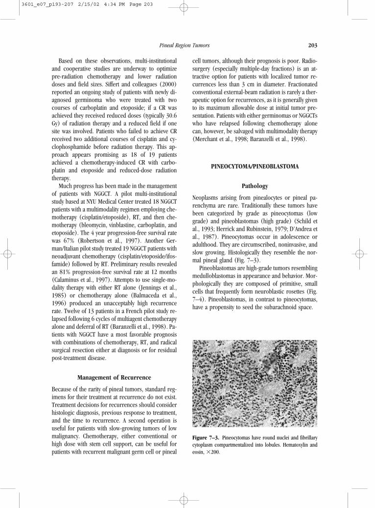
Neoplasms arising from pinealocytes or pineal pa-renchyma are rare. Traditionally these tumors havebeen categorized by grade as pineocytomas (lowgrade) and pineoblastomas (high grade) (Schild etal., 1993; Herrick and Rubinstein, 1979; D’Andrea etal., 1987). Pineocytomas occur in adolescence oradulthood. They are circumscribed, noninvasive, andslow growing. Histologically they resemble the nor-mal pineal gland (Fig. 7–3).
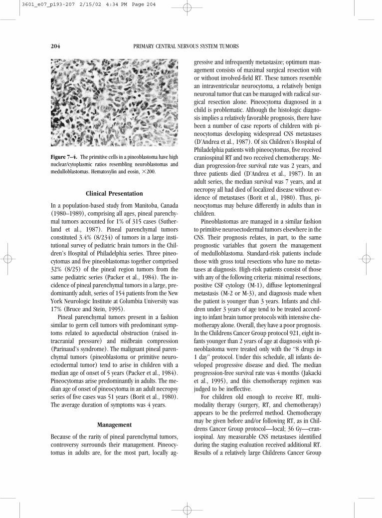
Pineoblastomas are high-grade tumors resemblingmedulloblastomas in appearance and behavior. Mor-phologically they are composed of primitive, smallcells that frequently form neuroblastic rosettes (Fig.7–4). Pineoblastomas, in contrast to pineocytomas,have a propensity to seed the subarachnoid space.
Pineal Region Tumors 203
Figure 7–3. Pineocytomas have round nuclei and fibrillarycytoplasm compartmentalized into lobules. Hematoxylin andeosin, �200.
3601_e07_p193-207 2/15/02 4:34 PM Page 203
Clinical Presentation
In a population-based study from Manitoba, Canada(1980–1989), comprising all ages, pineal parenchy-mal tumors accounted for 1% of 315 cases (Suther-land et al., 1987). Pineal parenchymal tumors constituted 3.4% (8/234) of tumors in a large insti-tutional survey of pediatric brain tumors in the Chil-dren’s Hospital of Philadelphia series. Three pineo-cytomas and five pineoblastomas together comprised32% (8/25) of the pineal region tumors from thesame pediatric series (Packer et al., 1984). The in-cidence of pineal parenchymal tumors in a large, pre-dominantly adult, series of 154 patients from the NewYork Neurologic Institute at Columbia University was17% (Bruce and Stein, 1995).
Pineal parenchymal tumors present in a fashionsimilar to germ cell tumors with predominant symp-toms related to aqueductal obstruction (raised in-tracranial pressure) and midbrain compression(Parinaud’s syndrome). The malignant pineal paren-chymal tumors (pineoblastoma or primitive neuro-ectodermal tumor) tend to arise in children with amedian age of onset of 5 years (Packer et al., 1984).Pineocytomas arise predominantly in adults. The me-dian age of onset of pineocytoma in an adult necropsyseries of five cases was 51 years (Borit et al., 1980).The average duration of symptoms was 4 years.
Management
Because of the rarity of pineal parenchymal tumors,controversy surrounds their management. Pineocy-tomas in adults are, for the most part, locally ag-
gressive and infrequently metastasize; optimum man-agement consists of maximal surgical resection withor without involved-field RT. These tumors resemblean intraventricular neurocytoma, a relatively benignneuronal tumor that can be managed with radical sur-gical resection alone. Pineocytoma diagnosed in achild is problematic. Although the histologic diagno-sis implies a relatively favorable prognosis, there havebeen a number of case reports of children with pi-neocytomas developing widespread CNS metastases(D’Andrea et al., 1987). Of six Children’s Hospital ofPhiladelphia patients with pineocytomas, five receivedcraniospinal RT and two received chemotherapy. Me-dian progression-free survival rate was 2 years, andthree patients died (D’Andrea et al., 1987). In anadult series, the median survival was 7 years, and atnecropsy all had died of localized disease without ev-idence of metastases (Borit et al., 1980). Thus, pi-neocytomas may behave differently in adults than inchildren.
Pineoblastomas are managed in a similar fashionto primitive neuroectodermal tumors elsewhere in theCNS. Their prognosis relates, in part, to the sameprognostic variables that govern the management of medulloblastoma. Standard-risk patients includethose with gross total resections who have no metas-tases at diagnosis. High-risk patients consist of thosewith any of the following criteria: minimal resections,positive CSF cytology (M-1), diffuse leptomeningealmetastasis (M-2 or M-3), and diagnosis made whenthe patient is younger than 3 years. Infants and chil-dren under 3 years of age tend to be treated accord-ing to infant brain tumor protocols with intensive che-motherapy alone. Overall, they have a poor prognosis.In the Childrens Cancer Group protocol 921, eight in-fants younger than 2 years of age at diagnosis with pi-neoblastoma were treated only with the “8 drugs in1 day” protocol. Under this schedule, all infants de-veloped progressive disease and died. The medianprogression-free survival rate was 4 months (Jakackiet al., 1995), and this chemotherapy regimen wasjudged to be ineffective.
For children old enough to receive RT, multi-modality therapy (surgery, RT, and chemotherapy)appears to be the preferred method. Chemotherapymay be given before and/or following RT, as in Chil-drens Cancer Group protocol—local; 36 Gy—cran-iospinal. Any measurable CNS metastases identifiedduring the staging evaluation received additional RT.Results of a relatively large Childrens Cancer Group
204 PRIMARY CENTRAL NERVOUS SYSTEM TUMORS
Figure 7–4. The primitive cells in a pineoblastoma have highnuclear/cytoplasmic ratios resembling neuroblastomas andmedulloblastomas. Hematoxylin and eosin, �200.
3601_e07_p193-207 2/15/02 4:34 PM Page 204
series (15 patients) treated on this randomized pro-tocol are more favorable, with a 3-year progression-free survival rate of 61% (Jakacki et al., 1995). It isdifficult to obtain comparative survival data fromother large studies. Most prior publications are casereports, and patients were managed in a variety ofways (i.e., involved-field RT, craniospinal RT, and RTplus chemotherapy). Craniospinal RT appears to bethe most effective treatment to date, and the addedbenefit of adjuvant chemotherapy can only be sur-mised from data concerning medulloblastoma clini-cal trials.
ASTROCYTOMAS
Astrocytomas are discussed briefly because they areone of the most common pineal region tumors. As-trocytomas are not pineal parenchymal tumors butarise in adjacent regions of the thalamus or midbrain.They are managed similarly to those arising elsewherein the CNS, except they are more surgically inacces-sible. Treatment guidelines are based on histologicgrading. Because radical resections are difficult toperform in this region, the prognosis for high-gradefibrillary astrocytomas of the pineal region is poorerthan the already dismal prognosis for high-grade as-trocytomas elsewhere in the brain. The prognosis oflow-grade astrocytomas is variable. The diffuse low-grade fibrillary astrocytoma behaves similarly to abrain stem glioma with a 3 year survival of less than5% (Reardon et al., 1998). Low-grade juvenile pilo-cytic astrocytomas have a more favorable outcome.One variant, the tectal or midbrain glioma, appearsto have a protracted course and may be managed withventriculoperitoneal shunt and deferral of surgery orRT (Squires et al., 1994). For children or adults withradiographic and clinical progression from a mid-brain or thalamic juvenile pilocytic astrocytoma, che-motherapy or RT may produce long-term palliation(Packer et al., 1993; Petronio et al., 1991).
CONCLUSIONS
It is clear that patients with symptomatic pineal re-gion tumors benefit from a surgical procedure to es-tablish a histologic diagnosis and control raised in-tracranial pressure. Modern multimodality therapyfor these uncommon malignant tumors should involveparticipation in cooperative group clinical trials.
REFERENCES
Allen JC. 1987. Management of primary intracranial germ celltumors of childhood. Pediatr Neurosci 13:152–157.
Allen JC. 1991. Controversies in the management of intracra-nial germ cell tumors. Neurol Clin 9:441–452.
Allen JC, DaRosso RC, Donahue B, Nirenberg A. 1994. A phaseII trial of preirradiation carboplatin in newly diagnosed ger-minoma of the central nervous system. Cancer 74:940–944.
Allen JC, Kim JH, Packer RJ. 1987. Neoadjuvant chemotherapyfor newly diagnosed CNS germ cell tumors of the centralnervous system. J Neurosurg 67:65–70.
Balmaceda C, Heller G, Rosenblum M, et al. 1996. Chemo-therapy without irradiation—a novel approach for newlydiagnosed CNS germ cell tumors: results of an internationalcooperative trial. The First International Central NervousSystem Germ Cell Tumor Study. J Clin Oncol 14:2908–2915.
Bamberg M, Kortmann R, Calaminus G, et al. 1999. Radiationtherapy for intracranial germinoma: results of the Germancooperative prospective trials MAKEI 83/86/89, J Clin On-col 17:2585–2592.
Baranzelli MC, Patte C, Bouffet E, et al. 1998. An attempt totreat pediatric intracranial �FP and �HCG secreting germcell tumors with chemotherapy alone. SFOP experience with18 cases. Societe Francaise d’Oncologie Pediatrique. J Neu-rooncol 37:229–239.
Bjornsson J, Scheithauer BW, Leech RW. 1986. Primary in-tracranial choriocarcinoma: a case report. Clin Neu-ropathol 5:242–245.
Bjornsson J, Scheithauer BW, Okazaki H, Leech RW. 1985. In-tracranial germ cell tumors: pathobiological and immuno-histochemical aspects of 70 cases. J Neuropathol Exp Neu-rol 44:32–46.
Borit A, Blackwood W, Mair WG, et al. 1980. The separationof pineocytoma from pineoblastoma. Cancer 45:1408–1418.
Bouffet E, Baranzelli MC, Patte C, et al. 1999. Combined treat-ment modality for intracranial germinomas: results of amulticentre SFOP experience. Societe Francaise d’Oncolo-gie Pediatrique. Br J Cancer 79:1199–1204.
Bruce J, Stein BM. 1993. Supracerebellar approaches in thepineal region. In: Apuzzo ML (ed), Brain Surgery: Compli-cation Avoidance and Management. New York: Churchill-Livingstone, 511 pp.
Bruce J, Stein BM. 1995. Surgical management of pineal re-gion tumors. Acta Neurochir (Wien) 134:130–135.
Buckner JC, Peethambaram PP, Smithson WA, et al. 1999.Phase II trial of primary chemotherapy followed by re-duced-dose radiation for CNS germ cell tumors. J Clin On-col 17:933–940.
Calaminus G, Andreussi L, Garre M, Kortmann RD, Schober R,Gobel U. 1997. Secreting germ cell tumors of the centralnervous system (CNS). First results of the cooperative German/Italian pilot study (CNS sGCT). Klin Pediatr 209:222–227.
Chandrasoma PT, Smith MM, Apuzzo MLJ. 1989. Stereotacticbiopsy in the diagnosis of brain masses: comparison of re-sults of biopsy and resected surgical specimen. Neuro-surgery 24:160–165.
Chik K, Li C, Shing MM, Leung T, Yuen PM. 1999. Intracranial
Pineal Region Tumors 205
3601_e07_p193-207 2/15/02 4:34 PM Page 205
germ cell tumors in children with and without Down syn-drome. J Pediatr Hematol Oncol 21:149–151.
D’Andrea AD, Packer RJ, Rorke LB, et al. 1987. Pineocytomasof childhood: a reappraisal of natural history and responseto therapy. Cancer 59:1353–1357.
Dattoli MJ, Newall J. 1990. Radiation therapy for intracranialgerminoma: the case for limited volume treatment. Int J Ra-diat Oncol Biol Phys 19:429–433.
Dearnaley DP, A’Hern RP, Whittaker S, Bloom HJ. 1990. Pinealand CNS germ cell tumors: Royal Marsden Hospital expe-rience 1962–1987. Int J Radiat Oncol Biol Phys 18:773–781.
Dempsey PK, Kondziolka D, Lunsford LD. 1992. Stereotacticdiagnosis and treatment of pineal region tumors and vas-cular malformations. Acta Neurochir (Wien) 116:14–22.
Edwards MS, Hudgins RJ, Wilson CB, Levin VA, Wara WM.1988. Pineal region tumors in children. J Neurosurg 68:689–697.
Felix I, Becker LE. 1990. Intracranial germ cell tumors in chil-dren: an immunohistochemical and electron microscopicstudy. Pediatr Neurosurg 16:156.
Ferrer E, Santamarta D, Garcia-Fructuoso G, Caral L, Rumia J.1997. Neuroendoscopic management of pineal region tu-mors. Acta Neurochir (Wien) 139:12–20.
Fuller BG, Kapp DS, Cox R. 1994. Radiation therapy of pinealregion tumors: 25 new cases and a review of 208 previ-ously reported cases. Int J Radiat Oncol Biol Phys 28:229–245.
Glanzmann C, Seelentag W. 1989. Radiotherapy for tumors ofthe pineal region and suprasellar germinomas. RadiotherOncol 16:31–40.
Gonzalez-Crussi F. 1979. The human yolk sac and yolk sac(endodermal sinus) tumors: a review. Perspect PediatrPathol 5:179–215.
Goodman R. 1993. Magnetic resonance imaging-directedstereotactic endoscopic third ventriculostomy. Neuro-surgery 32:1043–1047.
Haddock MG, Schild SE, Scheithauer BW, Schomberg PJ. 1997.Radiation therapy for histologically confirmed primary cen-tral nervous system germinoma. Int J Radiat Oncol Biol Phys38:915–923.
Hardenbergh PH, Golden J, Billet A, et al. 1997. Intracranialgerminoma: the case for lower dose radiation therapy. IntJ Radiat Oncol Biol Phys 39:419–426.
Herrick MK, Rubinstein LJ. 1979. The cytological differentiat-ing potential of pineal parenchymal neoplasms (truepinealomas): a clinicopathologic study of 28 tumors. Brain102:289–320.
Hoffman HJ, Yoshida M, Becker LE, et al. 1983. Pineal regiontumors in childhood: experience at the Hospital for SickChildren. In: Humphreys RP (ed), Concepts in PediatricNeurosurgery, vol 4. Basel: S. Karger, 360 pp.
Jakacki RI, Zeltzer PM, Boyett JM. 1995. Survival and prog-nostic factors following radiation and/or chemotherapy forprimitive neuroectodermal tumors of the pineal region ininfants and children: a report of the Childrens CancerGroup. J Clin Oncol 13:1377–1383.
Jenkin D, Berry M, Chan H, et al. 1990. Pineal region germi-nomas in childhood: treatment considerations. Int J RadiatOncol Biol Phys 18:541–545.
Jennings MT, Gelman R, Hochberg F. 1985. Intracranial germ-
cell tumors: natural history and pathogenesis. J Neurosurg63:155–167.
Lapras C, Patet JD. 1987. Controversies, techniques and strate-gies for pineal tumor surgery. In: Apuzzo MLJ (ed), Sur-gery of the Third Ventricle. Baltimore: Williams & Wilkins,p 649.
Legido A, Packer RJ, Sutton LN, et al. 1989. Suprasellar ger-minomas in childhood. A reappraisal. Cancer 63:340–344.
Linggood RM, Chapman PH. 1992. Pineal tumors. J Neuroon-col 12:85–91.
Linstadt D, Wara WM, Edwards MS, Hudgins RJ, Sheline GE.1988. Radiotherapy of primary intracranial germinomas:the case against routine craniospinal irradiation. Int J Ra-diat Oncol Biol Phys 15:291–297.
Malogolowkin MH, Mahour GH, Krailo M, Ortega JA. 1990.Germ cell tumors in infancy and childhood: a 45-year ex-perience. Pediatr Pathol 10:231–241.
Merchant TE, Davis BJ, Sheldon JM, Leibel SA. 1998. Radia-tion therapy for relapsed CNS germinoma after primary che-motherapy. J Clin Oncol 16:204–209.
Midgley AR, Pierce GB. 1962. Immnohistochemical localiza-tion of human chorionic gonadotropin. Proc Soc Exp BiolMed 115:289–294.
Neuwelt EA, Glasberg M, Frenkel E, Clark WK. 1979. Malig-nant pineal region tumors. J Neurosurg 51:597–607.
Packer R, Lange B, Ater J. 1993. Carboplatin and vincristinefor reccurent and newly diagnosed low-grade gliomas ofchildhood. J Clin Oncol 11:850–856.
Packer RJ, Sutton LN, Rosenstock JG, et al. 1984. Pineal re-gion tumors of childhood. Pediatrics 74:97–102.
Peragut JC, Dupard T, Graziani N, Sedan R. 1987. [Preventionof risk in stereotaxic biopsy of various tumors of the pinealregion. Apropos of 3 cases]. Neurochirurgie 33:23–27.
Petronio J, Edwards MS, Prados M, et al. 1991. Managementof chiasmal and hypothalamic gliomas of infancy and child-hood with chemotherapy. J Neurosurg 74:701–708.
Reardon D, Gajjar A, Sanford R, et al. 1998. Bithalamic in-volvement predicts poor outcome among children with thal-amic glial tumors. Pediatr Neurosurg 29:29–35.
Rich TA, Cassady JR, Strand RD, Winston KR. 1985. Radiationtherapy for pineal and suprasellar germ cell tumors. Can-cer 55:932–940.
Robertson PL, DaRosso RC, Allen JC. 1997. Improved prog-nosis of intracranial non-germinoma germ cell tumors withmultimodality therapy. J Neurooncol 32:71–80.
Rosenfeld JV, Murphy MA, Chow CW. 1990. Implantation me-tastasis of pineoblastoma after stereotactic biopsy. Case re-port. J Neurosurg 73:287–290.
Rubinstein LJ. 1970. Histological classification of pineal tu-mors. Tumors of the pineal region. In: Tumors of the Cen-tral Nervous System. Washington, DC: Armed Forces Insti-tute of Pathology, 269 pp.
Rueda-Pedraza ME, Heifetz SA, Sesterhenn IA, Clark GB. 1987.Primary intracranial germ cell tumors in the first two-decades of life: a clinical, light-microscopic, and immuno-histochemical analysis of 54 cases. Perspect Pediatr Pathol10:160–207.
Russell DS, Rubinstein LJ. 1989. Tumors and tumor-like le-sions of maldevelopmental origin. In: Russell DS, Rubin-stein LJ (eds), Pathology of Tumours of the Nervous Sys-tem. Baltimore: Williams & Wilkins, 664 pp.
206 PRIMARY CENTRAL NERVOUS SYSTEM TUMORS
3601_e07_p193-207 2/15/02 4:34 PM Page 206
Sano K. 1976. Diagnosis and treatment of tumours in the pinealregion. Acta Neurochir (Wien) 34:153–157.
Schild SE, Scheithauer BW, Schomburg PJ, et al. 1993. Pinealparenchymal tumors. Clinical, pathologic and therapeuticaspects. Cancer 72:870–880.
Siffert J, Robertson P, Jakacki R, Hukin J, Domnahue B, Ve-lasquez L, et al. 2000. Multiagent neoadjuvant chemother-apy followed by reduced dose radiotherapy for newly diag-nosed central nervous system germinoma: preliminaryresults of a multi-institutional phase II pilot. J Neurooncol2:294.
Squires LA, Allen JC, Abbott R, Epstein FJ. 1994. Focal tectal
tumors: management and prognosis. Neurology 44:953–956.
Stein BM, Bruce JN. 1992. Surgical management of pineal regiontumors (honored guest lecture). Clin Neurosurg 39:509–532.
Sung DI, Harisliadis L, Chang CH. 1978. Midline pineal tumorsand suprasellar germinomas: highly curable by irradiation.Radiology 128:745–751.
Sutherland GR, Florell R, Louw D, Choi NW, Sims AA, et al.1987. Epidemiology of primary intracranial neoplasms inManitoba, Canada. Can J Neurol Sci 14:586–592.
Tien RD, Barkovich AJ, Edwards MS. 1990. MR imaging ofpineal tumors. Am J Roentgenol 155:143–151.
Pineal Region Tumors 207
3601_e07_p193-207 2/15/02 4:34 PM Page 207