physiotherapy - keralascert.kerala.gov.in/images/2016/vhse/biology/18-physiotherapy.pdf · •...
TRANSCRIPT
Government of KeralaDepartment of Education
State Council of Educational Research and Training (SCERT),KERALA
2016
VOCATIONAL HIGHER SECONDARYEDUCATION (VHSE)
SECOND YEAR
PHYSIOTHERAPY
Reference Book
Prepared by :
State Council of Educational Research and Training (SCERT)Poojappura, Thiruvananthapuram 695012, Kerala
Website : www.scertkerala.gov.in e-mail : [email protected] : 0471 - 2341883, Fax : 0471 - 2341869
Typesetting and Layout : SCERT© Department of Education, Government of Kerala
List of Contributors
Academic Co-ordinatorDr.Gopalakrishnan N
Research Officer, SCERT
Participants1. MUBEEN.K.T
MSc (Psy), MPT (Neurological Physiotherapy)Vocational teacher in PhysiotherapyRahmaniya VHSS,Calicut
2. SEENATH.P.BPTDWH, FMTFI, CMTVocational instructor in physiotherapyRahmaniya VHSS, Calicut
3. RUSILABI. C.M.BPTPhysiotherapist Rahmaniya VHSS, Calicut.
Experts
1. LETO J.JOELMPT (musculoskeletal and spots)Assistant professorAWH special colleg, Kallai, Calicut.
2. MITHEN DEV M.P.T ORTHONavajeevan college of physiotherapyMarivanos vidyanagarNalanchira, Trivandrum.
ForewordDear Learners,This book is intended to serve as a ready reference for learners ofvocational higher secondary schools. It offers suggested guidelinesfor the transaction of the concepts highlighted in the course content.It is expected that the learners achieve significant learning outcomesat the end of the course as envisaged in the curriculum if it is followedproperly. In the context of the Right- based approach, quality education has tobe ensured for all learners. The learner community of Vocational HigherSecondary Education in Kerala should be empowered by providingthem with the best education that strengthens their competences tobecome innovative entrepreneurs who contribute to the knowledgesociety. The change of course names, modular approach adopted forthe organisation of course content, work-based pedagogy and theoutcome focused assessment approach paved the way for achievingthe vision of Vocational Higher Secondary Education in Kerala. Therevised curriculum helps to equip the learners with multiple skillsmatching technological advancements and to produce skilledworkforce for meeting the demands of the emerging industries andservice sectors with national and global orientation. The revisedcurriculum attempts to enhance knowledge, skills and attitudes bygiving higher priority and space for the learners to make discussionsin small groups, and activities requiring hands-on experience.The SCERT appreciates the hard work and sincere co-operation ofthe contributors of this book that includes subject experts, industrialistsand the teachers of Vocational Higher Secondary Schools. Thedevelopment of this reference book has been a joint venture of theState Council of Educational Research and Training (SCERT) andthe Directorate of Vocational Higher Secondary Education.The SCERT welcomes constructive criticism and creative suggestionsfor the improvement of the book.
With regards,
Dr. P. A. FathimaDirector
SCERT, Kerala
Reference Book
4
1. About the course ---------------------------------------------------- 5
2. Major skills- with sub skills --------------------------------------- 5
3. Syllabus -------------------------------------------------------------- 7
4. Unit in detailed Module - 3 ---------------------------------------- 8
5. Over view of module - 3 ----------------------------------------- 12
6. List of Practical --------------------------------------------------- 78
7. Over view of module - 4 ----------------------------------------- 81
8. List of Practical -------------------------------------------------- 160
9. List of Standard Equipments tools ----------------------------- 166
10. Reference --------------------------------------------------------- 168
CONTENTS
Physiotherapy
5
ABOUT THE COURSEVocational higher secondary education is a project of central government to addressthe issues of unemployment. In vocational higher secondary students are guided todifferent work stream after their secondary education. Among the different vocationalcourses introduced, physiotherapy has great relevance.
In history it has its roots in the after effects of second world war-in the rehabilitationafter injuries .It gradually developed in to a separate branch of medicine. In the past,medical and surgical care was directed only towards the treatment of diseases.Now it is well recognized that attention needs to be paid also towards the aftereffects of diseases. It is widely accepted that by using physiotherapy treatmenttechniques and facilities recovery of a patient can be accelerated and the period ofconvalescence can be reduced and in certain cases permanent disability resultingfrom disease can be prevented or minimized. "A Physician add life to years and aPhysiotherapist add years to life"
This new concept has been the key factor to the development of a new branch ofmedicine i.e. rehabilitation medicine. Today physiotherapy has emerged as a popularbranch of Allied Health science. Its progress and rapid expansion had made it achief component of Rehabilitation medicine.
It has also a major role in sports medicine and physical fitness.
Vocational higher secondary course in Physiotherapy will enable the students toacquire various skills needed to assist a Physiotherapist in the different specialties. Italso offers an opportunity for higher studies in various medical, paramedical andallied health science courses.
Major skills (with Sub-skills)Module - 3Major skillLearner will be able to assist physiotherapist in electro therapy and exercise therapyclinics
Sub skillThe learner:
• Give general instructions to the clients prior to treatment.
• Prepare the patient for treatment
• Arrange the treatment set up
Reference Book
6
• Checking, cleaning and maintenance of physiotherapy equipments
• Assist the physiotherapist in providing the following treatment
- Passive movements
- Resisted exercise
- Frenkel’s exercise
- Relaxation techniques
- Stretching exercise
- Strengthening exercise
- Gait training
- Donning and doffing of orthotics.
Module - 4Major skills
Student will achieve basic knowledge about various neurological, orthopaedic,paediatric and geriatric conditions seen in physiotherapy clinic. Learner will be ableto assist the physiotherapist in clinical or hospital set up in treatment of variousconditions
Sub skill
The learner:
• Provide assistance in treating patients with various neurological conditionslike hemiplegia, paraplegia, parkinsonism etc
• Provide assistance in treating patients with various orthopaedic conditionslike fracture, arthritis etc
• Provide assistance in treating children with cerebral palsy
• Positioning of hemiplegic patients
• Transfer of patients from bed to chair and viseversa
• Provide assistance in treating geriatric patients

Physiotherapy
7
SYLLABUSModule - 3
Unit No. Unit Name Periods
3.1 Introduction to physiotherapy 25
3.2 Exercise therapy 150
3.3 Human locomotion (gait) 25
3.4 Hydro therapy 15
3.5 Suspension therapy 5
3.6 Relaxation 5
3.7 Chest physiotherapy 10
3.8 Electrotherapy 105
Total 340
Module - 4
Unit No Unit Name Periods
4.1 Introduction to Neurological Physiotherapy 90
4.2 Introduction to orthopaedic Physiotherapy 105
4.3 Introduction of paediatric Conditions and relevanceof physiotherapy 40
4.4 Introduction of physiotherapy in surgical condition 40
45 Orthotics and prosthetics 25
4.6 Geriatric physiotherapy 40
Total 340
Reference Book
8
MODULE-3
FUNDAMENTALS OF PHYSIOTHERAPUTICS
UNIT-3.1 .Introduction to physiotherapy• Definition
• Branches of physiotherapy
• Scope of physiotherapy
• General goals of physiotherapy
UNIT-3.2. Exercise TherapyUNIT-3.2.1.Introduction to Exercise Therapy
UNIT-3. 2.2 Movement and Types of Movements
1. Active movement
• Free exercise
• Assisted exercise
• Assisted-resisted exercise
• Resisted exercise
2. Passive movement
3. Reflex movements
UNIT 3.2.3 Fundamental Positions
• Standing
• kneeling,
• sitting
• lying
• hanging
UNIT3. 2.4 Derived Positions
• Standing
• kneeling,
• sitting

Physiotherapy
9
• lying
• hanging
UNIT3. 2.5 Manual Muscle Test of Major Muscle Group
• Shoulder-flexors ,extensors, abductors and adductors
• Elbow-flexors and extensors
• Hip-flexors ,extensors, abductors and adductors
• Knee-flexors and extensors
UNIT 3.2.6 Range of Joint Motion of Major Joints
• Shoulder-flexion ,extension, abduction and adduction
• Elbow-flexion and extension
• Hip-flexion ,extension, abduction and adduction
• Knee-flexion and extention
UNIT 3. 2.7 Posture
• Definition and Types (Good and Bad posture)
UNIT-3. 2.8 Exercise therapy equipments
• Suspension unit
• Static cycle
• Treadmill
• Quadriceps table
• Shoulder wheel
• Hand exerciser
• Medicine ball,
• Swiss ball
• Abduction ladder.
• Parallel bar
• Tilt table
• Wheelchair
• Crutches

Reference Book
10
UNIT-3.3 Human locomotion (GAIT)UNIT-3.3.1 Definition and Gait cycle
UNIT-3.3.2 Pathological gait
• Circumductory gait
• Scissoring gait
• Festinent gait
• Ataxic gait
• High stepping gait
• Antalgic gait
UNIT 3.3.3 Gait training
UNIT3. 3.4 Transfer techniques
• Wheel chair to bed
• Bed to wheelchair
UNIT-3.4 Hydrotherapy• Pooltherapy
• Contrast bath
• Cryotherapy
UNIT-3.5 Suspension therapy• Types and indication
UNIT-3.6 Relaxation• Definition, types and techniques
UNIT-3.7 Chest physiotherapy• Postural drainage
• Breathing exercise
UNIT-3.8 ElectrotherapyUNIT 3.8.1 Introduction of electrotherapy
UNIT 3.8.2 Classification -low frequency, medium frequency and highfrequency equipments
Physiotherapy
11
UNIT 3.8.3 Preparation and safety measures for patient andequipments
UNIT3. 8.4 Thermotherapy
• Physiological and Therapeutic effects of heat.
• Application of heating modality
• Indication and contraindication of the electrotherapy equipments.
UNIT 8.5 Brief descriptions of electrotherapy equipments-
• IRR-Infra red radiation
• US-Ultra sound
• SWD-Short Wave Diathermy
• IFT-Interferential therapy
• TENS-Transcutaneous Electrical Nerve Stimulator
• Cold pack
• Hot pack
• Wax bath
• Laser
Reference Book
12
PART –BOVER VIEW OF MODULE 3
Students have previous ideas about Anatomy and Physiology of Human body. Inthis module the student will be able to familiarize with commonly used physiotherapyequipments in exercise therapy and electro therapy. This module introduces differenttypes of equipments used in physiotherapy clinics with emphasise on its usage,indications and maintenance. The student also acquires the skills in preparation ofthe client for treatment and basic physiotherapeutic techniques including basicexercises, posture, gait training and transfer techniques.
After completion of this module, student will be able to assist the physiotherapist inusing various electrotherapy and exercise therapy equipments in a clinical setup.Also student will be able to assist the therapist in Suspension therapy, Hydrotherapyand Chest physiotherapy .
UNIT - 1INTRODUCTION TO PHYSIOTHERAPY
IntroductionPhysiotherapy is a form of treatment carried through the medium of physical forcessuch as heat, electricity, mechanical pressure and mechanical forces. Thus, inphysiotherapy heat,electrical current, water, soft tissue manipulation, and exerciseswith or without resistance are utilized.Physiotherapy is ahealth profession whoseprimary purpose is the promotion of optimal human health by the application ofscientific principles to prevent,assess,correct or alleviate acute or prolongedmovement dysfunction.
Learning outcomesThe learner ;
• Achieve basic knowledge about the physiotherapy
• Define physiotherapy
• Understand the branches of physiotherapy
• Understand the scope of physiotherapy
• Identify the the different goals of physiotherapy
Physiotherapy
13
Concepts (Detailing )Definition
Physical therapy provides services to individuals and populations to develop maintainand restore maximum movement and functional ability throughout the lifespan. Thisincludes providing services in circumstances where movement and function arethreatened by ageing, injury, pain, diseases, disorders, conditions or environmentalfactors. Functional movement is central to what it means to be healthy.
Physical therapy is concerned with identifying and maximizing quality of life andmovement potential within the spheres of promotion, prevention, treatment/intervention, habilitation and rehabilitation. This encompasses physical, psychological,emotional, and social wellbeing. Physical therapy involves the interaction betweenthe physical therapist, patients/clients, other health professionals, families, care giversand communities in a process where movement potential is assessed and goals areagreed upon, using knowledge and skills unique to physical therapists(WCPT)
Branches of physiotherapyPhysiotherapy is a complex specialty, as it deals with all disciplines of medicine.Development in this field has resulted in a variety of sub-specialization-.
1. Musculo-skeletal physiotherapy
The Musculo - Skeletal Physiotherapy is a branch, where application of physiotherapyis involved in the treatment of physical ailments concerned with structures surroundingthe joint region such as Bones, Muscles, Ligaments and Bursa. Some of the conditionsdealt in this branch are: Muscle pain, strain, Muscle tear Joint stiffness, Fracture,Ligament strain, sprain, tear, Inability to walk, Inflammation of tendons and bursa,Joint pain, poor posture, Joint inflammation in case of osteoarthritis or rheumatoidarthritis etc
2. Cardio-Thoracic Physiotherapy:
Cardio - Thoracic Physiotherapy is concerned with the care of physical fitness ofthe heart and lungs. This treatment specializes in clearing away any chest secretionsso as to enable and help an individual to breathe more normally, and help in adequateoxygen supply to the healing wound and the body. Some of the conditions treatedunder this branch are: Asthma, Increased chest secretions other respiratory infectionsetc
Reference Book
14
3. Neurological Physiotherapy
Neurological Physiotherapy is concerned with the treatment arising from the problemsin brain, Nerves of the brain, Spinal cord and nerves of the spinal cord and meninges.Some of the conditions which physiotherapy is essential part of treatment are: Stroke,ataxia, cerebral palsy, Spinal cord injury, peripheral nerve injuries, etc.
4. Physiotherapy in Rehabilitation
Physiotherapy and Rehabilitation goes hand in hand. Rehabilitation is nothing butbringing back a physically disabled individual to near normal condition by using hismaximum existing capacities. Rehabilitation in physiotherapy involves training andretraining of physical activity in a physically disabled individual.Different types ofrehabilitations, where physiotherapy is involved: Stroke Rehabilitation, Geriatric/Old age Rehabilitation, Cardiac Rehabilitation and Amputee (person who has lostthe limb) Rehabilitation
5. Physiotherapy in Obstetrics
Physiotherapy in obstetrics is concerned with postural care and physical fitness ofwomen during pregnancy and after child birth.
6. Sports Physiotherapy
Sports physiotherapy is specialized for sports persons. Guidance in sport activitieslike techniques of warming up period, cooling down period, guidance in liftingtechniques and also treatment of sport injuries are dealt. Some of the conditionsunder this are: Any Muscle spasm (pain and tightness of muscle),Muscle strain,Ligament sprain, tear, all other sports injuries.
7. Physiotherapy in fitness and postural care
This branch is concerned with guidance and care for physical fitness, good posturalcare and Body muscle built. Some of the conditions dealt are: Obesity/ Overweight,Poor Posture (Hunch back), Good muscle built etc
8. Pediatric Physiotherapy
This branch of physiotherapy treatment is for the children born with physical disabilitiesand also for the children undergoing any surgery, requiring physiotherapy care .Someof the conditions dealt here are: Club foot- before and after a surgery, Respiratoryinfections, Fractures in children, Cerebral palsy etc.

Physiotherapy
15
Scope of physiotherapyThe scope of physical therapy practice is dynamic and responsive to patient/clientand societal health needs. With the development of knowledge and technologicaladvances, periodic review is required to ensure that scope of practice reflects thelatest evidence base and continues to be consistent with current health needs.Research is continually providing new evidence upon which future practice will bebuilt.
The scope of physical therapy servicesPhysical therapy is an essential part of the health and community/welfare servicesdelivery system. Physical therapists practice independently of other health care/serviceproviders and also within interdisciplinary rehabilitation/habilitation programmes toprevent, gain, maintain or restore optimal function and quality of life in individualswith loss and disorders of movement.
Physical therapists are guided by their own code of ethical principles. Thus, theymay be concerned with any of the following purposes:
1. Promoting the health and well-being of individuals and the general public/soci-ety-Emphasizing the importance of physical activity and exercise
2. Preventing impairments, activity limitations, participatory restrictions and dis-abilities in individuals at risk of altered movement behaviors due to health ormedically related factors, socio-economic stressors, environmental factors andlifestyle factors
3. Providing interventions/treatment to restore integrity of body systems essentialto movement, maximize function and recuperation, minimize incapacity, andenhance the Quality of life, independent living and workability in individualsand groups of individuals with altered movement behaviors resulting from im-pairments, activity limitations, Participatory restrictions and disabilities modify-ing environmental, home and work access and barriers to ensure full participa-tion in one’s normal and expected societal roles .Physical therapists may alsocontribute to the development of local, national and international Health poli-cies and public health strategies.
General goals of physiotherapyThe goals of physiotherapy are to improve mobility and strength, to relieve pain andto restore physical function. This enables you to resume your regular activities ofdaily living including work, school, recreational activities, home-making and/or self-

Reference Book
16
care. If the injury or disease is severe in nature, the goal is to assist you in returningto your maximal function.
Physiotherapy provides benefits by use of a non-invasive approach. The Goals ofPhysiotherapy are to facilitate and maximize recovery and functional mobility followinga musculoskeletal or neurological injury. Physiotherapy reaches these goals through:
2. Reduction of pain.
3. Acceleration in healing of injured.
4. Maintenance or restoration of normal range of motion in affected joints.
5. Prevention of fibrosis or soft tissue contractures in injured, weak or para-lyzed limbs.
6. Prevention of disuse atrophy during healing phases of neurological andmusculoskeletal insults.
7. Improvement of strength and function in weak and paralyzed muscles.
8. Improved performance and quality of movement.
9. Positive psychological effects maximizing both pet and owner’s well-be-ing.
10. Provision of individualized home care program to maximize functional mo-bility and prevent injury.
Practical detailingVisit to a physiotherapy clinic and make a report about the visit
Assessment activitiesBrain storming
Collection
Discussion
Field visit report
TE Questions1. Identify the role of physiotherapist in rehabilitation team?
2. List out the different branches of physiotherapy
Physiotherapy
17
UNIT 3.2. EXERCISE THERAPYIntroductionThis unit provides a general idea about the exercise therapy including therapeuticmovements, fundamental and derived positions, posture, gait ,gait training and transfertechniques, and different types of equipments used in exercise therapy.
Learning outcomesThe learner;
• Achieve Basic knowledge about exercise therapy
• Identify the different types of movements
• Demonstrate the fundamental positions
• Demonstrate the derived positions
• Check the muscle strength of major muscle group
• Measure the range of motion of major joints
• Identify good and bad posture
• Identify commonly used exercise therapy equipments
Concepts (Detailing)EXERCISE THERAPY
Exercise therapy is a means of accelerating the patient’s recovery from injuries anddiseases which have altered his normal way of living. The aims of exercise therapy
1. To promote activity and minimize the effects of inactivity.
2. To increase the normal range of motion.
3. To strength the weak muscles.
4. To improve the performance in daily activities
Movement and types of movementsMOVEMENT is a fundamental characteristic of all animal life and the mean bywhich the organism adapts itself to the demands made up on it by the environment inwhich it lives.
Movement used in treatment may be classified as follows;

Reference Book
18
1. Active movementsa. Voluntary: Voluntary movement-movement performed or controlled by the vol-
untary action of muscle working in opposition to an external force.
b. Involuntary reflex
Classification of Active movement
i) Assisted exercise
When muscle strength or co ordination is in adequate to perform a movement anexternal force is applied to compensate for the deficiency. When the force exertedon one of the body livers by muscular action is insufficient for the production orcontrol of movement, an external force may be added to augment it. This externalforce must be applied in the direction of the muscle action but not necessarily at thesame point.
ii) Free exercise
The working muscles are subject only to the forces of gravity acting up on the partmoved or stabilized. Free exercise are those which are performed by the patient’sown muscular efforts without the assistance or resistance of any external force,other than that of gravity.
(iii) Assisted-Resisted exercise
This type of exercise constitutes a combination of assistance and resistance duringsingle movement
(iv) Resisted exercise
An external force may be applied to the body livers to oppose the force of musclecontraction. Tension is increased within the muscle by the opposing force and themuscle respond by an increase in their power and hypertrophy.
There are five factors which contribute to the development of muscular efficiency-Power, Endurance, Volume, Speed of contraction and Co-ordination
Resistance force: The physiotherapist, patient, Weights, Weight and pulley circuit,Springs and other elastic structures, Substances which are malleable and Water
2. Passive movementsPassive movements
These movements are produced by an external force during muscular inactivity orwhen muscular activity is voluntarily reduced as much as possible to permit movement.
Physiotherapy
19
Classification of Passive Movement
1- Relaxed Passive Movements, including accessory movements.
2. Forced Passive movement.
3. Continuous Passive movement.
1. Relaxed Passive Movement
These are movements performed accurately, rhythmical and smoothly by thephysiotherapist through available range of motion( according to anatomy of joints) .The movements are performed in the same range and direction as active movements.The joint is moved through the free range and within the limits of pain.
Principles Of Relaxed Passive Movement
1. Relaxation: The selection of a suitable starting position ensures comfort andsupport, for both patient and physiotherapist through the movement.
2. Fixation: Good fixation for the proximal and distal joint by the physiotherapistto ensure that the movement is localized to the movable joint.
3. Support: Full and comfortable support is given to the part to be moved, sothat the patient has confidence and will remain relaxed.
4. Traction: The fixation of the bone proximal to the joint providing an opposingforce to a sustained pull on the distal bone. Traction is thought to facilitate themovement by reducing inter- articular friction.
5. Range of movement : The range of movement is done in painless range toavoid spasm in the surrounding muscles.
6. Speed and Duration:As it is essential that relaxation is maintained throughoutthe movement, the speed must be slow and rhythmical, with suitable repititionsof the movement.
Effects And Uses Of Relaxed Passive Movements1. Maintain range of motion and prevent formation of adhesions.
2. Maintain the physiological properties of the muscle (extensibility, elasticity, etc.)and prevent shortening and contracture.
3. Help in preserving and maintain the memory of the movement pattern bystimulating the kinaesthetic receptors.
4. The mechanical pressure resulted from the stretching of the thin walled vesselswhich passing across the moved joint will assist the venous and lymphatic return( improving circulation).
Reference Book
20
5. Can be used in training of relaxation as the rhythmic continuous passivemovements can have a soothing effect and induce further relaxation and sleep.
6. Improving sense of position and sense of movement.
Indications Of Relaxed Passive Movement
1. In cases of paralysis, patient who is confined in bed for a long time orcomplete rest on bed.
2. When there is an inflammatory reaction and active movement is painful.
3. When the patient in coma.
4. In relaxation as a factor helping to reduce spasm in group of muscles.
I. Contra-Indications Of Relaxed Passive Movement
1. Unhealed fracture, recent fracture, at the site of fracture.
2. At site of effusion or swelling.
3. Immediately following surgical procedure to tendon, ligaments, joint capsule.
4. Immediately after recent tear to ligament, tendon.
5. When a bony block limits joint motion e.g. myosities ossificans.
6. Recent injuries
7. Severe muscle weakness.
8. Acute inflammation or infection as arthritis, osteomylities etc
II. Forced Passive Movement
An exercise performed on a subject by a partner who exerts an external force notonly to produce a passive movement, but also to increase the range of movement ofa joint. The partner presses the joint into its end-position (i.e. end of range), whilethe subject’s muscles that normally carry out the movements are completely relaxed.There is a danger of overextension beyond the range of movement and damage tothe joint if the exercise is not carried out carefully.
III. Continuous Passive Motion (CPM)
A continuous passive motion device maintains movement of a joint after limb sparingsurgery. This device is usually called a CPM. Continuous movement limits stiffnessand pain. It is very important to keep joints moving following surgery so that motionwill not become limited. The CPM will move the involved leg through its full range ofmotion. It is only used in bed, but can be used while relaxing, eating, or sleeping.
Physiotherapy
21
Fundamental positionsThe postures from which movement is initiated are known as Starting Position andthey may be either active or passive in character. There are five basic or fundamentalstarting position and all the others are derived from them,i.e. standing, kneeling,sitting, lying and hanging. Equilibrium and stability is maintained in these positions bya balance of forces acting upon the body, and when the force of muscular contractionis used for this purpose the contraction is isometric. The strength and distribution ofthis contraction is normally controlled by a series of reflexes known collectively asthe Postural Reflexes but, during the learning process of new patterns of posture,voluntary effort may be required.
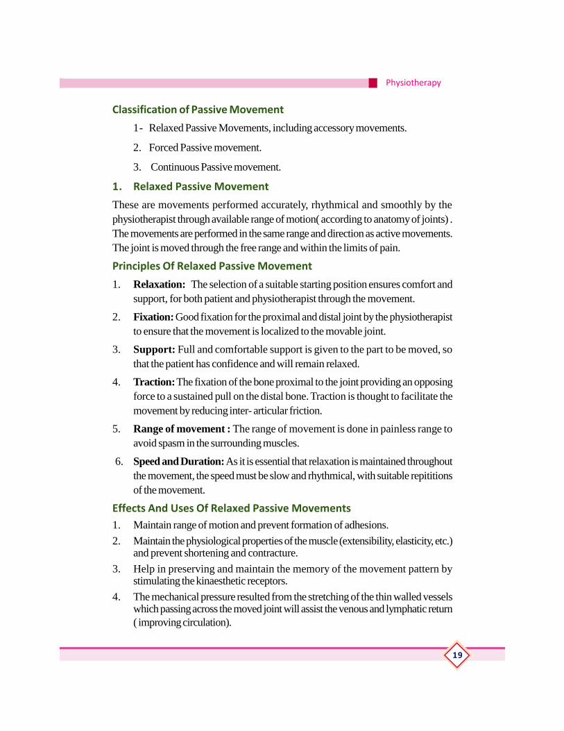
1. Standing
This is the most difficult of the fundamental positions to maintain, as the whole bodymust be balanced and stabilized in correct alignment on a small base by thecoordinated work of many muscle groups. The position may be described as follows:
Starting Positions
(i) The heels are together and on the same line, the toes slightly apart (so that theangle between the feet does not exceed 45°).
(ii) The knees are together and straight.
(iii) The hips are extended and laterally rotated slightly.
(iv) The pelvis is balanced on the femoral heads.
(v) The spine is stretched to its maximum length.
(vi) The vertex is thrust upwards, the ears are level and the eyeslook straight forwards.
(vii) The shoulders are down and back.
(viii) The arms hang loosely to the sides, palms facing inwardstowards the body.
It is usually preferable to modify the position of the legs to that inwhich the heels are slightly apart and the inner borders of the feetare parallel, as this is the natural functional position of the footwhen it is used as a lever to propel the body forwards.
Reference Book
22
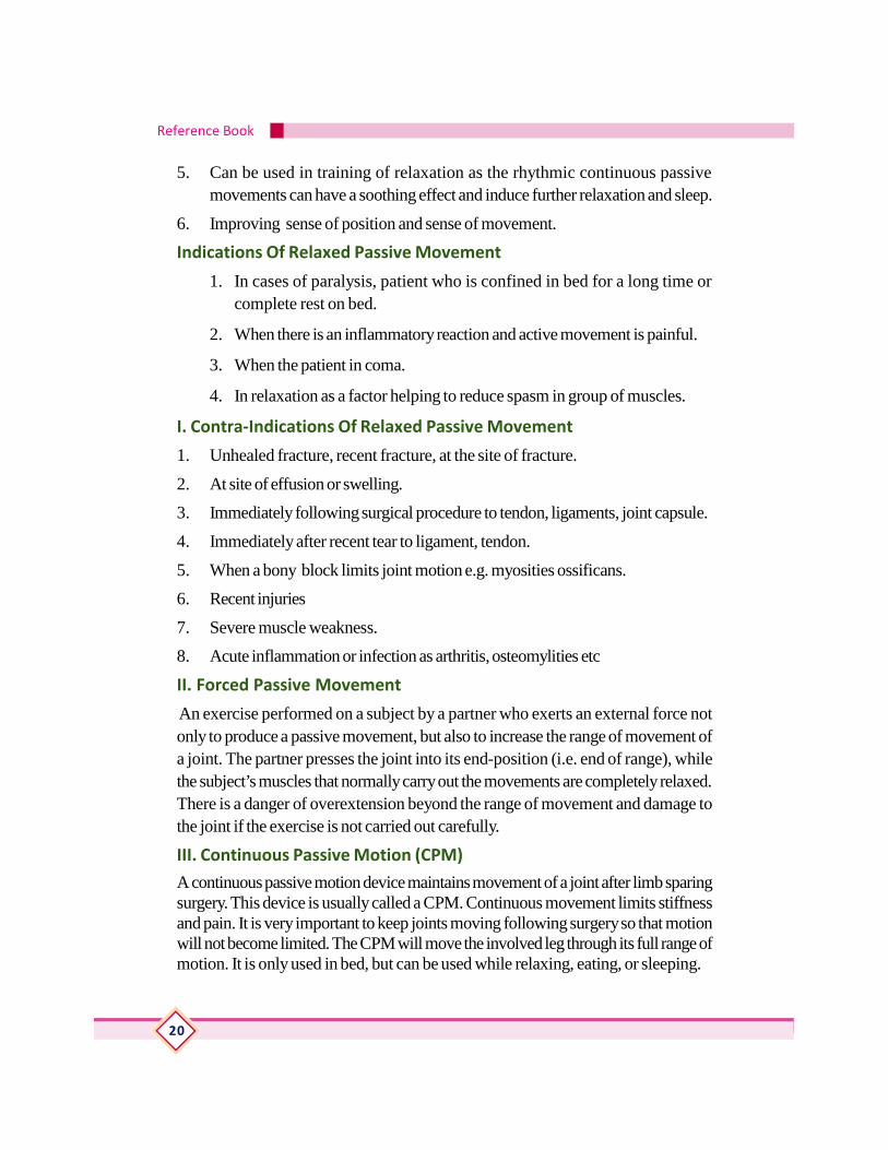
2. Kneeling
The body is supported on the knees which maybe together or slightlyapart. The lower leg rests on thefloor with the feet plantaflexed or, ifa plinth is used, thefeet may be in the mid-position over the edge.The restof the body is held as in standing.
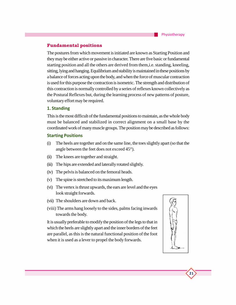
3. Sitting
The position is taken on a chair or stool, the height and width ofwhich allow the thighs to the fully supported and the hips and knees tobe flexed to a right angle. The knees are apart sufficiently to allow thefemora to be paralleland the feet rest on the floor with the heels verticallybelow the knees.
4. Lying
This is the easiest of the fundamental positions as the body can be completelysupported in the supine position and is as stable as is possible.
.
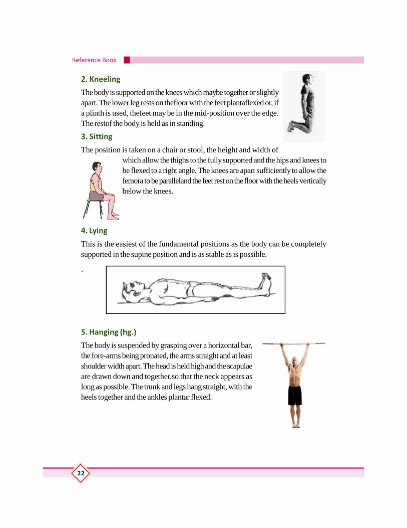
5. Hanging (hg.)
The body is suspended by grasping over a horizontal bar,the fore-arms being pronated, the arms straight and at leastshoulder width apart. The head is held high and the scapulaeare drawn down and together,so that the neck appears aslong as possible. The trunk and legs hang straight, with theheels together and the ankles plantar flexed.

Physiotherapy
23
Unit 3.2.4 Derived positionsDerived positions are positions used by modification of the arms, legs or trunk ineach of fundamental
position. The aims of derived positions are:
1. To increase or decrease the base of support.
2. To rise or lower the center of gravity (COG).
3. To gain local or general relaxation.
4. To gain fixation and good control of specific area.
5. To increase or decrease the muscle work required to maintain the position.
6. To increase or decrease the leverage.
Derived position from standing
By alteration of the legs
Achieved by change in the shaper size of the base.
1. Toe standing.
2. Stride standing.
3. Walk standing.
4. Half standing. Standing with trunk alteration
5. Stoop standing.
6. Lax stoop standing
Position Derived from Kneeling
1. Half Kneeling
2. Kneel Sitting
3. Prone Kneeling
Positions Derived from Sitting
1. Stride sitting.
2. Ride Sitting.
3. Crook Sitting.
4. Long sitting.
Reference Book
24
5. Cross Sitting
6. Side sitting.
7- High sitting
Position Derived from Lying
1. Crook Lying
2- Crook Lying with Pelvis Lifted
3. Half Lying
4. Prone Lying
5. Leg Prone Lying
6. Side Lying
7. Sit Lying
Position Derived from Hanging
Full Hanging
Manual muscle test of major muscle groupManual muscle testing is a procedure for the evaluation of the function and strengthof individual muscles and muscle groups based on the effective performance of amovement in relation to the forces of gravity and manual resistance.
Manual Muscle Test Grades
Grade 5 (Normal; 100%)- The patient or subject can complete the whole rangeof motion (movement) against gravity with maximum resistance appliedby the therapist at end-of-range.
Grade 4 (Good;75%)- The subject can complete the whole range of motionagainst gravity with moderate resistance applied by the physical therapist(PT) at end-range. Testing the uninvolved limb should always beconsidered to know whether you are applying too much force on theinvolved limb or not.
Grade 3+ (Fair+)- The patient can complete the motion against gravity with minimalresistance applied by the examiner at end-range.
Grade 3 (Fair;50%) - The patient can only complete the range of motion againstgravity. When external (outside) force is applied by the PT, the patientgives way.
Physiotherapy
25
Grade 2 (Poor;25%) - Your patient cannot perform the movement against gravity.But patient can do complete range of motion when pull of gravity iseliminated. No resistance is applied.
Grade 1 (Trace)-Patient is not able to move the joint even with gravity eliminated.However, closer examination by the therapist would reveal slight musclecontraction through palpation.
Grade 0 (Zero; No trace) - No contraction is noticed, even with physical therapist’spalpation (touch).
Note: Always tell your client about what procedure you are going to perform andwhat you are going to obtain from that procedure. Remember to check the uninvolvedside first and be consistent on where you apply the resistance. Also, instruct yourclient not to hold his or her breath as you apply force or resistance
Muscle testing procedures of major musclesDELTOID
Position of Patient:
With the patient sitting the elbow should be flexed to indicate the neutral position ofrotation.
Position of Therapist:
The therapist should stand at test side of patient. Place pressure against the dorsalsurface of the distal end of the humerus.
Test: The patient is to maintain the arm in abduction against gravity.
Instructions to Patient: “I am going to push down and I want you to resist me.Keep your arm up as I push down.”
BICEPSPosition of Patient:
With the patient sitting the elbow is flexed at a right angle, with forearm in supination.
Position of Therapist:
The therapist should stand in front of and at testing side of patient. The hand givingresistance is contoured over the flexor surface of the forearm just proximal to thewrist. The other hand is applied to the humerus to provide a counterforce.
Test: Patient flexes elbow against your applied force. If the biceps/brachialis areweak the patient will pronate the forearm before flexing the elbow.
Instructions to Patient: “Bend your elbow, hold it. Don’t let me pull it down.”
Reference Book
26
WRIST EXTENSORS
Position of Patient: With the patient sitting with the elbow and forearm supportedand forearm is in full pronation with the fingers flexed.
Position of Therapist: The therapist should stand or sit at a diagonal in front of thepatient.
Test: Support the patients forearm under the wrist while the other hand used forresistance is placed over the dorsal surface of the metacarpals. Do not permit fullextension of the fingers.
Sample Instructions to Patient: “Bring your wrist up, hold it. Don’t let me push itdown.”
QUADRICEPS
Position of Patient: With the patient sitting with the trunk approximately perpendicularto the floor, the leg is extended – but not locked – in extension at the knee. Trunkextension is allowed only if significant hamstring tightness precludes assuming therecommended testing position.
Position of Therapist: The therapist stands at the side of the tested limb and thetesting hand is placed over anterior surface of distal leg just above the ankle. Theother hand is placed under the distal thigh.
Test: The patient extends the knee through available range of motion but do notallow knee to “lock” into extension during the test.
Instructions to Patient: “Straighten your knee and hold it, don’t let me bend it.”
ANKLE DORSIFLEXORS
Position of Patient: With the patient sitting, the knee is flexed at 90°.
Position of Therapist: The therapist sits in front of testing limb and supports theleg just above the posterior aspect of the ankle joint.
Test: The patient dorsiflexes the ankle joint without extending the great toe. Pressureis applied on the dorsum of the foot (in the direction of plantar flexion and eversion).
Instructions to Patient: “Pull your foot up to the ceiling.”
GLUTEUS MEDIUS
Position of Patient: With the patient side lying, the test leg is superior to the supportingleg. The test limb is slightly extended beyond midline and pelvis is rotated slightlyforward. The supporting leg is flexed for stability.
Physiotherapy
27
Position of Therapist: The therapist stands behind patient and test hand is placedon lateral surface of knee or at the ankle and the other hand is just proximal togreater trochanter of femur.
Test: The patient abducts against the applied resistance without flexing or rotatingthe hip in either direction. Resistance by examiner is straight and downward.
Instructions to Patient: “I am going to push down on your leg and I want you to resistme.”
GLUTEUS MAXIMUS
Position of Patient: With the patient prone the knee is flexed to 90°.
Position of Therapist: The therapist stands on the side to be tested and the testinghand is placed over the posterior thigh just above the knee. The other hand maystabilize the pelvis at the upper buttocks.
Test: The patient extends the hip through the available range of motion
Instructions to Patient: knee flexion at 90°. Resistance is applied directly downwardtoward the floor.
Instructions to Patient ;”Lift your leg towards the ceiling and keep your knee bent.”
TRICEPS
Position of Patient: Supine or prone
Fixation
Shoulder abducted to 90°, nuetral with regard to rotation, & supported betweenthe shoulder & elbow by the table
Test
Extension of the elbow joint (to just short of full extension)Pressure against theforearm in the direction of flexion
Hamstrings
Range of Joint Motion of Major JointsRange of Motion is the measurement of movement around a specific joint or bodypart. To measure range of motion, physical therapists most commonly use agoniometer, which is an instrument that measures angle at a joint. Goniometers showdegrees of an angle from zero to 180 or 360 degrees and are available in differentshapes and sizes for the unique joints in the human body, example, when using agoniometer to measure knee flexion, the center of the tool will be at the side view of
Reference Book
28
the knee joint, and the arms of the goniometer are aligned in the center of the longbones above and below the knee. As the knee is bent or flexed the movable armsprovide a measure of the degree of movement.
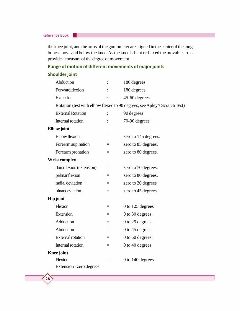
Range of motion of different movements of major joints
Shoulder joint
Abduction : 180 degrees
Forward flexion : 180 degrees
Extension : 45-60 degrees
Rotation (test with elbow flexed to 90 degrees, see Apley’s Scratch Test)
External Rotation : 90 degrees
Internal rotation : 70-90 degrees
Elbow joint
Elbow flexion = zero to 145 degrees.
Forearm supination = zero to 85 degrees.
Forearm pronation = zero to 80 degrees.
Wrist complex
dorsiflexion (extension) = zero to 70 degrees.
palmar flexion = zero to 80 degrees.
radial deviation = zero to 20 degrees
ulnar deviation = zero to 45 degrees.
Hip joint
Flexion = 0 to 125 degrees
Extension = 0 to 30 degrees.
Adduction = 0 to 25 degrees.
Abduction = 0 to 45 degrees.
External rotation = 0 to 60 degrees.
Internal rotation = 0 to 40 degrees.
Knee joint
Flexion = 0 to 140 degrees.
Extension - zero degrees
Physiotherapy
29
Ankle joint
Neutral position is with foot at 90 degrees to ankle.
Dorsiflexion is0 to 20 degrees;
Plantar flexion is 0 to 45 degrees.
Posture
Posture is the position the body adopts in response to the effects of gravity. It is theway you hold yourself in sitting, standing or lying down.
POSTURE
Posture is a term used to describe a position of the body or the arrangements ofbody parts relative to one another. Ideal postures are those assumed to perform anactivity in the most efficient manner utilizing the least amount of energy. All activitybegins with a posture and ends with a posture. The relationships between bodyparts can be controlled voluntarily but to do this would require too much concentration.During normal functioning one’s postures and adjustments to postures are automaticand occur quickly.
Good posture :Good posture is the attitude which, is assumed by Body parts tomaintain stability and balance with minimum effort and least strain during supportiveand non supportive positions.
CHARACTERISTICS OF GOOD POSTURE (Prerequisites of good posture):For good posture to be maintained the following must be obtained: The ability tomaintain ‘the body upright in good and erect position with less energy. The ability tomaintain balance in upright position via keeping the line of gravity near the center ofthe base of support.
Effects of good posture: Maintaining good posture has its values in different bodyfunctions and systems. The effects of good posture include:
1. Helps the muscles in the body to be unloaded and relaxed.
2. Improves respiratory and circulatory efficiency.
3. Prevents unnecessary strain and fatigue.
4. Decreases the incidence of diseases resulting from bad posture.
5. Improves the subject’s state; mentally or psychologically.:Poor posture: Poor posture is a position resulting from any deviation from ideallyaligned erect posture (good posture).
Causes of poor posture: Poor posture may occur due to:
Reference Book
30
1- Defects in: Joints: such as stiffness or immobilization. •Bones: such as shorteningor deformity. •Muscles: such as weakness, paralysis or contracture. •Vision orhearing.
2- Bad habits: either from early childhood or from occupational positions. Examplefor that is the workers that perform jobs which require continuous flexed positionof the trunk.
3- Pain, fatigue or bad psychological state.
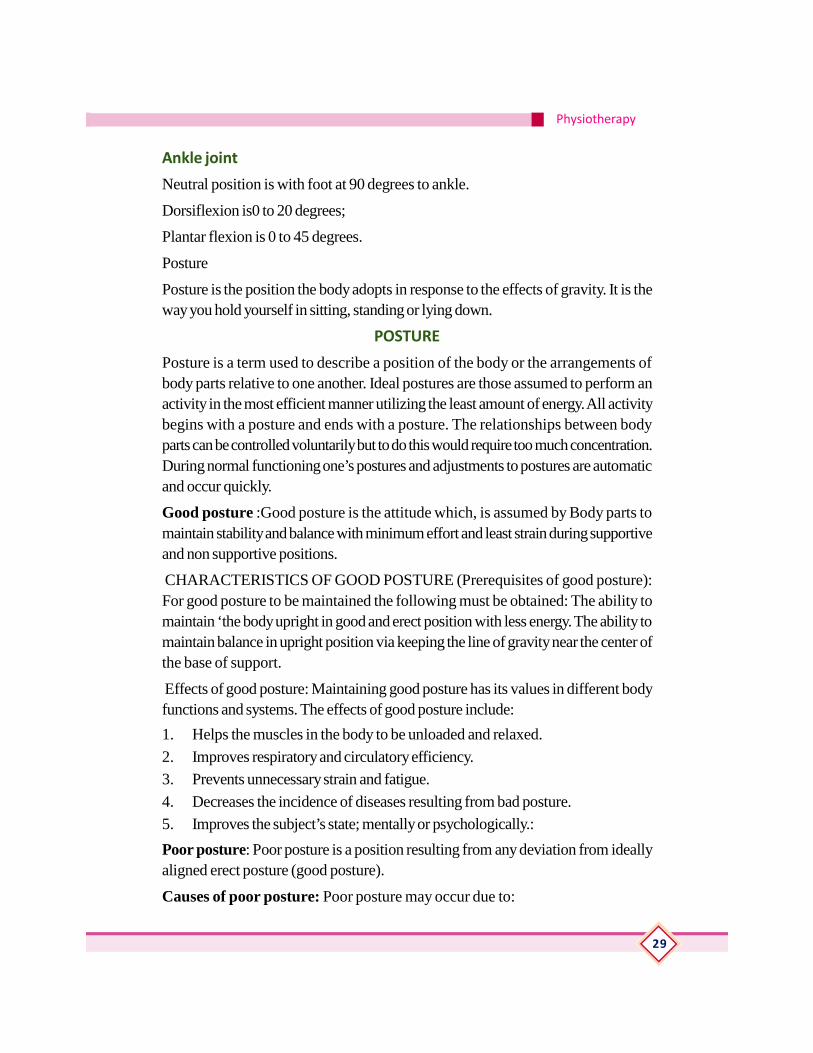
Effects of poor posture:
Poor posture causes deviations from the normally aligned posture.As a result the body functions are altered and this may lead todysfunction and diseases.
Poor posture may cause one or more of the following dysfunction:
1. Secondary deformities and compensatory postural defects.
2. Easy fatigability and high energy expenditure.
3. Decrease both respiratory and circulatory efficiency.
4. Pain, bad cosmetic appearance and psychologicaldisturbances.
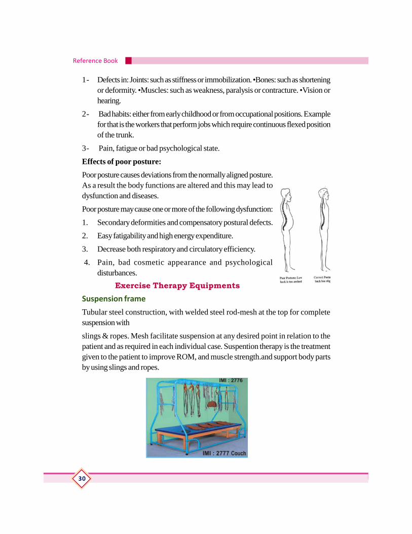
Exercise Therapy EquipmentsSuspension frame
Tubular steel construction, with welded steel rod-mesh at the top for completesuspension with
slings & ropes. Mesh facilitate suspension at any desired point in relation to thepatient and as required in each individual case. Suspention therapy is the treatmentgiven to the patient to improve ROM, and muscle strength.and support body partsby using slings and ropes.
Physiotherapy
31
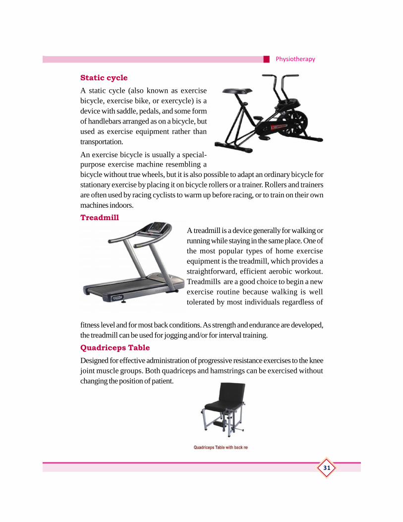
Static cycleA static cycle (also known as exercisebicycle, exercise bike, or exercycle) is adevice with saddle, pedals, and some formof handlebars arranged as on a bicycle, butused as exercise equipment rather thantransportation.
An exercise bicycle is usually a special-purpose exercise machine resembling abicycle without true wheels, but it is also possible to adapt an ordinary bicycle forstationary exercise by placing it on bicycle rollers or a trainer. Rollers and trainersare often used by racing cyclists to warm up before racing, or to train on their ownmachines indoors.
TreadmillA treadmill is a device generally for walking orrunning while staying in the same place. One ofthe most popular types of home exerciseequipment is the treadmill, which provides astraightforward, efficient aerobic workout.Treadmills are a good choice to begin a newexercise routine because walking is welltolerated by most individuals regardless of
fitness level and for most back conditions. As strength and endurance are developed,the treadmill can be used for jogging and/or for interval training.
Quadriceps TableDesigned for effective administration of progressive resistance exercises to the kneejoint muscle groups. Both quadriceps and hamstrings can be exercised withoutchanging the position of patient.
Reference Book
32
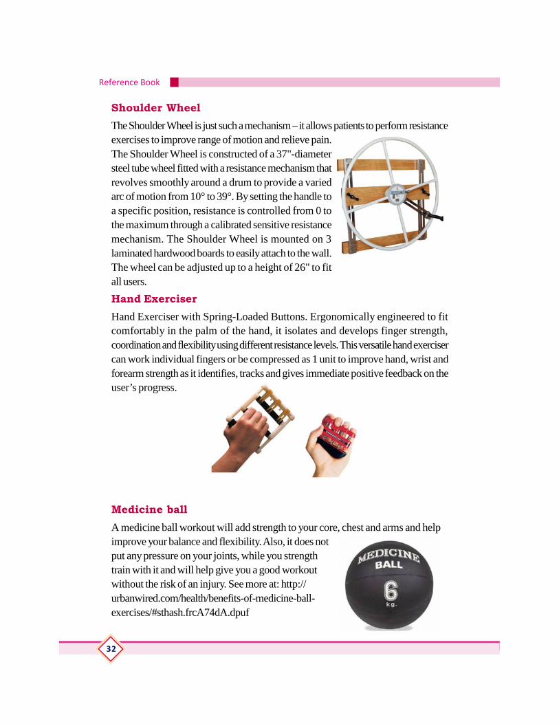
Shoulder WheelThe Shoulder Wheel is just such a mechanism – it allows patients to perform resistanceexercises to improve range of motion and relieve pain.The Shoulder Wheel is constructed of a 37"-diametersteel tube wheel fitted with a resistance mechanism thatrevolves smoothly around a drum to provide a variedarc of motion from 10° to 39°. By setting the handle toa specific position, resistance is controlled from 0 tothe maximum through a calibrated sensitive resistancemechanism. The Shoulder Wheel is mounted on 3laminated hardwood boards to easily attach to the wall.The wheel can be adjusted up to a height of 26" to fitall users.
Hand ExerciserHand Exerciser with Spring-Loaded Buttons. Ergonomically engineered to fitcomfortably in the palm of the hand, it isolates and develops finger strength,coordination and flexibility using different resistance levels. This versatile hand exercisercan work individual fingers or be compressed as 1 unit to improve hand, wrist andforearm strength as it identifies, tracks and gives immediate positive feedback on theuser’s progress.
Medicine ballA medicine ball workout will add strength to your core, chest and arms and helpimprove your balance and flexibility. Also, it does notput any pressure on your joints, while you strengthtrain with it and will help give you a good workoutwithout the risk of an injury. See more at: http://urbanwired.com/health/benefits-of-medicine-ball-exercises/#sthash.frcA74dA.dpuf

Physiotherapy
33
Swiss ballAn exercise ball, also known as a Swiss Ball, is a ball constructed of soft elastic witha diameter of approximately 35 to 85 centimeters (14 to 34 inches) and filled withair. The air pressure is changed by removing a valve stem and either filling with air orletting the ball deflate. It is most often used in physical therapy, athletic training andexercise. It can also be used for weight training. The ball, while often referred to asa Swiss ball, is also known by a number of different names, including balance ball,birth ball, body ball, ball, fitness ball, gym ball, gymnastic ball, physioball, pilatesball, Pezzi ball, sports ball, stability ball, Swedish ball, therapy ball, or yoga ball.
Abduction ladderShoulder Abduction Ladder - Ladder made of polished hard woodhaving Thirty numbered steps for shoulder abduction exercises. It isused to increase the range of motion of the shoulder.
Parallel barParallel bars are used in rehabilitation or therapy centers to helppatients regain strength, range of motion, balance and independenceas they learn to walk again or regain coordination. Parallel bars can
be used for patients who need to learn howto walk again, increase their range of motion, regain muscleand learn how to walk without the use of a walker, crutchesor cane. Parallel bars allow patients to slowly take theirfirst steps with the support of their hands and with theguidance of a physical therapist. As patients are able toregain their mobility, they will be able to transition fromusing the parallel bars to walking on the ground withoutthe assistance of a person or device.

Reference Book
34
Tilt tablePhysical therapists use tilt tables to provide early weight bearing experiences forpatients too weak to stand on their own. Tilt tables alsohelp patients with orthostatic hypotension—a significantdrop in blood pressure that occurs when they move from aprone to a sitting position. To use a tilt table, the patient lieson top of the table on her back. The physical therapistsecures the safety straps around the patient, then slowlyelevates the table, putting the patient into a standing position,while monitoring her blood pressure and heart ratethroughout the treatment.Tilt table treatments can preventosteoporosis via weight bearing, as well as anklecontractures, blood clots, pulmonary embolism and other bed rest complicationsfor the hospitalized patient.
WheelchairA wheelchair is a chair with wheels. The device comes in variations allowing eithermanual propulsion by the seated occupant turningthe rear wheels by hand, or electric propulsion bymotors. There are often handles behind the seatto allow it to be pushed by another person.Wheelchairs are used by people forwhom walking is difficult or impossible dueto illness, injury, or disability.
CrutchesA crutch is a mobility aid that transfers weight fromthe legs to the upper body. It is often used for people who cannot use their legs tosupport their weight, for reasons ranging from short-term injuries to lifelong disabilities.
Axillary crutches, elbow crutches and forearm crutches
Physiotherapy
35
Practical (Detailing)1. Free exercises
a. Upper limb
b. Lowerlimb
c. trunk
2. Resisted exercise
a. Shouher joint
i. Flexers and Extensors
ii. Adductors abductors
b. Elbow joint
i. Flexors
ii. Extensors
c. Wrist
i. Flexors
ii. Extensors
iii. Ulnar and radial deviators
d. Hand
i. Finger flexors and extensors
e. Hip joint
i. Flexors and extensors
ii. Abductors and adductors
f. Knee
i. Flexors and extensors
g. Ankle
i. Dorsi and plantar flexors
3. Passive movement
a. Shoulder girdle
b. Shoulder joint
c. Elbow joint
d. Wrist and fingers
e. Hip joint
Reference Book
36
f. Knee joint
g. Ankle
4. Fundamental position
a. Standing
b. Sitting
c. Kneeling
d. Lying
e. hanging
5. Derived position
a. Standing
b. Sitting
c. Kneeling
d. Lying
e. Hanging
6. Grading of muscle power of major muscles
a. Shouher
i. Flexers and Extensors
ii. Adductors abductors
b. Elbow
i. Flexors
ii. Extensors
c. Wrist
i. Flexors
ii. Extensors
iii. Ulnar and radial deviators
d. Hand
i. Finger flexors and extensors
e. Hip joint
i. Flexors and extensors
ii. Abductors and adductors
f. Knee
Physiotherapy
37
i. Flexors and extensors
g. Ankle
i. Dorsi and plantar flexors
7. Range of motion of major joints
a. Shoulder joint
b. Elbow joint
c. Wrist and fingers
d. Hip joint
e. Knee joint
f. Ankle
8. Good and bad posture.
a. Postural analysis of a given modal
9. Preparation and maintenance of exercise therapy equipment.
a. Static cycle
b. Treadmill
c. Quadriceps table
d. Shoulder wheel
e. Hand exerciser
f. Medicine ball,
g. Swiss ball
h. Abduction ladder
i. Parallal bar
j. Tilt table
k. Wheel chair
l. crutches
Assessment Activities• Seminar
• Assignments
• Chart preparation
• Poster Preparation
Reference Book
38
• Quiz
• Collection of related materials from magazine
TE Questions1. Differentiate between active and passive movement
2. Write about different types of active movement
3. Write about the fundamental positions.
4. Short note on
o Muscle power grading
o Ideal posture
o Types of suspension
o Free exercise
o Principles of passive movement

Physiotherapy
39
UNIT 3.3 HUMAN LOCOMOTION (GAIT)IntroductionGait or human locomotion is very important in physiotherapy. This chapter introducesgait cycle, pathological gait patterns and gait training. Also describes about the transfertechniques.
Learning outcomesThe learner:
• To understand the steps of normal human locomotion and to identify differentpathological gait patterns
• To assist the physiotherapist in different types of gait training
• To gain expertise in the transferring of the patients from bed to chair and viseversa.
Concepts (Detailing)Definition and Gait cycle
Human gait refers to locomotion achieved through the movement of human limbs.Human gait is defined as bipedal, biphasic forward propulsion of center of gravity ofthe human body, in which there are alternate sinuous movements of different segmentsof the body with least expenditure of energy.
When walking , One foot is always in contact with the ground .There is a briefperiod of double support during the gait cycle when both feet are in contact with theground .The 4 limbs move in a diagonal reciprocal pattern Eg. – the right arm andthe left leg move forward simultaneously, followed by the left arm and right leg.When running, There is a brief moment during the gait cycle when both feet are offthe ground
Different gait patterns are characterized by differences in limb movement patterns,overall velocity, forces, kinetic and potential energy cycles, and changes in the contactwith the surface (ground, floor, etc.). Human gaits are the various ways in which ahuman can move, either naturally or as a result of specialized training.
The Gait Cycle
The gait cycle is a repetitive pattern involving steps and strides. A step is one singlestep, a stride is a whole gait cycle. The step time is the time from one foot hitting thefloor to the other foot hitting the floor. Step width can be described as the mediolateral
Reference Book
40
space between the two feet.
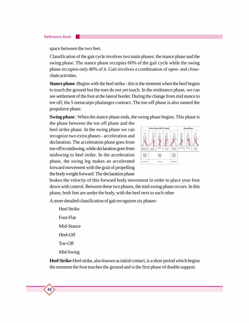
Classification of the gait cycle involves two main phases: the stance phase and theswing phase. The stance phase occupies 60% of the gait cycle while the swingphase occupies only 40% of it. Gait involves a combination of open- and close-chain activities.
Stance phase :Begins with the heel strike - this is the moment when the heel beginsto touch the ground but the toes do not yet touch. In the midstance phase, we cansee settlement of the foot at the lateral border. During the change from mid stance totoe off, the 5 metacarpo phalanges contract. The toe-off phase is also named thepropulsive phase.
Swing phase : When the stance phase ends, the swing phase begins. This phase isthe phase between the toe off phase and theheel strike phase. In the swing phase we canrecognize two extra phases - acceleration anddeclaration. The acceleration phase goes fromtoe-off to midswing, while declaration goes frommidswing to heel strike. In the accelerationphase, the swing leg makes an acceleratedforward movement with the goal of propellingthe body weight forward. The declaration phasebrakes the velocity of this forward body movement in order to place your footdown with control. Between these two phases, the mid-swing phase occurs. In thisphase, both feet are under the body, with the heel next to each other
A more detailed classification of gait recognizes six phases:
Heel Strike
Foot Flat
Mid-Stance
Heel-Off
Toe-Off
Mid-Swing
Heel Strike-Heel strike, also known as initial contact, is a short period which beginsthe moment the foot touches the ground and is the first phase of double support.

Physiotherapy
41
Foot Flat- In foot flat, or loading response phase, the body absorbs the impact ofthe foot by rolling in pronation
Midstance-In midstance the hip moves from 10° of flexion to extension bycontraction of the gluteus medius muscle.
Heel Off-Heel off begins when the heel leaves the floor. In this phase, the bodyweight is divided over the metatarsal heads.
Toe Off-.In toe-off, like the name says, the toes leave the ground
Mid Swing- In the midswing phase the hip flexes to 30° and the ankle becomesdorsiflexed due to a contraction of the tibialis anterior muscle. The knee flexes 60°but then extends approximately 30° due to contraction of the sartorius muscle.Thisextension is caused by the quadriceps muscles.
Pathological gait
Normal walking is the standard against which pathology is measured. Efficiency isoften reduced in pathology
Circumductory gait -Hemiplegic Gait- often seen as a result of a stroke. Theupper limb is in a flexed position, adducted and internally rotated at the shoulder.The lower limb is internally rotated, knee extended and the ankle inverted and plantarflexed. The gait is likely to be slow with circumduction or hip hitching of the affectedlimb to aid floor clearance
Scissoring gait-Diplegic Gait. Spasticity is normally associated with both lowerlimbs. Contractures of the adductor muscles can create a ‘scissor’ type gait with anarrowed base of support. Spasticity in the lower half of the legs results in plantarflexedankles presenting in ‘tip toe’ walking and often toe dragging. Excessive hip and kneeflexion is required to overcome this.
Festinent gait -Parkinsonian Gait often seen in Parkinson’s disease or associatedwith conditions which cause parkinsonisms. Rigidity of joints results in reduced armswing for balance. A stooped posture and flexed knees are a common presentation.
Reference Book
42
Bradykinesia causes small steps which are shuffling in presentation. There may beoccurrences of freezing or short rapid bursts of steps known as ‘festination’ andturning can be difficult
Ataxic Gait is seen as uncoordinated steps with a wide base of support and staggering/variable foot placement. This gait is associated with cerebellar disturbances and canbe seen in patients with longstanding alcohol dependency .People with‘Sensory’Disturbances may present with a sensory ataxic gait. Presentation is awide base of support, high steps and slapping of feet on the floor in order to gainsome sensory feedback. They may also need to rely on observation of foot placementand will often look at the floor during mobility due to lack of proprioception.
High stepping gait -NeuropathicGaits. High stepping gait to gain floor clearanceoften due to foot drop
Antalgic Gait Antalgic gait means that the pattern observed is a result of pain. Paincan cause a variety of responses, ranging from a lack of forceful activation up to afull blown flexor withdrawal reaction.
In antalgic gait, the problem is chronic to one degree or another and the patient isattempting tocompensate.eg : Degenerative Joint Disease(DJD) /Osteoarthritis(OA),bony or soft tissue trauma, heelspur, etc
Gait training
Gait training is a type of physical therapy that helps people improve their ability tostand and walk. One goal of gait training is preventing falls. Gait training may berecommended after an illness or injury, to help a patient regain independence inwalking, even if an adaptive device is needed. Gait training helps strengthen musclesand joints, improves balance, improves posture, develops muscle memory, buildsendurance, and retrains the legs for repetitive motion.The secondary benefit of gaittraining is a reduction of other illness, such as heart disease and osteoporosis, throughphysical activity and movement. People who choose gait training may becomehealthier overall than people who choose immobility.
Gait training is usually started as soon as possible after an injury or a healthcomplication. A doctor will prescribe it as part of physical therapy. The patient mustbe healthy enough for physical activity and movement and have joints that are strongenough to support the therapy.

Physiotherapy
43
Types of weight bearing precautions:
• Non-weight bearing : Do not apply any weight through involved leg.
• Touch down weight bearing: Allow only the ball of the foot to touch the floorfor balance purposes.
• Partial weight bearing: Allow a maximum of 50% body weight to be appliedto the involved leg.
• Weight bearing as tolerated: Allow as much weight as tolerated through theinvolved leg.
Use of CrutchesStanding up with crutches• Slide your hips forward to the edge of the chair, bed or toilet seat.
• Keep your injured/healing leg straight and your healthy leg beneath you.
• Place both crutches in the hand of your injured/ healing side, palm down.
• Your injured/healing leg should remain in front of you, bearing no weight.
• Once standing, reach with your free hand for the other crutch and place bothcrutches under your armpits with your hands firmly grasping the handgrips.
Walking with crutches• At the same time advance both crutches approximately one foot ahead while
balancing your weight on your healthy leg.
• Step forward with your injured/healing leg.
• Step forward with your healthy leg, bringing it through the crutches and pastthe injured/ healing leg.
• Move crutches forward to balance yourself.
• Remember to keep your weight on your hands, not your armpits.
• Keep in mind your crutches take up room on the sides and can get caught onthings, so keep a wide space around you.
• Look ahead to where you are walking; don’t look at your feet.
Reference Book
44
Climbing stairs with one crutch
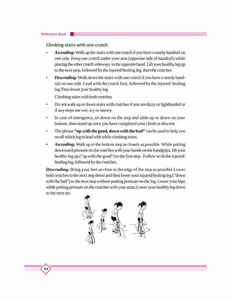
• Ascending: Walk up the stairs with one crutch if you have a sturdy handrail onone side. Keep one crutch under your arm (opposite side of handrail) whileplacing the other crutch sideways in the opposite hand. Lift your healthy leg upto the next step, followed by the injured/healing leg, then the crutches
• Descending: Walk down the stairs with one crutch if you have a sturdy hand-rail on one side. Lead with the crutch first, followed by the injured/ healingleg.Then lower your healthy leg.
Climbing stairs with both crutches
• Do not walk up or down stairs with crutches if you are dizzy or lightheaded orif any steps are wet, icy or snowy.
• In case of emergency, sit down on the step and slide up or down on yourbottom, then stand up once you have completed your climb or descent.
• The phrase “up with the good, down with the bad” can be used to help yourecall which leg to lead with while climbing stairs.
• Ascending: Walk up to the bottom step as closely as possible. While puttingdownward pressure on the crutches with your hands on the handgrips, lift yourhealthy leg up (“up with the good”) to the first step. Follow wi th the injured/healing leg, followed by the crutches.
Descending: Bring your feet as close to the edge of the step as possible.Lowerboth crutches to the next step down and then lower your injured/healing leg (“downwith the bad”) to the next step without putting pressure on the leg. Lower your hipswhile putting pressure on the crutches with your arms.Lower your healthy leg downto the next ste.

Physiotherapy
45
Transfer Techniques
Transferring in and out of your wheelchair puts higher stress on arms and shouldersthan anything else patients do on a regular basis. Learning the correct way to transferis extremely important in order to keep the arms functioning and pain-free.Everyoneneeds individualized transfer training to preserve function and avoid injury. Workwith a physical therapist to learn the best transfer technique . Transfer technique mayneed to be readjusted as years go by. If develop any problems or if livingcircumstances (e.g. pregnancy) or activities change, go back to your therapist foradvice.
Safe transfer rules and techniqueTechnique
Positioning/setup
Get as close as possible to the surface you want to move to.
Lock your wheels if transferring from a wheelchair.
Put your feet on floor (unless your therapist tells you not to).
Scoot to the edge of your chair.
Get your arm rest out of the way on the side next to the surface you are transferringto.
Lean your trunk forward.
When transferring, your head should move in the opposite direction of your hips.This is known as a head-hips relationship and can help with movement and clearingobstacles.To protect your shoulders, keep your arms as close to your body aspossible (about 30-45 degrees away from your body) while you are lifting yourweight.To protect your wrists, try to grip an edge or grab bar with your fingersrather than laying your hands flat. Keeping your hands flat and putting your weighton your palms is a dangerous position that can lead to wrist problems such as carpaltunnel syndrome down the road.
Lift-off
Make sure you are clearing obstacles (not bumping or rubbing) to avoid shearingand pressure sores.If you cannot perform the transfer in one smooth movementwhile keeping your arms close to your body, move in several small steps and/or usea transfer board.Be careful sliding across the transfer board because the motion candamage your skin. Use a pad or towel on the board when bare skin may come incontact with the board during the transfer.Alternate leading arms and direction of

Reference Book
46
transfers to keep your arm muscles balanced and reduce strain on one side.Maintainideal body weight. The more you weigh, the more weight you have to transfer andthe more stress you put on your shoulders and arms.If you are unable to perform atransfer safely or are at risk for developing arm pain, you should strongly considerusing one of the many kinds of patient lifts available.
Wheel chair to bedSteps for transferring from Wheelchair to Bed
1. Have the bed at the lowest level.
2. Park the wheelchair with the person’s strongest side next to the bed.3. Lock the wheelchair brakes and remove feet from foot rests.
4. Swing or remove foot rests from wheelchair.
5. Explain the sequence of lifting and pivoting into the wheelchair (example: on thecount of 3, I am going to help you stand up and turn to your strong side; eg rightside as in above example; and sit in the wheelchair).
6. Using the bear hug technique, ask the person to place his/her arms on yourshoulders as you place your arms around his/her trunk.
7. Bracket their feet with your feet to preventslipping.
8. Using your leg muscles, stand up and bringthe person upward in a slow steady risingmotion.
9. Seat the person on the bed
10. Assist in bring the person’s legs up onto thebed.
11. Position for comfort.
Bed to wheelchairSteps for transferring from Bed to Wheelchair
1. Remove clutter from area, including all scatter rugs
2. Discuss with the transferee, the process before and during the transfer
3. The amount of room available for transfer will dictate which side of the bed youwill be transferring from.
4. Determine if the transferee has a stronger side, as he/she will be better able toscoot to the edge of the bed on that side prior to transfer.

Physiotherapy
47
5. Position wheelchair on the transferee’s strongest side (for example if the rightside is strongest, you will be transferring from the right side of the bed.)
6. Assist person to be transferred to edge of bed and to sitting position first withfeet dangling and then with feet on floor
7. For ease of transfer, position the wheelchair next to the knee on his/her stron-gest side
8. Pull wheelchair within a foot of the person’s knee and lock the wheelchairbrakes
9. Explain the sequence of lifting and pivoting into the wheelchair (example: on thecount of 3, I am going to help you stand up and turn to your strong side; eg rightside as in above example; and sit in the wheelchair)
10. Using the bear hug technique, ask the person to place his/her arms on yourshoulders as you place your arms around his/her trunk
11. Bracket their feet with your feet to prevent slipping
12. Using your leg muscles, stand up and bringthe person upward in a slow steady risingmotion
13. If transferee is capable, have him/her reachfor the furthest wheelchair armrest
14. Pivot towards wheelchair seat, and lowerslowly
15. Attach or swing foot rests of wheelchairinto place
16. Place person’s feet onto foot rests ofwheelchair.
4. Practical (Detailing)1. Demonstration of gait and identification of its phases
a. Stance
b. swing
2. Demonstation of pathological gait
a. Cicum ductory
Reference Book
48
b. Scissoring
c. Festinant
d. Ataxic
e. High stepping
f. Antalgic
3. Crutch walking
a. Non Weight Bearing
b. Partial Weight Bearing
c. Full Weight Bearing
d. Ascending
e. Descending
4. Transfer technique
a. Wheel chair to bed
b. Bed to wheel chair
Assessment Activities• Chart preparation
• Poster Preparation
• Collection
• Model Preparation
• Practical presentation
TE Questions1. Define gait
2. Write about the phases of gait cycle
3. Short note on
• Scissoring gait
• Cicumdectory gait
Physiotherapy
49
UNIT3.4 HYDROTHERAPYIntroductionHydrotherapy is the use of water in the treatment of different conditions, includingarthritis and related rheumatic complaints. Hydrotherapy differs from swimmingbecause it involves special exercises that do in a warm-water pool. The watertemperature is usually 33–36ºC, which is warmer than a typical swimming pool.Learning OutcomesThe learner:• To gain expertise in the preparation of pool for treatment sessionand assisting the physiotherapist in the pool during treatment
• To gain expertise in the preparation of contrast bath session and assisting thephysiotherapist in treatment.
• To gain expertise in the preparation for cryo therapy treatment session andassisting the physiotherapist in treatment
Concepts (Detailing)Hydrotherapy, formerly called hydropathy, is a part of medicine and alternativemedicine, in particular of naturopathy, occupational therapy and physiotherapy, thatinvolves the use of water for pain relief and treatment. Hydrotherapy is the use ofwater to treat a disease or to maintain health. The theory behind it is that water hasmany properties that give it the ability to heal:
1. Water can store and carry heat and energy.
2. Water can dissolve other substances, such as minerals and salts.
3. Water cannot hurt you, even if you are sensitive to your surroundings.
4. Water is found in different forms, such as ice, liquid, or steam. Ice may be usedto cool, liquid is used in baths and compresses at varying pressures or tem-peratures, and steam is used in steam baths or when breathing in.
5. Water can help blood flow.
6. Water has a soothing, calming, and relaxing effect on people, whether in abath, shower, spray, or compress.
7. Exercise in water takes the weight off a painful joint while also providingresistance.
People use hydrotherapy to treat many illnesses and conditions, including acne;arthritis; colds; depression; headaches; stomach problems; joint, muscle, and nerve
Reference Book
50
problems; sleep disorders; and stress. People also use it for relaxation and to maintainhealth.You can also use hydrotherapy to reduce or relieve sudden or long-lastingpain.
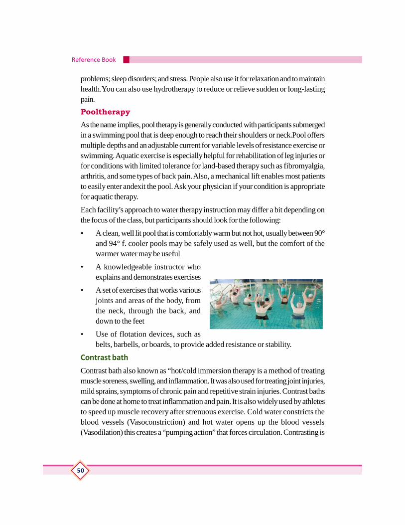
PooltherapyAs the name implies, pool therapy is generally conducted with participants submergedin a swimming pool that is deep enough to reach their shoulders or neck.Pool offersmultiple depths and an adjustable current for variable levels of resistance exercise orswimming. Aquatic exercise is especially helpful for rehabilitation of leg injuries orfor conditions with limited tolerance for land-based therapy such as fibromyalgia,arthritis, and some types of back pain. Also, a mechanical lift enables most patientsto easily enter andexit the pool. Ask your physician if your condition is appropriatefor aquatic therapy.
Each facility’s approach to water therapy instruction may differ a bit depending onthe focus of the class, but participants should look for the following:
• A clean, well lit pool that is comfortably warm but not hot, usually between 90°and 94° f. cooler pools may be safely used as well, but the comfort of thewarmer water may be useful
• A knowledgeable instructor whoexplains and demonstrates exercises
• A set of exercises that works variousjoints and areas of the body, fromthe neck, through the back, anddown to the feet
• Use of flotation devices, such asbelts, barbells, or boards, to provide added resistance or stability.
Contrast bath
Contrast bath also known as “hot/cold immersion therapy is a method of treatingmuscle soreness, swelling, and inflammation. It was also used for treating joint injuries,mild sprains, symptoms of chronic pain and repetitive strain injuries. Contrast bathscan be done at home to treat inflammation and pain. It is also widely used by athletesto speed up muscle recovery after strenuous exercise. Cold water constricts theblood vessels (Vasoconstriction) and hot water opens up the blood vessels(Vasodilation) this creates a “pumping action” that forces circulation. Contrasting is

Physiotherapy
51
also much easier and more practical and effective to apply to the limbs, so the bestcommon candidates for contrasting are: plantar fasciitis, shin splints, carpal tunnelsyndrome, tennis elbow and Achilles tendinitis.Contrast baths is used as a standardpart of many rehab facilities for treating musculoskeletal injuries, especially withrepetitive strain or overuse injuries
The Process
Before you start, you will need:
Two containers large enough and having the proper shape to allow the body part tobe treated
Water-bath thermometer
Drape sheet
Towels (for drying and to put under the basins)
Cold compress for the head (cold washcloths)
Pitcher to remove and add hot water
Means for heating water if not near a tub or sink
Ice for cold compress and cold bath
Procedure for hot and cold contrast bath
Place the body part(s) to be treated in hot water 104 degrees F. (40 degrees C.) for3-4 minutes. Apply a cold compress to the head.Then place the body part in icewater or tap water—45 to 70 degrees F. (7-21C) for 30 seconds to 1 minute).While they are in the cold, increase the temperature of the hot water each time, butdo not exceed 110 degrees F (43 degrees C). In other words, after each change,increase the contrast between the hot and cold. Make 6 to 8 changes and alwaysend with cold (except in cases of rheumatoid arthritis, in which case/in which youwould end in hot) and dry thoroughly.
As previously mentioned, warm and cool contrast baths are performed for diabeticindividuals, but should not be attempted by a layperson. Warm water is defined as92 degrees F to 100 degrees F. Cool water is between 70 and 80 degrees F. If theperson is a diabetic, do not exceed 102 degrees F (give only if the foot pulses arepalpable).

Reference Book
52
Cryotherapy
Cryotherapy in physical therapy is the application of cold for treatment of symptoms
of or problems associated with musculoskeletal conditions, such as strains and sprains.
Cryotherapy is also called cold therapy.
Health experts generally agree that cryotherapy should be the initial treatment for
acute injuries, such as muscle strains and sprains. Cold can help relieve your pain. In
addition, cold helps in vasoconstriction of blood vessels at the site of injury, thereby,
limiting bleeding and edema formation.
Side-effects of cryotherapy
Cold application is relatively safe when appropriately done. Prolonged exposure to
a cold modality may lead to frostbite injury. To avoid frostbite injury during physical
therapy treatment, the physical therapist should follow the recommended time of
cold application. If a commercial cold pack or cold hydrocollator pack is used, it
should be wrapped with several layers of towel before application.
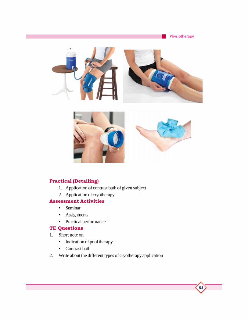
Types of Cryotherapy Application
Cryotherapy may be employed by way of any of the following: Ice massage, Ice
packs, Commercial cold gel (hydrocollator) packs, Cold sprays and Cold whirlpoolbaths.
Physiotherapy
53
Practical (Detailing)1. Application of contrast bath of given subject
2. Application of cryotherapyAssessment Activities
• Seminar
• Assignments
• Practical performanceTE Questions1. Short note on
• Indication of pool therapy
• Contrast bath
2. Write about the different types of cryotherapy application

Reference Book
54
UNIT 3.5 SUSPENSION THERAPYIntroductionTherapeutic exercises given to the patients to increase ROM and muscle powerwhile supporting body parts by using ropes, pulleys and slings
Learning OutcomesThe learner:
• The learner will be able to
• Gain expertise in the preparation and maintenance of the suspension unit andassisting the physiotherapist in
• Treating patients using suspension unit
Concepts (Detailing)The technique is used to free a body part from the resistance of friction while it ismoving the part.It is suspended in a sling attached to a rope that is fix to an appropriatepoint above the body segment. Suspension training is a tool that uses body weightand gravity to challenge the neuromuscular system in ways conventional exercisesare unable to accomplish. The suspension trainer provides an alternative to workingout with an exercise machine. The system-which uses a device that hangs from theceiling-was developed by Navy Seals.
Suspension training begins with an upside-down, Y-shaped strap system hangingsecurely overhead. Exercises involve the user holding on with one or two hands orfeet with one or two limbs contacting the ground. The user must always be in contactwith the ground, except when performing more advanced plyometric physiotherapyexercises.
Benefits of Suspension for rom Exercise• Active participation is required thus the patient learn to use the appropriate
muscle for desired movement
• Relaxation is promoted through secure support and smooth rhythmic motion
• Little work is required of stabilizing muscles because the part is supported
• Modification can be made to the system to provide grades of exercise resis-tance
• After instruction the patient can often work independently of a therapist
• Suspension therapy is a form of assisted exercise
Different types of Ropes, slings, pullies and clips are used to suspend body part

Physiotherapy
55
Types of suspensionVertical fixation
The rope is fixed so that it hangs vertically above the centre of gravity of the part tobe suspended. The centre of gravity of each part of the body is, at the junction ofthe upper and middle third. The point of attachment of the rope is over the center ofgravity of the moving segment. The part can then move like a pendulum. The movementis small range. This type is used for support.
Axial fixation
This occurs when all the ropes supporting a part are attached to one ‘S’ hook whichis fixed to a point immediately above the centre of the joint The point of attachmentof the all the rope supporting the part is above the axis of the joint to be moved. Thepart will move on a flat plan parallel to the floor this type of fixation allows for maximummovement of the joint.
Guthrie smith apparatus
Practical (Detailing)Preparation and positioning of axial and pendular suspension
Assessment Activities• Practical performance
TE QuestionsThe instrument used for suspension therapy is known as —————-
Reference Book
56
UNIT 3.6 RELAXATIONIntroductionPlanned relaxation calms anxiety and helps body and mind to recover from everydayrush and stress. Music, a long soak in the bath, or a walk in the park do the trick forsome people, but for others it’s not so easy.Learning outcomesThe learner:
• Student will be able to teach the patient the way of attaining good relaxationprior to the treatment
Concepts(Detailing)Relaxation techniques
A relaxation technique is any method, process, procedure, or activity that helps aperson to relax; to attain a state of increased calmness; or otherwise reduce levelsof pain, anxiety, stress or anger. Relaxation techniques are often employed as oneelement of a wider stress management program and can decrease muscle tension,lower the blood pressure and slower heart rate and breath rates.
Choose a quiet place where you won’t be interrupted. Before you start, do a fewgentle stretching exercises to relieve muscular tension. Make yourself comfortable,either sitting or lying down. Start to breathe slowly and deeply, in a calm and effortlessway. Gently tense, then relax, each part of your body, starting with your feet andworking your way up to your face and head. As you focus on each area, think ofwarmth, heaviness and relaxation. Push any distracting thoughts to the back of yourmind; imagine them floating away. Don’t try to relax; simply let go of the tension inyour muscles and allow them to become relaxed. Let your mind go empty. Somepeople find it helpful to visualise a calm, beautiful place such as a garden or meadow.Stay like this for about 20 minutes, then take some deep breaths and open youreyes, but stay sitting or lying for a few moments before you get up.
Contrast MethodPull your feet up towards you as hard as you can then relax them
Point your toes away from you then relax them
Either push your knees down into the pillow (if you are lying down) or push yourfeet down into the floor (if you are sitting down) then relax

Physiotherapy
57
Tighten your buttocks then relax them
Make a tight fist with both hands then relax them
Stretch both hands out into star shapes then relax them
Tighten the muscles around your elbows and either push down into the bed or ontothe arms of the chair then relax them
Pull your shoulders up towards your ears then relax them
Push your head back into the pillow or head rest then relax
Now your muscles are in a better state of relaxation. Stay in the position from yourcontrast method and begin to focus on your breathing. Try not to change yourbreathing pattern. With every breath in feel your chest rise, and with every breathout feel your chest fall. As your chest falls feel yourself sink slightly further into thechair or bed. Try to do this for at least 10 minutes.
Practical (Detailing)Preparation and positioning of the given subject for relaxation
Assessment activitiesAttention on practical procedures
TE QuestionsWrite about definition and technique of relaxation
Reference Book
58
UNIT-3.7 CHEST PHYSIOTHERAPYIntroductionChest physiotherapy also known as chest physical therapy (CPT ) is a treatmentthat helps to remove the excess secretions (also called mucus, phlegm, sputum )from inside the lungs, by physical means. It is used to assists a cough, re- educatebreathing muscles to improve ventilation of the lungs. For some people, this treatmentis only needed for a short time after a lung illness. Others with chronic lung diseaseswill need to perform this treatment daily because they have a lot of mucus to theupper airways where it can be coughed out. This reduces the chance of lung infectionand makes breathing more comfortable
Learning OutcomesThe learner:
• understand the importance of breathing exercise
• demonstrate inspiratory and expiratory breathing exercise
• understand the techniques of postural drainage
Concepts (Detailing)Aims of chest physiotherapy:
1. To prevent accumulation of secretion.
2. Drainage of secretion.
3. To reduce the work done in breathing.
4. To maintain and improve chest wall mobility.
5. To improve breathing pattern.
6. To educate normal and efficient breathing pattern.
7. To develop respiratory muscles endurance.
8. To improve cardiopulmonary exercise tolerance.
Chest physical therapy includes postural drainage (bronchial drainage therapy), activecycle of breathing technique (ACBT), autogenic drainage,positive expiratory pressuretechnique (PEPT), chest percussion, chest vibration, turning, breathing exercisesforced expiratory techniques (FET) eg.coughing and huffing and incentive spirometry
Postural drainage
Postural drainage technique in which the patient assumes one or morepositions thatwill facilitate the drainage of secretions from the bronchial airways.The procedureuses gravity to move secretions toward the trachea, where theycan be coughed up
Physiotherapy
59
more easily. External manipulation of the thorax includes percussion (or "clapping,")and vibration, often done in conjunction with postural drainage
Percussion involves rhythmic strikingof the chest wall over the area being drained.The manual method is done.
Withhands cupped, fingers flexed, and thumbs held tightly against index fingers. Ifdone properly, a hollow sound is heard and there is no discomfort to the patient.
Vibration is done immediately after percussion and is directed to the same area.Whilethe patient performs a prolonged exhalation through pursed lips, thetherapist pressesthe flat of the hands or the mechanical device
against the thorax in a downward movement toward the midline of the body. This isrepeated four or five times.
The purpose of both activities is to dislodge plugs of mucus,allowing air to penetratebehind them and thus aid in their removal.
Breathing exercise
Exercise that enhances the respiratory system by improving ventilation, strengtheningrespiratory muscles, and increasing endurance. It is used in pulmonary rehabilitation
Goals
• To strengthen the abdominal muscles and diaphragm.
• To decrease the use of the accessory muscles of respiration.
• To decrease the work of breathing.
• To increase the efficiency of breathing.
• To increase thoracic cage mobility and tidal volume.
• To assist in removal of secretions.
• To increase chest wall compliance.
• To increase exercise tolerance.
Indications Breathing exercises are indicated in any pathological state, which causesthe patient to use his muscles of respiration inefficiently resulting in an impairment ofpulmonary function; generally, any patient with an abnormal pattern of breathing orincreased work of breathing. They are done in conjunction with cough effort toassure total lung clearing.

Reference Book
60
Contraindications: Voluntary breathing exercises are contraindicated:
• In acute medical or surgical emergencies.
• In a patient whose level of consciousness does not allow his full cooperation.
• Significant pain or discomfort: although most definitely not a contraindication totherapy, pain or discomfort should be considered by the therapist and all pos-sible steps taken to relieve it if it interferes with the patient’s cooperation.
Step Action
1 Check patient’s medical record for physician’s orders.
2 Wash hands.
3 Explain purpose/goals of breathing exercise(s) to the patient.
4 Position patient for most efficient breathing pattern
5 Auscultate patient’s chest.
6 Instruct patient on specific breathing exercise.
7 Have patient repeat exercise until he/she is performing it adequatelywithout supervision.
8 Have patient cough. Auscultate chest.
9 Therapist may demonstrate by exaggerated pantomime.
10 Document outcome/effectiveness of breathing exercise(s) on RCStreatment card.
11. Maintains proper medical and departmental records, per RCS Poli-cies 7.1.1 and 7.1.2.
Pursed-Lip Breathing - Detailed Purpose
Physiotherapy
61
Helps to maintain higher and expiratory pressure in the airways. Prevents alveolarcollapse and extends alveolar collapse time onset
Step Action
1 Instruct patient to pucker his/her lips in a whistling position.
2 Ask patient to exhale slowly and completely.
3 Have patient repeat exercise.
4 May be used in conjunction with diaphragmatic breathing.
Diaphragmatic Breathing - Detailed Purpose
Used to help strengthen and train the diaphragm and other respiratory abdominalmuscles. To increase tidal volume.
Step Action
1 Position the patient in a 45? relaxed sitting position with the back and headsupported.
2 The therapist places hand on the patient’s upper abdomen.
3 Have the patient inhale through nose, letting the therapist’s hand rise duringinspiration.
4 Then have the patient exhale through pursed-lips, while the therapist’s handpresses inward on the abdomen.
5 Have the patient perform this exercise with patient’s hand on upperabdomen.
6 Have patient repeat exercise until adequate expansion is achieved.
7 The patient should relax upper chest and shoulders.
Lateral Costal Excursion - Detailed Purpose
Mobilize the thoracic cage. Help relieve splinting from incision or abdominalpain.
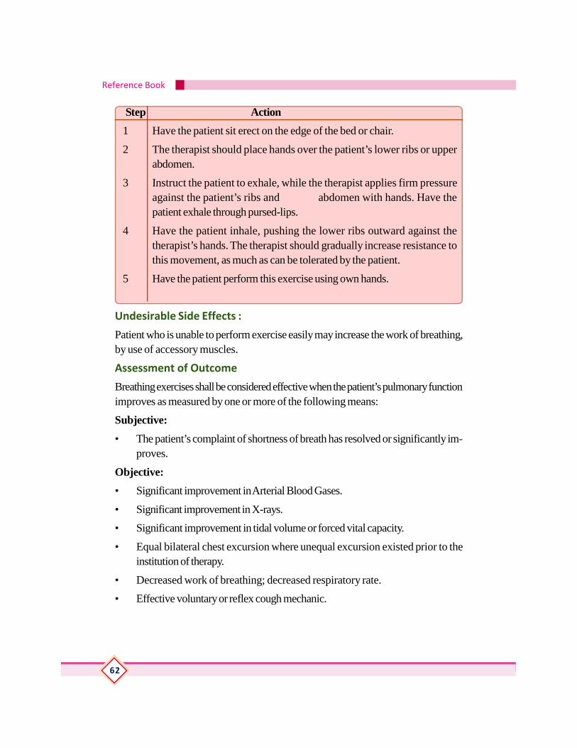
Reference Book
62
Step Action
1 Have the patient sit erect on the edge of the bed or chair.
2 The therapist should place hands over the patient’s lower ribs or upperabdomen.
3 Instruct the patient to exhale, while the therapist applies firm pressureagainst the patient’s ribs and abdomen with hands. Have thepatient exhale through pursed-lips.
4 Have the patient inhale, pushing the lower ribs outward against thetherapist’s hands. The therapist should gradually increase resistance tothis movement, as much as can be tolerated by the patient.
5 Have the patient perform this exercise using own hands.
Undesirable Side Effects :
Patient who is unable to perform exercise easily may increase the work of breathing,by use of accessory muscles.
Assessment of Outcome
Breathing exercises shall be considered effective when the patient’s pulmonary functionimproves as measured by one or more of the following means:
Subjective:
• The patient’s complaint of shortness of breath has resolved or significantly im-proves.
Objective:
• Significant improvement in Arterial Blood Gases.
• Significant improvement in X-rays.
• Significant improvement in tidal volume or forced vital capacity.
• Equal bilateral chest excursion where unequal excursion existed prior to theinstitution of therapy.
• Decreased work of breathing; decreased respiratory rate.
• Effective voluntary or reflex cough mechanic.
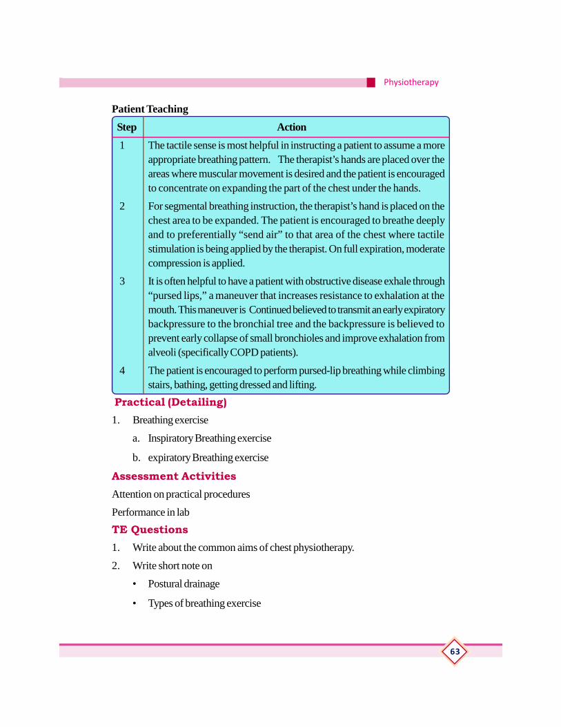
Physiotherapy
63
Patient Teaching
Step Action
1 The tactile sense is most helpful in instructing a patient to assume a moreappropriate breathing pattern. The therapist’s hands are placed over theareas where muscular movement is desired and the patient is encouragedto concentrate on expanding the part of the chest under the hands.
2 For segmental breathing instruction, the therapist’s hand is placed on thechest area to be expanded. The patient is encouraged to breathe deeplyand to preferentially “send air” to that area of the chest where tactilestimulation is being applied by the therapist. On full expiration, moderatecompression is applied.
3 It is often helpful to have a patient with obstructive disease exhale through“pursed lips,” a maneuver that increases resistance to exhalation at themouth. This maneuver is Continued believed to transmit an early expiratorybackpressure to the bronchial tree and the backpressure is believed toprevent early collapse of small bronchioles and improve exhalation fromalveoli (specifically COPD patients).
4 The patient is encouraged to perform pursed-lip breathing while climbingstairs, bathing, getting dressed and lifting.
Practical (Detailing)1. Breathing exercise
a. Inspiratory Breathing exercise
b. expiratory Breathing exercise
Assessment ActivitiesAttention on practical procedures
Performance in lab
TE Questions1. Write about the common aims of chest physiotherapy.
2. Write short note on
• Postural drainage
• Types of breathing exercise
Reference Book
64
UNIT-3.8ELECTROTHERAPY
Introduction
Electrotherapy is a form of medical treatment, which uses small electrical impulsesto repair tissue, stimulate muscles and increase sensations and muscle strength.Thereare several different forms of electrotherapy; these include ultrasound, interferentialtherapy, transcutaneous electrical nerve stimulation (TENS), laser therapy and musclestimulation. Ultrasound uses sound waves to speed up the healing process, whileinterferential therapy and TENS reduce pain by manipulating the nerves which reducesthe sensation of pain and produces a tingling feeling. Laser therapy is sometimesused to repair damaged tissue; using lasers means the treatment can be both accurateand intense.
Learning OutcomesThe learner:
• Understand the advantages of electrotherapy
• Categorize the electrotherapy equipments
• Prepare the patient and equipments for treatment
• Understand the types and importance of heating modalities
• Understand the Physiological and Therapeutic effects of heat.
• To know the application of modalities
• To know where to use and not to use electrotherapy
• To gain expertise in maintain and checking various electrotherapy equipmentsand assist the physiotherapist in electrotherapy treatment.
Concepts (Detailing)Effects and benefits of electrotherapy
The benefits of electrotherapy are wide-ranging and include a more advanced healingprocess, a more effective circulatory system and increased muscle tone. The effectsof electrotherapy include a reduction in pain, increased strength, increased range ofmovement, increased speed and strength of muscle contractions and an increasedrate of absorption. Classification -low frequency, medium frequency and highfrequency equipments
Physiotherapy
65
Classification of therapeutic currents according to frequency
Low Frequency
Current in which the direction of electron flow changes periodically with a frequencyof 1-1000 Hz. The treatment promotes local blood circulation and muscle build-up,and inhibits inflammation and reduces swellings. Above all, in the event of weakenedand partially paralysed musculature, the function of the muscle can be maintainedand the weakness can be reduced.
High frequency
Those current of 10,000 Hz or more.
Medium Frequency
Current in which alternate the direction of flow of electrons with a frequency between1000-10.000 Hz . Depending on the form of application, the primary effect is thealleviation of pain or the stimulating impact with muscle contraction and subsequentmuscle relaxation. In the process, circulation improves, swellings are reduced andthe musculature is relaxed.
I. Low & ll-Medium Frequency Modalities
TENS (transcutaneous Electrical Nerve Stimulation)
High-Voltage pulsed stimulation
Neuromuscular Electrical Stimulation
Galvanic Stimulation
Russian current
Faradic current
Iontophoresis
Interferential Therapy (I.F.T)
III. High Frequency Modalities
Shortwave Diathermy (S.W.D)Microwave Diathermy (M.W.D)Ultra Sound Therapy(U.S)Shockwave therapy
IV-Actinotherapy
Infra - red Radiations (I.R.R)Ultraviolet Radiations (U.V.R)Laser Therapy

Reference Book
66
Other Modalities
Continuous Passive Mobilizer (C.P.M)Traction unit (Cervical & LumbarTraction)Paraffin Wax Bath (P.W.D)Hydrocollator UnitHydrotherapy – WhirlpooltherapyFluido – Therapy
Preparation and safety measures for patient and equipments
Electrotherapy Patient Preparation
Examine the skin for any wounds and clean the skin. Apply the electrodes to thetreatment area. Ensure the electrodes are applied securely to the skin. Ensure goodcontact between each electrode and the skin. Check the electrode contact regularlyduring the treatment. Examine the skin again after the treatment. Choose electrodesthat fit the anatomy.
Avoid Water-A critical precaution when working with electrical equipment is stayingout of water.
Check for Live Wires-Before beginning work, check to ensure what you’re workingon is not energized. Use an electrical current tester to check for live or hot wires.
Prevent Shocks-Prevent shocks from happening by covering the bare ends of awire in a service panel with a small plastic cap. If you touch the end by mistake, thecap works as an insulator for the copper and eliminates being shocked.
ThermotherapyThe external application of heat is thermotherapy. Heat can be transferred into thebody by conduction, convection, and radiation. Conduction is the exchange ofthermal energy, while the body’s surface is in contact with the thermal agent, such ashot packs.Convection is a faster process and involves contacting the body’s surfacewith heat energy through a fluid or gaseous method; such as a whirlpool bath or asteam bath.
Radiation is the transfer of heat energy in electromagnetic rays through a conductingmedium, such as infrared lamps.
Moist heat is transferred by conduction and convection. Dry heat is transferredby radiation. Moist heat is the best form of heat for enhancing general relaxation.Physiologic responses to heat are superficial. When our bodies encounter heat, wereact in several ways.
• Sweating is stimulated and evaporation cools the body.
• Blood vessels near the skin surface begin to dilate and excess heat is lost throughthe skin. This flushes the skin.
Physiotherapy
67
• Our behavior will be altered. Feeling hot makes us uncomfortable and wemake environmental adjustments; such as turning on the air conditioner or turn-ing down the thermostat.
Benefits of Thermotherapy
1. Alleviates pain by reducing endorphins when applied.
2. Increases white blood cells, stimulating an immune system response.
3. Soothes, comforts, and sedates as long as inflammation is not present.
4. Reduces muscle spasms.
5. Increases blood volume, oxygenation, and nutrition.
6. Distends and softens superficial fascia.
Contraindications of Thermotherapy
1. Tumor or malignancy
2. Recent injury
3. Recent contusion
4. Phlebitis
5. Diabetes
6. Hypertension
7. Cardiac impairment
8. Recent burn or sunburn
9. Edema
10. Directly over eyes or external genitalia
11. Cerebrovascular accident
12. Fever
13. Open wounds or skin infections
14. Sensory impairment
15. Pregnancy
Thermotherapy Applications
Heat therapy or thermotherapy applications include, but are not limited to thefollowing:
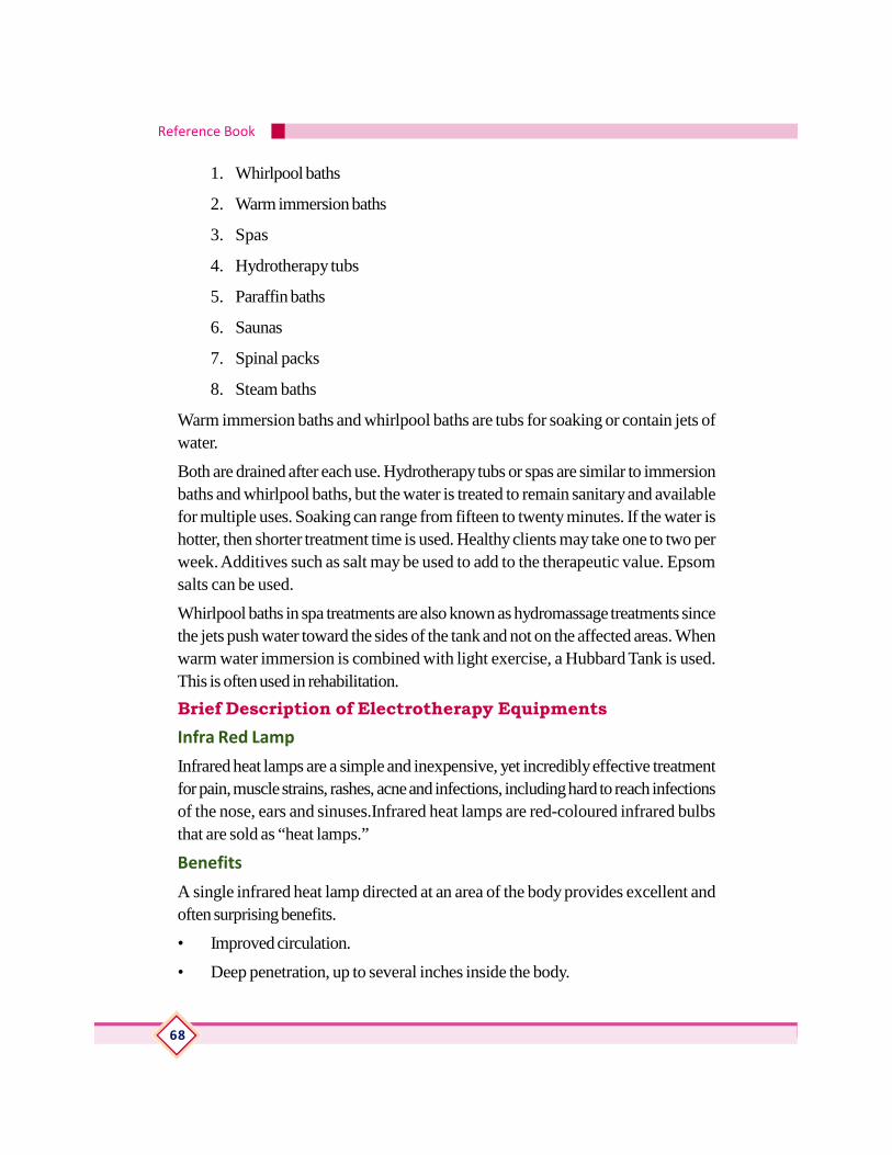
Reference Book
68
1. Whirlpool baths
2. Warm immersion baths
3. Spas
4. Hydrotherapy tubs
5. Paraffin baths
6. Saunas
7. Spinal packs
8. Steam baths
Warm immersion baths and whirlpool baths are tubs for soaking or contain jets ofwater.
Both are drained after each use. Hydrotherapy tubs or spas are similar to immersionbaths and whirlpool baths, but the water is treated to remain sanitary and availablefor multiple uses. Soaking can range from fifteen to twenty minutes. If the water ishotter, then shorter treatment time is used. Healthy clients may take one to two perweek. Additives such as salt may be used to add to the therapeutic value. Epsomsalts can be used.
Whirlpool baths in spa treatments are also known as hydromassage treatments sincethe jets push water toward the sides of the tank and not on the affected areas. Whenwarm water immersion is combined with light exercise, a Hubbard Tank is used.This is often used in rehabilitation.
Brief Description of Electrotherapy EquipmentsInfra Red Lamp
Infrared heat lamps are a simple and inexpensive, yet incredibly effective treatmentfor pain, muscle strains, rashes, acne and infections, including hard to reach infectionsof the nose, ears and sinuses.Infrared heat lamps are red-coloured infrared bulbsthat are sold as “heat lamps.”
Benefits
A single infrared heat lamp directed at an area of the body provides excellent andoften surprising benefits.
• Improved circulation.
• Deep penetration, up to several inches inside the body.
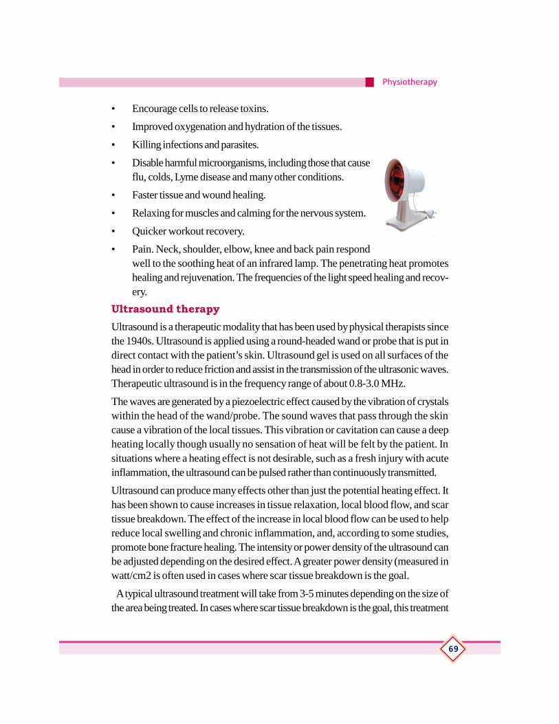
Physiotherapy
69
• Encourage cells to release toxins.
• Improved oxygenation and hydration of the tissues.
• Killing infections and parasites.
• Disable harmful microorganisms, including those that causeflu, colds, Lyme disease and many other conditions.
• Faster tissue and wound healing.
• Relaxing for muscles and calming for the nervous system.
• Quicker workout recovery.
• Pain. Neck, shoulder, elbow, knee and back pain respondwell to the soothing heat of an infrared lamp. The penetrating heat promoteshealing and rejuvenation. The frequencies of the light speed healing and recov-ery.
Ultrasound therapyUltrasound is a therapeutic modality that has been used by physical therapists sincethe 1940s. Ultrasound is applied using a round-headed wand or probe that is put indirect contact with the patient’s skin. Ultrasound gel is used on all surfaces of thehead in order to reduce friction and assist in the transmission of the ultrasonic waves.Therapeutic ultrasound is in the frequency range of about 0.8-3.0 MHz.
The waves are generated by a piezoelectric effect caused by the vibration of crystalswithin the head of the wand/probe. The sound waves that pass through the skincause a vibration of the local tissues. This vibration or cavitation can cause a deepheating locally though usually no sensation of heat will be felt by the patient. Insituations where a heating effect is not desirable, such as a fresh injury with acuteinflammation, the ultrasound can be pulsed rather than continuously transmitted.
Ultrasound can produce many effects other than just the potential heating effect. Ithas been shown to cause increases in tissue relaxation, local blood flow, and scartissue breakdown. The effect of the increase in local blood flow can be used to helpreduce local swelling and chronic inflammation, and, according to some studies,promote bone fracture healing. The intensity or power density of the ultrasound canbe adjusted depending on the desired effect. A greater power density (measured inwatt/cm2 is often used in cases where scar tissue breakdown is the goal.
A typical ultrasound treatment will take from 3-5 minutes depending on the size ofthe area being treated. In cases where scar tissue breakdown is the goal, this treatment
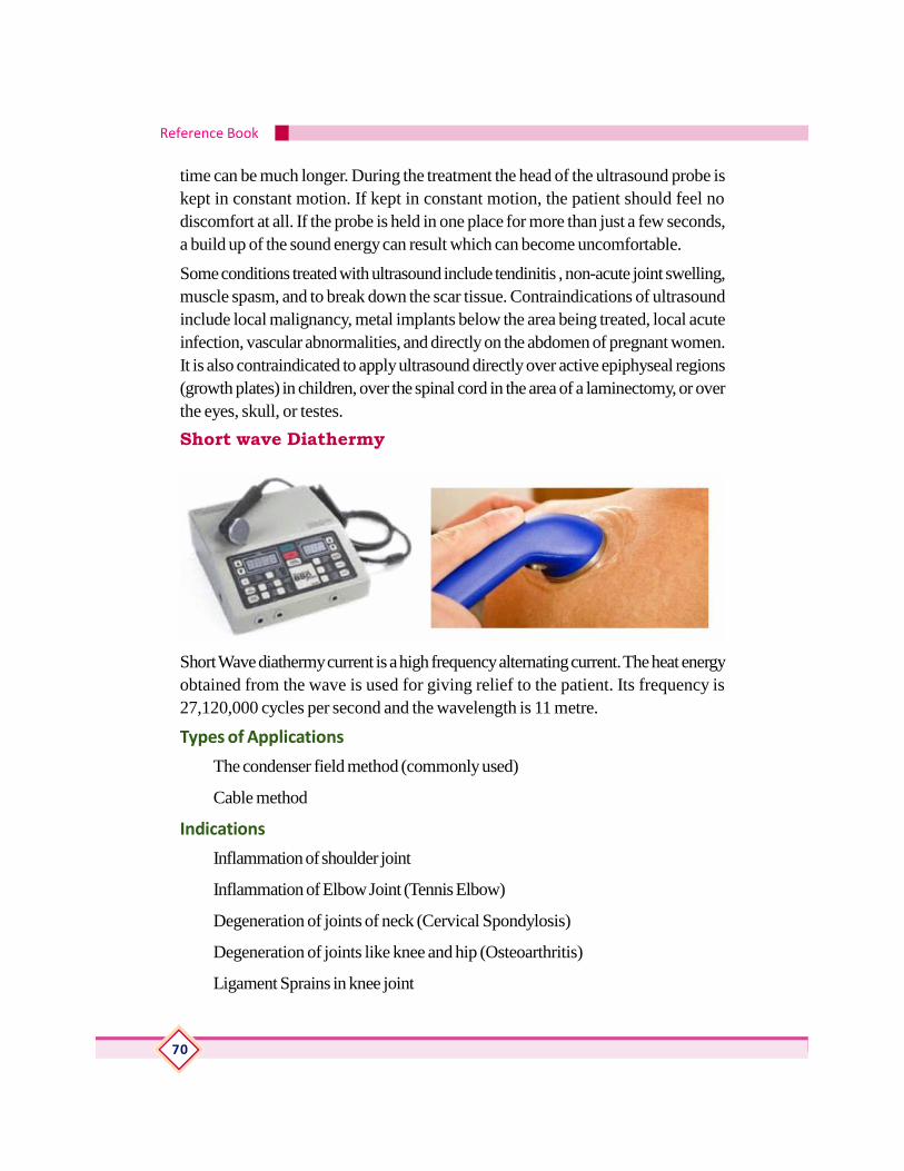
Reference Book
70
time can be much longer. During the treatment the head of the ultrasound probe iskept in constant motion. If kept in constant motion, the patient should feel nodiscomfort at all. If the probe is held in one place for more than just a few seconds,a build up of the sound energy can result which can become uncomfortable.
Some conditions treated with ultrasound include tendinitis , non-acute joint swelling,muscle spasm, and to break down the scar tissue. Contraindications of ultrasoundinclude local malignancy, metal implants below the area being treated, local acuteinfection, vascular abnormalities, and directly on the abdomen of pregnant women.It is also contraindicated to apply ultrasound directly over active epiphyseal regions(growth plates) in children, over the spinal cord in the area of a laminectomy, or overthe eyes, skull, or testes.
Short wave Diathermy
Short Wave diathermy current is a high frequency alternating current. The heat energyobtained from the wave is used for giving relief to the patient. Its frequency is27,120,000 cycles per second and the wavelength is 11 metre.
Types of Applications
The condenser field method (commonly used)
Cable method
Indications
Inflammation of shoulder joint
Inflammation of Elbow Joint (Tennis Elbow)
Degeneration of joints of neck (Cervical Spondylosis)
Degeneration of joints like knee and hip (Osteoarthritis)
Ligament Sprains in knee joint
Physiotherapy
71
Low Back Ache
Plantar fascitis (Heel Pain)
Sinusitis
Contraindications -General
High Fever
Fluctuating Blood Pressure
Very sensitive Skin
Persons with Untreated Fits
Persons using Cardiac Pace Maker
Severe kidney and heart problems
Pregnant Women
Mentally Retarded Individuals
Tuberculosis of Bone
Malignant cancer
Local
If the treatment area has:
Open wounds
Skin disease
Unhealed scars
Recent burns
Treatment Time
Initial Stage - 5-10 minutes
Moderate Stage - 10-20 minutes
Severe State - 20-30 minutes
Advantages
Relaxation of the muscles
Effective in bacterial infections

Reference Book
72
Relief of pain
Disadvantages
Burns
Scalds (Boils)
Overdose
Shock
Electric Sparking
Faintness
Interferential TherapyInter Ferential Therapy (IFT) uses a Mid frequency - current for treatment. Thebasic principle of Interferential Therapy (IFT) is to utilise the significant physiologicaleffects of low frequency (<250pps) electrical stimulation of nerves without theassociated painful and somewhat unpleasant side effects sometimes associated withlow frequency stimulation
The physiological effects include:
1. An increase in localized blood flow which can improve healing by reducingswelling and as a result helps remove damaged tissue and bring nutrients nec-essary for healing to the injured area

Physiotherapy
73
2. The stimulation of local nerve cells that can have a pain reducing/anaestheticeffect due to potentially blocking the transmission of the pain signals (pain gatemechanism) or by stimulating the release of pain reducing endorphins (opiodmechanism)
3. Some degree of muscle stimulation as muscle contraction can be achievedthrough external application of an electrical current, overcoming some of themuscle inhibition often caused by local injury and swelling
The electrical current is applied to the affected area using four electrodes. The fourelectrodes are placed in such a way that the two currents produced cross eachother in the affected area. For example, if it is a knee injury that is being treated, thetwo currents can be applied so that they cross deep within the actual knee jointitself. Where the two currents meet, they actually ‘interfere’ with each other; hencethe name ‘interferential.’
Indications
Individuals with
Pain
Muscles spasm (pain and tightness)
Swelling
Muscles strain
Contraindications -If the Treatment area has:
Localised wounds
Unhealed scar
Recent cuts
Skin infection
Transcutaneous Electrical Nerve StimulatorTranscutaneous electrical nerve stimulation (TENS or TNS) is the use of electriccurrent produced by a device to stimulate the nerves for therapeutic purposes. TENS,by definition, covers the complete range of transcutaneously applied currents usedfor nerve excitation .The unit is usually connected to the skin using two or moreelectrodes. A typical battery-operated TENS unit is able to modulate pulse width,frequency and intensity. Generally TENS is applied at high frequency (>50 Hz) with
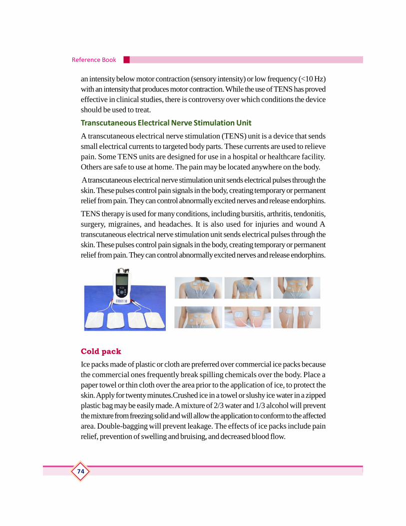
Reference Book
74
an intensity below motor contraction (sensory intensity) or low frequency (<10 Hz)with an intensity that produces motor contraction. While the use of TENS has provedeffective in clinical studies, there is controversy over which conditions the deviceshould be used to treat.
Transcutaneous Electrical Nerve Stimulation Unit
A transcutaneous electrical nerve stimulation (TENS) unit is a device that sendssmall electrical currents to targeted body parts. These currents are used to relievepain. Some TENS units are designed for use in a hospital or healthcare facility.Others are safe to use at home. The pain may be located anywhere on the body.
A transcutaneous electrical nerve stimulation unit sends electrical pulses through theskin. These pulses control pain signals in the body, creating temporary or permanentrelief from pain. They can control abnormally excited nerves and release endorphins.
TENS therapy is used for many conditions, including bursitis, arthritis, tendonitis,surgery, migraines, and headaches. It is also used for injuries and wound Atranscutaneous electrical nerve stimulation unit sends electrical pulses through theskin. These pulses control pain signals in the body, creating temporary or permanentrelief from pain. They can control abnormally excited nerves and release endorphins.
Cold packIce packs made of plastic or cloth are preferred over commercial ice packs becausethe commercial ones frequently break spilling chemicals over the body. Place apaper towel or thin cloth over the area prior to the application of ice, to protect theskin. Apply for twenty minutes.Crushed ice in a towel or slushy ice water in a zippedplastic bag may be easily made. A mixture of 2/3 water and 1/3 alcohol will preventthe mixture from freezing solid and will allow the application to conform to the affectedarea. Double-bagging will prevent leakage. The effects of ice packs include painrelief, prevention of swelling and bruising, and decreased blood flow.
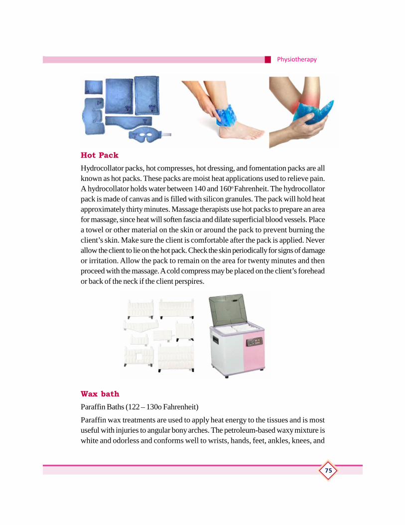
Physiotherapy
75
Hot PackHydrocollator packs, hot compresses, hot dressing, and fomentation packs are allknown as hot packs. These packs are moist heat applications used to relieve pain.A hydrocollator holds water between 140 and 160oFahrenheit. The hydrocollatorpack is made of canvas and is filled with silicon granules. The pack will hold heatapproximately thirty minutes. Massage therapists use hot packs to prepare an areafor massage, since heat will soften fascia and dilate superficial blood vessels. Placea towel or other material on the skin or around the pack to prevent burning theclient’s skin. Make sure the client is comfortable after the pack is applied. Neverallow the client to lie on the hot pack. Check the skin periodically for signs of damageor irritation. Allow the pack to remain on the area for twenty minutes and thenproceed with the massage. A cold compress may be placed on the client’s foreheador back of the neck if the client perspires.
Wax bathParaffin Baths (122 – 130o Fahrenheit)Paraffin wax treatments are used to apply heat energy to the tissues and is mostuseful with injuries to angular bony arches. The petroleum-based waxy mixture iswhite and odorless and conforms well to wrists, hands, feet, ankles, knees, and

Reference Book
76
elbows. Paraffin is used for pain relief and softening the skin. Paraffin baths are usedfor arthritic joints or bursitis, except when joints are hot and swollen.
Paraffin wax is kept at 122 to 130o Fahrenheit. In order to prevent the wax fromsticking to the skin, heavy mineral oil is added to the waxy mixture in a five to onesolution (five pounds of paraffin wax to one pint mineral oil). This molten wax can beapplied as a pack, a dip, or painted on the affected area.The client dips the bodypart in and out of the mixture quickly, allowing the part to cool and promote dryingbetween dipping. The process may be repeated six to twelve times or until a layer ofwax ½ inch thick is opaque. Alcohol or lotion may be applied to the area to removehardened wax. After dipping, wrap the area with a plastic sheet and then a towel.Allow the client to rest the area up to thirty minutes or until the client reports that hecan no longer feel heat. Inform the client to keep the fingers or toes in a relaxedposition and to avoid movement to prevent cracks in the paraffin “glove.” Ask theclient to remove the wax covering and form a wax ball that the client can use forfinger exercise, or replace the paraffin for your next client.
Never use the paraffin bath with open lesions or with peripheral vascular disease.
LaserLaser Therapy is a therapeutic modality used in physiotherapy to accelerate tissuehealing and for pain relief.
How does Laser Therapy Work?
Laser light is exposed directly over the target tissue. Once the laser light reaches thetarget tissue, it has several effects that contribute to its ability to accelerate woundhealing and relieve pain.
• Increase energy production: Laser can turn on proteins that help speed up yourbody’s ability to make energy.
• Initiate breakdown of damaged tissue: Laser increases processes within thebody that break down damaged and infected tissue.
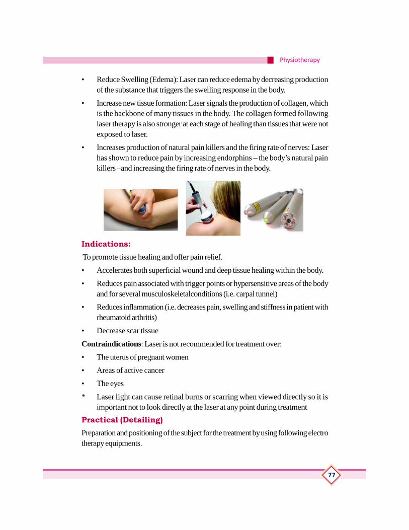
Physiotherapy
77
• Reduce Swelling (Edema): Laser can reduce edema by decreasing productionof the substance that triggers the swelling response in the body.
• Increase new tissue formation: Laser signals the production of collagen, whichis the backbone of many tissues in the body. The collagen formed followinglaser therapy is also stronger at each stage of healing than tissues that were notexposed to laser.
• Increases production of natural pain killers and the firing rate of nerves: Laserhas shown to reduce pain by increasing endorphins – the body’s natural painkillers –and increasing the firing rate of nerves in the body.
Indications: To promote tissue healing and offer pain relief.
• Accelerates both superficial wound and deep tissue healing within the body.
• Reduces pain associated with trigger points or hypersensitive areas of the bodyand for several musculoskeletalconditions (i.e. carpal tunnel)
• Reduces inflammation (i.e. decreases pain, swelling and stiffness in patient withrheumatoid arthritis)
• Decrease scar tissue
Contraindications: Laser is not recommended for treatment over:
• The uterus of pregnant women
• Areas of active cancer
• The eyes
* Laser light can cause retinal burns or scarring when viewed directly so it isimportant not to look directly at the laser at any point during treatment
Practical (Detailing)Preparation and positioning of the subject for the treatment by using following electrotherapy equipments.
Reference Book
78
a. IRR
b. SWD
c. ULTRASOUND
d. IFT
e. TENS
f. EMS
g. Hot packAssessment Activities
Practical presentation
Care of equipments
Approach to clients
Record work
Class testList of practicals
1. Free exercise
2. Resisted exercise
3. Passive movements
4. Fundamental position
5. Derived position
6. Grading of muscle power of major muscles
7. Range of motion of major joints
8. Good and bad posture
Preparation and maintenance of exercise therapy equipment
9. Static cycle
10. Treadmill
11. Quadriceps table
12. Shoulder wheel
13. Hand exerciser
14. Medicine ball
15. Swiss ball
16. Abduction ladder
Physiotherapy
79
Demonstration of pathological gate
17. circumductory gait
18. Scissoring gait
19. Festinent gait
20. Ataxic gait
21. High stepping gait
Demonstration of gait training
22. Transfer techniques
23. Cryo therapy
24. Contrast bath
25. Relaxation
26. Breathing exercise
Preparation and application of electro therapy modalities
27. IRR
28. SWD
29. ULTRASOUND
30. IFT
31. TENS
32. EMS
TE Questions
1. Classify the electrotherapy equipment based on frequency with suitable example.
2. List out the therapeutic effect of heat
3. Short note on
4. Indication and Contra indication of thermotherapy
5. Indication and contra indications of SWD
6. Expand the term
Reference Book
80
• SWD
• TENS
• IFT
7. Answer in one word
8. An example of Electro therapy equipments that produces superficial heat.
9. Temperature range of paraffin bath
At the end of module
Extended activities
Physiotherapy
81
Over view of Module 4Students have already achieved knowledge about basics of physiotherapy This moduleEnable the students to acquire the knowledge about the various conditions in medicaland surgical disciplines like neurology, orthopaedics, paediatrics, and geriatrics. Thelearner also acquires the concept, types and indications of orthotics and prosthetics.
On successful completion of the fourth module the learner will develop an overallidea of different disease conditions and role of physiotherapist in management ofthese conditions. The learner will be able to do the preparation of the client, basicphysiotherapy techniques based on the condition under physiotherapist’ssupervision.The learner can also assist physiotherapist in management of variousneurological, orthopaedic ,paediatric and geriatric conditions in various rehabilitationcentres like physiotherapy clinics ,old age home , special schools, palliative centresetc.
UNIT 4.1 INTRODUCTION TO NEUROLOGICALPHYSIOTHERAPY
IntroductionNeurological physiotherapy is the treatment of individuals who have neurologicalimpairments; for example Traumatic Brain Injury or Stroke; Multiple Sclerosis, SpinalCord Injury and Parkinson’s disease. Neuro Physiotherapists specialise in assessingand treating individuals with such neurological impairments to enhance or maximisetheir functional ability.
Physiotherapy can be carried out within home or clinic. Home based treatmentsessions are limited to mat based exercises and walking practise. Clinic basedtreatment sessions are able to incorporate a larger range of physiotherapy exercisesusing different machines to enhance strengthening programme.
Neuro Physiotherapy objectives
Neuro Physiotherapy aims to identify and maximise movement potential and qualityof life. Treatment programme is specific and based on goals identified during initialassessment session. These may include: education, training, posture awareness,strengthening, stretching, sitting balance, standing balance, and walking practice (ifapplicable). Address issues proactively to prevent long term problems fromdeveloping.
When clients present with progressive neurological conditions such as Parkinson’sdisease or Multiple Sclerosis, aim to provide advice and treatment during each stage
Reference Book
82
of the condition. If early intervention can be provided, it can help to prevent theprogression of a progressive condition and reduce the degree of secondarycomplications. In some cases, clients can still make physical gains with improvementsin posture, muscle strength and flexibility helping to improve their functionalindependence and their quality of life.
Neuro Physiotherapists have a special interest in working with clients withneurological impairments. If have a neurological impairment, treatment should beprovided by a Specialist Neuro Physiotherapist to ensure that the latest techniquesare used within your treatment sessions.
Learning Outcomes
The learner:
• Learner will be able to
• Understand the concept of neurological physiotherapy
• Assist physiotherapist in management of paraplegia, paraparesis, quadripegiaand quadriparesis
• Assist physiotherapist in management of Parkinson patient
• Assist physiotherapist in management of hemi paresis and hemiplegia
• Assist physiotherapist in management of ataxic patient
• Identify the conditions like, Epilepsy, Guillianbarre syndrome, Bell’s palsy,Multiple sclerosis ,Alzheimer’s disease
Concepts (Detailing)
Stroke
A stroke is a medical emergency, and treatment must be sought as quickly aspossible.It is the 5th leading cause of death in the US, with one person dying every4 minutes as a result. For black people, stroke is the 3rd leading cause of death.Strokes occur due to problems with the blood supply to the brain: either the bloodsupply is blocked or a blood vessel within the brain ruptures, causing brain tissue todie.
There are three main kinds of stroke.
Ischemic strokes
Hemorrhagic strokes
Physiotherapy
83
Transient ischemic attacks (TIAs), also referred to as mini-strokes
Ischemic stroke
Ischemic stroke is the most common form of stroke, accounting for around 85% ofstrokes. This type of stroke is caused by blockages or narrowing of the arteries thatprovide blood to the brain, resulting in ischemia - severely reduced blood flow.These blockages are often caused by blood clots, which can form either in thearteries connecting to the brain, or in other blood vessels before being swept throughthe bloodstream and into narrower arteries within the brain. Clots can be caused byfatty deposits within the arteries called plaque.
Hemorrhagic stroke
Hemorrhagic stroke are caused by arteries in the brain either leaking blood or burstingopen. The leaked blood puts pressure on brain cells and damages them. Bloodvessels can burst or spill blood in the middle of the brain or near the surface of thebrain, sending blood into the space between the brain and the skull.The ruptures canbe caused by conditions such as hypertension, trauma, blood-thinning medicationsand aneurysms (weaknesses in blood vessel walls).
Intracerebral haemorrhage is the most common type of hemorrhagic stroke andoccurs when brain tissue is flooded with blood after an artery in the brain bursts.Subarachnoid haemorrhage is the second type of hemorrhagic stroke and is lesscommon. In this type of stroke, bleeding occurs in the subarachnoid space - thearea between the brain and the thin tissues that cover it.
Transient ischemic attack (TIA)
TIAs are different from the aforementioned kinds of stroke because the flow ofblood to the brain is only briefly interrupted. TIAs are similar to ischemic strokes inthat they are often caused by blood clots or other debris. TIAs should be regardedas medical emergencies just like the other kinds of stroke, even if the blockage ofthe artery is temporary. They serve as warning signs for future strokes and indicatethat there is a partially blocked artery or clot source in the heart. According to theCanters for Disease Control and Prevention (CDC), over a third of people whoexperience a TIA go on to have a major stroke within a year if they have not receivedany treatment .Between 10-15% will have a major stroke within 3 months of a TIA.
Symptoms of stroke
Confusion, including trouble with speaking and understanding
Headache, possibly with altered consciousness or vomiting
Reference Book
84
Numbness of the face, arm or leg, particularly on one side of the body
Trouble with seeing, in one or both eyes
Trouble with walking, including dizziness and lack of co-ordination.
Bladder or bowel control problems
Pain in the hands and feet that gets worse with movement and temperature changes
Paralysis or weakness on one or both sides of the body
Trouble controlling or expressing emotions.
Treatments for stroke
As the two main different kinds of stroke, ischemic and hemorrhagic, are caused bydifferent factors, both require different forms of treatment. It is not only importantthat the type of stroke is diagnosed quickly to reduce the damage done to the brain,but also because treatment suitable for one kind of stroke can be harmful to someonewho has had a different kind.
Ischemic strokes are caused by arteries being blocked or narrowed and so treatmentfocuses on restoring an adequate flow of blood to the brain. Treatment can beginwith drugs to break down clots and prevent further ones from forming. Aspirin canbe given, as can an injection of a tissue plasminogen activator (TPA). TPA is veryeffective at dissolving clots but needs to be injected within 4.5 hours of strokesymptoms manifesting themselves.
Surgeons are able to remove plaque and any other obstructions from the carotidartery through surgery. Emergency procedures include administering TPA via catheterdirectly into an artery in the brain or using a catheter to physically remove the clotfrom its obstructive position. There are other procedures that can be carried out todecrease the risk of future strokes or TIAs. A carotid endarterectomy involves asurgeon opening the carotid artery and removing any plaque that might be blockingit. Alternatively, an angioplasty involves a surgeon inflating a small balloon in a narrowedartery via catheter and then inserting a stent (a mesh tube) into the opening in orderto prevent the artery from narrowing again.
Hemorrhagic strokes are caused by bleeding into the brain and so treatment focuseson controlling the bleeding and reducing the pressure on the brain that it is causing.Treatment can begin with drugs being given to reduce the pressure in the brain,overall blood pressure, prevent seizures and prevent sudden constrictions of bloodvessels. Surgery can be used to repair any problems with blood vessels that have

Physiotherapy
85
led or could lead to hemorrhagic strokes. Surgeons can place small clamps at thebase of aneurysms or fill them with detachable coils to stop blood flow to them andprevent rupture.
Surgery can also be used to remove small arteriovenous malformations (AVMs) ifthey are not too big and not too deep within the brain. AVMs are tangled connectionsbetween arteries and veins that are weaker and burst more easily than other normalblood vessels.
Rehabilitation
Strokes are life-changing events that can affect a person both physically andemotionally, temporarily or permanently. After a stroke, successful recovery willoften involve specific rehabilitative activities such as:
Speech therapy - to help with problems producing or understanding speech. Practice,relaxation and changing communication style, using gestures or different tones forexample, all help
Physical therapy - to help a person relearn movement and co-ordination. It isimportant to get out and about, even if it is difficult at first
Occupational therapy - to help a person to improve their ability to carry out routinedaily activities, such as bathing, cooking, dressing, eating, reading and writing
Joining a support group - to help with common mental health problems such asdepression that can occur after a stroke. Many find it useful to share commonexperiences and exchange information
Support from friends and family - to provide practical support and comfort. Lettingfriends and family know what can be done to help is very important.
Prevention of stroke
The best way to prevent a stroke is to address the underlying causes. This is bestdone by living healthily, which means:
Eating a healthy diet
Maintaining a healthy weight
Exercise regularly
Not smoking
Avoiding alcohol or moderating consumption.
Eating a healthy diet means getting plenty of fruits, vegetables and healthy whole
Reference Book
86
grains, nuts, seeds and legumes; eating little or no red or processed meat; limitingintake of cholesterol and saturated fat (typically found in foods of animal origin);and minimizing salt intake so as to support healthy blood pressure
Other measures taken to help reduce the risk of stroke include: Keeping bloodpressure under control, Managing diabetes well and treating obstructive sleepapnoea (if present).
A health care provider can help to reduce the risk of future strokes through prescribinganti-coagulant and anti-platelet medication. In addition to this, the arterial surgerycan also be used to lower the risk of repeat strokes.
Spinal cord injury (SCI)Spinal cord injury (SCI) involves damage to the nerves within the bony protectionof the spinal canal. The most common cause of SCI is trauma, although damage canoccur from various diseases acquired at birth or later in life. The spinal cord and thebrain together make up the central nervous system (CNS). The spinal cordcoordinates the body’s movement and sensation. Therefore, an injured cord losesthe ability to send and receive messages from the brain to the body’s systems thatcontrol sensory, motor, and autonomic function below the level of injury. Often, thisresults in some level of paralysis.
Spinal cord trauma is more than a single event. The initial blunt force damages orkills spinal nerve cells. However, in the hours and days after injury a cascade ofsecondary events, including loss of oxygen and the release of toxic chemicals at thesite of injury, further damage the cord.
Acute care following an injury may involve surgery if the spinal cord appears to becompressed by bone, a herniated disk, or a blood clot. Traditionally, surgeons waitedfor several days to decompress the spinal cord, believing that operating immediatelycould worsen the outcome. More recently, many surgeons advocate immediate earlysurgery.
Complete vs. incomplete SCIThose with an incomplete injury have some sensory or motor function below thelevel of injury – the spinal cord was not totally damaged or disrupted. In a completeinjury, nerve damage obstructs all signals coming from the brain to the body belowthe injury.People with incomplete injuries have a better chance of getting more return.
The sooner muscles start working again, the better the chances are of additionalrecovery. When muscles come back later, after the first several weeks, they are
Physiotherapy
87
more likely to be in the arms than in the legs.
As long as there is some improvement and additional muscles recover function, thechances are better that more improvement is possible. The longer there is noimprovement, the lower the odds it will start to happen on its own.
Parkinson’s diseaseParkinson’s disease affects the nerve cells in the brain that produce dopamine.Parkinson’s disease symptoms include muscle rigidity, tremors, and changes in speechand gait. After diagnosis, treatments can help relieve symptoms, but there is no cure.
Symptoms of Parkinson’s disease differ from person to person. They also changeas the disease progresses. Symptoms that one person gets in the early stages of thedisease, another person may not get until later-or not at all. Symptoms typicallybegin appearing between the ages of 50 and 60. They develop slowly and often gounnoticed by family, friends, and even the person who has them. The disease causesmotor symptoms and non-motor symptoms. Motor symptoms are those that haveto do with how you move. The most common one is tremor.
• Tremor, or shaking, often in a hand, arm, or leg, occurs when you’re awakeand sitting or standing still (resting tremor), and it gets better when you movethat body part.Tremor is often the first symptom that people with Parkinson’sdisease or their family members notice. At first the tremor may appear in justone arm or leg or only on one side of the body. The tremor also may affect thechin, lips, and tongue. As the disease progresses, the tremor may spread toboth sides of the body. But in some cases the tremor remains on just one side.Emotional and physical stress tends to make the tremor more noticeable. Sleep,complete relaxation, and intentional movement or action usually reduce or stopthe tremor..
• Stiff muscles (rigidity) and aching muscles. One of the most common earlysigns of Parkinson’s is a reduced arm swing on one side when you walk. Thisis caused by rigid muscles. Rigidity can also affect the muscles of the legs, face,neck, or other parts of the body. It may cause muscles to feel tired and achy.
• Slow, limited movement, especially when you try to move from a restingposi-tion. For instance, it may be hard to get out of a chair or turn over in bed.
• Weakness of face and throat muscles. It may get harder to talk and swallow.You may choke, cough, or drool. Speech becomes softer and monotonous.Loss of movement in the muscles in the face can cause a fixed, vacant facial
Reference Book
88
expression, often called the “Parkinson’s mask.”• Difficulty with walking and balance. A person with this disease is likely to take
small steps and shuffle with his or her feet close together, bend forward slightlyat the waist, and have trouble turning around. Balance and posture problemsmay cause frequent falls. But these problems usually don’t happen until lateron.
• Freezing, a sudden, brief inability to move. It most often affects walking.
• A small number of people have symptoms on only one side of the body thatnever move to the other side.
Treatment OverviewNo known treatment can stop or reverse the breakdown of nerve cells thatcauses Parkinson’s disease. But there are many treatments that can reduce yoursymptoms and improve your quality of life. Age, work status, family, and living situationcan all affect decisions about when to begin treatment, what types of treatment touse, and when to make changes in treatment. As medical condition changes, needregular changes in treatment to balance quality-of-life issues, side effects of treatment,and treatment costs.
Treatments for Parkinson’s include:Medicines, such as levodopa and dopamine agonists. This is the most commontreatment for Parkinson’s disease.Home treatment. There are many steps you can take at home to make dealing withthe symptoms of Parkinson’s disease easier, such as getting regular exercise andeating a healthy diet.
Surgery. Brain surgery, for example deep brain stimulation (DBS), may be consideredwhen medicine fails to control symptoms of Parkinson’s disease or causes severe ordisabling side effects.
Speech therapy. Speech therapists use breathing and speech exercises to help youovercome the soft, imprecise speech and monotone voice that develop in advancedParkinson’s disease.
Physical therapy. Therapists may help you improve your walking and reduce yourrisk of falling.
Occupational therapy. Therapists can help you learn new ways to do things foryourself so you can stay independent longer.
Physiotherapy
89
Treatment for mental problems. Family members may notice that you begin to haveproblems with memory, problem solving, learning, and other mental functions. Whenthese problems keep you from doing daily activities, it is called dementia. There aremedicines that can help treat dementia in people with Parkinson’s.
Cerebral PalsyCerebral palsy is refers to a group of disorders affecting a person’s ability to move.It is due to damage to the developing brain either during pregnancy or shortly afterbirth.Cerebral palsy affects people in different ways and can affect body movement,muscle control, muscle coordination, muscle tone, reflex, posture and balance.Although cerebral palsy is a permanent life-long condition, some of these signs ofcerebral palsy can improve or worsen over time.People who have cerebral palsymay also have visual, learning, hearing, speech, epilepsy and intellectualimpairments.Cerebral palsy is the result of a combination of events either before,during or after birth that can lead to an injury in a baby’s developing brain. There isno single cause of cerebral palsy. For most babies born with cerebral palsy, thecause remains unknown. Researchers now know that only a very small percentageof cases of cerebral palsy are due to complications at birth (e.g. asphyxia or lack ofoxygen). It is accepted that cerebral palsy usually arises from a series of causalpathways, i.e. a sequence of events that when combined can cause or accelerateinjury to the developing brain.
Stroke is the most common cause in babies who acquire cerebral palsy after 1month of age. The stroke may occur spontaneously or arise from surgical or heartcomplications.
Risk factors
Risk factors do not cause cerebral palsy. However, the presence of some risk factorsmay lead to an increased chance of a child being born with cerebral palsy.
Premature birth (less than 37 weeks)
Low birth weight (small for gestational age)
Blood clotting problems (thrombophilia)
Inability of the placenta to provide the developing feotus with oxygen and nutrients
RH or A-B-O blood type incompatibility between mother and baby
Infection of the mother with German measles or other viral diseases in early pregnancy
Bacterial infection of the mother, foetus or baby that directly or indirectly attacks the

Reference Book
90
infant’s central nervous systemProlonged loss of oxygen during the pregnancy or birthing process, or severe jaundiceshortly after birth.
Types of Cerebral Palsy
Cerebral palsy can be described by the way it affects people’s movement, the partof the body affected and by how severe the affects are.
QUADRIPLEGIA: Both arms and legs are affected. The muscles of the trunk, faceand mouth are often also affected.
DIPLEGIA: Both legs are affected. The arms may be affected to a lesser extent.
HEMIPLEGIA: One side of the body (one arm and one leg) is affected.
Other classifications
DYSKINETIC: Characterised by involuntary movements (i.e. out of a person’scontrol)
ATAXIC: Characterised by shaky movements. Affects balance and sense ofpositioning in space
MIXED: Although cerebral palsy is a lifelong disability, there are many interventionsthat can help reduce its impact on the body and the individual’s quality of life. Anintervention is a service that aims to improve the condition of cerebral palsy and theday-to-day experience of the person living with it.
Children with cerebral palsy may be supported by a team of professionals includinghealth professionals and community-based support services who work together tohelp the child and family reach their goals.
Through their late teens and early 20s, many young people take increasingresponsibility for their own health and wellbeing. Their team may include healthprofessionals and mainstream community providers such as fitness trainers.At present,there is no way to totally prevent or cure cerebral palsy.
Interventions for movement difficulties
Medication: Medical specialists may prescribe medications that assist movementissues. Some medications are taken orally (e.g. diazepam) and others are injectedor delivered through surgically implanted pumps (e.g. Baclofen). Many children withcerebral palsy benefit from Botulinum toxin type A injections into muscles affectedby spasticity. This intervention is used from about two years of age and is mosteffective when used in conjunction with therapy.
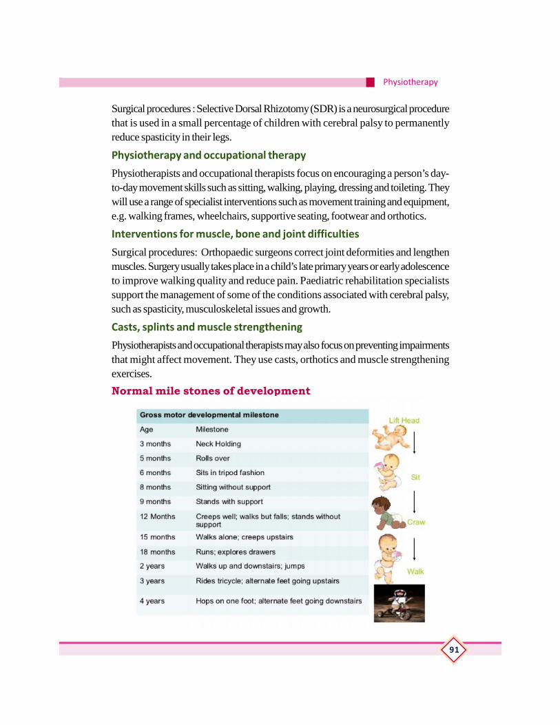
Physiotherapy
91
Surgical procedures : Selective Dorsal Rhizotomy (SDR) is a neurosurgical procedurethat is used in a small percentage of children with cerebral palsy to permanentlyreduce spasticity in their legs.
Physiotherapy and occupational therapy
Physiotherapists and occupational therapists focus on encouraging a person’s day-to-day movement skills such as sitting, walking, playing, dressing and toileting. Theywill use a range of specialist interventions such as movement training and equipment,e.g. walking frames, wheelchairs, supportive seating, footwear and orthotics.
Interventions for muscle, bone and joint difficulties
Surgical procedures: Orthopaedic surgeons correct joint deformities and lengthenmuscles. Surgery usually takes place in a child’s late primary years or early adolescenceto improve walking quality and reduce pain. Paediatric rehabilitation specialistssupport the management of some of the conditions associated with cerebral palsy,such as spasticity, musculoskeletal issues and growth.
Casts, splints and muscle strengthening
Physiotherapists and occupational therapists may also focus on preventing impairmentsthat might affect movement. They use casts, orthotics and muscle strengtheningexercises.
Normal mile stones of development
Reference Book
92
AtaxiaAtaxia is a lack of muscle coordination which may affect speech, eye movements,the ability to swallow, walking, picking up objects and other voluntary movements.A person with persistent ataxia may have damage in the part of the brain that controlsmuscle coordination - the cerebellum. Ataxia may have several causes, includingmultiple head injury, alcohol abuse, stroke, and cerebral palsy,
Types of ataxias
The following are some of the more common types of ataxias.
Cerebellar ataxia This is ataxia caused by a dysfunction of the cerebellum - aregion in the brain which is involved in the assimilation of sensory perception,coordination and motor control. Cerebellar ataxia causes some basic neurologicalproblems, such as:
• Floppiness (hypotonia).
• Lack of co-ordination between organs, muscles, limbs or joints (asynergy).
• Impaired ability to control distance, power, and speed of an arm, hand, leg oreye movement (dysmetria).
• Difficulty in accurately estimating how much time has passed (dyschronometria).
• An inability to perform rapid, alternating movements (dysdiadochokinesia).
• How the patient is affected depends on which parts of the cerebellum arelesioned, and whether lesions occur on one side (unilateral) or both sides (bi-lateral).
Sensory ataxia : This is ataxia due to loss of proprioception. Proprioception is thesense of the relative position of neighbouring parts of the body. It is a sense thatindicates whether the body is moving with the required effort, and also where thevarious parts of the body are located in relation to each other. A patient with sensoryataxia typically has an unsteady stamping gait, with the heel striking hard as it touchesthe ground with each step. Postural instability becomes worse in poorly litenvironments. If a doctor asks the patient to stand with eyes closed and feet togetherhis/her instability will clearly worsen. This is because loss of proprioception makesthe patient much more reliant on visual data. The patient may find it hard to performsmoothly coordinated voluntary movements with the limbs, trunk, pharynx, larynx,and eyes.

Physiotherapy
93
Vestibular ataxia
The vestibular system is affected. In acute (sudden) unilateral cases the patient mayexperience vertigo, nausea, and vomiting. In slow-onset chronic bilateral cases thepatient may only experience unsteadiness, giddiness.
Causes of ataxia
• Vitamin B12 deficienc - as well as other neurological abnormalities, vitamin b12deficiency can cause overlapping cerebellar and sensory ataxia.
• Inherited ataxia: this means the ataxia is caused by a genetic fault inheritedeither from the mother or father, or both. a faulty gene that is passed downthrough generations can cause cerebellar ataxia - in some cases its severityworsens from one generation to the next and the age of onset gets younger. thechances of inheriting ataxia from a parent depends on the type of ataxia thatparent has.
• Non-inherited ataxia: even without a family history of ataxia some people maystill develop the condition. in some cases, experts never find out what the causewas. the following procedures and circumstances can cause ataxia:
• Brain surgery.
• Head injury.
• Alcohol abuse.
• Drug abuse.
• Infections, such as chicken pox (usually goes away after infection has gone).
• A brain tumour.
• Exposure to toxic chemicals.
• Multiple sclerosis, cerebral palsy, and some other neurological conditions.
• Malformation of the cerebellum while the baby is still in the womb.
Symptoms of ataxia
Symptoms may vary depending on the severity and type of ataxia. Initial ataxiasymptoms usually include:
Poor limb coordination.
Reference Book
94
Dysarthria - slurred and slow speech that is difficult to produce. The patient mayalso have difficulties controlling volume, rhythm, and pitch.
If the ataxia advances other symptoms may also appear:
Swallowing difficulties, which may sometimes result in choking or coughing?
Facial expressions become less apparent.
Tremors - parts of the body may shake or tremble unintentionally.
Nystagmus - involuntary rapid rhythmic repetitious eye movement. Movements maybe vertical, horizontal, or circular.
Pes cavus - a foot with too high an arch.
Cold feet - because of a lack of muscle activity.
Problems with balance.
Walking difficulties - in severe cases the patient may need a wheelchair.
Vision problems.
Hearing problems.
Diagnosis of Ataxia
A doctor who suspects the patient may have ataxia will refer him/her to a neurologist-a doctor specialized in diagnosis, treatment and prevention of conditions and illnessesrelated to the nervous system.The specialist will check the patient’s medical historyfor cause factors, such as brain injury, as well as the patient’s family history forindications of inheritance. The following tests may also be ordered:
Magnetic resonance imaging (MRI) or Computerized Tomography (CT) scan - todetermine whether there is any brain damage.
Genetic tests - to determine whether the patient has inherited ataxia.
Blood tests - some types of ataxia affect the makeup of blood.
Urine tests - urinalysis may suggest specific systemic abnormalities that are linked tosome forms of ataxia. If the specialist suspects Wilson’s disease the patient may beasked for a 24-hour urine collection to determine how much copper is in the system.
Treatments for ataxia
Although ataxia is not curable, a great deal can be done to ease symptoms andimprove the quality of life of the patient. Treatment for coordination and balanceproblems usually involves the use of adaptive devices which help the patient attain
Physiotherapy
95
as much independence as possible. These may include the use of a cane (walkingstick), crutches, walker or a wheelchair. Symptoms such as tremor, stiffness,spasticity, sleep disorders, muscle weakness, depression (or frustration, sadnessand anger) may be addressed with targeted physical therapy, speech therapy,medications and counselling.
Occupational therapy - the occupational therapist can help the patient manage betteraround the house and work. This may involve some home adaptations, wheelchairassessments, and making the kitchen more practical for the patient.
Speech therapy - the speech therapist can help with swallowing, coughing, chokingand speech problems. If speech becomes very difficult the speech therapist can helpthe patient learn how to use speech aids.
Orthopaedic care - this can help patients with curvature of the spine (scoliosis).
Physical therapy (physiotherapy) - a physical therapist (physiotherapist) can helpmaintain strength and improve mobility.
Counselling - patients with ataxia commonly become frustrated and depressed; thisusually results from having to cope with some symptoms which affect physical mobilityand coordination. Talking to a well qualified counsellor, such as a psychotherapistcan help.
Supplements and nutrition - some patients with ataxia have very low levels of vitaminE and require supplements and/or a special diet. As sensitivity to gluten is morecommon among ataxia patients, a gluten-free diet also helps.
Medication - some patients with ataxia telangiectasia are prescribed gamma-globulininjections to boost their immune systems. There are also drugs for muscle spasmsand uncontrollable eye movements.
Peripheral nerve injuryPeripheral nerves link brain and spinal cord to the other parts of body, such as yourmuscles and skin. Peripheral nerves are fragile and easily damaged. A nerve injurycan interfere with brain’s ability to communicate with muscles and organs. If youfeel tingling or numbness in your leg, arm, shoulder or hand, you may have injuredone or more nerves when you had an accident or broke a bone. You may alsoexperience numbness or tingling if a nerve is being compressed due to factors suchas a narrow passageway, tumour or other diseases. It’s important to get medicalcare for a peripheral nerve injury as soon as possible because nerve tissue can berepaired. Early diagnosis and treatment can prevent complications and permanentinjury.
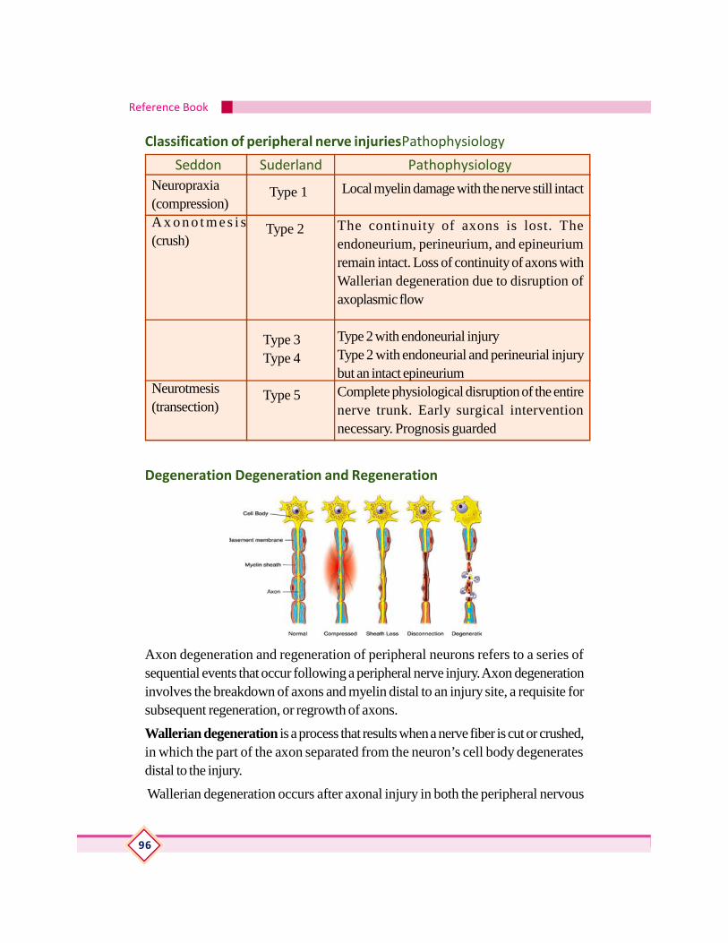
Reference Book
96
Neuropraxia(compression)A x o n o t m e s i s(crush)
Neurotmesis(transection)
Type 1
Type 2
Type 3Type 4
Type 5
Local myelin damage with the nerve still intact
The continuity of axons is lost. Theendoneurium, perineurium, and epineuriumremain intact. Loss of continuity of axons withWallerian degeneration due to disruption ofaxoplasmic flow
Type 2 with endoneurial injuryType 2 with endoneurial and perineurial injurybut an intact epineuriumComplete physiological disruption of the entirenerve trunk. Early surgical interventionnecessary. Prognosis guarded
Classification of peripheral nerve injuriesPathophysiology
Seddon Suderland Pathophysiology
Degeneration Degeneration and Regeneration
Axon degeneration and regeneration of peripheral neurons refers to a series ofsequential events that occur following a peripheral nerve injury. Axon degenerationinvolves the breakdown of axons and myelin distal to an injury site, a requisite forsubsequent regeneration, or regrowth of axons.
Wallerian degeneration is a process that results when a nerve fiber is cut or crushed,in which the part of the axon separated from the neuron’s cell body degeneratesdistal to the injury.
Wallerian degeneration occurs after axonal injury in both the peripheral nervous

Physiotherapy
97
system (PNS) and central nervous system (CNS). It occurs in the axon stump distalto a site of injury and usually begins within 24–36 hours of a lesion. Prior todegeneration, distal axon stumps tend to remain electrically excitable. After injury,the axonal skeleton disintegrates, and the axonal membrane breaks apart. The axonaldegeneration is followed by degradation of the myelin sheath and infiltration bymacrophages. The macrophages, accompanied by Schwann cells, serve to clearthe debris from the degeneration.
Neuroregeneration refers to the regrowth or repair of nervous tissues, cells or cellproducts. When an axon is damaged, the distal segment undergoes Walleriandegeneration, losing its myelin sheath. The proximal segment can either die byapoptosis or undergo the chromatolytic reaction, which is an attempt at repair.Peripheral nervous system has an intrinsic ability for repair and regeneration,Neuroregeneration in the peripheral nervous system (PNS) occurs to a significantdegree. Axonal sprouts form at the proximal stump and grow until they enter thedistal stump. The growth of the sprouts is governed by chemotactic factors secretedfrom Schwann cell. Regeneration is rapid in PNS, allowing for rates of up to 1millimeter a day of regrowth.
General Principles of management
The management of a peripheral nerve injury varies depending on the cause, typeand degree of the nerve injury. If a nerve is not healing properly surgery may berequired to repair the damaged section. Physiotherapy is very important to promotethe recovery of peripheral nerve injuries regardless of whether surgery is required toMaintain range muscle length / joint range of movement
Maintain nerve integrity
Increase muscle strength
Increase sensation
Manage neuropathic pain
Clinical features and Physiotherapy Management of:
Brachial plexus injury-Erb’s palsy and Klumpke’s palsy
Brachial plexus injury
The brachial plexus is a group of nerves that come from the spinal cord in the neckand travel down the arm. These nerves control the muscles of the shoulder, elbow,wrist and hand, as well as provide feeling in the arm. Some brachial plexus injuries
Reference Book
98
are minor and will completely recover in several weeks. Other injuries are severeenough and could cause some permanent disability in the arm.
CAUSES: These nerves can be damaged by stretching, pressure or cutting.Stretching can occur when the head and neck are forced away from the shoulder,such as during a motorcycle fall or car accident. If severe enough, the nerves cantear out of the spinal cord in the neck. Pressure could occur from the crushing of thebrachial plexus between the collarbone and first rib, which can happen during afracture or dislocation. Swelling in this area from excessive bleeding or injured softtissues can also cause an injury.
Signs and symptoms: Nerve injuries can stop signals to and from the brain,preventing the muscles of the arm and hand from working properly, and causing lossof feeling in the area.
Treatment: Many adult injuries will not recover on their own, and early evaluationby physicians who have experience treating these problems is essential. Some injuriescan recover with time and therapy. The time for recovery can be weeks or months.When an injury is unlikely to improve, several surgical techniques can be used toimprove the recovery.In some cases, when nerve recovery will not happen, a tendontransfer surgery may be performed.
Recovery: The patient must do several things to keep up muscle activity and preventthe joints from getting stiff. If the joints become stiff, they will not move even aftermuscles begin to work again, like a hinge that has rusted.
When a sensory nerve has been injured, the patient must be extra careful not to burnor cut fingers while there is no feeling in the affected area. During nerve recovery, thebrain may not interpret the new nerve signals properly, and a procedure called sensoryre-education may be needed to optimize muscle control and feeling in the hand orfingers. Factors that may affect results after a brachial plexus injury include patientage and the type, severity and location of the injury. Though these injuries will resultin lasting problems for the patient, care by a hand surgeon and proper therapy canmaximize function.
Types of Brachial Plexus Injuries
Erb’s Palsy: Erb’s palsy is caused by damage to the upper C5 and C6 nerves. Children with Erb’s palsy have partial or full paralysis of the arm, possibly involvingloss of sensation. The affected arm hangs to the side, and cannot be fully raised.
Physiotherapy
99
Klumpke’s Palsy: Klumpke’s palsy involves paralysis of the forearm and handmuscles, caused by damage to the lower C8 and T1 nerves. This primarily affectsthe wrist and fingers, and often appears as a “clawed” hand.
Diagnosing and Treating a Brachial Plexus Injury
Brachial plexus injuries are usually diagnosed by a baby’s paediatrician. If the baby’sparents or doctor notices arm weakness, the doctor may order tests, including anMRI, electromyogram (EMG) or a nerve conduction study (NCS). These testshelp to determine whether there is any damage to the nerves.
Treatment options fall into two categories. These injuries can be treated surgically—an initial exploratory surgery on the nerves is performed, which may bring backsome amount of function. Additional follow-up surgeries may be able to remedycontinued weakness. The other option is daily physical therapy, where a therapistand the baby’s parents manually move the affected limb to keep it limber and healthy. This physical therapy can begin very soon after birth.
Most brachial plexus injuries resolve within six months. However, about 10% ofthese injuries cause some degree of permanent impairment. These injuries, affectingthe shoulders, arms, wrists or fingers, may cause some loss of sensation, or maycause full paralysis.
Children with brachial plexus injuries require extensive medical treatment, possiblyincluding surgery and physical therapy, to achieve their maximum potential. Somechildren with brachial plexus injuries have emotional and self-esteem difficulties causedby the injury. Those children can benefit from counseling
Radial nerve injury - wrist drop
The radial nerve runs down the underside of the arm and controls movement of thetriceps (the muscle located at the back of the upper arm). The radial nerve is alsoresponsible for extending the wrist and fingers, and it controls sensation in part ofthe hand.Injury to the radial nerve may result in radial neuropathy, or radial nervepalsy. Radial nerve injury may be due to physical trauma, infection, or even exposureto toxins. It often causes numbness and tingling or burning pain. It can also be painless.In many cases, this condition will improve if the underlying cause of the injury istreated.
Causes of Radial Nerve Injury• Injury to the radial nerve has a variety of possible causes. These include:
• Fracture the humerus
• Sleeping with the upper arm in an awkward position, pressure from leaning the
Reference Book
100
arm over the back of a chair (called “Saturday night palsy” when caused byconsuming too much alcohol and falling asleep in this position)
• using crutches
• falling on or receiving a blow to the arm
• long-term constriction of the wrist (for example, by wearing a tight bracelet orwatch)
Symptoms of an Injury to the Radial Nerve
• Injury to the radial nerve usually causes symptoms in the back of the hand,near the thumb, and in the index and middle fingers. Symptoms may in-clude:
• Sharp or burning pain
• Numbness or tingling
• Trouble straightening the arm
• Trouble moving the wrist and fingers”drooping” of the wrist and fingers(not being able to extend or straighten the wrist and fingers), also called“wrist drop” and “finger drop”
• Weak hand grip
Treatment Options
The goal of treatment for radial nerve injury is to relieve symptoms while allowingmovement of the wrist and hand. Treatment will depend on the underlying cause. Insome cases, symptoms will go away slowly without intervention. The goal of treatmentis to allow to use the hand and arm as much as possible.
Surgery to repair the nerve or remove something that is pressing on the nerve mayhelp.
Braces, splints, or other devices to help you use the hand in severe cases
Physical therapy to help maintain muscle strength
Occupational therapy and vocational therapy or counselling to suggest changes atthe worksite may be needed.
II. Ulnar nerve Inuary Claw HandUlnar nerve runs all the way from your shoulder to your little finger. The ulnar nervemanages the muscles that allow to make fine movements with fingers. It also controls

Physiotherapy
101
some of the muscles of forearm that allow you to grip things tightly. Unlike most ofother nerves, the ulnar nerve isn’t protected by muscle or bone throughout its course.In some areas, it’s near the surface of skin. This means that injuries to the ulnar nervearen’t uncommon.
May lose sensation and have muscle weakness if damage ulnar nerve. This is knownas ulnar nerve palsy or ulnar neuropathy. This condition can affect ability to makefine movements and perform many routine tasks. In severe cases, ulnar nerve palsycan cause muscle wasting, or atrophy, that makes the hand look like a claw. Surgeryis sometimes necessary to correct this.
Symptoms of Ulnar Nerve Palsy
Ulnar nerve palsy is typically a progressive condition, meaning it gets worse overtime.The symptoms associated with ulnar nerve palsy include:
• A loss of sensation in hand, especially in ring and little fingers
• A loss of coordination in fingers
• A tingling or burning sensation in hand
• Pain
• Hand weakness that may get worse with physical activity
• A loss of grip strength: The lack of strength in hand can affect daily activities,such as gripping a glass and holding a pencil.
• Over time, the lack of control and sensation can cause the muscles in hand totighten, leading to a claw-like deformity. This usually only occurs in severecases of ulnar nerve palsy.
Causes Ulnar Nerve Palsy
The cause of ulnar nerve palsy isn’t always known. However, damage to the ulnarnerve can occur due to:
• An illness that damages your nerve
• An injury to the nerve
• Excess pressure on the nerve
• Nerve pressure due to swelling
• An elbow fracture or dislocation
Reference Book
102
• Damage to the ulnar nerve is like cutting a telephone cord. The messagesfrom your brain can’t be properly transmitted to their targets in your handand arm nor can they be received from the hand.
Treatments of Ulnar Nerve Palsy
Nerve tissues usually heal much more slowly than other types of tissues. However,some ulnar nerve palsy symptoms may get better without treatment.
Medications to reduce nerve spasms, such as gabapentin, carbamazepine, orphenytoin
Corticosteroids to reduce inflammation
A splint to support the hand and reduce painful symptoms
Physical therapy to increase muscle strength and function
Occupational therapy to minimize further injury
Doctor may also recommend surgery if the nerve damage is extensive, extremelypainful, or not improving. Surgery is also often necessary if it makes difficult daily lifedue to the nerve palsy.
One surgical procedure involves tendon transfer. During a tendon transfer surgery, afunctioning tendon is moved from its original bone attachment to a new one. Thiscan help restore muscle function, allowing you to perform routine activities onceagain.
To prevent further injury need to wear a cast, splint, or brace for support.
Common peroneal nerve injury- foot drop
The common peroneal nerve branches from the sciatic nerve. It includes the deepand superficial peroneal branches. These nerves provide sensation to the anterior(front) and lateral (side) parts of the legs and to the top of the feet. They innervatemuscles in the legs which lift the ankle and toes upward (dorsi flexion).
A peroneal nerve injury (also called foot drop or drop foot), is a peripheral nerveinjury that affects a patient’s ability to lift the foot at the ankle. While foot drop injuryis a neuromuscular disorder, it can also be a symptom of a more serious injury, suchas a nerve compression or herniated disc.
Causes of peroneal nerve injury (foot drop)
Foot drop injury can be caused by an injury to the spinal cord or from other underlyingdiseases, such as amyotrophic lateral sclerosis (ALS), multiple sclerosis (MS), or
Physiotherapy
103
Parkinson’s disease. Sometimes, drop foot is a complication from hip replacementsurgery, or other injuries (e.g., knee or joint dislocation or fracture, herniated disc).
Symptoms of peroneal nerve injury (foot drop)
• Inability to point toes toward the body (dorsi flexion)
• Pain
• Weakness
• Numbness (on the shin or top of the foot)
• Loss of function of foot
• High-stepping walk (called steppage gait or footdrop gait)
Treatment for peroneal nerve injury (foot drop)
Nonsurgical treatment for peroneal nerve injury (foot drop)
Orthotics, including braces or foot splints, which may be custom-built into the patient’sshoe.
Physical therapy, including gait training.
Surgery for peroneal nerve injury (foot drop).
Surgical treatment for drop foot depends on the cause of the injury. A physician mayrecommend decompression surgery, nerve sutures, nerve grafting, or nerve transferor tendon transfer.
Awareness of the following neurological conditions1. Epilepsy
Epilepsy is the fourth most common neurological disorder and affects people of allages.Epilepsy means the same thing as “seizure disorders” .Epilepsy is characterizedby unpredictable seizures and can cause other health problems .Epilepsy is a spectrumcondition with a wide range of seizure types and control varying from person-to-person.Epilepsy is a chronic disorder, the hallmark of which is recurrent, unprovokedseizures. Many people with epilepsy have more than one type of seizure and mayhave other symptoms of neurological problems as well.
Although the symptoms of a seizure may affect any part of the body, the electricalevents that produce the symptoms occur in the brain. The location of that event,how it spreads and how much of the brain is affected, and how long it lasts all haveprofound effects. These factors determine the character of a seizure and its impact
Reference Book
104
on the individual. Having seizures and epilepsy also can also affect one’s safety,relationships, work, driving and so much more. How epilepsy is perceived or howpeople are treated (stigma) often is a bigger problem than the seizures.
2. Guillianbarre syndrome
The precise cause of Guillain-Barre is unknown. According to the Centers for DiseaseControl and Prevention (CDC), about two-thirds of people with Guillain-Barredevelop it soon after they’ve been sick with diarrhoea or a respiratory infection.This suggests that the disorder may be triggered by an improper immune responseto the previous illness. Anyone can get Guillain-Barre, but older adults and men aremost likely to contract it.
Symptoms of Guillain-Barre Syndrome?
In Guillain-Barre syndrome, immune system attacks peripheral nervous system.The nerves in peripheral nervous system connect brain to the rest of your body andtransmit signals to muscles. The muscles won’t be able to respond to signals theyreceive from brain if these nerves are damaged.
The symptoms of Guillain-Barre include:
Tingling or prickly sensations in fingers and toes
Muscle weakness in legs that travels to upper body and gets worse over time
Difficulty walking steadily
Difficulty moving your eyes or face, talking, chewing, or swallowing
Severe lower back pain
Loss of bladder control
Fast heart rate
Difficulty breathing
Paralysis
Treatment
Everybody with Guillain-Barre should be admitted to a hospital for close observation.The symptoms can quickly get worse and can be fatal if they aren’t treated. Insevere cases, people with Guillain-Barre can develop full-body paralysis. Guillain-Barre can be life-threatening if paralysis affects the diaphragm or chest muscles,preventing proper breathing. Guillain-Barre can’t be cured. The goal of treatment is
Physiotherapy
105
to lessen the severity of your symptoms and keep your body functioning while yournervous system recovers
3. Bell’s palsy
Bell’s palsy is a paralysis or weakness of the muscles on one side of face. Damageto the facial nerve that controls muscles on one side of the face causes that side offace to droop .The nerve damage may also affect sense of taste. This conditioncomes on suddenly, often overnight, and usually gets better on its own within a fewweeks.
Bell’s palsy is not the result of a stroke or a transient ischemic attack (TIA).While stroke and TIA can cause facial paralysis, there is no link between Bell’s palsyand either of these conditions. But sudden weakness that occurs on one side of yourface should be checked by a doctor right away to rule out these more serious causes.
Causes Bell’s palsy
The cause of Bell’s palsy is not clear. Most cases are thought to be caused bythe herpes virus that causes cold sores. In most cases of Bell’s palsy, the nerve thatcontrols muscles on one side of the face is damaged by inflammation.
Symptoms of Bell’s palsy
Sudden weakness or paralysis on one side of your face that causes it to droop. Thisis the main symptom. It may make it hard for you to close your eye on that side ofyour face.
Drooling.
Eye problems, such as excessive tearing or a dry eye.
Loss of ability to taste.
Pain in or behind your ear.
Numbness in the affected side of your face.
Increased sensitivity to sound.
Treatment Of Bell’s PalsySteroids have been shown to be effective
Eye protection drops, patch and eye lid weights (available on prescription)
Physiotherapy treatment for Bell’s Palsy may consist of facial massage, exercises,acupuncture and electrical stimulation
Reference Book
106
4. Multiple sclerosis
Multiple sclerosis (MS) is a potentially disabling disease of the brain and spinal cord(central nervous system).
In MS, the immune system attacks the protective sheath (myelin) that covers nervefibers and causes communication problems between brain and the rest of body.Eventually, the disease can cause the nerves themselves to deteriorate or becomepermanently damaged.
Signs and symptoms of MS vary widely and depend on the amount of nerve damageand which nerves are affected. Some people with severe MS may lose the ability towalk independently or at all, while others may experience long periods of remissionwithout any new symptoms.There’s no cure for multiple sclerosis. However,treatments can help speed recovery from attacks, modify the course of the diseaseand manage symptoms.
5. Alzheimer’s disease
Alzheimer’s disease is a progressive disease that destroys memory and otherimportant mental functions. It’s the most common cause of dementia — a group ofbrain disorders that results in the loss of intellectual and social skills. These changesare severe enough to interfere with day-to-day life.
Dementia is a general term for a decline in mental ability severe enough to interferewith daily life. Memory loss is an example. Alzheimer’s is the most common type ofdementia. Most people with Alzheimer’s disease have “late-onset” Alzheimer’s, whichusually develops after age 60.
Signs and symptoms
The degenerative changes that occur with Alzheimer’s disease affect the areas of thebrain that control thought, memory and language resulting in gradual signs andsymptoms related to a person’s behaviour and mental function. Often, physicalfunctions such as bowel and bladder control are also affected. With Alzheimer’sdisease there is great individual variability as to the nature of symptoms experiencedand the speed at which deterioration occurs. The types of behaviour change and thelength of time symptoms are present are different for each person. The symptoms ofAlzheimer’s disease typically develop quite slowly. The time between the onset ofthe disease and death can range from five to 20 years.
Symptoms commonly experienced during the early stages of Alzheimer’s diseaseinclude:

Physiotherapy
107
• Mild forgetfulness – especially short-term memory loss
• Mood changes, including irritability and anxiety
• Difficulty processing new information and learning new things
• Loss of spontaneity and initiative
• Confusion about time and place
• Communication difficulties
• Decline in ability to perform routine tasks.
Treatment
As there is no known cure for Alzheimer’s disease, treatment focuses on managingsymptoms and supporting the person and their family. This may include:
Treating medical conditions that may contribute to confusion or physical decline eg:lung disease or anaemia.
Encouraging stimulating activities in order to encourage the person to continue theirnormal activities as much as possible.
Providing memory aids and memory triggers such as calendars and written reminders.
Encouraging social interaction to help prevent feelings of loneliness and depression.
Contacting support groups that may be able to offer family/caregivers assistance.
Encouraging regular routine to reduce confusion.
Not smoking.
Practical (detailing)1. Positioning of the hemiplegic patient
• Positionining by using pillows and sand bags.
Supine lying
Affected side
Unaffected side
• Sitting
2. Passive movements
Upper limb
Lower limb

Reference Book
108
3. Positioning of the paraplegic patient
To demonstrate the positioning and passive movement of the spinal cord injury patient.
• Positioning
• Lying position
• Sitting
• Passive movement
• Transfer technique
• Bed to wheel chair
• Wheel chair to beD
4. To demonstrate the exercise in parkinsonism
• Range of motion/flexibility exercise
• 5.To demonstrate the exercise in Ataxia
• Co ordination exercise –frenkal’s exercise• Upper lib
• Lower limb
• Trunk
6. Demonstration of exercise in cerebral palsy.
• Special care activities for reducing possible complication.
• Child carrying
• Sitting
• Positioning
• Stretching
7. Peripheral nerve injury
• Donning and doffing of following
• Cock op splint
• Afo
• Aero plane splint
8. Preparation and positioning of the subject for electro therapy treatment
Stimulator
TENS
Physiotherapy
109
Assessment ActivitiesSeminar
Assignments
Magazine preparation
Chart preparation
Poster Preparation
Quiz
Collection
Model Preparation
TE Questions1. List out the risk factors of stroke
2. Differentiate the stroke and TIA
3. Write about the symptoms of stroke
4. The special type of exercise given in ataxic patient is known as ————5. Classification of peripheral nerve injury.
6. Write short notes on
a. Degeneration and regeneration
b. Erb’s palsy
c. Wrist drop
d. Bell’s palsy
Reference Book
110
INTRODUCTION TO ORTHOPAEDICPHYSIOTHERAPY
Introduction
Orthopaedics is a branch of medicine focused on the muscular and skeletal systems.The musculoskeletal system is composed of bone, cartilage, ligaments, muscle,tendons, synovium, bursae and fascia. These tissues have evolved to serve twobasic functions: structural integrity and stable mobility. The tissues are compositematerials made up of cells lying within the extracellular matrix they produce.
Orthopaedic physical therapists diagnose, manage, and treat disorders and injuriesof the musculoskeletal system as well as rehabilitate patients post orthopaedic surgery.This specialty of physical therapy is most often found in the out-patient clinical setting.Orthopedic therapists are trained in the treatment of post operative joints, acute sportsinjuries, arthritis, and amputations just to name a few. Joint mobilizations, strengthtraining, hot/cold packs, and electrical stimulation are modalities often used to expediterecovery in the orthopaedic setting. Those who have suffered injury or diseaseaffecting the muscles, bones, ligaments, or tendons of the body will benefit fromassessment and treatment by a physical therapist specialized in orthopaedics
Learning Outcomes
The learner:
• describes the orthopedic physiotherapy
• lists out the classifcation,clinical features and complications of fracture.
• explaines the management of different types of fracture ,Myositis ossificansand management of injury to lateral ligament of Ankle joint as an example
• describes clinical features and general physiotherapy management of Rheuma-toid arthritis
• describes clinical features and general physiotherapy management of Osteoar-thritis
• describes clinical features and general physiotherapy management of Ankylosingspondylitis
• idenfies Cervical and lumbar spondylosis Per arthritis shoulder Tennis elbow
Physiotherapy
111
• Golfers elbow
• Plantar fasciitis To explain the indication, types and levels of amputation andcare of stump
• Assist physiotherapist in management of amputee patient
Concepts (Detailing)FRACTURE
A fracture is defined as a break in the bone.A significant percentage of bone fracturesoccur because of high force impact or stress
Types of bone fracture
• Greenstick fracture - the bone partly fractures on one side, but does not breakcompletely because the rest of the bone can bend. More common among chil-dren, whose bones are softer and more elastic
• Closed fracture-A closed fracture is one where the fracture haematoma doesnotcommunicate with the outside
• Open fracture(compound fracture) - is one where the fracture haematomacommunicate with the outside through an open wound.
• Pathological fracture – It is a fracture occurring after a trivial violence in a boneweakened by some pathological lesion.
• Stress fracture - more common among athletes. A bone breaks because ofrepeated stresses and strains
• Birth fracture-It is a fracture in the new born child due to injury during delivery
Anatomical types of fracture—Various names are given to the fracture accordingto type of Fracture line-as transverse fracture, Oblique fracture and Spiral fracture
• Comminuted fracture - the bone is shattered into many pieces
• Stellate fracture_ Occur in flat bones of skull and patella where the fracturelinerun in various directions from one point
• Avulsion fracture - a muscle or ligament pulls on the bone, fracturing it
• Compression (crush) fracture - generally occurs in the spongy bone in thespine
Reference Book
112
• Fracture dislocation - a joint becomes dislocated, and one of the bones ofthe joint has a fracture
Clinical features of a bone fracture
Can vary wildly depending on the affected region and severity.
• Pain
• Swelling
• Bruising
• Discoloured skin around the affected area
• Angulations - the affected area may be bent at an unusual angle
• The patient is unable to put weight on the injured area
• The patient cannot move the affected area
• The affected bone or joint may have a grating sensation
• If it is an open fracture there may be bleeding.
Treatment of bone fracture.
Bone healing is a natural process which in most cases will occur automatically. Fracturetreatment is usually aimed at making sure there is the best possible function of theinjured part after healing. Treatment also focuses on providing the injured bone withthe best circumstances for optimum healing (immobilization).For the natural healingprocess to begin, the ends of the broken bone need to be lined up - this is known asreducing the fracture. The patient is usually asleep under a general anesthetic whenfracture reduction is done. Fracture reduction may be done by manipulation, closedreduction (pulling the bone fragments), or surgery.
Immobilization - as soon as the bones are aligned they must stay aligned while theyheal. This may include:
Plaster casts or plastic functional braces - these hold the bone in position until it hashealed.
Metal plates and screws - current procedures use minimally invasive techniques.
Intra-medullary nails - Internal steel rods are placed down the center of longbones. Flexible wires may be used in children.
External fixators- these may be made of metal or carbon fiber; they have steelpins that go into the bone directly through the skin. They are a type of scaffoldingoutside the body.
Physiotherapy
113
Usually the fractured bone area is immobilized for between two to eight weeks. Theduration depends on which bone is affected and whether there are any complications,such as a blood supply problem or an infection.
Healing - If a broken bone has been aligned properly and kept immobile the healingprocess is usually straightforward. Osteoclasts (bone cells) absorb old and damagedbone while osteoblasts (other bone cells) are used to create new bone. Callus isnew bone which is formed around a fracture. It forms on either side of the fractureand grows towards each end until the fracture gap is filled. Eventually the excessbone smoothes off and the bone is like it used to be before.
Factors influencing bone healing: The patient’s age, which bone is affected, the typeof fracture, as well as the patient’s general health is all factors which influence howrapidly the bone heals. If the patient smokes regularly the healing process will takelonger.
Physical therapy - after the bone has healed it may be necessary to restore musclestrength as well as mobility to the affected area. If the fracture occurred near orthrough a joint there is a risk of permanent stiffness - the individual may not be ableto bend that joint as well as before.
Surgery - if there was damage to the skin and soft tissue around the affected boneor joint, plastic surgery may be required.
Complications of a bone fracture
Immediate complication
Shock, injury to vessels, injury to nerves and viscera in the vicinity
Delayed complication
These are complications setting in after a few days up to few weeks.
Infection,fat embolism, volkmann’s ischemia ,delayed nerve injury and myositisossificans
Late complications
These occurs as late results of injury or of its mismanagement.This include
• Malunion:-Anatomically malunited with angulation,rotation or over riding ofthe fragments.
• Nonunion:-Failure of the fracture to unite by bony continuity.
Reference Book
114
• Cross union:- proximal fragment of one bone unite with the distal fragment ofthe other bone
• Stiffness and contracture of joints
• Myositis ossificans and Volkmann’s ischemic contractureMyositis ossificansIs a condition where there is new bone formation in soft tissues around joint followingtrauma. Myositis ossificans is a rare condition that may develop following Contusionsthat are severe and resulting in a large loss of movement. Treatment is initiallyconservative, as some patients’ calcifications will spontaneously be reabsorbed, andothers will have minimal symptoms. In occasional cases, surgical debridement of theabnormal tissue is required, although success of such therapy is limited.
Treatment of myositis ossificans:
• Rest
• Reduction
• Immobilization
• Anti-inflammatory drugs
• Physiotherapy management
Volkmann contracture
Is a deformity of the hand, fingers, and wrist caused by injury to the muscles of theforearm.Volkmann contracture occurs when there is a lack of blood flow (ischemia)to the forearm. This usually occurs when there is increased pressure due to swelling,a condition called compartment syndrome.Injury to the arm, including a crush injuryor fracture, can lead to swelling that presses on blood vessels and can decreaseblood flow to the arm. A prolonged decrease in blood flow will injure the nerves andmuscles, causing them to become stiff (scarred) and shortened.When the muscleshortens, it pulls on the joint at the end of the muscle just as it would if it werenormally contracted. But because it is stiff, the joint remains bent and stuck. Thiscondition is called a contracture.There are three levels of severity in Volkmanncontracture:
• Mild — contracture of two or three fingers only, with no or limited loss offeeling
• Moderate — all fingers are bent (flexed) and the thumb is stuck in the palm; the
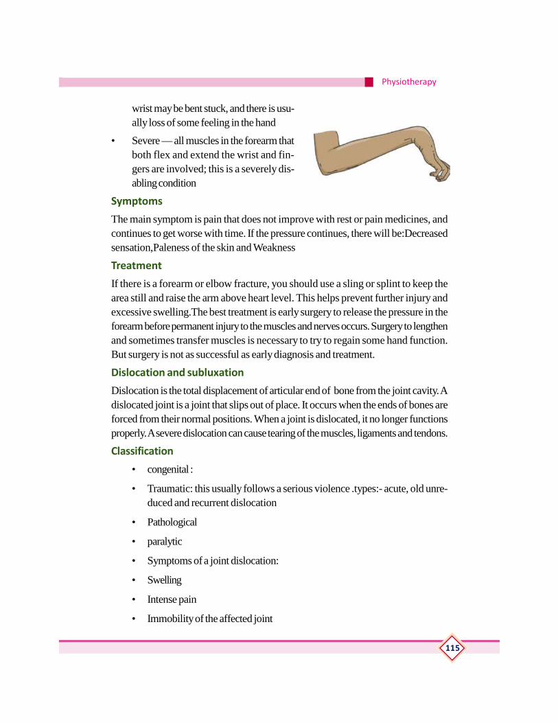
Physiotherapy
115
wrist may be bent stuck, and there is usu-ally loss of some feeling in the hand
• Severe — all muscles in the forearm thatboth flex and extend the wrist and fin-gers are involved; this is a severely dis-abling condition
Symptoms
The main symptom is pain that does not improve with rest or pain medicines, andcontinues to get worse with time. If the pressure continues, there will be:Decreasedsensation,Paleness of the skin and Weakness
Treatment
If there is a forearm or elbow fracture, you should use a sling or splint to keep thearea still and raise the arm above heart level. This helps prevent further injury andexcessive swelling.The best treatment is early surgery to release the pressure in theforearm before permanent injury to the muscles and nerves occurs. Surgery to lengthenand sometimes transfer muscles is necessary to try to regain some hand function.But surgery is not as successful as early diagnosis and treatment.
Dislocation and subluxation
Dislocation is the total displacement of articular end of bone from the joint cavity. Adislocated joint is a joint that slips out of place. It occurs when the ends of bones areforced from their normal positions. When a joint is dislocated, it no longer functionsproperly. A severe dislocation can cause tearing of the muscles, ligaments and tendons.
Classification
• congenital :
• Traumatic: this usually follows a serious violence .types:- acute, old unre-duced and recurrent dislocation
• Pathological
• paralytic
• Symptoms of a joint dislocation:
• Swelling
• Intense pain
• Immobility of the affected joint

Reference Book
116
Treatment
Treatment of the dislocation depends on the site and severity of injury and mayinclude:
• Reduction. During this process, doctor may try some gentle maneuvers to helpbones back into position. Depending on the amount of pain and swelling, needa local anesthetic or even a general anesthetic before manipulation of bones.
• Immobilization. After bones are back in their right positions, doctor may im-mobilize joint with a splint or sling for several weeks. Use of the splint or slingdepends on the joint involved and the extent of damage to nerves, blood ves-sels and supporting tissues.
• Surgery. May need surgery if doctor can’t move dislocated bones back intotheir correct positions or if the nearby blood vessels, nerves or ligaments havebeen damaged. Surgery may also be necessary if recurring dislocations, espe-cially of shoulder.
• Rehabilitation. After splint or sling is removed, begin a gradual rehabilitationprogram designed to restore joint’s range of motion and strength.
A subluxation is a Partial dislocation of a joint. Subluxation is often the result of atraumatic or acute injury. In these cases, a subluxation of a joint can be quite painful. Subluxations may lead to damage of the normal structures that support a joint. Forexample, when an ACL tear occurs, often the joint is starting to dislocate, and theligaments that normally support the knee joint are stretched too far, causing theknee joint to sublux.
Some of the common symptoms of a joint subluxation include:
• Pain around the injured joint
• A sensation of wanting to give-out or being unstable
• Swelling of the joint
• Limited mobility of the joint
Subluxations most often occur after an acute injury to the joint. Injuries can includesports injuries, car crashes, falls from a height, or even just simple twisting or reachingmovements. Often patients describe hearing a pop or snap of the joint when theinjury occurs. Subluxations can also occur as a result of loose joints. In patients withgeneralized joint laxity, their body may allow for joint subluxations without any traumaor injury.
Physiotherapy
117
Treatment of Joint Subluxation
Early treatment of a joint subluxation should begin by ensuring the joint is in properposition. Once the joint alignment is confirmed, treatment can be focused at reducinginflammation of the affected joint. Steps that can be helpful to reduce inflammationinclude:
• Rest: Resting the joint may require the use of a splint, sling, or crutches. De-pending on which joint is injured will determine what devices may help you torest the joint. Limiting the exertion of the joint and allowing the injured joint orest will help allow the inflammation subside.
• Ice: Icing an injured joint can be a very helpful step to reduce inflammation. Applying ice packs to the area frequently can help quite down an injured joint.
• Elevate: Elevation is helpful to reduce swelling and pain. Placing the injuredjoint above the level of the heart, and lying down can help allow the inflamma-tion to subside.
• Anti-Inflammatory Medications: Anti-inflammatory medications, often calledNSAIDs, are helpful tools to alleviate swelling and inflammation. By limitingthe inflammation they can also help to reduce pain.
• Splints/Supports/Braces: Braces are often helpful at controlling excessive mo-tion of an unstable joint. Until there can be a more definitive treatment plan, abrace can help to stabilize an unstable joint.
• Once the acute inflammation has subsided, a long-term treatment plan can bepursued. Many joint subluxations are one-time events, and patients will beable to recover full function of the joint. However, some other injuries cancause long-term and recurrent problems. If your symptoms of instability per-sist, then your should seek medical help to determine a longer-term plan.
Soft tissue injuryThe most common soft tissues injured are muscles, tendons, and ligaments. Theseinjuries often occur during sports and exercise activities, but sometimes simpleeveryday activities can cause an injury.Sprains, strains, and contusions, as well astendinitis and bursitis, are common soft-tissue injuries. Even with appropriatetreatment, these injuries may require a prolonged amount of time to heal.
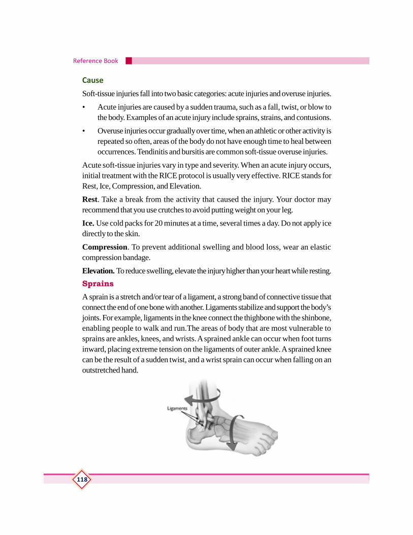
Reference Book
118
Cause
Soft-tissue injuries fall into two basic categories: acute injuries and overuse injuries.
• Acute injuries are caused by a sudden trauma, such as a fall, twist, or blow tothe body. Examples of an acute injury include sprains, strains, and contusions.
• Overuse injuries occur gradually over time, when an athletic or other activity isrepeated so often, areas of the body do not have enough time to heal betweenoccurrences. Tendinitis and bursitis are common soft-tissue overuse injuries.
Acute soft-tissue injuries vary in type and severity. When an acute injury occurs,initial treatment with the RICE protocol is usually very effective. RICE stands forRest, Ice, Compression, and Elevation.
Rest. Take a break from the activity that caused the injury. Your doctor mayrecommend that you use crutches to avoid putting weight on your leg.
Ice. Use cold packs for 20 minutes at a time, several times a day. Do not apply icedirectly to the skin.
Compression. To prevent additional swelling and blood loss, wear an elasticcompression bandage.
Elevation. To reduce swelling, elevate the injury higher than your heart while resting.
SprainsA sprain is a stretch and/or tear of a ligament, a strong band of connective tissue thatconnect the end of one bone with another. Ligaments stabilize and support the body’sjoints. For example, ligaments in the knee connect the thighbone with the shinbone,enabling people to walk and run.The areas of body that are most vulnerable tosprains are ankles, knees, and wrists. A sprained ankle can occur when foot turnsinward, placing extreme tension on the ligaments of outer ankle. A sprained kneecan be the result of a sudden twist, and a wrist sprain can occur when falling on anoutstretched hand.

Physiotherapy
119
A twisting force to the lower leg or foot is a common cause of ankle sprains.
Sprains are classified by severity:
Grade 1 sprain (mild): Slight stretching and some damage to the fibers (fibrils) ofthe ligament.
Grade 2 sprain (moderate): Partial tearing of the ligament. There is abnormallooseness (laxity) in the joint when it is moved in certain ways.
Grade 3 sprain (severe): Complete tear of the ligament. This causes significantinstability and makes the joint nonfunctional.
While the intensity varies, pain, bruising, swelling, and inflammation are common toall three categories of sprains. Treatment for mild sprains includes RICE andsometimes physical therapy exercises. Moderate sprains often require a period ofbracing. The most severe sprains may require surgery to repair torn ligaments.
Strains
A strain is an injury to a muscle and/or tendons. Tendons are fibrous cords of tissuethat attach muscles to the bone. Strains often occur in your foot, leg (typically thehamstring) or back. Similar to sprains, a strain may be a simple stretch in yourmuscle or tendon, or it may be a partial or complete tear in the muscle-and-tendoncombination. Typical symptoms of a strain include pain, muscle spasm, muscleweakness, swelling, inflammation, and cramping.
The recommended treatment for a strain is the same as for a sprain: rest, ice,compression and elevation. This should be followed by simple exercises to relievepain and restore mobility. Surgery may be required for a more serious tear.
PRICE therapy
PRICE stands for:
Protection – protect the affected area from further injury by using a support or, inthe case of an ankle injury, wearing shoes that enclose and support your feet, suchas lace-ups.
Rest – stop the activity that caused the injury and rest the affected joint or muscle.Avoid activity for the first 48 to 72 hours after injuring yourself. Your GP mayrecommend you use crutches.
Ice – for the first 48 to 72 hours after the injury; apply ice wrapped in a damp towelto the injured area for 15 to 20 minutes every two to three hours during the day.

Reference Book
120
Don’t leave the ice on while you’re asleep, and don’t allow the ice to touch yourskin directly because it could cause a cold burn.
Compression – compress or bandage the injured area to limit any swelling andmovement that could damage it further. You can use a simple elastic bandage or anelasticated tubular bandage available from a pharmacy. It should be wrapped snugglyaround the affected area, but not so tightly that it restricts blood flow. Remove thebandage before you go to sleep.
Elevation – keep the injured area raised and supported on a pillow to help reduceswelling. If your leg is injured, avoid long periods of time where your leg isn’t raised.
Mechanism, Clinical features and management of injury to lateral ligament of Anklejoint as an example.
The ankle is a hinge joint between the leg and the foot, and allows up and downmovement. The bones of the leg (tibia and fibula) form a slot, and the talus bone ofthe foot fits between them. The talus is held to the tibia and fibula by strong bands oftissue called ligaments. Each ligament is made of many strands or fibres of a materialcalled collagen, which is extremely strong.The ligament on the inside of the ankle(the deltoid ligament) has two layers; the deepest one is most important. This ligamentis mainly torn in association with severe fractures of the ankle bones. Sporting injuriesof this ligament are rare.The ligament on the outside of the ankle (lateral ligament) ismade up of three separate bands: one at the front (anterior talo-fibular ligament),one in the middle (calcaneo-fibular ligament) and one at the back (posterior talo-fibular ligament). The front and middle bands are the ligaments injured in a sprain.
Most ankle ligament injuries are caused when the foot twists inwards. All of thebody’s weight is then placed on the lateral ankle ligaments. Most ankle sprains arefairly minor injuries, which will get better with simple self-care treatment. The wordRICE reminds us of the basic treatment of a sprained joint.
Normally a sprained ankle will recover within 6-8 weeks, although it may tend toswell for a few months longer.
ArthritisArthritis is acute or chronic inflammation of a joint, often accompanied by pain andstructural changes and having diverse causes, as infection, crystal deposition, orinjury. Arthritis is more common among adults aged 65 years or older, but people ofall ages (including children) can be affected. There are more than 100 different typesof arthritis and related conditions. People of all ages, sexes and races can and dohave arthritis, and it is the leading cause of disability
Physiotherapy
121
Causes of arthritis
There is no single cause of all types of arthritis; the cause or causes in any given casevary according to the type or form of arthritis. Potential causes for arthritis mayinclude:
• njury - leading to degenerative arthritis
• Abnormal metabolism - leading to gout and pseudo gout
• Inheritance - such as in osteoarthritis
• ·Infections - such as in the arthritis of Lyme disease
• mmune system dysfunction - such as in RA and SLE
Common arthritis joint symptoms include swelling, pain, stiffness and decreased rangeof motion. Symptoms may come and go. They can be mild, moderate or severe.They may stay about the same for years, but may progress or get worse over time.Severe arthritis can result in chronic pain, inability to do daily activities and make itdifficult to walk or climb stairs. Arthritis can cause permanent joint changes.
Types of arthritis:
• Degenerative Arthritis
• Rheumatoid arthritis
• Inflammatory Arthritis
• Infectious Arthritis
• Metabolic Arthritis
Clinical features and general management of Rheumatoid arthritis
Rheumatoid arthritis is an autoimmune disease that can cause chronic inflammationof the joints and other areas of the body. Rheumatoid arthritis is characterized byperiods of disease flares and remissions. In rheumatoid arthritis, multiple joints areusually, but not always, affected in a symmetrical pattern. Chronic inflammation ofrheumatoid arthritis can cause permanent joint destruction and deformity. Damageto joints can occur early and does not always correlate with the severity of RA
Rheumatoid arthritis symptoms and signs include
• Joint pain in the feet, hands, and knees,
• Swollen joints,

Reference Book
122
• Fever,
• Tender joints,
• Loss of joint function,
• Stiff joints,
• Fatigue,
• Joint redness,
• Rheumatoid nodules,
• Joint warmth
• Joint deformity.
There is no cure for RA. The treatment of rheumatoid arthritis optimally involves acombination of patient education, rest and exercise, joint protection, medications,and occasionally surgery. Medications used in the treatment of rheumatoid arthritisinclude NSAIDs, DMARDs, TNF alpha inhibitors, IL-6 inhibitors, immunesuppressants, and steroids. Early RA treatment results in a better prognosis..
Rehabilitation Management for Rheumatoid Arthritis Patients
Rest: Rest and energy conservation can be helpful for locally inflamed joints, butshould be avoided long-term due to the potential deleterious side effects
Exercise : A structured exercise program can be greatly beneficial to the overallwell-being and functioning of the individual with rheumatoid arthritis. Such a programshould focus on stretching, strengthening and aerobic conditioning while conservingenergy
Stretching : Acutely, inflamed joints should be rested to prevent exacerbation ofsymptoms. For non-inflamed joints, active or active-assisted stretching of all majorjoints is essential to prevent contracture formation and maintain the current range ofmotion to perform most activities of daily living.
Strengthening: Strengthening exercises should be utilized in non-inflamed joints;while isometric exercises can help maintain strength to prevent injury or facilitatefatigue.
Areobic Conditioning: Several studies have shown the benefit from an aerobicconditioning or aquatic exercise program. in-water exercise would decrease theforces against joints, and the warmth could help decrease joint pain and decreasemuscle spasm. They noted an increase in strength and endurance from the waterexercise program..
Physiotherapy
123
Superficial Heat: Superficial heat has its greatest effect on the skin and subcutaneoustissues (Feibel 1976). It is especially useful in circumstances where the goal is toheat joints that are covered by little soft tissue such as those in the hands and feet
Deep Heat: Deep heating increases the tissue temperature at a deeper level withoutoverheating skin and subcutaneous fat
Cryotherapy : Cold therapy has several physiologic effects that enhance or suppressnormal responses to certain stressors. Hemodynamic effects include reflexivevasoconstriction followed by delayed vasodilation. Neuromuscular effects includeslowing of nerve conduction velocity, and decreased firing of the muscle spindles,which have been shown by some to reduce spasticity.
Electrical Nerve Stimulation: The primary delivery mechanism is via transcutaneouselectrical nerve stimulation .
Clinical features and general management of OsteoarthritisOsteoarthritis is the most common form of arthritis, affecting millions of peopleworldwide. It occurs when the protective cartilage on the ends of your bones wearsdown over time.Although osteoarthritis can damage any joint in your body, thedisorder most commonly affects joints in your hands, knees, hips andspine.Osteoarthritis often gradually worsens, and no cure exists. But staying active,maintaining a healthy weight and other treatments may slow progression of the diseaseand help improve pain and joint function.
Osteoarthritis occurs when the cartilage that cushions the ends of bones in yourjoints gradually deteriorates. Cartilage is a firm, slippery tissue that permits nearlyfrictionless joint motion. In osteoarthritis, the slick surface of the cartilage becomesrough. Eventually, if the cartilage wears down completely, you may be left with bonerubbing on bone.
Osteoarthritis symptoms often develop slowly and worsen over time. Signs andsymptoms of osteoarthritis include:
• Pain. Your joint may hurt during or after movement.
• Tenderness. Your joint may feel tender when you apply light pressure to it.
• Stiffness. Joint stiffness may be most noticeable when you wake up in the morningor after a period of inactivity.
• Loss of flexibility. You may not be able to move your joint through its full rangeof motion.

Reference Book
124
• Grating sensation. You may hear or feel a grating sensation when you use thejoint.
• Bone spurs. These extra bits of bone, which feel like hard lumps, may formaround the affected joint.
Treatments:There’s no known cure for osteoarthritis, but treatments can help reduce pain andmaintain joint movement.
Physical therapy. A physical therapist can work to create an individualized exerciseprogram that will strengthen the muscles around joint, increase range of motion andreduce pain.
Braces or shoe inserts. Doctor may recommend shoe inserts or other devicesthat can help reduce pain when stand or walk. These devices can immobilize orsupport your joint to help take pressure off it.
Surgical and other procedures:If conservative treatments don’t help, you maywant to consider procedures such as: Cortisone shots, Lubrication injections,Realigning bones and Joint replacement.
Clinical features and general management of AnkylosingspondylitisAnkylosing spondylitis is an inflammatory disease that can cause some of the vertebraein your spine to fuse together. This fusing makes the spine less flexible and can resultin a hunched-forward posture. If ribs are affected, it may be difficult to breathedeeply. Ankylosing spondylitis affects men more often than women. Signs andsymptoms of ankylosing spondylitis typically begin in early adulthood. There is nocure for ankylosing spondylitis, but treatments can decrease your pain and lessenyour symptoms.
Symptoms: The most common early symptoms of ankylosing spondylitis include:
• Pain and stiffness: Constant pain and stiffness in the low back, buttocks, andhips that continue for more than three months. Spondylitis often starts aroundthe sacroiliac joints, where the sacrum (the lowest major part of the spine)joins the ilium bone of the pelvis in the lower back region.
• Bony fusion: Ankylosing spondylitis can cause an overgrowth of the bones,which may lead to abnormal joining of bones, called “bony fusion.” Fusionaffecting bones of the neck, back, or hips may impair a person’s ability toperform routine activities. Fusion of the ribs to the spine or breastbone maylimit a person’s ability to expand his or her chest when taking a deep breath.
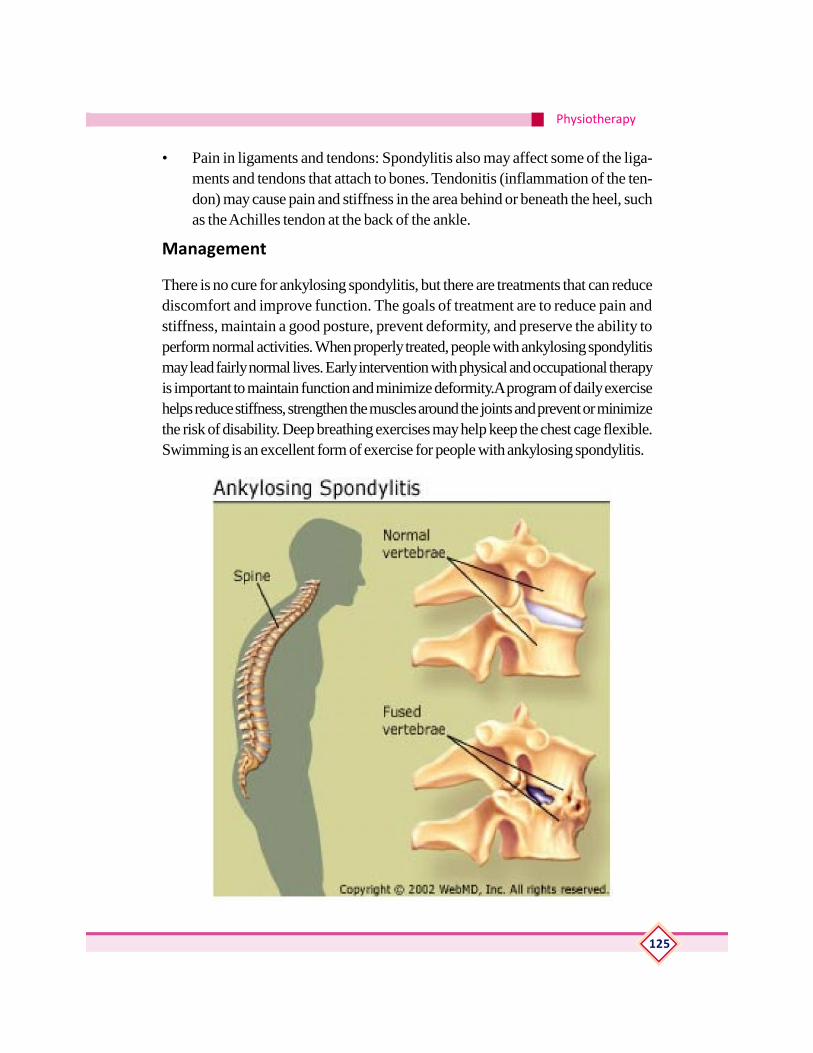
Physiotherapy
125
• Pain in ligaments and tendons: Spondylitis also may affect some of the liga-ments and tendons that attach to bones. Tendonitis (inflammation of the ten-don) may cause pain and stiffness in the area behind or beneath the heel, suchas the Achilles tendon at the back of the ankle.
Management
There is no cure for ankylosing spondylitis, but there are treatments that can reducediscomfort and improve function. The goals of treatment are to reduce pain andstiffness, maintain a good posture, prevent deformity, and preserve the ability toperform normal activities. When properly treated, people with ankylosing spondylitismay lead fairly normal lives. Early intervention with physical and occupational therapyis important to maintain function and minimize deformity.A program of daily exercisehelps reduce stiffness, strengthen the muscles around the joints and prevent or minimizethe risk of disability. Deep breathing exercises may help keep the chest cage flexible.Swimming is an excellent form of exercise for people with ankylosing spondylitis.

Reference Book
126
Awareness of the following orthopaedic conditions1. Cervical and lumbar spondylosis
Cervical spondylosis is a general term for age-related wear and tear affecting thespinal disks in your neck. As the disks dehydrate and shrink, signs of osteoarthritisdevelop, including bony projections along the edges of bones (bone spurs). Cervicalspondylosis is very common and worsens with age. More than 85 percent of peopleolder than age 60 are affected by cervical spondylosis. Most people experience nosymptoms from these problems. When symptoms do occur, (include pain and stiffness)in the neck nonsurgical treatments often are effective. Sometimes, cervical spondylosisresults in a narrowing of the space needed by the spinal cord and the nerve rootsthat pass through the spine to the rest of your body. If the spinal cord or nerve rootsbecome pinched, you might experience:
• Tingling, numbness and weakness in your arms, hands, legs or feet
• Lack of coordination and difficulty walking
• Loss of bladder or bowel control
Causes
As age increases, the bones and cartilage that make up your backbone and neckgradually develop wear and tear. These changes can include:Dehydrated disks,Herniated disks,Bone spurs and Stiff ligaments.
Treatment
Treatment for cervical spondylosis depends on the severity of your signs andsymptoms. The goal of treatment is to relieve pain, help you maintain your usualactivities as much as possible, and prevent permanent injury to the spinal cord andnerves.
A physical therapist can teach exercises to help stretch and strengthen the muscles inneck and shoulders. Some people with cervical spondylosis benefit from the use oftraction, which can help provide more space within the spine if nerve roots are beingpinched.
Acupuncture: Recommend trying acupuncture to reduce pain.
Surgery: If conservative treatment fails or if neurological signs and symptoms —such as weakness in your arms or legs — worsen, might need surgery to createmore room for spinal cord and nerve roots.
The surgery might involve: Removing a herniated disk or bone spurs, Removing partof a vertebra and Fusing a segment of the neck using bone graft .

Physiotherapy
127
Lumbar spondylosis
Spondylosis is degenerative osteoarthritis of the joints between the spinal vertebraeand/or neural foramina. Lower back spondylosis or osteoarthritis is known as lumbarspondylosis.
Lumbar spondylitis symptoms and treatment: Symptoms of spondylitis varydepending on the individual and the location and severity of the condition. Thecommon symptoms of spondylitis include:
• Stiffness and pain in the neck or back
• Loss of mobility in the spine
• Inability to twist and turn, or to do so without pain
• ·Chronic pain and discomfort when standing, sitting, or walking
• Deformity or abnormal spine curvature
• There are several conservative treatments to reduce chronic pain, includingphysical therapy, yoga and medication.
2.Periarthritis shoulder
Periarthritis is a condition charecterised by pericapsular adhesions following a minortrauma to the degenerated rotator cuff.Patient past middle age, presents with diffusepain in the shoulder with radiation down to the middle of the upper arm.It is commonerin patients with diabetes and myocardial ischemia. On examination there will betenderness in the subacromial region and in the anterior joint line. There is markedlimitation of abduction and external rotation of the shoulder.
When the condition involves the whole rotator cuff, it results in total restriction of allmovements of joint. The condition is then termed as frozen shoulder .Treatmentafter controlling the pain with analgesics and short wave diathermy is vigorousshoulder mobilization exercises. Resisted cases are treated by local infiltration ofhydrocortisone and manipulation under anaesthesia.

Reference Book
128
3. Tennis elbow
Tennis elbow or lateral epicondylitis is a condition in which the outer part of theelbow becomes sore and tender. The forearm muscles and tendons become damagedfrom overuse — repeating the same strenuous motions again and again. This leadsto pain and tenderness on the outside of the elbow. Any activity, including playingtennis, which involves the repetitive use of the extensor muscles of the forearm cancause acute or chronic tendonitis of the tendinous insertion of these muscles at thelateral epicondyle of the elbow. The condition is common in carpenters and otherlaborers who swing a hammer or other tool with the forearm.
4. Golfers elbow
Golfer’s elbow is a condition that causes pain where the tendons of forearm musclesattach to the bony bump on the inside of elbow. Golfer’s elbow is characterized by.• Pain and tenderness on the inner side of elbow: Sometimes the pain extends
along the inner side of forearm. Pain typically worsens with certain movements.
• Stiffness: Elbow may feel stiff, and it may hurt to make a fist.
• Weakness: May have weakness in hands and wrists.
• Numbness or tingling. These sensations might radiate into one or more fingers— usually the ring and little fingers.
Treatment:
The sooner you begin treatment, the sooner you’ll be able to return to your usualactivities.
Rest.:Put golf game or other repetitive activities on hold until the pain is gone. Ifreturn to activity too soon, may make it worse.
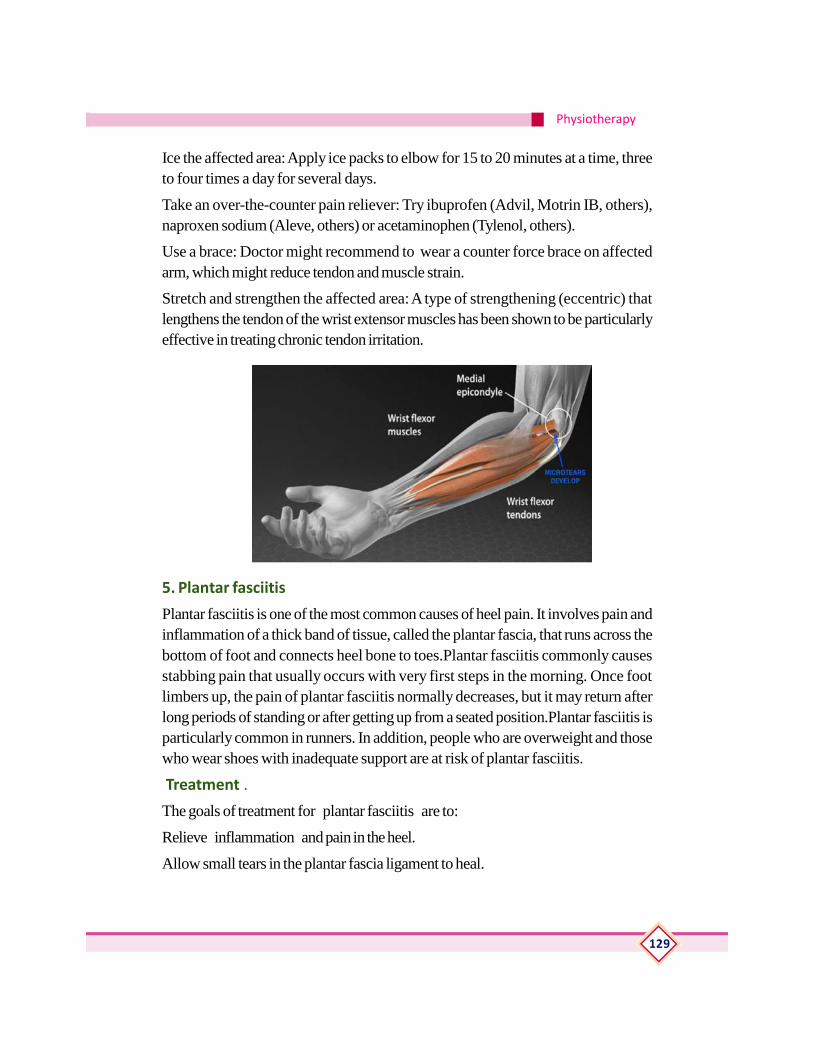
Physiotherapy
129
Ice the affected area: Apply ice packs to elbow for 15 to 20 minutes at a time, threeto four times a day for several days.
Take an over-the-counter pain reliever: Try ibuprofen (Advil, Motrin IB, others),naproxen sodium (Aleve, others) or acetaminophen (Tylenol, others).
Use a brace: Doctor might recommend to wear a counter force brace on affectedarm, which might reduce tendon and muscle strain.
Stretch and strengthen the affected area: A type of strengthening (eccentric) thatlengthens the tendon of the wrist extensor muscles has been shown to be particularlyeffective in treating chronic tendon irritation.
5. Plantar fasciitis
Plantar fasciitis is one of the most common causes of heel pain. It involves pain andinflammation of a thick band of tissue, called the plantar fascia, that runs across thebottom of foot and connects heel bone to toes.Plantar fasciitis commonly causesstabbing pain that usually occurs with very first steps in the morning. Once footlimbers up, the pain of plantar fasciitis normally decreases, but it may return afterlong periods of standing or after getting up from a seated position.Plantar fasciitis isparticularly common in runners. In addition, people who are overweight and thosewho wear shoes with inadequate support are at risk of plantar fasciitis.
Treatment .
The goals of treatment for plantar fasciitis are to:
Relieve inflammation and pain in the heel.
Allow small tears in the plantar fascia ligament to heal.

Reference Book
130
Improve strength and flexibility and correct foot problems such asexcessive pronation so that don’t stress the plantar fascia ligament.
Rest feet. Limit or, if possible, stop daily activities that are causing heel pain. Try toavoid running or walking on hard surfaces, such as concrete.
To reduce inflammation and relieve pain, put ice on heel. Try a nonsteroidal anti-inflammatory drug (NSAID) such asibuprofen or naproxen . NSAIDs come in pillsand in a cream that rub over the sore area.
Wear shoes with good shock absorptionand the right arch support for foot. Athleticshoes or shoes with a well-cushioned soleare usually good choices.
Try heel cups or shoe inserts to help cushionheel.
Put on your shoes as soon as you get outof bed. Going barefoot or wearing slippersmay make your pain worse.
AmputationAmputation is the removal of the whole or part of a limb by cutting through bone orjoint.
Indication of amputation
1. Traumatic conditions: Crush injuries to the limb
2. Vascular conditions: ischemic condition of the limb-Atherosclerosis, gan-grene etc
3. Neoplastic conditions: Malignant tumors of bone or soft tissue
4. Infective conditions: Leprosy, filarial elephantiasis etc
5. Congenital conditions: when the limb is grossly deformed and useless.
Types of amputation
Classified according to the surgical technique or the emergency of situation
1. Guillotine Amputation: Used when primary healing is unlikely or delayedbecause of infection, ischemia, or inadequate wound debridement. It is doneas an emergency procedure , to save the life of the patient.
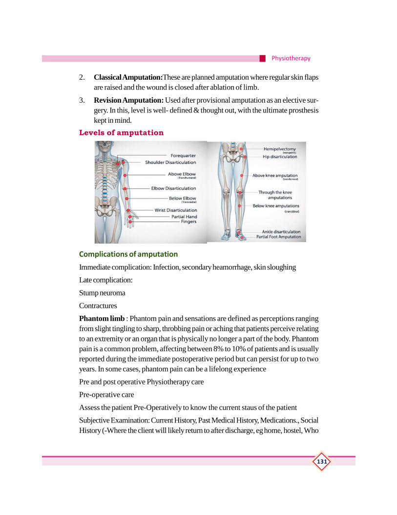
Physiotherapy
131
2. Classical Amputation:These are planned amputation where regular skin flapsare raised and the wound is closed after ablation of limb.
3. Revision Amputation: Used after provisional amputation as an elective sur-gery. In this, level is well- defined & thought out, with the ultimate prosthesiskept in mind.
Levels of amputation
Complications of amputation
Immediate complication: Infection, secondary heamorrhage, skin sloughing
Late complication:
Stump neuroma
Contractures
Phantom limb : Phantom pain and sensations are defined as perceptions rangingfrom slight tingling to sharp, throbbing pain or aching that patients perceive relatingto an extremity or an organ that is physically no longer a part of the body. Phantompain is a common problem, affecting between 8% to 10% of patients and is usuallyreported during the immediate postoperative period but can persist for up to twoyears. In some cases, phantom pain can be a lifelong experience
Pre and post operative Physiotherapy care
Pre-operative care
Assess the patient Pre-Operatively to know the current staus of the patient
Subjective Examination: Current History, Past Medical History, Medications., SocialHistory (-Where the client will likely return to after discharge, eg home, hostel, Who
Reference Book
132
is at home to assist in ADL’s, or could participate / supervise in their rehabilitationetc ) Vocational activities, Premorbid Mobility Levels, Pain etc
Objective Examination: Joint Range of motion, Muscle Lengths, Muscle Strength,Condition of intact limb, Arm Function, Functional Assessments etc
Instructions on Post-Operative Exercises and Positioning
Short term goals include: Maintenance of respiratory function, Maintenance ofstrength, Maintenance of range of motion & muscle lengths and Maintenance ofcardiovascular fitness.
Post-operative care
Maintain function in the remaining leg and stump to maintain peripheral circulation.Maintain respiratory function Mobility Aids: The choice of mobility aids depends onthe level of fitness, strength, balance skills of the individual: Walking frame, Axillarycrutches, Elbow crutches, Walking stick and For bilateral lower limb amputees awheelchair is indicated
Stump and its Management
Stump is the residual part of the limb which is left after amputation
Amputees will be taught to care for their stump by either a nurse or a physiotherapist.Itis extremely important that to master the techniques of caring stump. If utmostattention is not given to this care then small easily reversible problems may developinto large ones which become difficult to treat.
Care of the Stump – Keep the stump clean, dry, and free from infection at all times.– If fitted with a prosthesis, you should remove it before going to sleep. – Inspectand wash the stump with mild soap and warm water every night, then dry thoroughlyand apply talcum powder. – Do not use the prosthesis until the skin has healed. –The stump sock should be changed daily, and the inside of the socket may be cleanedwith mild soap.
RESUDIAL LIMB WRAPPING Eary wrapping provides a number of positivebenefits: Decrease odema & venous stasis , Assist in shaping , Help in counteractcontracture , Provide skin protection , Reduce redundant tissue problems, Reducephantom limb sensation and discomfort and Desensitize the residual limb with localpain.
Physiotherapy
133
Practical1. Application of price therapy in soft tissue injury
2. Application of elastocrepe bandage in lateral ligament injury
3. Demonstration of exercise for rheumatoid arthritis
• Joint mobility exercise
• Exercises for knee joint osteoarthritis
• Isometric
• Mobility
• Strengthening
4. Demonstration of exercises for cervical and lumbar exercises
• Isometric neck exercise
• Neck mobility exercise
• Spinal extension /flexion exercise
5. Preparation and positioning of client for electro therapy.
• IRR for per arthritis
• US for tennis/golfers/plantar fasciitis
Assessment ActivitiesSeminar
Assignments
Field visit
Chart preparation
Quiz
Collection
Model Preparation
Reference Book
134
TE Questions1. Write any four differences between RA and OA
2. Briefly explain about stump management
3. List out the complications of fracture
4. Partial displacement of articular surfaces of joint is known as ————-
5. Differentiate between
• Dislocation and subluxation
• Sprain and strain-
6. Short note on
• Plantar fasciitis
• Tennis elbow
• Ankylosing spondylitis
• Periarthritis shoulder

Physiotherapy
135
INTRODUCTION OF PAEDIATRIC CONDITIONS ANDRELEVANCE OF PHYSIOTHERAPY
IntroductionPaediatric physiotherapists work with people of varying ages from premature babiesto adolescents to ensure optimal physical function and development. Like allphysiotherapists, they are concerned with movement, co-ordination, posture andthe cardiorespiratory system. The aim of the paediatric physiotherapist is to providea program that the client will enjoy, while encouraging them to participate and becomeindependent.
Paediatric physiotherapists aim to minimise the effects of physical impairment topromote optimum function and musculoskeletal development. Advice on activitiesand stretches offered by the physiotherapist can assist in maintaining full range ofmovement and prevention of contracture. Paediatric Physiotherapists treat manyconditions similar to those in general physiotherapy practice. However, there aremany conditions which are related almost entirely to paediatrics. Some of these arerecognised at birth, others may become apparent later in the child’s life.Learning outcomesThe learner:
• To understand the condition Cerebral palsy
• To assist the physiotherapist in management of cerebral palsy
• To do the corrective movement of CTEV
• To identify and assist in the management of Erb’s Palsy ,CDH ,Spina Bifidaand Torticollis
ConceptsCerebral palsy
Cerebral palsy is refers to a group of disorders affecting a person’s ability tomove. It is due to damage to the developing brain either during pregnancy or shortlyafter birth.Cerebral palsy affects people in different ways and can affect bodymovement, muscle control, muscle coordination, muscle tone, reflex, posture andbalance. Although cerebral palsy is a permanent life-long condition, some of thesesigns of cerebral palsy can improve or worsen over time.People who have cerebral

Reference Book
136
palsy may also have visual, learning, hearing, speech, epilepsy and intellectualimpairments.Cerebral palsy is the result of a combination of events either before,during or after birth that can lead to an injury in a baby’s developing brain. There isno single cause of cerebral palsy. For most babies born with cerebral palsy, thecause remains unknown. Researchers now know that only a very small percentageof cases of cerebral palsy are due to complications at birth (e.g. asphyxia or lack ofoxygen). It is accepted that cerebral palsy usually arises from a series of causalpathways, i.e. a sequence of events that when combined can cause or accelerateinjury to the developing brain.
Stroke is the most common cause in babies who acquire cerebral palsy after 1month of age. The stroke may occur spontaneously or arise from surgical or heartcomplications
CTEVCongenital talipes equino varus also known as ‘club foot’, is a congenital foot deformitypresent at birth. It is one of the most common congenital deformities. Incidencevaries between ethnic groups.
Clinical Presentation : Foot plantar flexed at ankle,Inverted at subtaloid joint,Adductedat forefoot
Foot become smaller than normal, Heel poorly devoloped, Crease across the medialborder, Lateral malleolus prominent. Medial malleolus burried in to depression. Footcannot be dorsiflexed and everted .Wasting of calf muscle.
Principle of treatment
Correct the deformity-hold the foot in corrected position
Treatment should begin in the first few weeks of life
Different component of deformity should be corrected in the proper sequences,
Fore foot adduction first, Invertion next, equinus last-failure to follow –break thefoot—use minimum force
Conservative treatment
1st week-mother is taught to mould the infants foot twice a day,3 weeks to 3months-surgeon manipulate once in 2weeks,maintained by pop,After 4or 5manipulation correction is maintained by splint-deni‘s brown splint
Physiotherapy
137
Surgical treatment
If correction is not achieved by POP, surgery is indicated.Soft tissue operation-postero medial soft tissue release, complete subtaloid release,Tendo achiluslengthening,alligaments and soft tissue release,Bone operation-lateral wedgeresection,calcaneo cuboid fusion.
CDHCongenital hip dislocation also known as dysplasia of the hip is a condition in whicha child is born with a hip problem. Congenital hip dislocation is when the formationof the hip joint is abnormal. The ball at the top of the thighbone which is known asthe femoral head is not stable within the socket (which is also known as theacetabulum). This abnormality may cause the ligaments of the hip to be loose orstretched. This condition is usually diagnosed once the baby is born; it mostly affectsthe left side of the hip in first-born children, girls, and babies born in a breech position.Girls are four times as likely to have hip dysplasia compared to boys. The cause ofthis condition is still unknown; however, some factors of congenital hip dislocationare through heredity and racial background. It is also thought that the higher incidencein some ethnic groups
The early sign of congenital hip dislocation is when a person is able to hear “clicking”sounds when the legs are moved apart from one another. This condition can betreated if detected early. If this condition goes undetected it can cause one leg tolook shorter than its counterpart and the buttock folds are also not symmetricalwhich causes more creases to be present on the affected side, and skin folds at thethigh are uneven.
Treatment
Aim-Reduce the hip and maintain it in the reduced position.Treatment is age relatedand depend on the age at which child is brought.
Below 3 month-von rosen splin,
3- 6 month –reduced by gentle manipulation under general anaesthesia.
6 - 2 month-preliminary traction and gradual abduction
2 - 3 weekplaster spica applied
1 - 3 years-surgical open reduction
3 - 6 years-open reduction and osteotomy of femur,hip bone
Reference Book
138
Spina bifidaSpina bifida is a birth defect where there is incomplete closing of the backbone andmembranes around the spinal cord. There are three main types: spina bifida occulta,meningocele, and myelomeningocele. The most common location is the lower back,but in rare cases it may be the middle back or neck.
Signs and symptoms
• Physical signs of spina bifida may include:
• Leg weakness and paralysis
• Orthopedic abnormalities (i.e., club foot, hip dislocation, scoliosis)]
• Bladder and bowel control problems, including incontinence, urinary tract in-fections, and poor kidney function
• Pressure sores and skin irritations
• Abnormal eye movement
Treatment
There is no known cure for nerve damage caused by spina bifida. Standard treatmentis surgery after delivery. This surgery aims to prevent further damage of the nervoustissue and to prevent infection
TorticollisTorticollis is the Deformity of the neck charecterised by tilting of head to one side,Rotation of the occiput to one side and chin to opposite side. Contracture ofsternocledomastoid on one side may be due to myodysplasia of genetic origin, Birthtrauma, hematoma,or arterial ischemia
Clinical feature: Firm swelling at the junction of midle and distal third ofsternomastoid muscle
In left torticolis—the chin is tilted upward to the right side and the occiput is tilteddown on the left side
In neglected cases contracture is severe and cannot be stretched passively
TreatmentMild deformity: -physiotherapy, Heat modality, Passive movement to cervical spine,Passive stretching of sternocledomastoid.
After correction –maintained by passive hold.Home treatment-train the mother.
During sleep-posisioning by pillows and sand bags
Physiotherapy
139
Practical1. Demonstrate the correction of deformity in the following conditions
CTEV
TORTICOLLIS
Assessment ActivitiesSeminar
Assignments
Field visit
Chart preparation
Quiz
Collection
Model Preparation
TE QuestionsDefine cerebral palsy
Write about the position of neck in right side torticollis
Write about the deformity in CTEV
Name the splint used in
CDH
CTEV
Reference Book
140
INTRODUCTION OF PHYSIOTHERAPY INSURGICAL CONDITION
IntroductionThe physiotherapist has an important role to play in assessing which patients beingprepared for surgery are at risk of developing complications that she may help toprevent. The physiotherapist must take into consideration the effects of the anestheticon secretions in the respiratory tract. The physiotherapist will explain to the patientwhy treatment is necessary and teach the patient the exercises that he will be requiredto perform post–operatively.Learning out comes• To familiarize the importance of physiotherapy in Head injury
• To familiarize with the importance of physiotherapy in Spinal cord injury
• To familiarize with the importance of physiotherapy in Burns
ConceptsHead injury
A head injury is any sort of injury to brain, skull, or scalp. This can range from a mildbump or bruise to a traumatic brain injury. Common head injuries include concussions,skull fractures, and scalp wounds. The consequences and treatments vary greatly,depending on what caused head injury and how severe it is.Head injuries may beeither closed or open. A closed head injury is any injury that doesn’t break skull. Anopen, or penetrating, head injury is one in which something breaks skull and enterbrain.It can be hard to assess how serious a head injury is just by looking. Someminor head injuries bleed a lot, while some major injuries don’t bleed at all. It’simportant to treat all head injuries seriously and get them assessed by a surgeon.
Causes
In general, head injuries can be divided into two categories based on what causesthem. They can either be head injuries due to blows to the head or head injuries dueto shaking.
Head injuries caused by shaking are most common in infants and small children, butthey can occur any time due to violent shaking.
Head injuries caused by a blow to the head are usually associated with:
motor vehicle accidents
falls
Physiotherapy
141
physical assaults
sports-related accidents
Major Types of Head Injuries
Hematoma
A hematoma is a collection, or clotting, of blood outside the blood vessels. It can bevery serious if a hematoma occurs in the brain. The clotting can cause pressure tobuild inside skull, which can cause to lose consciousness.
Hemorrhage
A hemorrhage is uncontrolled bleeding. There can be bleeding in the space aroundbrain, which is a subarachnoid hemorrhage, or bleeding within brain tissue, which isan intracerebral hemorrhage.
Subarachnoid bleeds often cause headaches and vomiting.
Concussion
A concussion is a brain injury that occurs when brain bounces against the hard wallsof skull. Generally speaking, the loss of function associated with concussions istemporary. However, repeated concussions can eventually lead to permanent damage.
Edema
Any brain injury can lead to edema, or swelling. Many injuries cause swelling of thesurrounding tissues, but it’s more serious when it occurs in brain. Skull can’t stretchto accommodate the swelling, which leads to a buildup of pressure in brain. This cancause brain to press against skull.
Skull Fracture
Skull doesn’t have bone marrow. This makes the skull very strong and difficult tobreak. A broken skull is unable to absorb the impact, making it more likely that therewill also be damage to brain.
Diffuse Axonal Injury
A diffuse axonal injury, or sheer injury, is an injury to the brain that doesn’t causebleeding but does damage brain cells. Though it isn’t as outwardly visible as otherforms of brain injury, diffuse axonal injury is one of the most dangerous types ofhead injuries and can lead to permanent brain damage and even death.

Reference Book
142
Symptoms of a Head InjuryCommon symptoms of a minor head injury include:
• Headache
• Mild confusion
• Nausea
• Temporary ringing in the ears
• Loss of consciousness
• Seizures
• Vomiting
• Balance or coordination problems
• Abnormal eye movements
• A loss of muscle control
• A persistent or worsening headache
• Memory loss
• Changes in mood
Treatment
The treatment for head injuries depends on both the type and the severity of theinjury.With minor head injuries, there are often no symptoms other than pain at thesite of the injury. If have an open cut, may use sutures or staples to close it. Even ifinjury seems minor, watch condition to make sure it doesn’t get worse. May needto be hospitalized if serious head injury occur. The treatment at the hospital willdepend on diagnosis.
The treatment for severe head injuries can include:Medication, Surgery andRehabilitation
PhysiotherapyPhysiotherapy helps restore movement and function to as near normal as possiblewhen someone is affected by a brain injury. Neuromuscular (brain, nervous systemand musculoskeletal system) specialist physiotherapist play an essential role in themultidisciplinary team, working with brain injured persons. The role of aphysiotherapist starts right from the acute stage (initial days after the injury) of braininjury through the rehabilitation phase to the long term care.
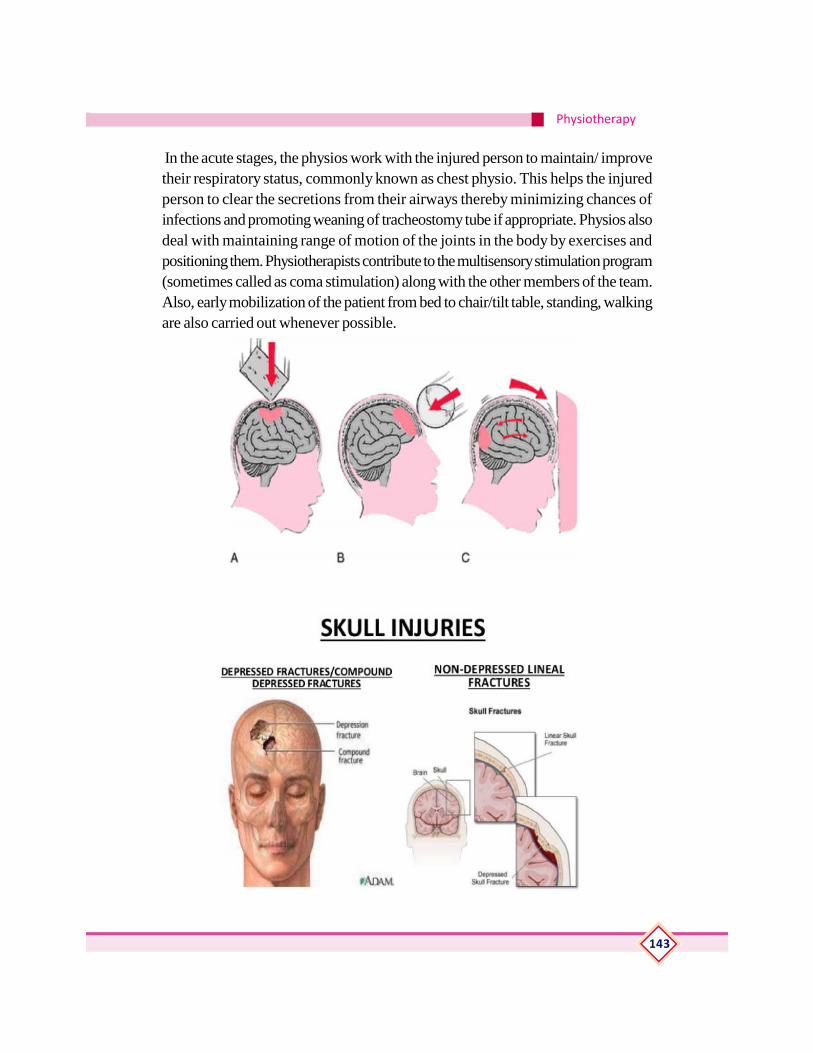
Physiotherapy
143
In the acute stages, the physios work with the injured person to maintain/ improvetheir respiratory status, commonly known as chest physio. This helps the injuredperson to clear the secretions from their airways thereby minimizing chances ofinfections and promoting weaning of tracheostomy tube if appropriate. Physios alsodeal with maintaining range of motion of the joints in the body by exercises andpositioning them. Physiotherapists contribute to the multisensory stimulation program(sometimes called as coma stimulation) along with the other members of the team.Also, early mobilization of the patient from bed to chair/tilt table, standing, walkingare also carried out whenever possible.
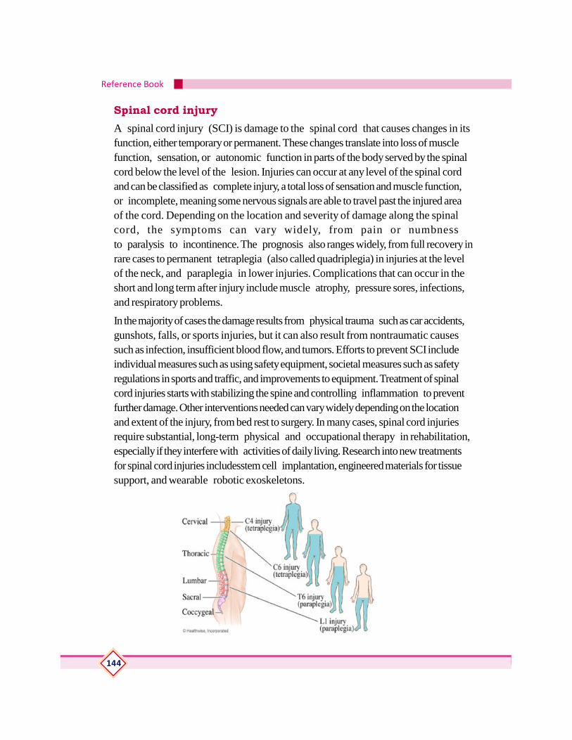
Reference Book
144
Spinal cord injuryA spinal cord injury (SCI) is damage to the spinal cord that causes changes in itsfunction, either temporary or permanent. These changes translate into loss of musclefunction, sensation, or autonomic function in parts of the body served by the spinalcord below the level of the lesion. Injuries can occur at any level of the spinal cordand can be classified as complete injury, a total loss of sensation and muscle function,or incomplete, meaning some nervous signals are able to travel past the injured areaof the cord. Depending on the location and severity of damage along the spinalcord, the symptoms can vary widely, from pain or numbnessto paralysis to incontinence. The prognosis also ranges widely, from full recovery inrare cases to permanent tetraplegia (also called quadriplegia) in injuries at the levelof the neck, and paraplegia in lower injuries. Complications that can occur in theshort and long term after injury include muscle atrophy, pressure sores, infections,and respiratory problems.
In the majority of cases the damage results from physical trauma such as car accidents,gunshots, falls, or sports injuries, but it can also result from nontraumatic causessuch as infection, insufficient blood flow, and tumors. Efforts to prevent SCI includeindividual measures such as using safety equipment, societal measures such as safetyregulations in sports and traffic, and improvements to equipment. Treatment of spinalcord injuries starts with stabilizing the spine and controlling inflammation to preventfurther damage. Other interventions needed can vary widely depending on the locationand extent of the injury, from bed rest to surgery. In many cases, spinal cord injuriesrequire substantial, long-term physical and occupational therapy in rehabilitation,especially if they interfere with activities of daily living. Research into new treatmentsfor spinal cord injuries includesstem cell implantation, engineered materials for tissuesupport, and wearable robotic exoskeletons.
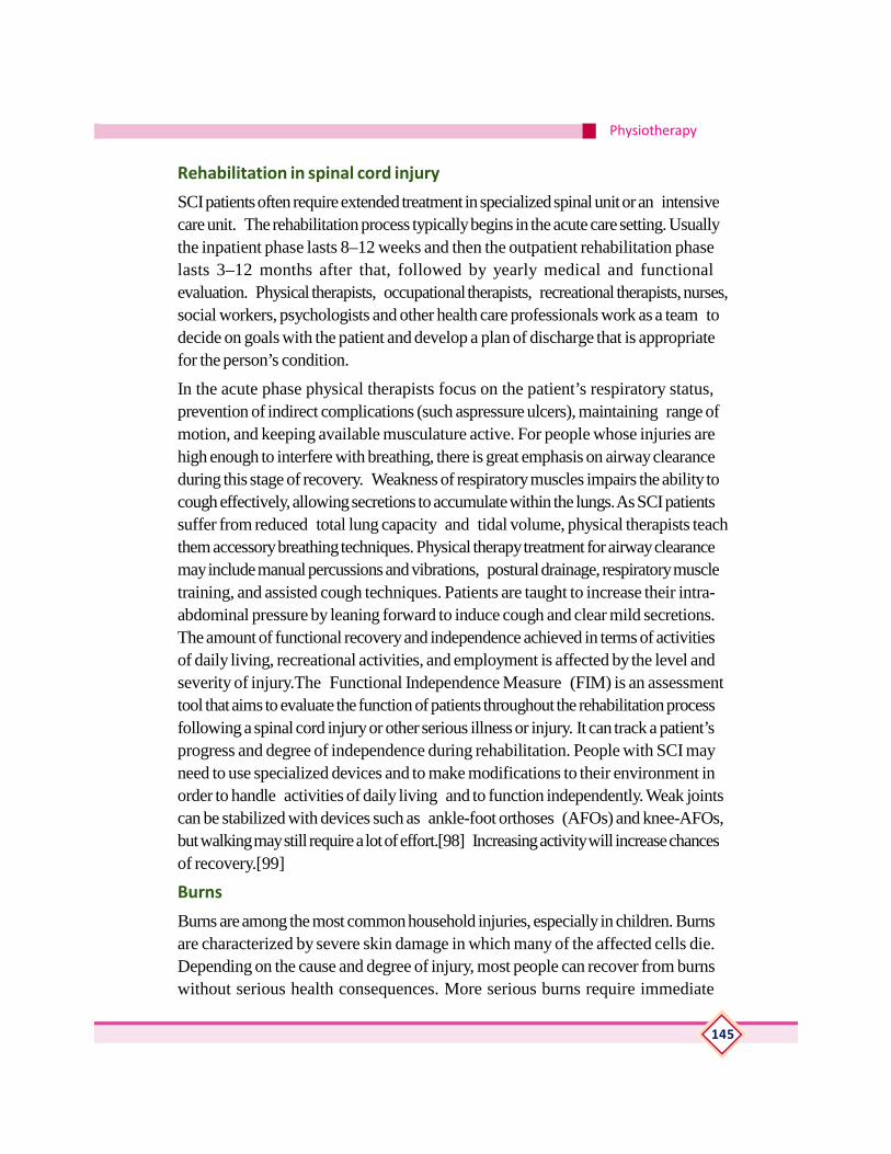
Physiotherapy
145
Rehabilitation in spinal cord injury
SCI patients often require extended treatment in specialized spinal unit or an intensivecare unit. The rehabilitation process typically begins in the acute care setting. Usuallythe inpatient phase lasts 8–12 weeks and then the outpatient rehabilitation phaselasts 3–12 months after that, followed by yearly medical and functionalevaluation. Physical therapists, occupational therapists, recreational therapists, nurses,social workers, psychologists and other health care professionals work as a team todecide on goals with the patient and develop a plan of discharge that is appropriatefor the person’s condition.
In the acute phase physical therapists focus on the patient’s respiratory status,prevention of indirect complications (such aspressure ulcers), maintaining range ofmotion, and keeping available musculature active. For people whose injuries arehigh enough to interfere with breathing, there is great emphasis on airway clearanceduring this stage of recovery. Weakness of respiratory muscles impairs the ability tocough effectively, allowing secretions to accumulate within the lungs. As SCI patientssuffer from reduced total lung capacity and tidal volume, physical therapists teachthem accessory breathing techniques. Physical therapy treatment for airway clearancemay include manual percussions and vibrations, postural drainage, respiratory muscletraining, and assisted cough techniques. Patients are taught to increase their intra-abdominal pressure by leaning forward to induce cough and clear mild secretions.The amount of functional recovery and independence achieved in terms of activitiesof daily living, recreational activities, and employment is affected by the level andseverity of injury.The Functional Independence Measure (FIM) is an assessmenttool that aims to evaluate the function of patients throughout the rehabilitation processfollowing a spinal cord injury or other serious illness or injury. It can track a patient’sprogress and degree of independence during rehabilitation. People with SCI mayneed to use specialized devices and to make modifications to their environment inorder to handle activities of daily living and to function independently. Weak jointscan be stabilized with devices such as ankle-foot orthoses (AFOs) and knee-AFOs,but walking may still require a lot of effort.[98] Increasing activity will increase chancesof recovery.[99]
Burns
Burns are among the most common household injuries, especially in children. Burnsare characterized by severe skin damage in which many of the affected cells die.Depending on the cause and degree of injury, most people can recover from burnswithout serious health consequences. More serious burns require immediate
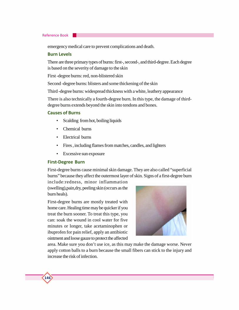
Reference Book
146
emergency medical care to prevent complications and death.
Burn Levels
There are three primary types of burns: first-, second-, and third-degree. Each degreeis based on the severity of damage to the skin
First -degree burns: red, non-blistered skin
Second -degree burns: blisters and some thickening of the skin
Third -degree burns: widespread thickness with a white, leathery appearance
There is also technically a fourth-degree burn. In this type, the damage of third-degree burns extends beyond the skin into tendons and bones.
Causes of Burns
• Scalding from hot, boiling liquids
• Chemical burns
• Electrical burns
• Fires , including flames from matches, candles, and lighters
• Excessive sun exposure
First-Degree Burn
First-degree burns cause minimal skin damage. They are also called “superficialburns” because they affect the outermost layer of skin. Signs of a first-degree burninclude:redness, minor inflammation(swelling),pain,dry, peeling skin (occurs as theburn heals).
First-degree burns are mostly treated withhome care. Healing time may be quicker if youtreat the burn sooner. To treat this type, youcan: soak the wound in cool water for fiveminutes or longer, take acetaminophen oribuprofen for pain relief, apply an antibioticointment and loose gauze to protect the affectedarea. Make sure you don’t use ice, as this may make the damage worse. Neverapply cotton balls to a burn because the small fibers can stick to the injury andincrease the risk of infection.
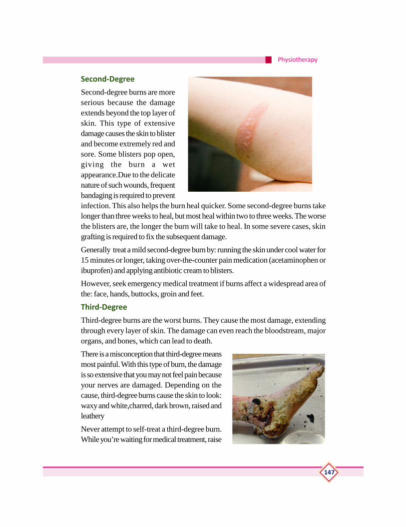
Physiotherapy
147
Second-Degree
Second-degree burns are moreserious because the damageextends beyond the top layer ofskin. This type of extensivedamage causes the skin to blisterand become extremely red andsore. Some blisters pop open,giving the burn a wetappearance.Due to the delicatenature of such wounds, frequentbandaging is required to preventinfection. This also helps the burn heal quicker. Some second-degree burns takelonger than three weeks to heal, but most heal within two to three weeks. The worsethe blisters are, the longer the burn will take to heal. In some severe cases, skingrafting is required to fix the subsequent damage.
Generally treat a mild second-degree burn by: running the skin under cool water for15 minutes or longer, taking over-the-counter pain medication (acetaminophen oribuprofen) and applying antibiotic cream to blisters.
However, seek emergency medical treatment if burns affect a widespread area ofthe: face, hands, buttocks, groin and feet.
Third-Degree
Third-degree burns are the worst burns. They cause the most damage, extendingthrough every layer of skin. The damage can even reach the bloodstream, majororgans, and bones, which can lead to death.
There is a misconception that third-degree meansmost painful. With this type of burn, the damageis so extensive that you may not feel pain becauseyour nerves are damaged. Depending on thecause, third-degree burns cause the skin to look:waxy and white,charred, dark brown, raised andleathery
Never attempt to self-treat a third-degree burn.While you’re waiting for medical treatment, raise
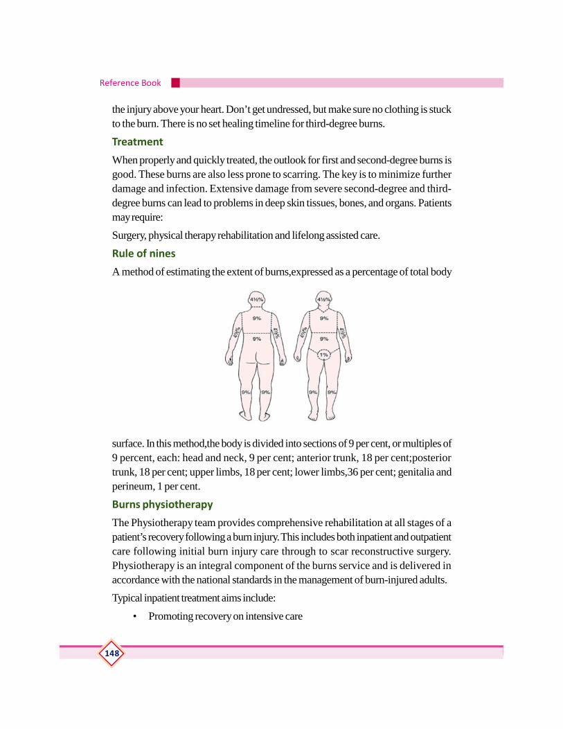
Reference Book
148
the injury above your heart. Don’t get undressed, but make sure no clothing is stuckto the burn. There is no set healing timeline for third-degree burns.
Treatment
When properly and quickly treated, the outlook for first and second-degree burns isgood. These burns are also less prone to scarring. The key is to minimize furtherdamage and infection. Extensive damage from severe second-degree and third-degree burns can lead to problems in deep skin tissues, bones, and organs. Patientsmay require:
Surgery, physical therapy rehabilitation and lifelong assisted care.
Rule of nines
A method of estimating the extent of burns,expressed as a percentage of total body
surface. In this method,the body is divided into sections of 9 per cent, or multiples of9 percent, each: head and neck, 9 per cent; anterior trunk, 18 per cent;posteriortrunk, 18 per cent; upper limbs, 18 per cent; lower limbs,36 per cent; genitalia andperineum, 1 per cent.
Burns physiotherapy
The Physiotherapy team provides comprehensive rehabilitation at all stages of apatient’s recovery following a burn injury. This includes both inpatient and outpatientcare following initial burn injury care through to scar reconstructive surgery.Physiotherapy is an integral component of the burns service and is delivered inaccordance with the national standards in the management of burn-injured adults.
Typical inpatient treatment aims include:
• Promoting recovery on intensive care

Physiotherapy
149
• Regaining movement, strength and function
• Regaining mobility
• Improving exercise tolerance
• Scar managementImproving exercise tolerance
Treatment may include positioning, stretching, an exercise programme, and mayrequire a splint(s) to maintain joints in an optimum position during recovery. When awound is healed, an individualised skin care and scar management programme maybe required.
Practical1. To demonstrate the therapeutic intervension for preventing complications of
chronic bed ridden patients
• Positioning
• Passive movement
• Stretching
2. Demonstration of physiotherapy management of burn
• Positioning
• Stretching
Assessment ActivitiesSeminar
Assignments
Field visit
Quiz
Collection
TE Questions• Types of head injury
• List out the different causes of burning
• Classify the burn according to skin level/thickness
• Write about the main goals of physiotherapy in burn rehabilitation.
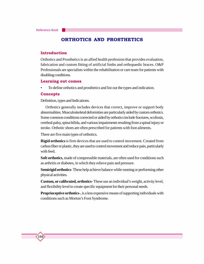
Reference Book
150
ORTHOTICS AND PROSTHETICS
IntroductionOrthotics and Prosthetics is an allied health profession that provides evaluation,fabrication and custom fitting of artificial limbs and orthopaedic braces. O&PProfessionals are specialists within the rehabilitation or care team for patients withdisabling conditions.
Learning out comes• To define orthotics and prosthetics and list out the types and indication.
ConceptsDefinition, types and Indications.
Orthotics generally includes devices that correct, improve or support bodyabnormalities. Musculoskeletal deformities are particularly aided by custom orthotics.Some common conditions corrected or aided by orthotics include fractures, scoliosis,cerebral palsy, spina bifida, and various impairments resulting from a spinal injury orstroke. Orthotic shoes are often prescribed for patients with foot ailments.
There are five main types of orthotics.
Rigid orthotics is firm devices that are used to control movement. Created fromcarbon fiber or plastic, they are used to control movement and reduce pain, particularlywith feed.
Soft orthotics, made of compressible materials, are often used for conditions suchas arthritis or diabetes, in which they relieve pain and pressure.
Semirigid orthotics- These help achieve balance while running or performing otherphysical activities.
Custom, or calibrated, orthotics- These use an individual’s weight, activity level,and flexibility level to create specific equipment for their personal needs.
Proprioceptive orthotics-, is a less expensive means of supporting individuals withconditions such as Morton’s Foot Syndrome.
Physiotherapy
151
KAFO-Knee Ankle foot orthosis andAFO-Ankle foot
orthosis Spinal Orthotics
Prosthetics are artificial extensions of the body. They are used to replace body partsthat are missing from birth, lost through an injury or disease, or which require removalfor medical purposes. It is an artificial device that replaces a missing body part,which may be lost through trauma, disease, or congenital conditions. Prosthesis canalso supplement an existing but defective body part. Some prosthetics include artificiallungs, knees, eyes, hearts, hips, limbs, heart valves, ankles, dentures, and gastricbands.
Prosthetic amputee rehabilitation is primarily coordinated by a prosthetist and aninter-disciplinary team of health care professionals including psychiatrists, surgeons,physical therapists, and occupational therapists.
Types
A person’s prosthesis should be designed and assembled according to the patient’sappearance and functional needs. The patient’s future goals and economicalcapabilities may help them choose between one or more devices.
Craniofacial prostheses include intra-oral and extra-oral prostheses. Extra-oralprostheses are further divided into hemifacial, auricular (ear), nasal, orbital and ocular.Intra-oral prostheses include dental prostheses such as dentures, obturators, anddental implants.
Limb prostheses include both upper- and lower-extremity prostheses.
Orthotics and prosthetics can be the practice of designing and creating suchequipment. People who work in orthotics and prosthetics evaluate patients’ needs;fabricate products according to those needs, and custom fit orthopedic braces and
Reference Book
152
artificial limbs. Careers opportunities in this health profession can include job titlessuch as direct practitioner, orthotist, fitter, assistant, pedorthist, or technician. Manysciences have contributed to the study of orthotics and prosthetics. These includeanatomy and physiology, engineering, psychology, and gait analysis. Orthotics andprosthetics are also part of the biomechatronics field of science. This field includesthe fusion of mechanical devices with actual human body parts within the nervous,muscular, and skeletal systems. Many people with abnormalities, deformities, ormissing body parts can be helped with orthotics and prosthetics. These are piecesof medical equipment that can help replace or improve body parts. They includebraces, inserts, artificial limbs, and much other medical equipment fabrication
Practical1. Application of orthotics and prosthetics
• Upper limb
• Lower limb
• Neck and trunk
Assessment ActivitiesSeminar
Assignments
Field visit
Chart preparation
Quiz
Collection
Model Preparation
TE QuestionsExpand the following term
KAFO
AFO
Artificial appliances used to replase original body parts is known as —————Write any two examples of internal and external orthotics and prosthetics.
List out the indications of orthotics.
Odd man out KAFO OSTIN MOOR
K WIRE BRACES

Physiotherapy
153
GERIATRIC PHYSIOTHERAPYIntroductionGeriatric medicine is a specialty that focuses on health care of elderly people. Itaims to promote health by preventing and treating diseases and disabilities in olderadults. Geriatric physical therapy covers a wide area of issues concerning people asthey go through normal adult aging but is usually focused on the older adult. Geriatricphysical therapy was defined as a medical specialty in 1989 and covers a broadarea of concerns regarding people as they continue the process of aging, although itcommonly focuses on older adults. “Physical therapy intervention can prevent life-long disability, restoring the person’s level of functioning to its highest level.”Learning outcomeThe learner:
• familiarizes with common problems of geriatric patients
• understands the history,concept and methods of geriatric physiotherapy
• understands the role of physiotherapist, exercise, manual therapy and educationin geriatric physiotherapy
• familiarizes with Aim of geriatric physiotherapy and different settings
• understands the concept of assessment ,goal setting, treatment intervention andre-assessment
ConceptsIntroduction to geriatric Physiotherapy.
Geriatric physical therapy is a proven means for older adults from every level ofphysical ability to improve their balance and strength, build their confidence, andremain active. A number of people are familiar with physical therapy as a form oftreatment to pursue after an accident, or in relation to a condition such as a stroke.Physical therapy is useful for many additional reasons, such as improving balance,strength, mobility, and overall fitness. All of these are factors which older adults maybenefit from, contributing to their physical abilities and helping to maintain theirindependence for longer periods of time. Physical therapy can also help older adultsto avoid falls, something that is crucial to this population.
Falling is one of the greatest risks older adults face, often leading to things such aship fractures which then lead to a downward health spiral. In fact, falling is such anissue among older adults that the Center for Disease Control and Prevention has
Reference Book
154
reported that one-third of all people over the age of sixty-five fall every year, makingfalls the leading cause of injury among people from this age group. Hundreds ofthousands of older adults experience falls and resulting hip fractures every year, withresulting hospitalizations. Most of the people who experience a hip fracture stay inthe hospital for a minimum of one week, with approximately twenty-percent dyingwithin a year due to the injury. Unfortunately, a number of the remaining eighty-percent do not return to their previous level of functioning. Physical therapy can helpolder adults to remain strong and independent, as well as productive.
Common condition seen in old age
1. Musculoskeletal: Osteoarthritis (inflammation of joints due to wear and tear)and Osteoporosis (bone loss).
2. Hormonal : Diabetes (impaired control of blood glucose), Menopause, thy-roid dysfunction, high blood cholesterol, Slower overall metabolism.
3. Neurologic :Dementia (Alzheimer or other types), Parkinson’s disease, Stroke,hearing impairment and balance problem
4. Visual : macular degeneration, glaucoma, cataracts and Diabetes and hyper-tension related eye disease.
5. Cardiovascular disease: heart attacks, congestive heart failure, irregular heartrhythm (atrial fibrillation), high blood pressure (hypertension) , atherosclerosis(hardening and narrowing of blood vessels) and Peripheral vascular disease orperipheral artery disease (poor blood flow as a result of narrow blood ves-sels).
6. Lungs: Chronic obstructive pulmonary disease (COPD) and Loss of lung vol-ume.
7. Kidneys: Poor kidney function (kidney or renal disease) from long standingdiabetes and hypertension.
8. Skin and Hair: Hair loss, dry skin, itching, infections.
9. Cancers: Prostate, colon, lung, breast, skin, bladder, ovary, brain, pancreas,etc
10. Bone marrow and immune system: Inability to produce sufficient bloodcells (anemia, myelodysplasia).
11. Gastrointestinal: Stomach ulcers, Diverticulitis (small pockets forming in thewall of colon, Swallowing difficulty (dysphagia) , Constipation and Bowel in-continence
Physiotherapy
155
12. Urinary: Urinary incontinence, urinary urgency, difficulty urinating.
13. Oral and dental: gum disease, dry mouth, loss of teeth, poorly fitting den-tures.
14. Infections: urinary tract infection, pneumonia, skin infection,
15. Psychiatric: Depression, anxiety, sleep disturbance, insomnia.
16. General problems:Fatigue, general reconditioning, forgetfulness, medicationside effects, diminished appetite, weight loss, falls.
Forms of Geriatric Physical TherapyPhysical Therapists
Physical therapists provide people with a variety of services. They work with peopleindividually, evaluating their physical capabilities and designing specific programs ofexercise, education and wellness for them. Physical therapists also work with otherhealth care providers to coordinate the person’s care. The potential for age-relatedbodily changes to be misunderstood can lead to limitations of daily activities. Theusual process of aging does not need to result in pain, or decreased physical mobility.A physical therapist can be a source of information for understanding changes in thebody, they can offer assistance for regaining lost abilities, or for development of newones. A physical therapist can work with older adults to help them understand thephysiological and anatomical changes that occur with the aging process.
Physical therapists help injured or ill people improve their movement and managetheir pain. These therapists are often an important part of rehabilitation and treatmentof patients with chronic conditions or injuries.
Physical therapist assistants may deliver treatment and physical interventions forpatients and clients under a care plan established by and under the supervision of aphysical therapist.
Physical therapists evaluate and develop specifically designed, therapeutic exerciseprograms. Physical therapy intervention can prevent life-long disability, restoring theperson’s level of functioning to its highest level. A physical therapist uses things suchas treatments with modalities, exercises, educational information, and screeningprograms to accomplish a number of goals with the person they are working with,such as:
• Reduce pain• Improve sensation, joint proprioception• Increase overall fitness through exercise programs
Reference Book
156
• Suggest assistive devices to promote independence
• Recommend adaptations to make the person’s home accessible and safe
• Prevent further decline in functional abilities through education, energyconservation techniques, joint protection
• Increase, restore or maintain range of motion, physical strength, flexibility,coordination, balance and endurance
• Teach positioning, transfers, and walking skills to promote maximum functionand independence within the person’s capability
• There are various common conditions that can be effectively treated throughphysical therapy. Among the specific diseases and conditions that might affectolder adults which can be improved with physical therapy are arthritis,osteoarthritis, stroke, Parkinson’s disease, cancer, amputations, urinary andfecal incontinence, and cardiac and pulmonary diseases. Conditions such asAlzheimer’s disease, dementia’s, coordination and balance disorders, jointreplacements, hip fractures, functional limitations related to mobility, orthopedicor sports injuries can also be improved through geriatric physical therapy.
Exercise
Exercise is defined as any form of physical activity that is beyond what the persondoes while performing their daily tasks. Exercise is something that is designed toboth maintain and improve a person’s coordination, muscle strength, flexibility andphysical endurance, as well as their balance. It is meant to increase their mobilityand lessen their chance of injury through falling. Exercise in relation to geriatric therapymight include activities such as stretching, walking, weight lifting, aquatic therapy,and specific exercises that are geared towards a particular injury or limitation. Aphysical therapist works with the person, teaching them to exercise on their own, sothey may continue their exercise program at home.
Manual Therapy
Manual therapy is applied with the goals of improving the person’s circulation andrestoring mobility they may have lost due to an injury or lack of use. This form oftherapy is also used to reduce pain. Manual therapy can include manipulation of theperson’s joints and muscles, as well as massage.
Education
Education is important to the success and effectiveness of geriatric physical therapy.People are taught ways of performing daily tasks safely. Physical therapists also

Physiotherapy
157
teach people how to use assistive devices, as well as how to protect themselvesfrom further injury. Older adults can utilize physical therapy as a means for regainingtheir independence. Physical therapy can help seniors to feel better, as well as toenjoy a higher quality of life.
Principles of Geriatric Physical TherapyAims of Geriatric Assessment
Geriatric physical therapy has been identified as a physical therapy specialization inorder to acknowledge the advanced-level skills of physical therapists who seek toaddress the unique medical and functional problems of older persons. Geriatricphysical therapists encounter a wide spectrum of elderly patients, ranging from thosewho are frail and institutionalized to those who are functionally independent butrequire attention in outpatient departments.
Geriatric physical therapy in different settings
The role of geriatric physical therapy is important in all care settings. Geriatric physicaltherapists are providing quality care to patients in multiple settings that include acutecare hospitals, skilled nursing facilities, outpatient departments, rehabilitation centres,home health agencies and hospice settings. The over-all condition of a patient willdecide the type of care setting.
Geriatric Physical Therapy in Acute Care Hospitals
This is particularly important for the elderly having multiple co morbid conditions.However, a patient with single event like stroke may also need acute care in hospital.The patient is usually monitored by the interdisciplinary team. Physical therapy shouldbe started as early as the elderly is able to tolerate the therapeutic interventions,especially exercises. The early physical therapy intervention can prevent secondaryfunctional loss and promote early restoration of function and thereby reduce thelength of hospital stay. The patient should receive therapy for two hrs per day tohave significant improvement.
Geriatric Physical Therapy in Skilled Nursing Facilities
A skilled nursing facility has staff and equipment to provide skilled nursing care andother health services. Geriatric physical therapist may provide her specialized servicesas an employee of a skilled nursing facility or as an independent contractor. Elderlypatients who are not suitable for acute care in hospitals may be treated in a skillednursing facility. The patient should be employed two half-hourly sessions of physicaltherapy per day. The treatment time may be increased after the reassessment of apatient.
Reference Book
158
Geriatric Physical Therapy at Home
Many patients prefer to have physical therapy at their home. Physical therapist mayvisit the patient for once or twice a day. The main advantage of this kind of provisionis that it saves the time of caregivers. The caregivers do not have to bother to take apatient to physiotherapy clinic. However, there is a disadvantage in the sense thatthe necessary equipment cannot be used at home.
Geriatric Physical Therapy in Outpatient Departments
Outpatient department is the best option for the patients having the transport facility.However, it is not the suitable option for the frail elderly. Easy access to equipmentand peer interaction are the main advantages for the patients being treated in outpatientdepartments.
Geriatric Physical Therapy Program
The geriatric population is unique in its wide variation from individual to individual inthe effects of both aging and disease processes. The effective administration ofPhysical Therapy program can make the quality of life better for the patients belongingto this complex group. The important components of this program are:
Assessment
A comprehensive geriatric assessment is often helpful before the initiation of a physicaltherapy program to assist with setting realistic goals with each patient. Nevertheless,modification in the assessment may be required in some specific cases. For example,assessment of communication skill is must while working with the older patient withcognitive deficits so as to know the difficulties of patient in problem solving and self-care.
Goal-Setting
Functional independence is the ultimate goal of physical therapy intervention. This isparticularly important in geriatric care, because the presence of acute as well aschronic illness in elderly individuals is often associated with loss of day-to-day function.To achieve this long-term goal, a physical therapist should establish several short-term goals:
• To improve or maintain ROM of different joints. For example, a geriatric pa-tient should
Have enough ROM at shoulder to dress up or to reach dishes in the cupboard.
• To improve or maintain strength and endurance of muscles. For example, the

Physiotherapy
159
patient should have sufficient muscle strength to lift a jug of milk, to make abed, to make chapatti or to wash clothes.
• To improve or maintain cardiovascular endurance so that a geriatric patient isable to do strenuous activities such as fast walk, cycling or swimming.
• To improve or maintain ambulatory status of a patient so that a patient can goto toilet or for shopping independently.
• To relieve pain. It has been estimated that over 85 percent of older adults haveat least one chronic disease that may give rise to the feeling of discomfort orpain.24 Acute pain following surgery is also becoming quite common in geriatricpatients. The most common therapeutic interventions to relieve pain are exercise,orhtotics, heat and cold modalities; and electrical stimulating currents.
Therapeutic Intervention
A number of physical therapy interventions may be employed in order to attaintreatment goals:
• Range-of-motion exercise
• Stretching exercise
• Mobilization exercise
• Strengthening exercise
• Aerobic exercises
• Gait training
• Orthotics
• Electrotherapeutic modalities
Practical1. To demonstrate the exercise in geriatrics
• Mobility exercise
• Recreational activities for geriatrics.
Assessment activities• Seminar
• Assignments

Reference Book
160
TE Questions1. List out the common geriatric conditions.
2. A medical specialty that focuses on health care of elderly people known as-
3. Explain about the role of physical therapist in a geriatric centre.
4. List out the main goals of physiotherapy in geriatric care
At the end of each module
Extended activities
• Visit to special school
• Visit to orthotic center
• Visit to old age home
List of practicals1. Free exercises
a. Upper limb
b. Lowerlimb
c. trunk
2. Resisted exercise
a. Shouher joint
• Flexers and Extensors
• Adductors abductors
b. Elbow joint
• Flexors
• Extensors
c. Wrist
• Flexors
• Extensors
• Ulnar and radial deviators
d. Hand
• Finger flexors and extensors
e Hip joint
Physiotherapy
161
• Flexors and extensors
• Abductors and adductors
f. Knee
• Flexors and extensors
g. Ankle
• Dorsi and plantar flexors
3. Passive movement
• Shoulder girdle
• Shoulder joint
• Elbow joint
• Wrist and fingers
• Hip joint
• Knee joint
• Ankle
4. Fundamental position
• Standing
• Sitting
• Kneeling
• Lying
• hanging
5. Derived position
• Standing
• Sitting
• Kneeling
• Lying
• Hanging
6. Grading of muscle power of major muscles
1. Shouher
• Flexers and Extensors
• Adductors abductors
Reference Book
162
2. Elbow
• Flexors
• Extensors
3. Wrist
• Flexors
• Extensors
• Ulnar and radial deviators
4. Hand
• Finger flexors and extensors
5. Hip joint
• Flexors and extensors
• Abductors and adductors
6. Knee
• Flexors and extensors
• Ankle
• Dorsi and plantar flexors
7. Range of motion of major joints
a. Shoulder joint
b. Elbow joint
c. Wrist and fingers
d. Hip joint
e. Knee joint
f. Ankle
8. Good and bad posture
• Postural analysis of a given modal
9. Preparation and maintenance of exercise therapy equipment.
a. Static cycle
b. Treadmill
Physiotherapy
163
c. Quadriceps table
d. Shoulder wheel
e. Hand exerciser
f. Medicine ball,
g. Swiss ball
h. Abduction ladder
i. Parallal bar
j. Tilt table
k. Wheel chair
l. crutches
10. Demonstration of gait and identification of its phases
a. Stance
b. swing
11. Demonstation of pathological gait
a. Cicum ductory
b. Scissoring
c. Festinant
d. Ataxic
e. High stepping
f. Antalgic
12. Crutch walking
a. Non weight bearing
b. Partial weight bearing
c. full weight bearing
d. ascending
e. descending
13. Transfer technique
a. wheel chair to bed
b. bed to wheel chair
14. Application of contrast bath of given subject
15. Application of cryotherapy
Reference Book
164
16. Preparation and positioning of axial and pendular suspension
17. Preparation and positioning of the given subject for relaxation
18. Breathing exercise
a. Inspiratory Breathing exercise
b. expiratory Breathing exercise
19. Electrotherapy
Preparation and positioning of the subject for the treatment by using following electrotherapy equipments.
h. IRR
i. SWD
j. ULTRASOUND
k. IFT
l. TENS
m. EMS
n. Hot pack
Module- IV20. Positioning of the hemiplegic patient
Positionining by using pillows and sand bags.
Supine lying
Affected side
Unaffected side
Sitting
21. Passive movements
Upper limb
Lower limb
22. Positioning of the paraplegic patient
23. To demonstrate the positioning and passive movement of the spinalcord injury patient.
Positioning
Lying position
Sitting

Physiotherapy
165
Passive movement
24. Transfer technique
Bed to wheel chair
Wheel chair to bed
25. To demonstrate the exercise in parkinsonism
Range of motion/flexibility exercise
To demonstrate the exercise in Ataxia
Co ordination exercise –frenkal’s exerciseUpper lib
Lower limb
Trunk
26. Demonstration of exercise in cerebral palsy
Special care activities for reducing possible complication.
Child carrying
Sitting
Positioning
Stretching
27. Peripheral Nerve Injury
Application of splint
Donning and doffing
Cock op splint
Afo
Aero plane splint
28. Preparation and positioning of the subject for electro therapy treatment
Stimulator
TENS
29. Application of price therapy in soft tissue injury
Application of elastocrepe bandage in lateral ligament injury
30 Demonstration of exercise for rheumatoid arthritis
joint mobility exercise

Reference Book
166
31. Exercises for knee joint osteoarthritis
Isometric
Mobility
Strengthening
32. Demonstration of exercises for cervical and lumbar exercises
Isometric neck exercise
Neck mobility exercise
Spinal extension e/flexion exercise
33. Preparation and positioning of client for elecctro therapy.
IRR for periarthritis
US for tennis/golfers/plantar fasciitis
34. Demonstrate the correction of deformity in thre following conditions
CTEV
TORTICOLLIS
35. To demonstrate the therapeutic intervention for preventingcomplications of chronic bed ridden patients
Positioning
Passive movement
Stretching
36. Demonstration of physiotherapy management of burn
Positioning
stretching
37. Application of orthotics and prosthetics
upper limb
lower limb
neck and trunk
38. To demonstrate the exercise in geriatrics
General Mobility exercise
List of Standard Equipments and Tools White board with marker
Lcd with laptope
Physiotherapy
167
Stethoscope
BP apparatus
Bone and skeletal set
Goneometer
Inclinometer
Thera band
Spirometer
Theraputty
Exercise mat
Thermom1eter
Measuring1 tape
Plinth
Dumb bells 2,3,4,5,kg
Barbells
Free weights
Cables
Tubing's
Weight cuffs
Static cycle
Treadmill
Elliptical trainer
Ice packs
Wax bath
Hydro collator packs
First aid box
Slings\ Splints
Crepe bandage
Multi gym
Reference Book
168
ReferencesPrinciples of exercise therapy M. Dena GardinerElectro Therapy explained - Principle and Practice John Low & Ann ReedClayton’s Electro Therpy - Theory and Practice Forster and PalastangaCash’s Text book of Neurology for Physiotherapist Patricia. A DownieCash’s Text book of Orthopaedics and Rheumatology Patricia .A DownieCash’s Text book of Chest, Heart and Vascular Disorders Patricia. A DownieCash’s Text book of General Medical and Surgical Conditions Patricia. A DownieNatarajan’s Text book of Orthopaedic and Traumatology M. Natarajan & N.MayilvahananEssentials of Orthopaedic and applied physiotherapy Jayanth Joshi & PrakashKotwalTherapeutic Exercise - Foundation and Techniques Carolyn KlisnerLecture note on Rehabilitation L. SundarAids to Physiotherapy Jennifar M. LeePhysical Rehabilitation Assessments and Treatment Sussan O. SullivanTidy’s Physiotherapy-Ann Thomson, Alison Skinner & Joan PiercyGoel’s Physiotherapy Vol 1 &11..Dr.R.N.Goel
Websiteswww.google.com
www.pubmed.com
www.physiobase.com
www.ebpt.com
www.apta.com
www.physio-pedia.com
http://www.medindia.net/patients/patientinfo/physiotherapytypes_wave.htm#ixzz457Y0SUfN
http://www.utmb.edu/policies_and_procedures/Non-IHOP/Respiratory/Respiratory_Care_Services/07.03.05%20Breathing%20Exercises.pdf
http://www.msktc.org/sci/factsheets/safe-transfer-technique#sthash.hEchFzIR.dpuf
https://www.niehs.nih.gov/research/resources/assets/docs/mmt8_grading_and_testing_procedures_for_the_abbreviated_8_muscle_groups_508.pdf
https://crcjs.med.utah.edu/sma_fitness/pdfs/manualmuslce_tests_upper_lower_PST.pdf