photometry iii anu
TRANSCRIPT
PHOTOMETRY - III
Presenter: Dr.Anurag Yadav
Moderator: Mr.Arun Kumar
CONTENT
ATOMIC ABSORPTION SPECTROPHOTOMETRY
FLAME EMISSION SPECTROPHOTOMETRY
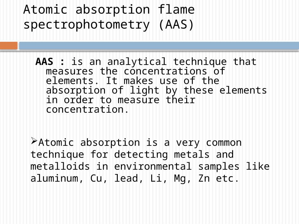
Atomic absorption flame spectrophotometry (AAS)
AAS : is an analytical technique that measures the concentrations of elements. It makes use of the absorption of light by these elements in order to measure their concentration.
Atomic absorption is a very common technique for detecting metals and metalloids in environmental samples like aluminum, Cu, lead, Li, Mg, Zn etc.
Atomic absorption flame spectrophotometry (AAS)
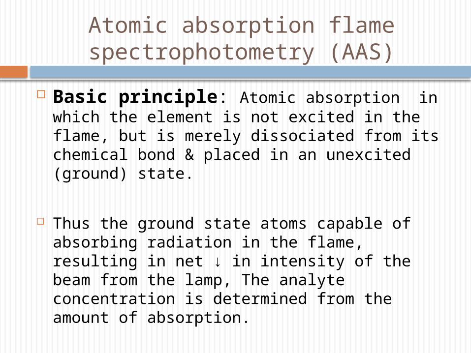
Basic principle: Atomic absorption in which the element is not excited in the flame, but is merely dissociated from its chemical bond & placed in an unexcited (ground) state.
Thus the ground state atoms capable of absorbing radiation in the flame, resulting in net ↓ in intensity of the beam from the lamp, The analyte concentration is determined from the amount of absorption.
Atomic absorption flame spectrophotometry (AAS)
Concentration measurements are usually determined from a working curve after calibrating the instrument with standards of known concentration.
Absorption bands- .001 - .01 nm.
Entire absorption spectrum of atoms – line spectrum.
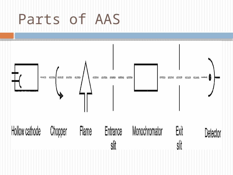
Parts of AAS
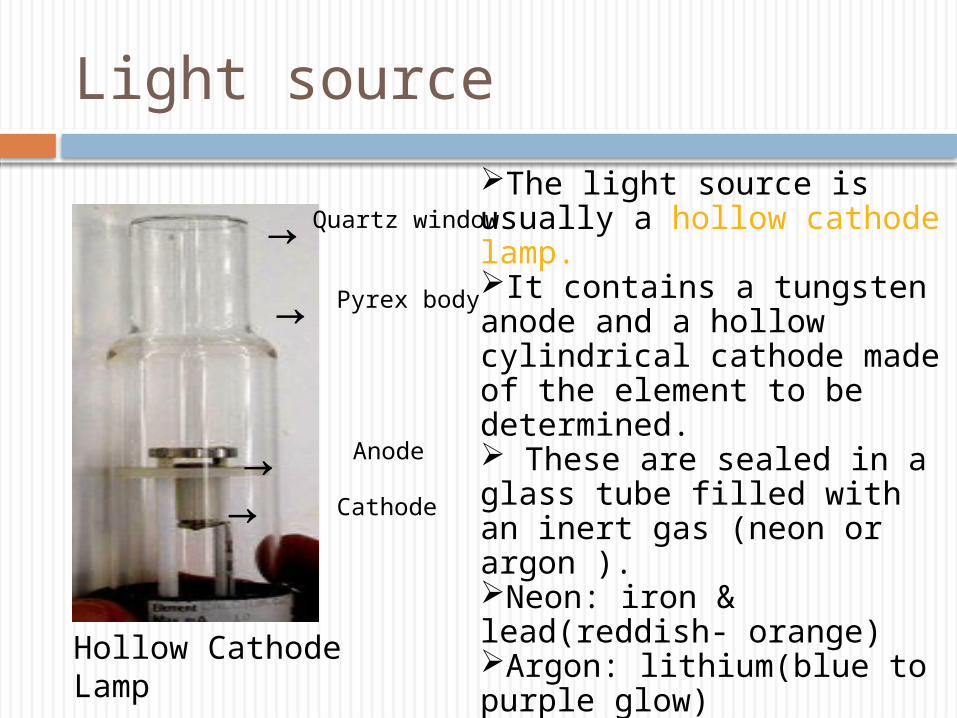
Light source
→→
→→
Quartz window
Pyrex body
Anode
Cathode
The light source is usually a hollow cathode lamp.It contains a tungsten anode and a hollow cylindrical cathode made of the element to be determined. These are sealed in a glass tube filled with an inert gas (neon or argon ). Neon: iron & lead(reddish- orange)Argon: lithium(blue to purple glow)Each element has its own unique lamp which must be used for that analysis.
Hollow Cathode Lamp
How it works
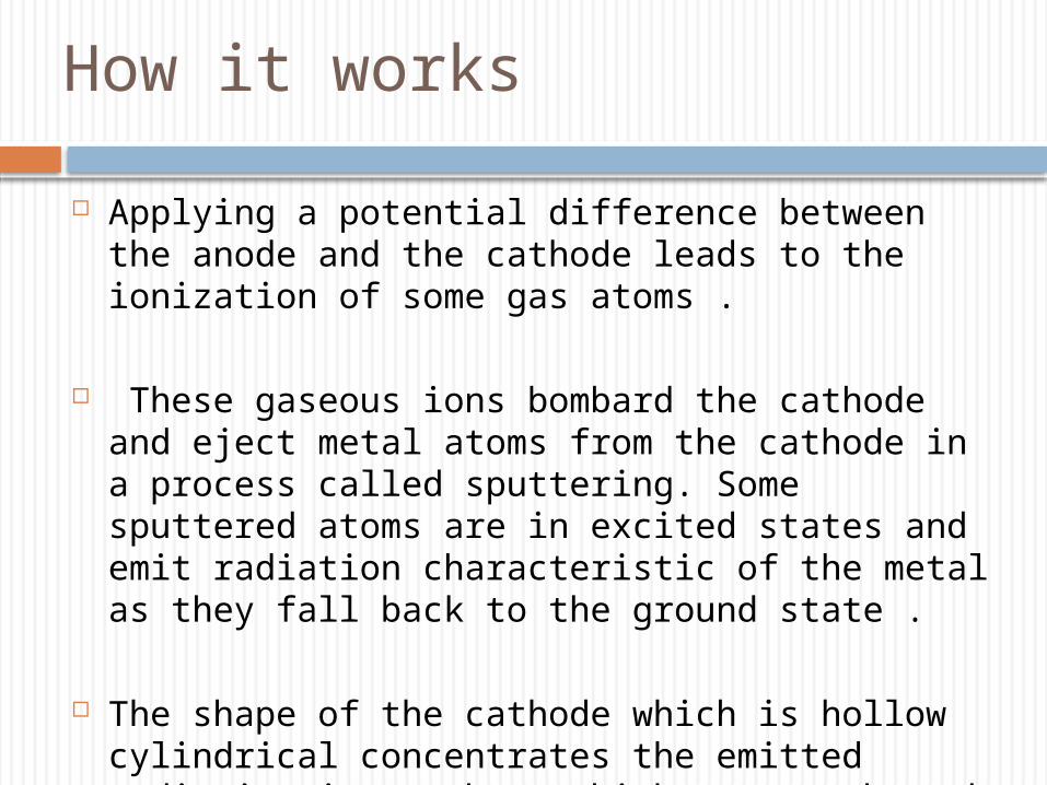
Applying a potential difference between the anode and the cathode leads to the ionization of some gas atoms .
These gaseous ions bombard the cathode and eject metal atoms from the cathode in a process called sputtering. Some sputtered atoms are in excited states and emit radiation characteristic of the metal as they fall back to the ground state .
The shape of the cathode which is hollow cylindrical concentrates the emitted radiation into a beam which passes through a quartz window all the way for absorbtion by ground state atoms in the flame.
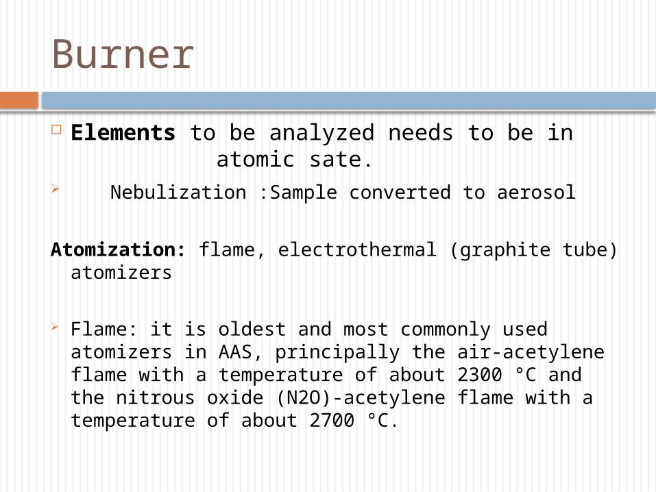
Burner
Elements to be analyzed needs to be in atomic sate.
Nebulization :Sample converted to aerosol
Atomization: flame, electrothermal (graphite tube) atomizers
Flame: it is oldest and most commonly used atomizers in AAS, principally the air-acetylene flame with a temperature of about 2300 °C and the nitrous oxide (N2O)-acetylene flame with a temperature of about 2700 °C.
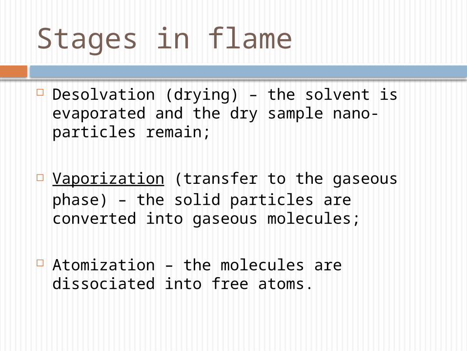
Stages in flame
Desolvation (drying) – the solvent is evaporated and the dry sample nano-particles remain;
Vaporization (transfer to the gaseous phase) – the solid particles are converted into gaseous molecules;
Atomization – the molecules are dissociated into free atoms.
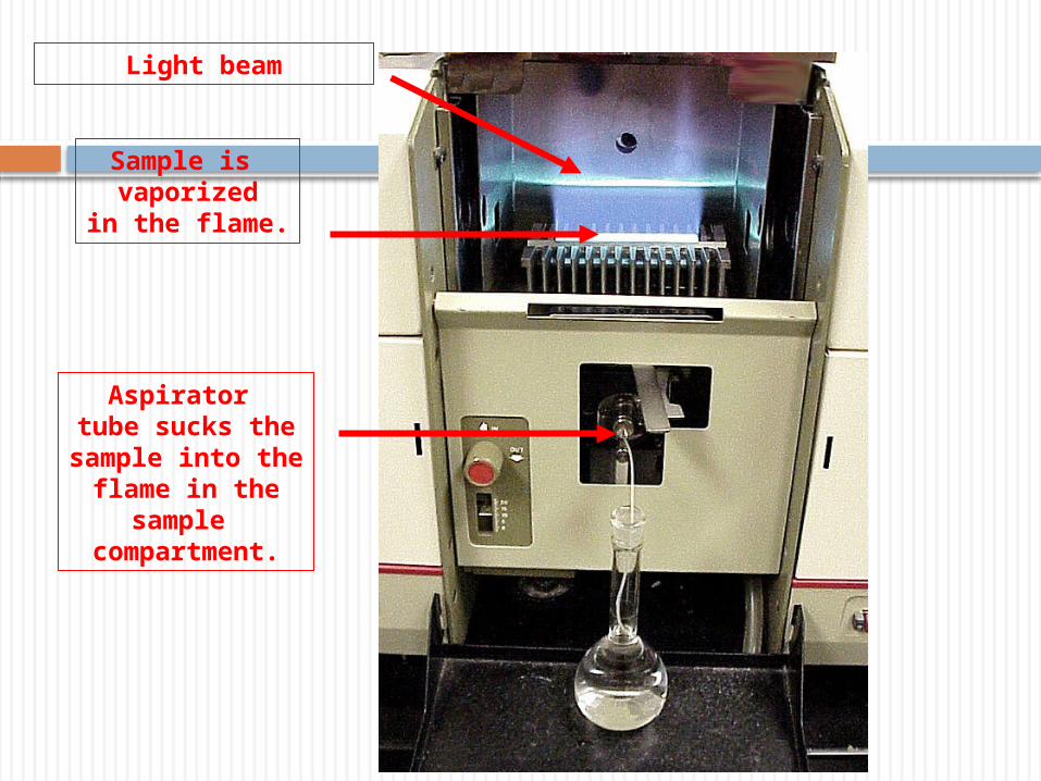
Sample is vaporized
in the flame.
Aspirator tube sucks thesample into the
flame in thesample
compartment.
Light beam
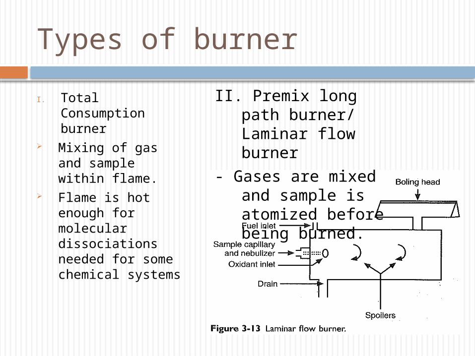
Types of burner
I. Total Consumption burner
Mixing of gas and sample within flame.
Flame is hot enough for molecular dissociations needed for some chemical systems
II. Premix long path burner/ Laminar flow burner
- Gases are mixed and sample is atomized before being burned.
Advantages of long path burner
Larger droplets go waste and only the fine mist enters the flame thus produces less noisy signal
Path length through the flame of the burner is longer then the total consumption burner – greater absorption and increases sensitivity of measurement.
Flame is not as hot as that of total consumption burner - cant dissociate certain metal complexes in flame- Ca- phosphate complexes.
Disadvantages of long path burner
Monochromator
The monochromater in AAS is placed between flame and detector
Used to select the specific wavelength of light which is absorbed by the sample, and to exclude other wavelengths.
To allow the single line in the spectrum of analyte.
To minimize the emission from the flame itself because detector detects photons over a wide wavelength range.
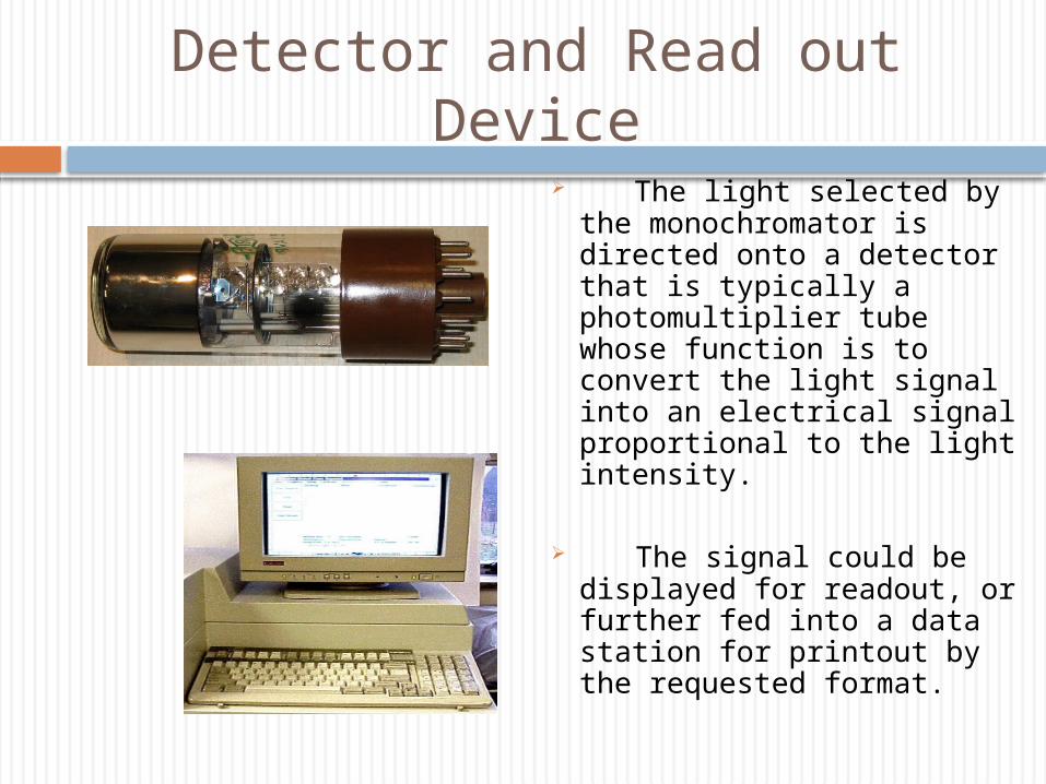
Detector and Read out Device
The light selected by the monochromator is directed onto a detector that is typically a photomultiplier tube whose function is to convert the light signal into an electrical signal proportional to the light intensity.
The signal could be displayed for readout, or further fed into a data station for printout by the requested format.
Calibration Curve
A calibration curve is used to determine the unknown concentration of an element in a solution. The instrument is calibrated using several solutions of known concentrations. The absorbance of each known solution is measured and then a calibration curve of concentration vs absorbance is plotted.
The sample solution is fed into the instrument, and the absorbance of the element in this solution is measured .The unknown concentration of the element is then calculated from the calibration curve
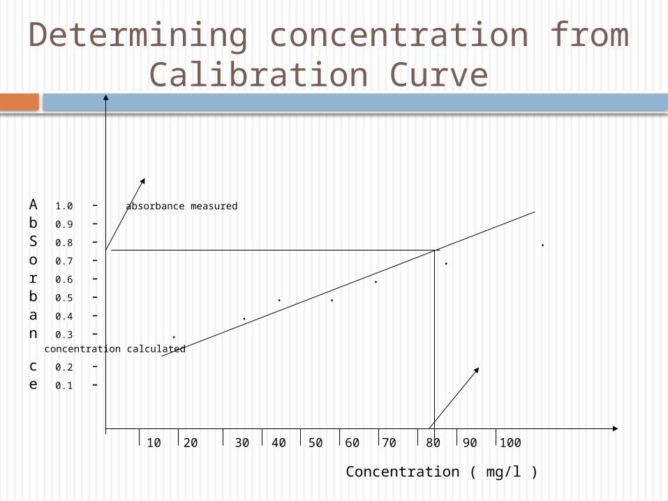
Determining concentration from Calibration Curve
A 1.0 - absorbance measured
b 0.9 - S 0.8 - .o 0.7 - .r 0.6 - . b 0.5 - . .a 0.4 - .n 0.3 - . concentration calculated c 0.2 - e 0.1 -
10 20 30 40 50 60 70 80 90 100
Concentration ( mg/l )
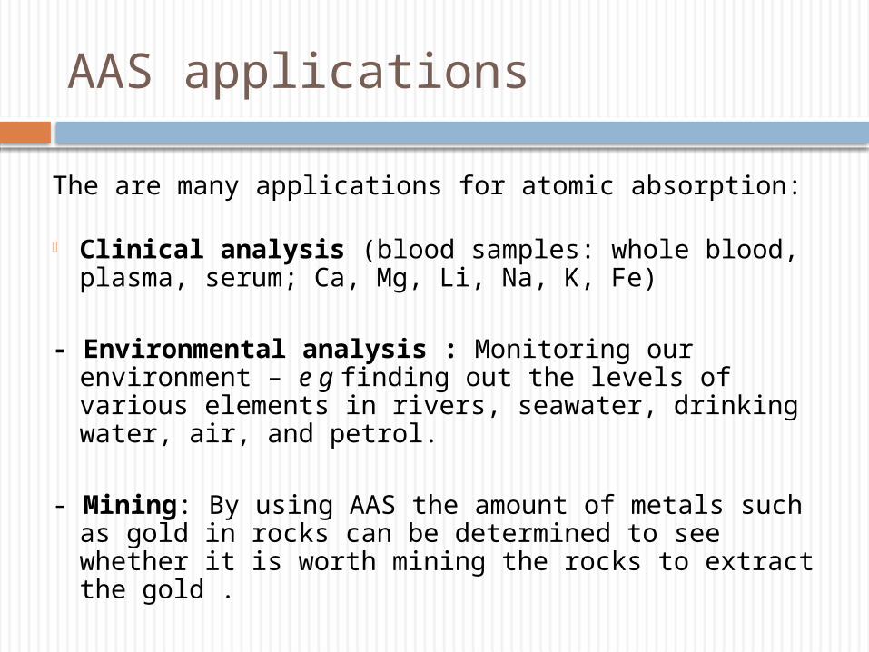
AAS applications
The are many applications for atomic absorption: - Clinical analysis (blood samples: whole blood, plasma,
serum; Ca, Mg, Li, Na, K, Fe)
- Environmental analysis : Monitoring our environment – e g finding out the levels of various elements in rivers, seawater, drinking water, air, and petrol.
- Mining: By using AAS the amount of metals such as gold in rocks can be determined to see whether it is worth mining the rocks to extract the gold .
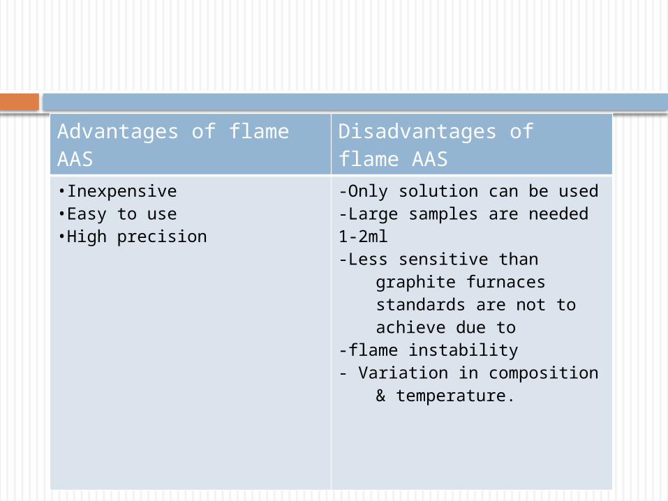
Advantages of flame AAS Disadvantages of flame AAS
•Inexpensive •Easy to use •High precision
-Only solution can be used-Large samples are needed 1-2ml-Less sensitive than graphite furnaces
standards are not to achieve due to
-flame instability- Variation in composition &
temperature.
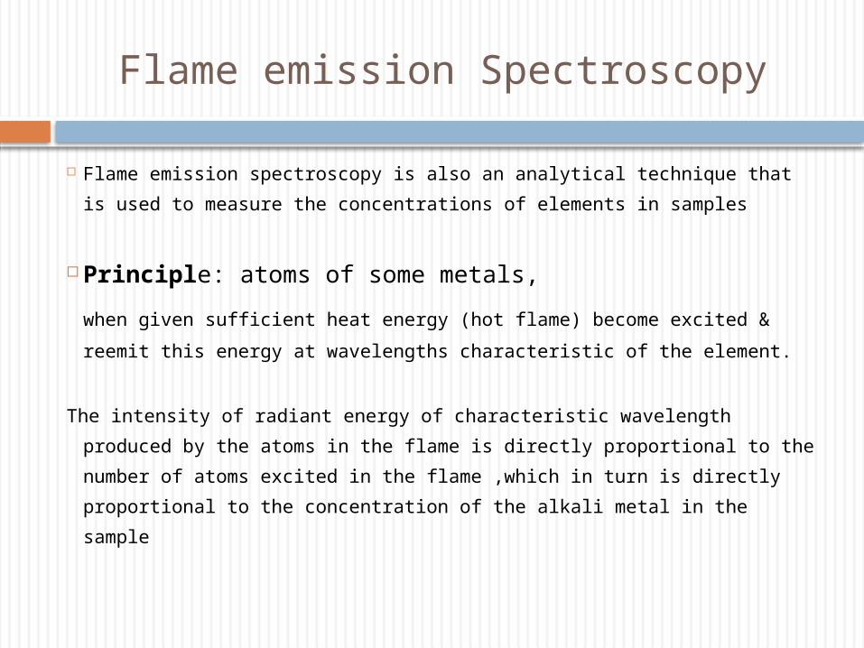
Flame emission Spectroscopy
Flame emission spectroscopy is also an analytical technique that is used
to measure the concentrations of elements in samples
Principle: atoms of some metals,
when given sufficient heat energy (hot flame) become excited &
reemit this energy at wavelengths characteristic of the element.
The intensity of radiant energy of characteristic wavelength produced by
the atoms in the flame is directly proportional to the number of atoms
excited in the flame ,which in turn is directly proportional to the
concentration of the alkali metal in the sample
Flame emission Spectroscopy
The excited atoms decay back to lower levels by emitting light . Emissions are passed through monochromators or filters prior to detection by photomultiplier tubes.
Alkali metals are easy to excite by flame Li- red emission Na – yellow emission K- red violet emission Rubidium- red emission Mg- blue emission
Flame emission Spectroscopy
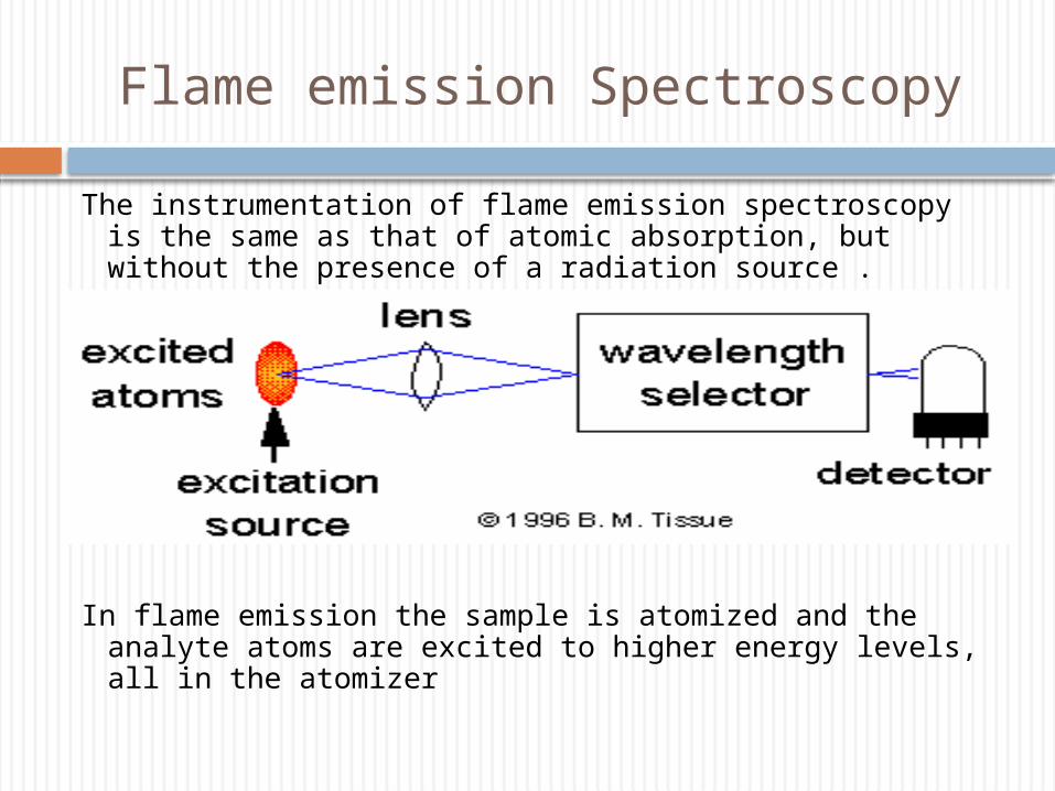
The instrumentation of flame emission spectroscopy is the same as that of atomic absorption, but without the presence of a radiation source .
In flame emission the sample is atomized and the analyte atoms are excited to higher energy levels, all in the atomizer
Flame emission Spectroscopy
The source of energy in Atomic Emission could be a flame like the one used in atomic absorption, or an inductively coupled plasma ( ICP ) .
The flame ( 1700 – 3150 oC ) is most useful for elements with relatively low excitation energies like sodium, potassium and calcium.
The ICP ( 6000 – 8000 oC) has a very high temperature and is useful for elements of high excitation energies.
Application of flame emission spectroscopy
1.Electrons of alkali metals like sodium, potassium, lithium become easily excited hence preferentially analyzed by flame photometry.
2.Used in clinical laboratory to determine concentrations of sodium and potassium in biological fluids like serum, urine and sweat.
3.Serum lithium levels – therapeutic monitoring.
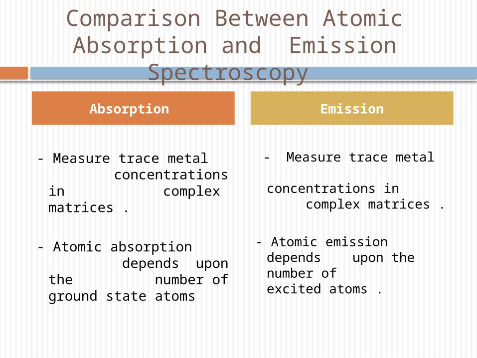
Comparison Between Atomic Absorption and Emission Spectroscopy
- Measure trace metal
concentrations in complex matrices .
- Atomic absorption depends upon the number of ground state atoms
- Measure trace metal concentrations in complex matrices .
- Atomic emission depends upon the number of excited atoms .
Absorption Emission
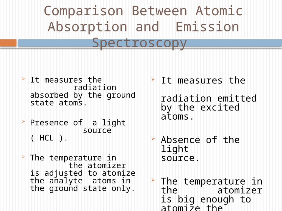
Comparison Between Atomic Absorption and Emission Spectroscopy
It measures the radiation absorbed by the ground state atoms.
Presence of a light source ( HCL ).
The temperature in the atomizer is adjusted to atomize the analyte atoms in the ground state only.
It measures the radiation emitted by the excited atoms.
Absence of the light source.
The temperature in the atomizer is big enough to atomize the analyte atoms and excite them to a higher energy level.
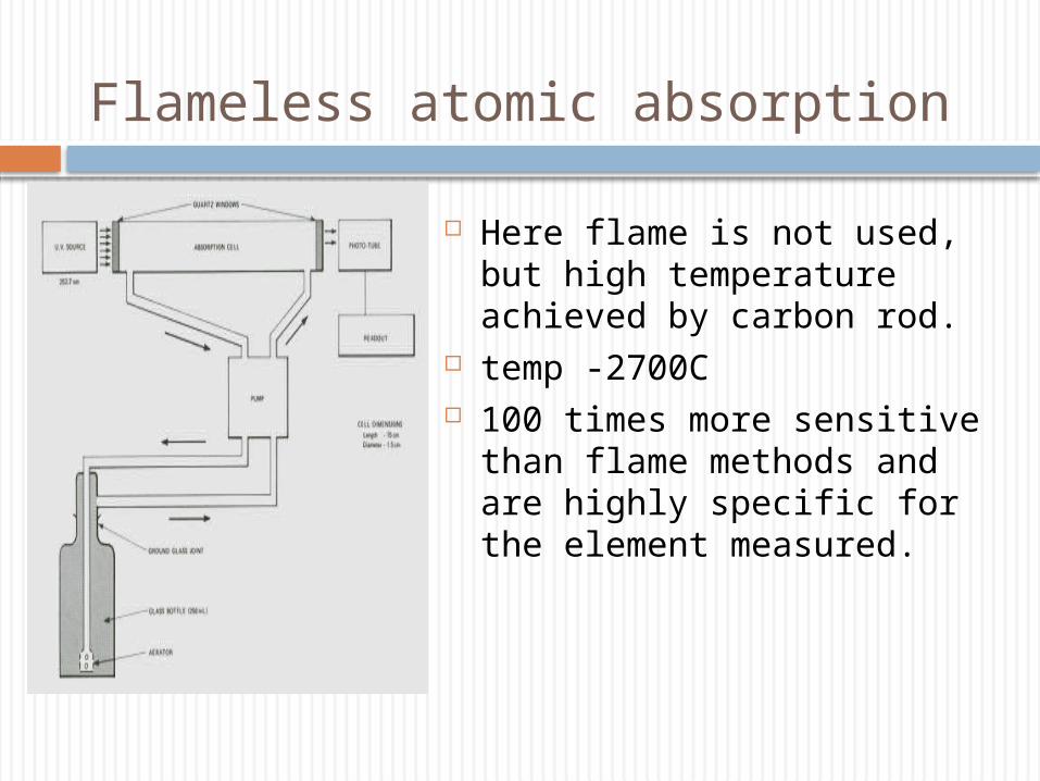
Flameless atomic absorption
Here flame is not used, but high temperature achieved by carbon rod.
temp -2700C 100 times more sensitive than flame
methods and are highly specific for the element measured.
Flameless atomic absorption
Atomization techniques- electrothermal
1. Drying : Sample is dried on carbon support by removing solvent.
2. Pyrolysis- majority of matrix constituents are removed
3. Atomization: the analyte element is released to the gaseous phase
4. Cleaning- residues in the graphite tube removed by high temperatures
Flameless atomic absorption
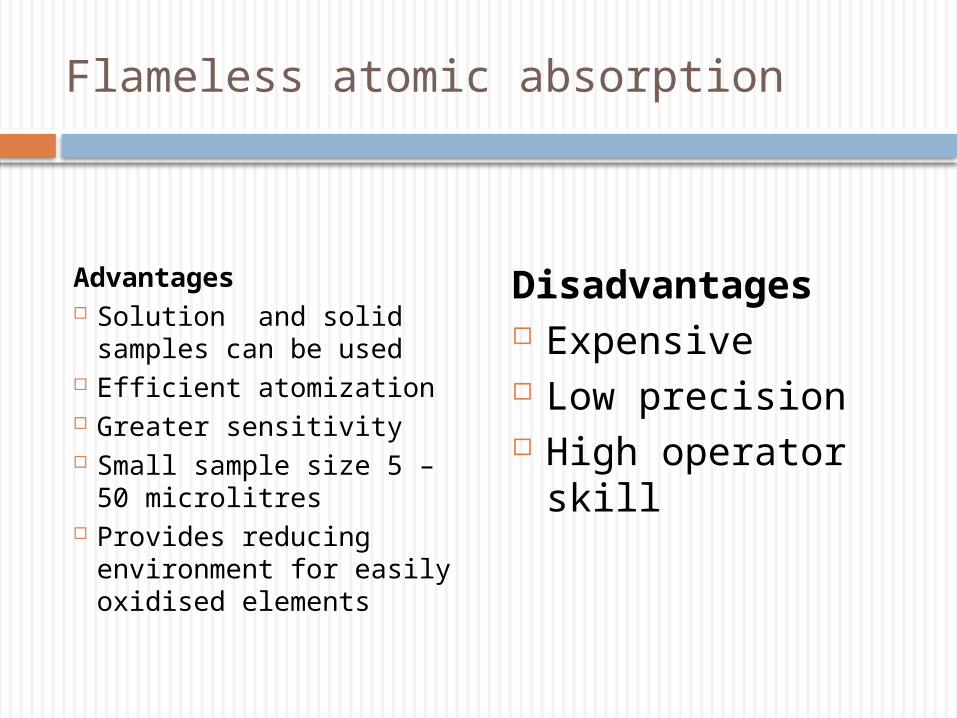
Advantages Solution and solid samples
can be used Efficient atomization Greater sensitivity Small sample size 5 – 50
microlitres Provides reducing
environment for easily oxidised elements
Disadvantages Expensive Low precision High operator skill
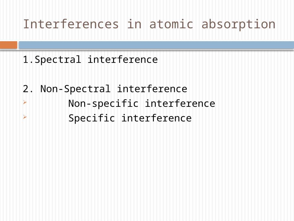
Interferences in atomic absorption
1.Spectral interference
2. Non-Spectral interference Non-specific interference Specific interference
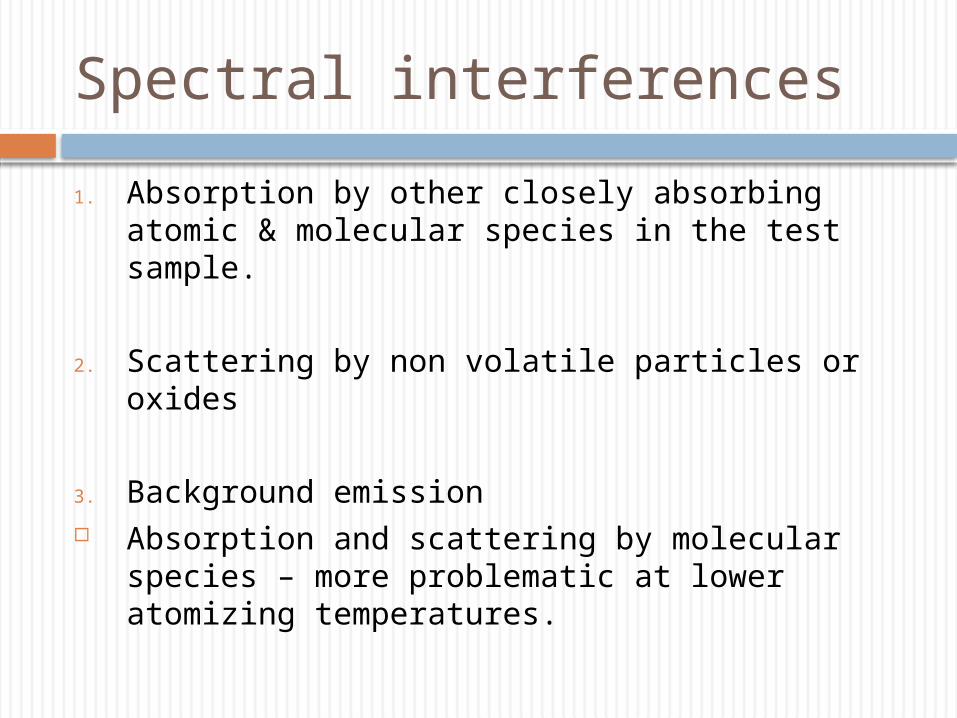
Spectral interferences
1. Absorption by other closely absorbing atomic & molecular species in the test sample.
2. Scattering by non volatile particles or oxides
3. Background emission Absorption and scattering by molecular species – more
problematic at lower atomizing temperatures.
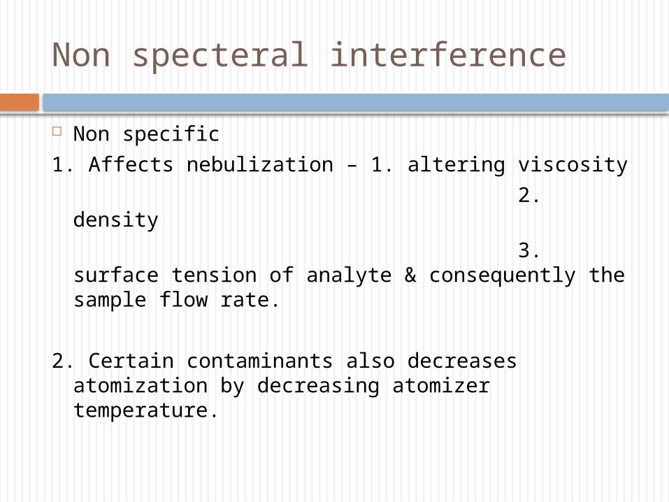
Non specteral interference
Non specific
1. Affects nebulization – 1. altering viscosity
2. density
3. surface tension of analyte & consequently the sample flow rate.
2. Certain contaminants also decreases atomization by decreasing atomizer temperature.
Specific/chemical interferences
Anions – form compounds that’s not completely dissociated
(decreasing the no of ground state atoms )
Eg : phosphate interference by calcium phosphate complexes.
Interference eliminated – Lanthanum, strontium (cation)
Ionization interference
Atoms are ionized instead of being in ground state- not absorb incident light – apparent decrease in analyte concentration.
This interference is minimized by operating flame at low temp.
Emission /excitation interference
Atoms excited by the flame, emit photons of same wavelength as of incident light measured – enhances the signal received – translated as ↓ A – low concentration of analyte.
Minimized by using a chopper or pulsing the light to the hollow cathode lamp.
Burner problems
Essential to have a steady flame.
Burner head should be clean.
In flameless AA carbon rod should be changed after a no of
firings.

Reference
Clinical chemistry: Kaplan Clinical chemistry: TIETZ