phlebotomy for medical laboratory professionals
TRANSCRIPT

Phlebotomy
Ravi KumudeshMSc/BSc / Dip(MLT)/PG Dip(SMgt)
Sri Lanka Society for Medical Laboratory Science
slsmls.org

Modern Phlebotomy
• Diagnosis and management of disease
• Remove blood for transfusions
• Therapeutic reasons:– Polycythemia– Hemochromatosis
2

Blood Function:
1. Supplies nutrients to tissues:
O2, hormones, glucose
2. Removes end-products of metabolism:
CO2, urea, creatinine
3. Provides defense mechanism:
WBC, antibodies
4. Prevents blood loss:
platelets, coagulation proteins
3
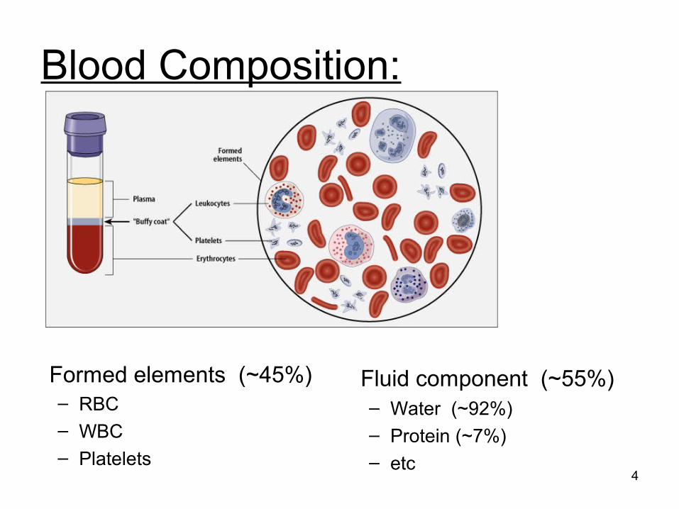
Blood Composition:
Formed elements (~45%)– RBC– WBC– Platelets
4
Fluid component (~55%)– Water (~92%)– Protein (~7%)– etc
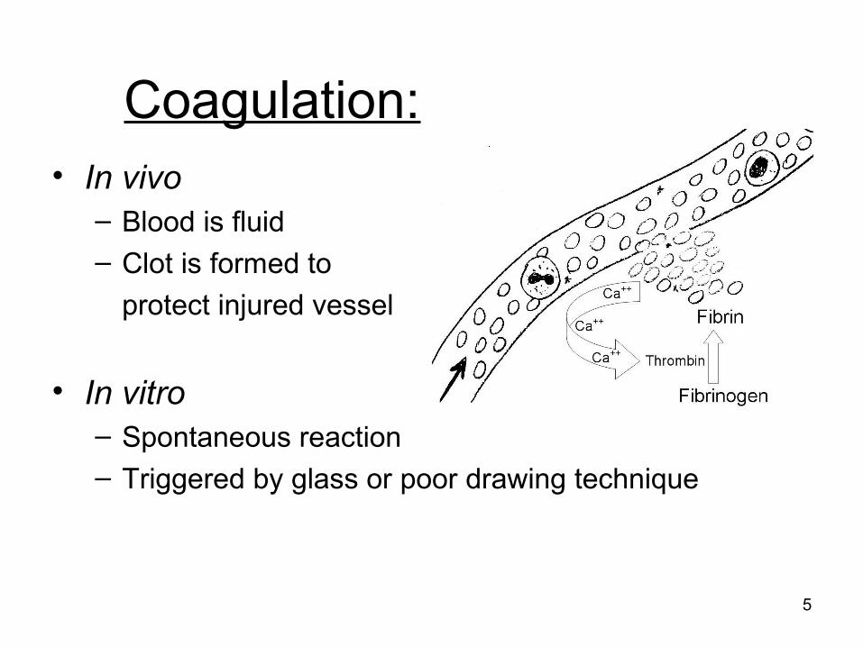
Coagulation:• In vivo
– Blood is fluid– Clot is formed to
protect injured vessel
• In vitro– Spontaneous reaction– Triggered by glass or poor drawing technique
5
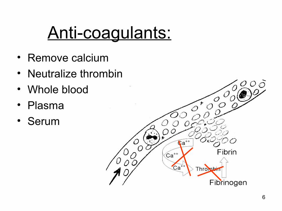
Anti-coagulants:• Remove calcium• Neutralize thrombin• Whole blood• Plasma• Serum
6
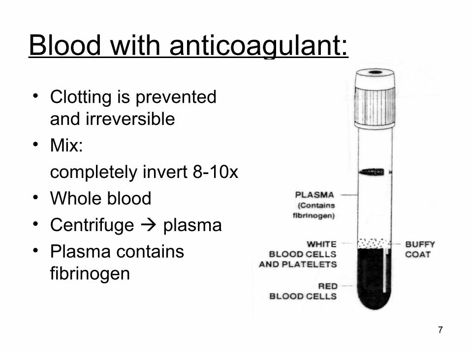
Blood with anticoagulant:
• Clotting is prevented and irreversible
• Mix:
completely invert 8-10x • Whole blood• Centrifuge plasma• Plasma contains
fibrinogen
7
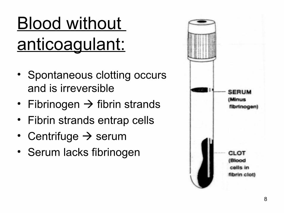
Blood without anticoagulant:
• Spontaneous clotting occurs and is irreversible
• Fibrinogen fibrin strands• Fibrin strands entrap cells • Centrifuge serum• Serum lacks fibrinogen
8
Appearance
• Normal: clear and ‘yellow’
• Abnormal:– Hemolyzed = pink to red (ruptured RBC)– Icteric = dark orange-yellow (bilirubin)– Lipemic = cloudy (fat, triglycerides)
9
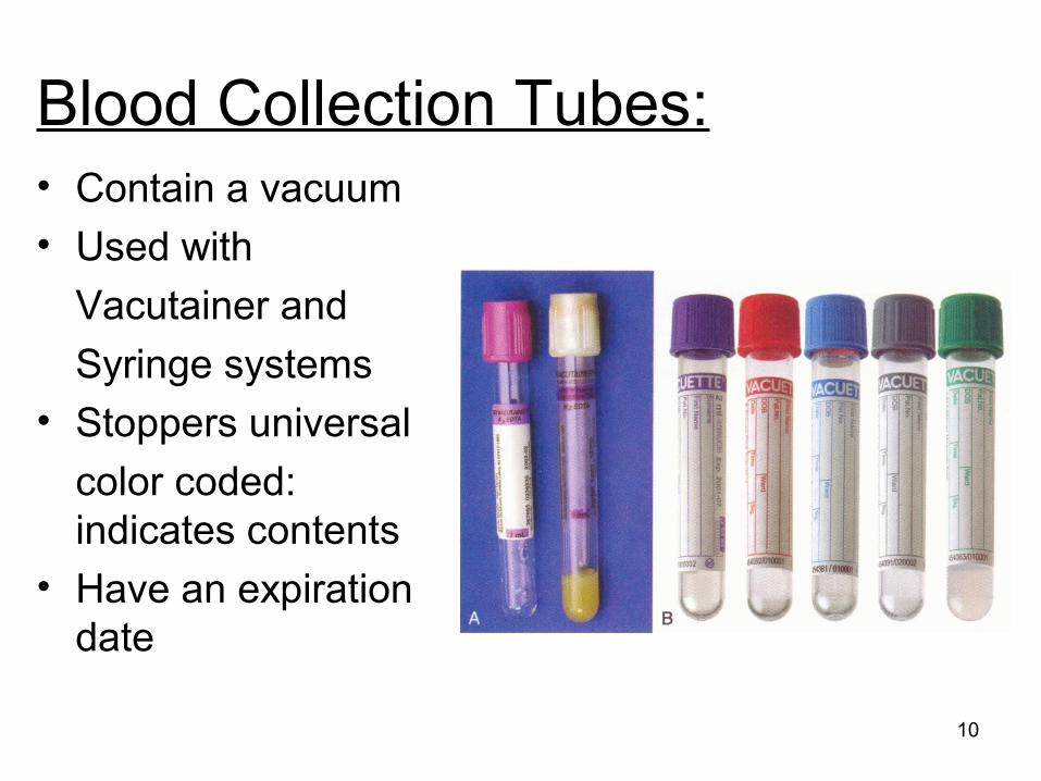
Blood Collection Tubes:• Contain a vacuum• Used with
Vacutainer and
Syringe systems• Stoppers universal
color coded: indicates contents
• Have an expiration date
10
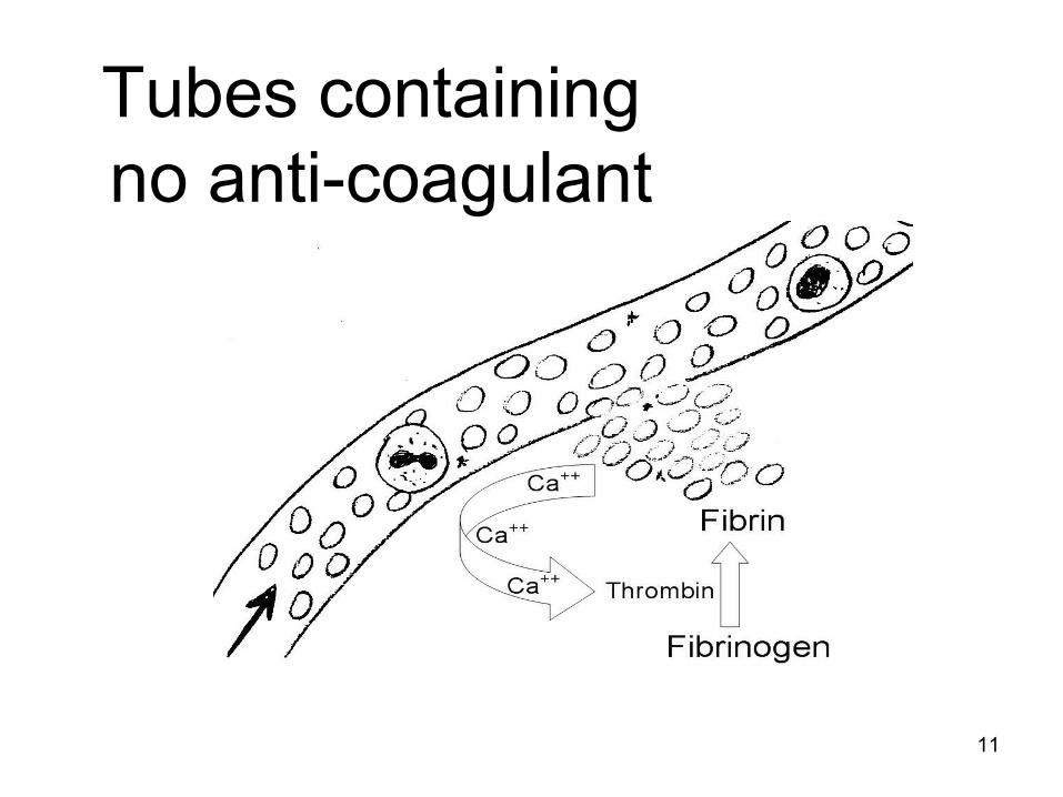
Tubes containing no anti-coagulant
11
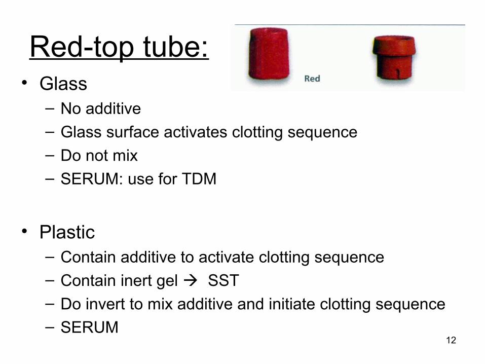
Red-top tube:• Glass
– No additive
– Glass surface activates clotting sequence
– Do not mix
– SERUM: use for TDM
• Plastic– Contain additive to activate clotting sequence– Contain inert gel SST– Do invert to mix additive and initiate clotting sequence– SERUM
12
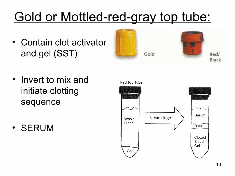
Gold or Mottled-red-gray top tube:
• Contain clot activator and gel (SST)
• Invert to mix and initiate clotting sequence
• SERUM
13
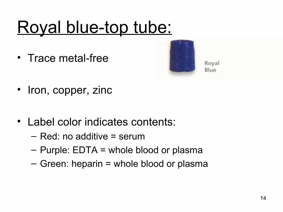
Royal blue-top tube:
• Trace metal-free
• Iron, copper, zinc
• Label color indicates contents:– Red: no additive = serum
– Purple: EDTA = whole blood or plasma
– Green: heparin = whole blood or plasma
14
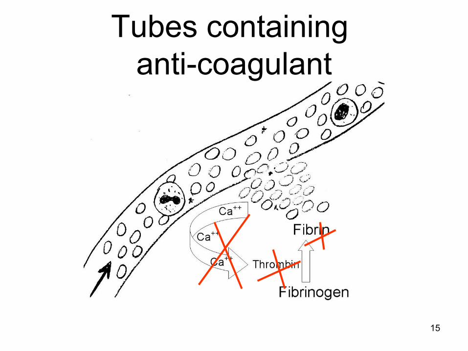
Tubes containing anti-coagulant
15
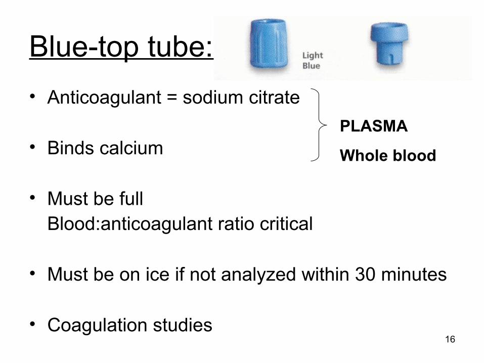
Blue-top tube:
• Anticoagulant = sodium citrate
• Binds calcium
• Must be fullBlood:anticoagulant ratio critical
• Must be on ice if not analyzed within 30 minutes
• Coagulation studies16
PLASMA
Whole blood
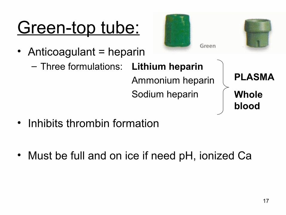
Green-top tube:• Anticoagulant = heparin
– Three formulations: Lithium heparin
Ammonium heparin
Sodium heparin
• Inhibits thrombin formation
• Must be full and on ice if need pH, ionized Ca
17
PLASMA
Whole blood
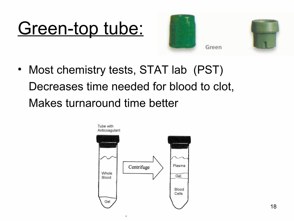
Green-top tube:
• Most chemistry tests, STAT lab (PST)
Decreases time needed for blood to clot,
Makes turnaround time better
18
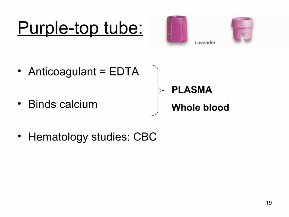
Purple-top tube:
• Anticoagulant = EDTA
• Binds calcium
• Hematology studies: CBC
19
PLASMA
Whole blood
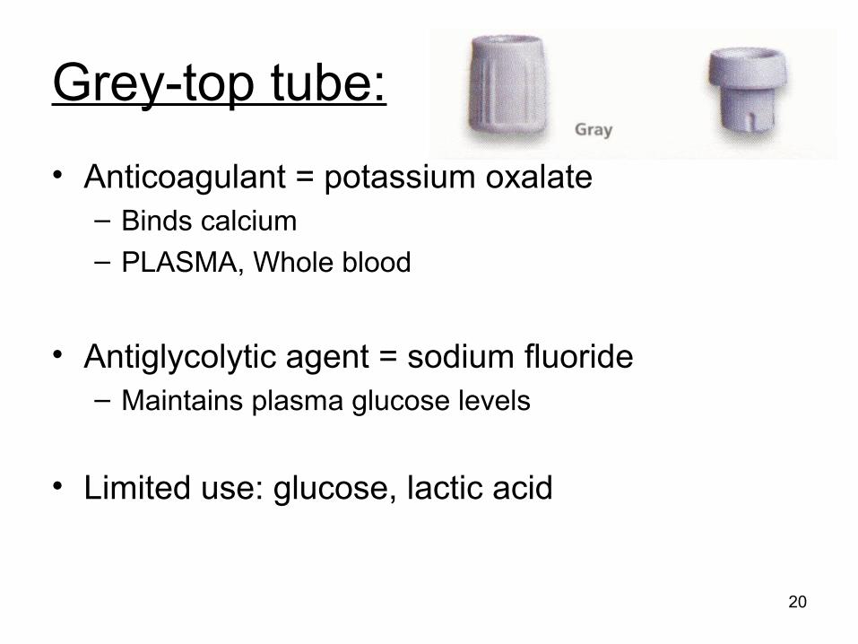
Grey-top tube:
• Anticoagulant = potassium oxalate– Binds calcium– PLASMA, Whole blood
• Antiglycolytic agent = sodium fluoride– Maintains plasma glucose levels
• Limited use: glucose, lactic acid
20
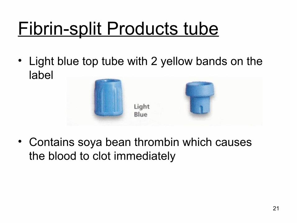
Fibrin-split Products tube
• Light blue top tube with 2 yellow bands on the label
• Contains soya bean thrombin which causes the blood to clot immediately
21
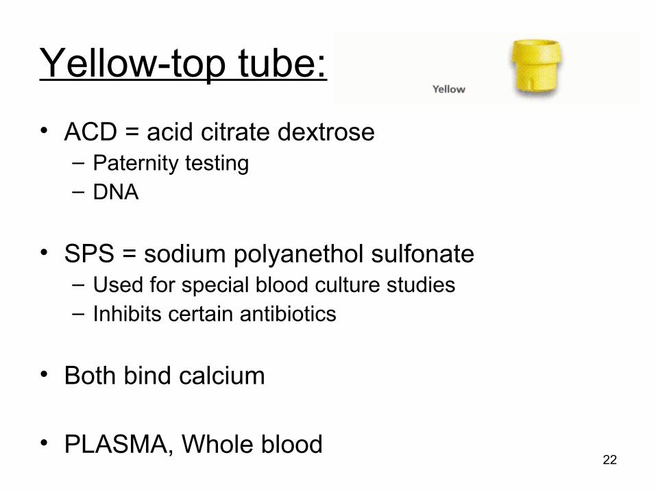
Yellow-top tube:
• ACD = acid citrate dextrose– Paternity testing– DNA
• SPS = sodium polyanethol sulfonate– Used for special blood culture studies– Inhibits certain antibiotics
• Both bind calcium
• PLASMA, Whole blood22
Type and Amount of Specimen:• Dependent upon
– Test
Whole blood: EDTA or heparin?
Plasma: EDTA or heparin?
Serum: trace free? Separator gel interference?
– Amount of sample needed to perform test
– Multiple labs needing the same specimen at the same time
23
Valid Test Results Require:
• Trained personnel– Causes of pre-analytical error– Invalid test results
• Quality control• Quality assurance• Sophisticated
instruments
24
Safety Practices:
For infection to spread:
1. Infectious substance: HBV, HCV, HIV
2. Mode of transmission
3. Susceptible host
25

Modes of Transmission:
• Parenteral: any route other than the digestive tract– Intramuscular– Intravenous– Subcutaneous– Mucosal
• Ingestion
26
Non-intact skin: chapped hands, cuts, cuticles
Percutaneous: needles, sharps
Permucosal: mouth, nose, eyes
Safety Practices:
Infection Control: stop the spread of infection
27
Safety: Infection Control• Hand washing
– Primary means of preventing spread of infection (especially nosocomial)
– Minimum 15 seconds, soap, friction– Wash hands before and after each blood draw
• PPE– Lab coat– Gloves– Mask
• Standard precautions at all times 28
Equipment:
1. PPE: gloves, lab coat, mask
2. Cleaning agent– Alcohol pads: routine– Povidone iodine: blood culture collection and
blood gases– Soap and water: alcohol testing, allergies
1. Cotton balls, gauze29
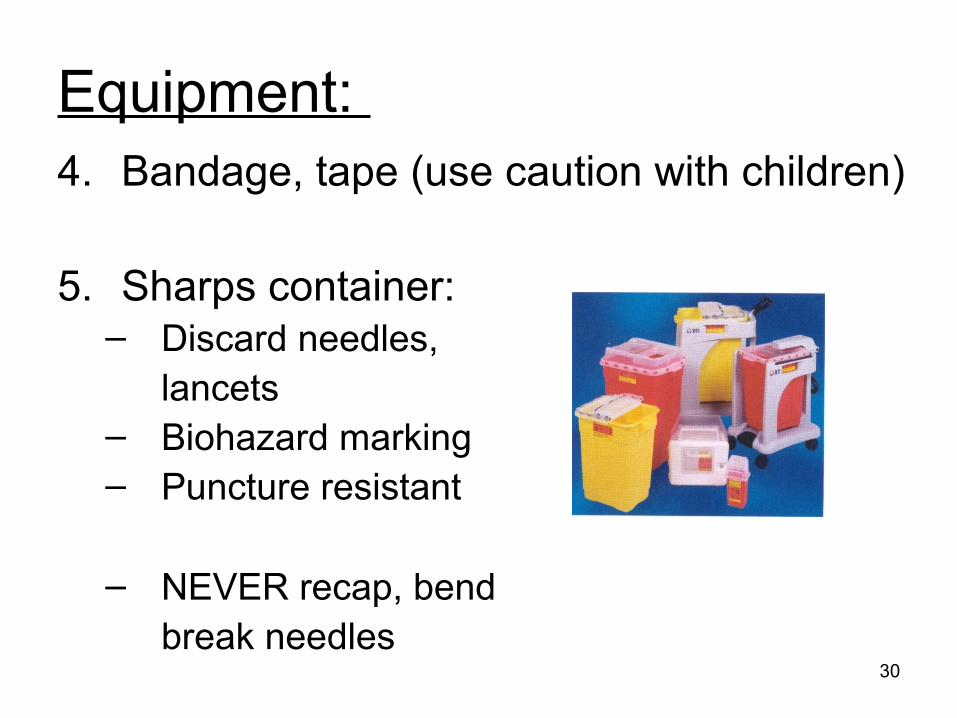
Equipment: 4. Bandage, tape (use caution with children)
5. Sharps container: – Discard needles,
lancets– Biohazard marking– Puncture resistant
– NEVER recap, bendbreak needles
30
Equipment:
6. Tourniquets:– Slows venous blood flow down– Causes veins to become more prominent– NEVER leave on for >1 minute – AVOID rigorous fist clenching or hand
pumping (potassium, lactic acid, LD)– Latex allergy
31
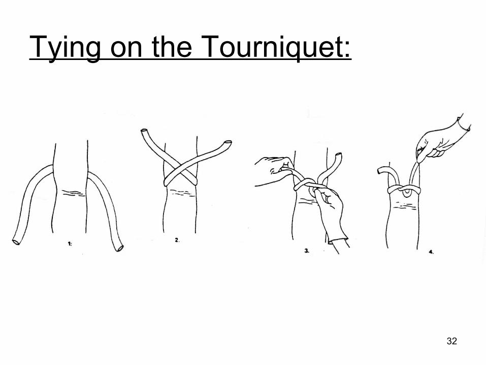
Tying on the Tourniquet:
32
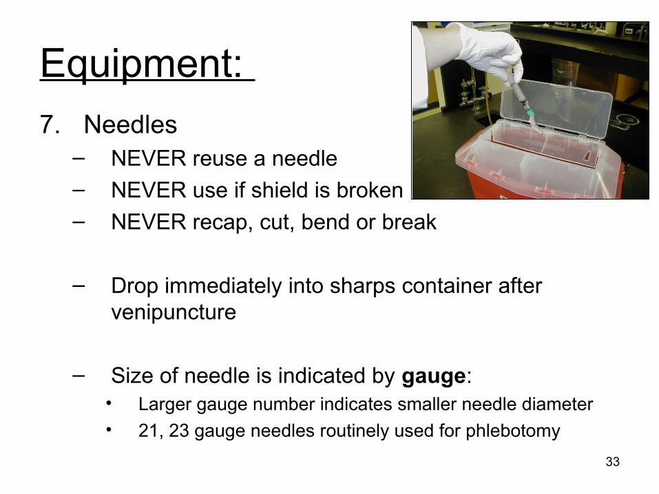
Equipment:
7. Needles– NEVER reuse a needle– NEVER use if shield is broken
– NEVER recap, cut, bend or break
– Drop immediately into sharps container after venipuncture
– Size of needle is indicated by gauge:• Larger gauge number indicates smaller needle diameter• 21, 23 gauge needles routinely used for phlebotomy
33
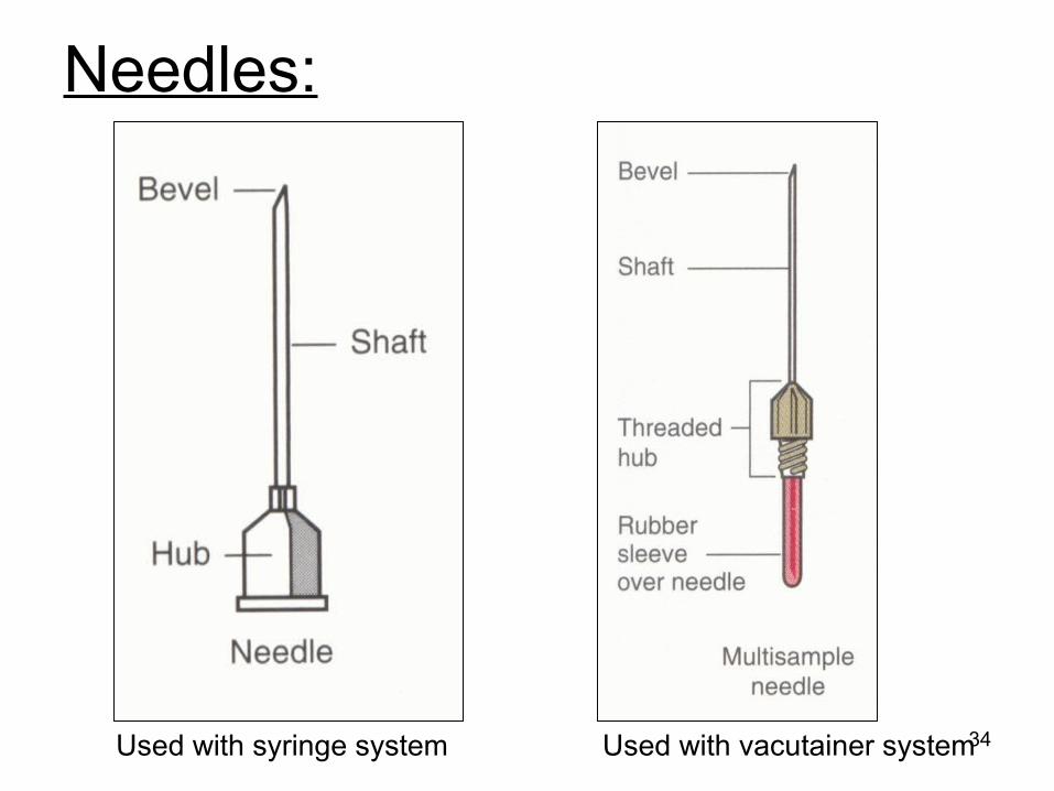
Needles:
34Used with syringe system Used with vacutainer system
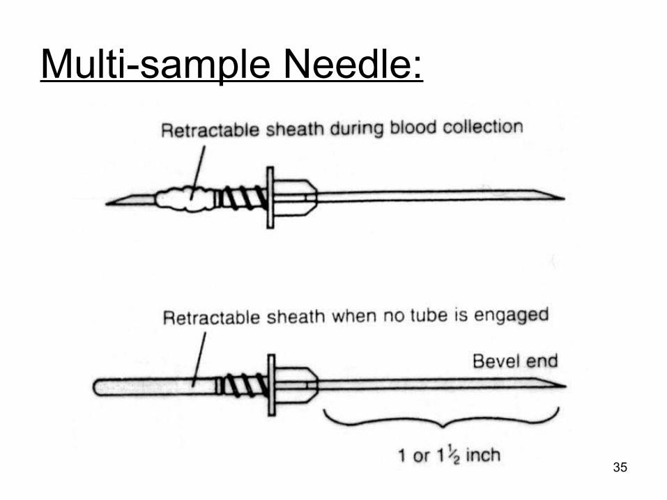
Multi-sample Needle:
35
Butterfly Needle:
36
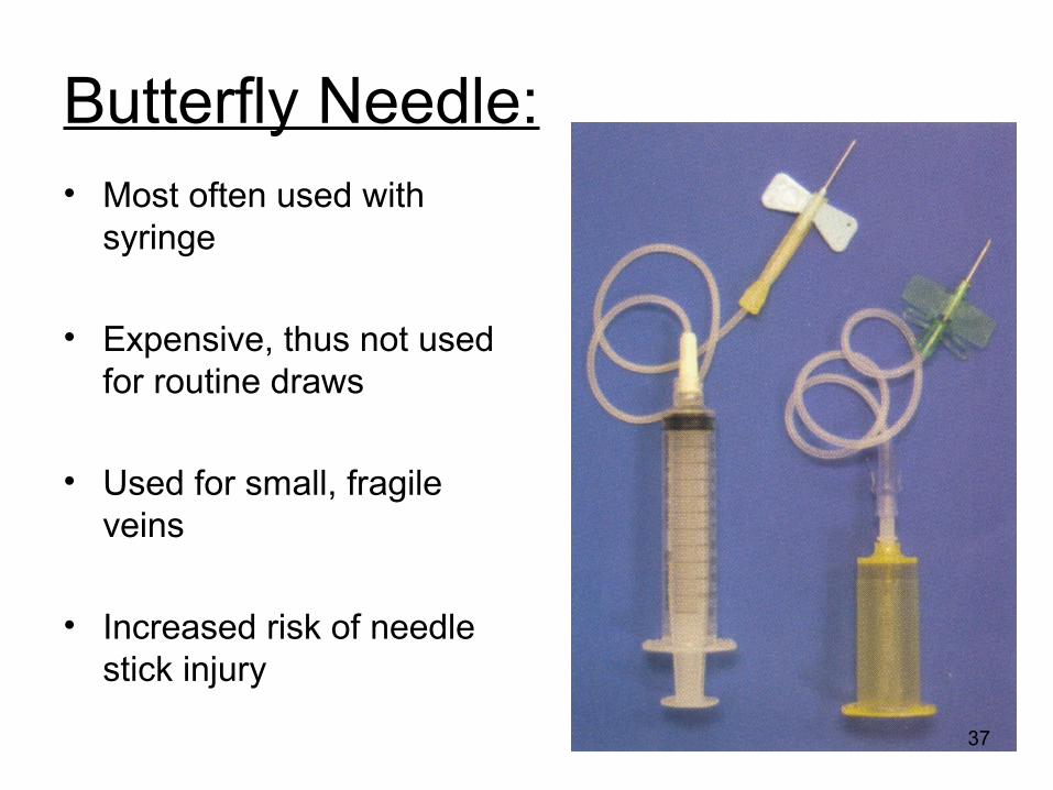
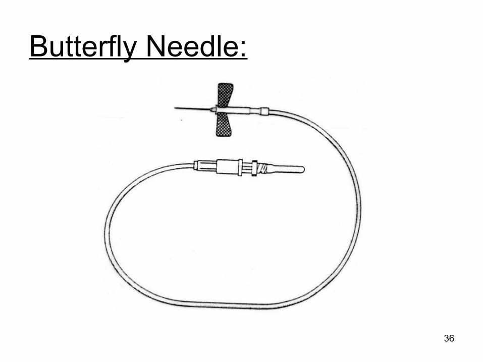
Butterfly Needle:• Most often used with
syringe
• Expensive, thus not used for routine draws
• Used for small, fragile veins
• Increased risk of needle stick injury
37
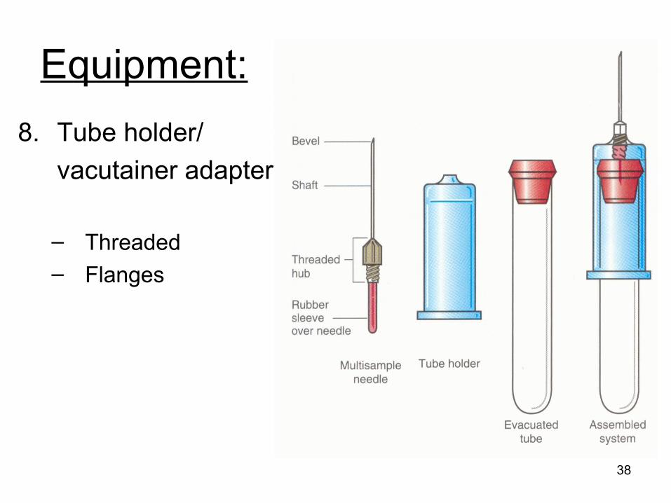
Equipment:
8. Tube holder/
vacutainer adapter
– Threaded– Flanges
38
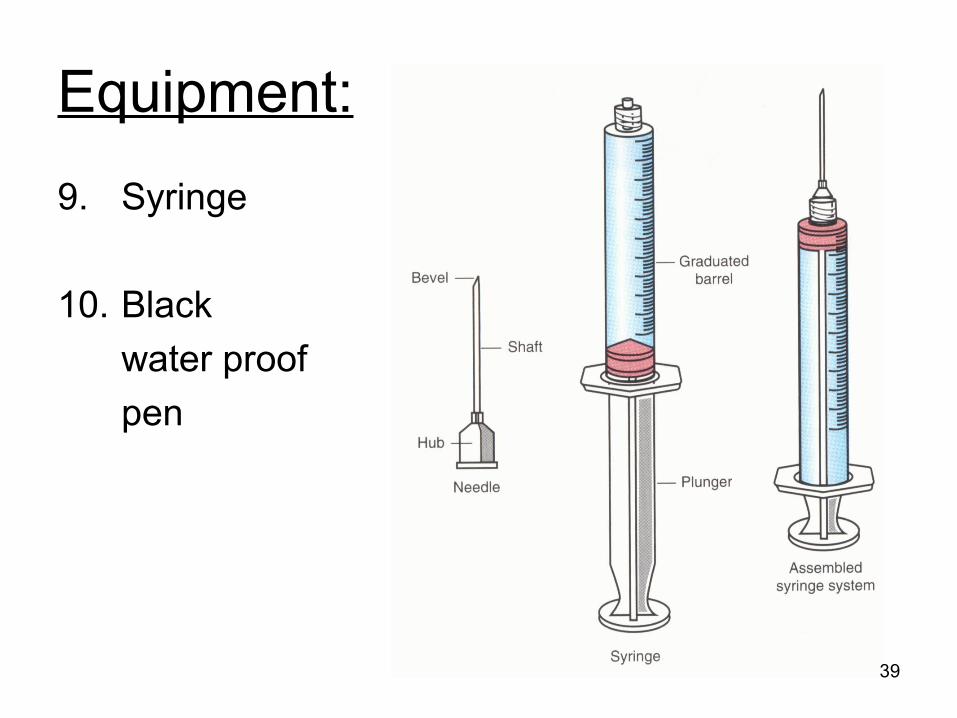
Equipment:
9. Syringe
10. Black
water proof
pen
39
Labeling Blood Collection Tubes:• Black indelible marker (water proof)
– Never pencil– Legal document– Print legibly
• Required information: 5 items– Patient name– Identification number– Date of draw (mm,dd,yyyy)– Time of draw (military time)– Phlebotomist signature: first initial, last name
40
Vacutainer or Syringe?
• Vacutainer– Most often used– Most economical– Quick– Least risk of accidental needle stick
• Syringe– More control– Reposition easily– Will see ‘flash’ of blood in syringe hub when
vein successfully entered 41
The Patient:
• Approach• Communication• Empathy• Handling special situations• Patient identification
– Arm band
– Legal document
• Prepare patient for blood draw– Latex allergy?
42
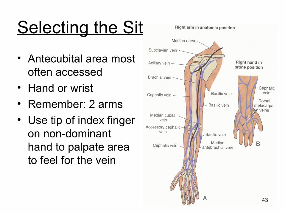
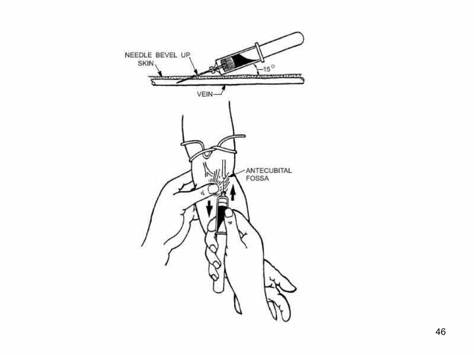
Selecting the Site:
• Antecubital area most often accessed
• Hand or wrist• Remember: 2 arms• Use tip of index finger
on non-dominant hand to palpate area to feel for the vein
43
Collection Site Problems:
• Indwelling lines:– Hickman catheters– Heparin locks
• Used to administer medication
• Only nurse may access these lines
• Can obtain blood: called a ‘line draw’
• Must clear line of heparin contamination by discarding first 5-10 cc of blood
44
Inserting the Needle:
• Anchor the vein– Grasp arm with your
non-dominant hand
– Use thumb to pull skin taut
• Smoothly and confidently insert the needle bevel up– 15-30 degree angle
45
46
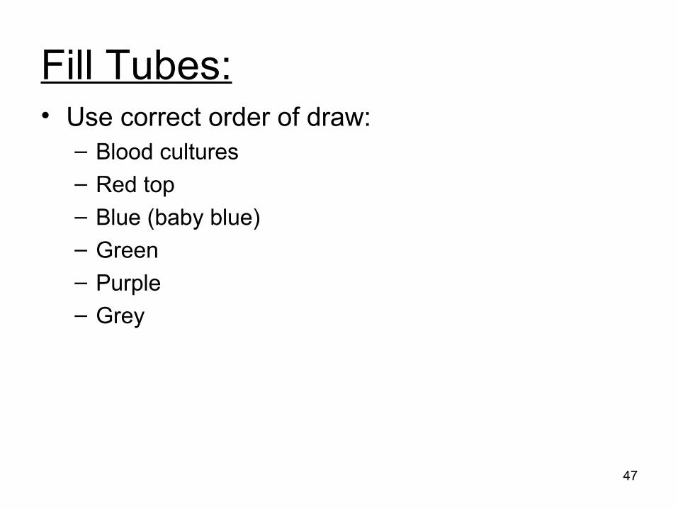
Fill Tubes:• Use correct order of draw:
– Blood cultures
– Red top
– Blue (baby blue)
– Green– Purple– Grey
47
Be careful not to:
• Push needle further into vein when engaging evacuated tube
• Pull needle out of vein when disengaging tube
• Pull needle out of vein as you pull back on the plunger
• Pull up or press down when needle in vein
• Forget to mix additive tubes 8-10 times
48
Withdraw Needle:
• First release tourniquet
• Disengage tube
• Place cotton directly over needle, without pressing down
• Withdraw needle in swift, smooth motion
• Immediately apply pressure to wound
• Do not bend arm
49
Label Tubes Immediately:
• In sight of patient• Patient name• Identification number• Date of draw• Time of draw
(military time)• Your initials
50
Recheck Draw Site:
51
Failure to Obtain Blood:
• Check tube position and vacuum– Always have back up tubes near by
• Needle position
• Collapsed vein
52
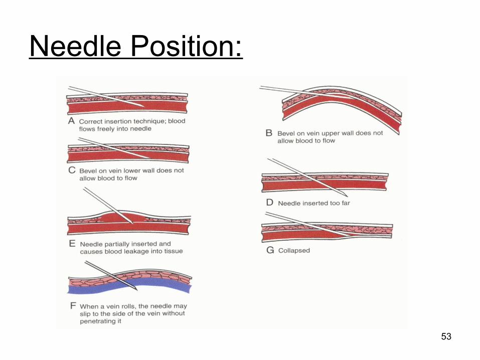
Needle Position:
53
You should try again
• Look at alternate site– Other arm– Hand
• Use clean needle• Use fresh syringe if
contaminated
• Only try twice
54
Poor Collection Techniques:• Venous stasis
– Prolonged application of tourniquet (>1 min)
• Hemodilution– Drawing above IV– Short draw (blood to anticoagulant ratio)
• Hemolysis– Traumatic stick
– Too vigorous mixing
– Alcohol still wet– Using too small of needle– Forcing blood into syringe
55
Poor Collection Techniques:• Clotted sample
– Inadequate mixing– Traumatic stick
• Partially filled tubes– Short draw– Sodium citrate tube draw volume critical
• Using wrong anticoagulant
56
Poor Collection Techniques:• Specimen contamination
– Using incorrect cleanser– Alcohol still wet– Powder from gloves– Drawing above IV
• Specimen handling– Exposure to light– Pre-chilled tube– Body temperature
57
Venipuncture Procedure
• Remain calm
• Organize yourself
• Organize your equipment:
STICK TO ELEVEN
58
Equipment: Stick to Eleven
• Gloves• Lab coat• Alcohol wipe• Cotton ball• Bandage/tape• Sharps container• Tourniquet
• Needle• Syringe or vacutainer
holder• Collection tubes with
backup tubes• Water-proof marker
59
Venipuncture Procedure:
• Wash hands
• Put on gloves
• Identify patient
• Latex allergy?
• Position arm
• Apply tourniquet
60
Accidental Needle Stick:
• Remain calm• Cleanse wound with alcohol• Wash wound thoroughly• Notify supervisor, instructor• Follow site protocol• Page OUCH hotline: 1-402-888-OUCH
1-402-888-6824• Complete incident report
61

Skin Puncture:
• Method of choice for infants, children under 1 year
• Adults– Scarred – Fragile veins– Hardened veins– Home glucose monitoring (POCT)– Patients with IV
62
Capillary Blood• Mixture of arterial, venous, capillary
blood and fluid from surrounding tissues
• Fluid from surrounding tissues may interfere and/or contaminate the specimen
• Warming skin puncture site increases arterial blood flow to the area
• Reference ranges often differ from venous
63
Skin Puncture Equipment:
1. PPE
2. Cleaning agent– Alcohol pads: routine– Soap and water: alcohol testing,
allergies– DO NOT use providone iodine
1. Cotton balls, gauze
64
Skin Puncture Procedure:1. Wash hands
2. Approaching the patient
3. Patient identification
4. Latex allergy?
5. Bedside manner
6. Site selection7. Cleanse site: DO NOT use providone- idodine
8. Perform puncture: Wipe away first drop of blood
9. Label the specimen 68
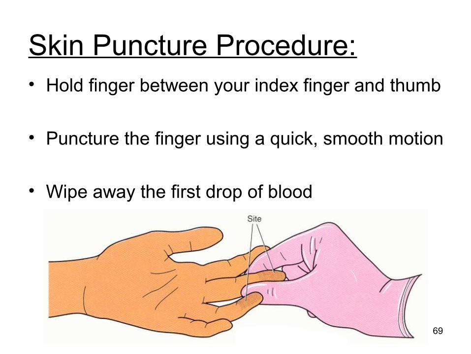
Skin Puncture Procedure:• Hold finger between your index finger and thumb
• Puncture the finger using a quick, smooth motion
• Wipe away the first drop of blood
69
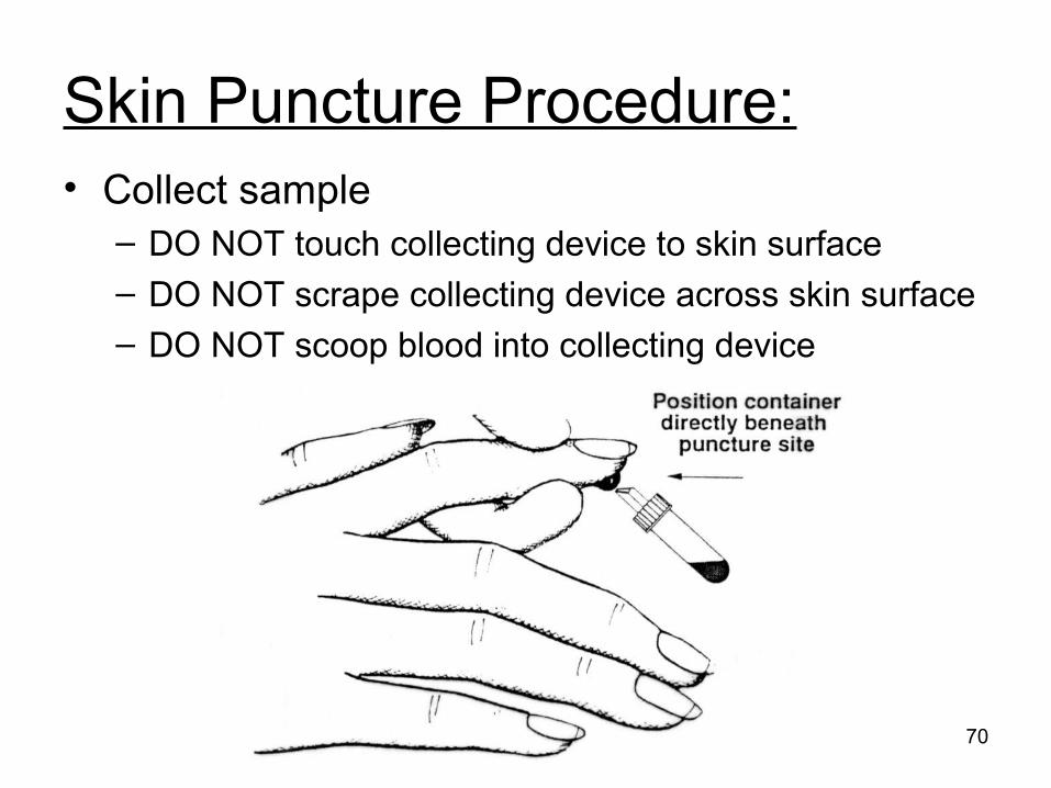
Skin Puncture Procedure:• Collect sample
– DO NOT touch collecting device to skin surface– DO NOT scrape collecting device across skin surface– DO NOT scoop blood into collecting device
70
Skin Puncture Procedure:• Order of draw is critical: platelets
accumulate at puncture site causing clot formation– Blood smear– EDTA– Heparin
– Serum
• Apply pressure to puncture site• Label specimen in sight of patient (indelible
marker)
71
72
Thank You !
73