philosophical trans1~ctions
TRANSCRIPT

I
PHILOSOPHICAL TRANS1~CTIONS.
I. -- The eed-Fu11g11s of Lolium ternulentum, L., the Dame{.
By E. I. F.REEMA ·, Jl.S., Unii:er::;ity of .Jlinne ·ota. cJ
Conimimicaled by PrnfosHor lAR 'UALL YV AHD, F.R.1 '.
Heceived J tulC 6,- Read J u11e Hl, Hl02.
[Pr.ATES 1-3. J
Introductory.
lT lias long been known* that the grains of the Darn 1 contain a poi onou ho(ly (Lolium), which can lie extracted by ther. This ·ub tance ha marked toxic action on rabbits and certain carnivorous auimals, and i · ·aid t induce vomitinrr ancl other unplea ant ·ymptom · in man, but to aff ct pig , cattl , ancl e ·e but littl , or not at all.J
In 1898t attention wa · drawn to the fad that a lar<Te p 1·c 11tacr ( 0- 100 p r cent.) of the grain of this grass contain a deiinit fuurru -m •celium, alw, 'S ituat d in a definite layer of the seed, i .e., iu lhe remains of the nuc llu JU outside the aleuroue-layer of the endosperm.
lthough certain details were made out re<rarding the natm" and po. ilio11 of th hyphc:e, their relation · to th eed and se dling and to th poi. ouous prop •rti referred to, almo t nothing was discov r cl r arclina the ' t ma.tic p , iti 11 of th fuugu , the course of its lifi -hi tory, or v n how it obtain it ntry into th Loliwn.
In the following account of an inve. tigation pm u cl in th ambrid otauical Laboratory during the pa. t e.' ·ion, I lrnxe . ucce d cl in carr incr our know] dcr' of this r markable fungu con iderably further, and p cially in rend rina clear the principal point concernina it lifi in th plant, and th mocle of infi ction of th embryo. N everthele s, all attempt· to grn' the fuu<ru ' out id it ho t-plant, or to induce it to form spore , have failed with me a· with oth r investicrator and allhourrh some suggestive fact have been obtained which may help iu fuime ~fforl to tablish
According to G ElUN (5) since Roman times. t HACKEL in Engler n.nd Prantl, 'x,~tiirliche PAanzenfamilien,' II. Th. 2 Abth., 1 ' 7, p. 76. + By VOGL, GUERl~, H.\:\.\t:::>EK, and -.i:: 'TLJm, in four papers rnforred lo below.
VOL. CXCVJ.--B 214. .8 ~. 1.03.
2 MR. E. !\I. FREEMAN ON THE SEED-FUNGUS
the systematic position of the fungus, we are still utterly baffled as to whether it '.s au talagine, a U recline, or an Ascomycete of the nature of ergot 01· other toxic sp cies as yet unknown.
The evidence so far points to the present case being an interesting example of syrnbio ·is.
Histo1·ical Summary.
Previous to 1898 the research work on Lolium ternulent11m was entirely of a chemical
nature, dealing especially with the toxic constituents. In January, 1898, VoGL ~2) announced the pre ence of a layer of fungus hyphrn in the crushed nucellar remams just outside the aleurnne layer, and later on in the same year three other papers appeared ou the subject.
In Augu t, GUERIN ( 5) published the result of his investigations, the object of which was to determine the cause of the toxicity of Darnel."'' He described and
iignred a densely interwoven layer of hyphrn between the exterior wall of the aleuro~e la er and the hyaline layer of the grain. He found the hyphrn lining the entire grain, except alono- the groove on the inner side and in the vicinity of the embryo.
o mention is made of the fungus in the early stages of the gl'Owing plant. He b erved the fungus in the young ovary before the fertilisation of the egg-cell, as
al ·o the growth of the endosperm, which results in the comprnssion of the layer of hyphro found in the mature grain. Lolium ai·vense, With., and L. linicolwn, Sond.,
were als ob erved to contain the fungus, and since these three species are those to which poi onous properties are assigned, Gu1ht1N suggested that their toxicity m~y he du to the fungus found in them. Lolium italicum, Braun, did not contam hypl~a~ at all, and of L. perenne, L. , only a single example with the fungus was
obh med. amples of L. teniulentum from South America, Asia, Africa, and Europe, w~re e~amined, and only three were found to be devoid of the hyphrn. The nature
of ~h fu~g.u c~uld. not be determined. Gu.ERIN points out the pronounced differences wluch ch hngmsh it from Endoco11idium temulentum, described by PRILLIEUX and
ELA ROIX (10). The germination of these grains of Lolium ternulenturn was found to be xcell nt, and th fungus was said to be never met with in the neighbourhood
of th mbryo. Finally, G ERIN suggests that the presence of the hyphal layer in Darnel o-rains indicates a case of symbiosis rather than of true parasitism. The
rearr nt u cl by GUERIN were chloral hydrate or lactic acid fior swellino- and "coton bl " fi . . O' u or tammg.
The IJapers of HANAUs"'·K d N (3) . . • ·A an ESTLER appeared m September. HANAUSEK
cl<'~cnlP~l the position of the fungus essentially as <lid GUERrN. He also fouud the hvpln:e m the nucellus f" tl · (" K .. l ")
·• . ' 0 1e young ovules, where it produced knots naue ' ' n1ch , however develop cl f' tl FI . . . . · · l 1 U tila-' e no LU' 1er. e pornted out sm11lanties wit 1 t le s c
* In a ub cquenL noLc (11) " . ·. . 1 · · h th L of \ • ' \.J:CL!Ul\ c aims Lhat his work was at least contemporaneous wit a Ol.r,, although published later.
~ ' . '

OF LOLI M TE 1ULE JTU M, L., THE I> R~ TEL. 3
ginere, and waR of opinion that there is much evidence in support of the vi w that the fungus is an Ustilagin , which rarely forms spores. No hyphre were found m L. perenne.
NESTLER ( 4) agrees with the previou · author · as to the po. ition of the h yphre m the grain. Darnel grains were allowed to germinate under various condition after treatment with ether. On the eighth clay some hyphm w r found in the growing point of the seedling st m and in the base of the young leaf rudiment . Th y were subsequently found also in the internode , and eventually in th v g tative con ancl in the youngest leaf rudiments, occupying the intercellular pace . Th y w r even traced through the axis of the inflorescence into th . ovular rudiment, ent ring through the funicle, and were found in abundance in the nuc llus. NE. TLER'.· description of the subsequent development agrees with that of Er:.IN. NE TLER also placed pieces of th h phre from the nucellar l.ayer of the mature grains in various nutrient solutions of different strength ; but the hyphre would not grow. The examination of the grain· during germination show cl, after a few clay , a fonnation of isolated hyphre, with rounded cells at the end or in the micldl of th b ·phre (" Sporenbildung "). Later on, littl but imprint.· on the aleuron c lls w r to be found. vVhen the culm · w r about l clecim. hiah , the enclo. perm of th min wa founcl to contain numerous thin, long, .·egmented hyphm with p rp ndicular branche . Th . e occurred even in partially sterilised grain . NE TLER adds that the id ntity of the. with the nucellar hyphre is not proved. A to th method of penetration, the author suggests that the funau is in th vea tative con from th beainnin of g rmination, but he found it in the growing point of the embryo in onl r a sinale in tance. He al. o argued that penetration could not hav taken plac from the ext rior, a hi Cc r ful manipulation mu t have pr vented uch an ntrance. No <l finite sy t matic po. it ion is assigned to th fungus, but a . imilarity betw n the toxic phy. iological action of Darnel and of \VORO~IN's (12) "Taum lrogO' n,, is . uggestecl c po.' ibl r indicating relationship . The areat diffi r nc . in appearanc and h haviour of the fungi which cau e " Taumelroaa n ' ar , however, point cl out. TE- TLlm mad u e of chloral hydrate and potas. ium hydrate a cl arina reag nt..
No fungu layer was found in the oth r .·p ci . of Loli1rni. Th speci . xamin cl were L. perenne, L. , L. rnultiflo1'um, Lam. (= L. italic1w1, A. Br. = L. Boucl1ean11m, Kun th) L. 1·e11iotum, ch rank ( = L. w·ven:e "chracl. = L. linicolum, A. r. ), L. festucaceum, Link. ( = L. perenne, L. X Fe.·tuca elatiol', L.), and oth i..
hort paper hy 11 HELETTI appeared la. t year (1901), and cl als almo t ntir ly
with th toxicity of Darnel (6).
Jfethods.
For the demonstration of the presence of the funO'US in the O'rowina point of the plant, the arains were placed in an ordinary O'erminating chamber, and the embryo or seedlings dissected out at different stage . FLE:\fMINO s weak solution (7. p. 4 l)
B 2

4 l\fR. E. 1\1. FREEMAN ON THE SEED-FUNUUS
and chromic acid, 1 per cent. and t per cent., were used as fixing fluids. The material was cut, in the usual way, in paraffin. Aniline-water-safranin (8, p. 185) . and HAIDE~HAIN's hrnmatoxylin proved most useful for staining. The latter is particularly helpful in the detection of the hyphre by means of the nucleoli, . wh~ch stain deeply, and the method proved quite successful, especially in the exammat10n of the seedling. Analine-water-safranin is very useful in the study of the ovary. Chloral hydrate, potassium hydrate, and lactic acid are useful in cel'tain cases in the examination of the grains, but they are not suitable for the study of the growing point; their use probably accounts for the failure of previous writers to di cover the hyphre in the earliest stages of germination. As the alcoholic dehydration and high temperature of the paraffin-bath make the starchy endosperm unfit for the preparation of paraffin sections, the grains were cut in an ether-freezingmicrotome after fixing in the usual way.* Of sections so cut, some were examined without further treatment, and others were treated with the various reagents above mention d.
Attempts at cultures of the nucellar hyphai taken from the grain were made in various media, in hanging drops in the Marshall Ward cell (9, p. 131). The difficulties of obtaining mounts free from bacteria and from other fungi are considerable, and details of the cultures will be described later on.
umerous grafts of embryos of various species of Loliwn upon the endosperm of other pecie w re also successfully obtained. In the early part of this investigation the mall percentage of grains of Lolium temulentum which do not contain the fungus,
contrasted with the similarly small number of grains of L. perenne which do c ntain it, occa. ioned some difficulty, especially in view of the erratic occurrence and variation, a explained below. This difficulty was completely obviated later by examininO' sections from the stigmatic end of all grains used, so that the condition of ach grain (as regards pre ence or absence of fungus) worked with was accurately
known. In the grafting and other experiments of a similar nature, no meth(ld was d vi eel which could ensure absolute freedom from various moulds and bacterial forms without at the same time injuring the embryo, but several were found helpful in ch ckinO' the growth of these forms, and in some cases perfectly clean seeds were appar nt~y obtain d. ry heat at 95-98° C. killed the embryos; immersion in ether for 15 mmutes (th method of NES'l'L~~R) gave fairly satisfactory results in checking th growth of mould., but failed to completely prevent contamination; the embryos in almo tall c,v .· survived the treatment. One per cent. corrosive sublimate applied to .the dry. grains for 10-15 minutes usually cleaned the grains, but the embryos wh1~h sm~viYed ~uch. severe treatment were comparatively few in number. A ?-mmut~ nnmers10n i~ the same solution, however, proved satisfactory, at least m checkmg, and often m completely preventing, foreign growths ; the growth of the
* I am great!}' inuebted to Mr A W H D · · th' part of the work. · • • ILL, elJlon$lrntor ln Botany, for the use of apparatus 111 is

OF LOLIUM TE fULENTU 1, L., THE DAR TEL. 5
embryos ·wa1> at most but slightly retarded. In all cases where anti ptic solutions w re u1>ccl the grains were subsec1uently thoroughly washed in boiled cli tilled water.
The Grain and 1'ts Fungus.
The cross-section of the average grain of Lolium temulentum shows a layer ot densely woven fungus hyphre just outside the aleurone layer in the crushed remnants of the nucellus, of which the outer cell-rows form th "hyaline " layer. This is the hyphal layer which has been <le crib d by NE TLER, G ERIN, HA AU EK and oth rs.
Previous writers have found but a very small per cent. of the grains of L. temulenttt?n devoid of the hyphre. HAr AU EK states that he examined many hundreds of grains and found none without the fungus! The number devoid of the fungus found by G1 ERIN was very small, and NE TLER also reports only a few. I have found tho proportion between those grain with and tho1> without th fungu exce dincrly variable. In one package of grain crrown at the niver ity Botanical Gard ns in Cambridge, 15 per cent. (l2 out of 76) wer devoid of the fnngu , and in a package received from Upsala, out of ten crrain1> examine<l eiO'ht did not contain hyphre. Another lot of thirty-five from th niver ity Botanical ardens at ambridg gave thirty-three with and two without hyphre. The latter probably approximat th usual proportion. The error which might ari · from an admixtur of oth r p ci 1s practically nil, on account of the marked character of the grain. of Darn 1. *
fter examining about tw nt grain ', 1 discovered that c rtain micro copic di:ffi rences were sufficient to enable me, eYen without the aid of a 1 n. , to pick out mo t of those grain deYoicl of the fungus, suhsec1u nt micr . copic examination in alm t every ca e confirming th lection.'. The grains without the fungu often app ar incompletely developed. Th y are u. ually more . lender in both lat ral a pect , and les swollen in the centre, and ar usually cl void of the y llow to dark-brown or gr y colour of those which contain the fungu . In many cru e th y ar mor or 1 . s y llowi. h-green in colour; but ometim . of a dark traw-y llow hu . v rth 1 . examples are to b m t with ' hich ar micro. copically indi ·tingui. habl from tho e which contain the funcru" Th latt r ar usually wollen con id rably in the c ntral region, and ar y llowish-brown or grey in colour, ldom crreen. Th e g neral diffi rences are based upon the xamination of material of 1901 grown in th Botanical +arden. at 1ambridge. The colour i, perhap. the mo. t u eful cliacrno. tic charact r. pparently grain!> k pt . ev ra.l years tend to lo. e the colour cliffi rence. I han
sp cimens of L. temulentwn Yar. C(,ri;ense which are . o dark in colour that at fir. t . ight one would pronounce them to he ergot cl; they are, howeYer perfi ctly health ' crn1.ins
containing a broad lay r of hyphre. For the exmnination of the fungu in the grain arnl embr 'O , grains of Darnel wer
placed in ordinary g rminating chamb r fi r l to 24 hour . In . uch grain. no
* cc MARSHALL 'VARD, 'Grasses,' p. 168, and authorities quoted.

6 MR. E. M. FREEMAN ON THE SEED-FUNGUS
actual growth of the embryo has as yet taken place, since very rarely indeed are mitotic figures found. The considerable increase in the size of the embryo is of course due simply to the absorption of moisture. These embryos were then dissected . out and cut by the paraffin method, while the endosperm was cut with an ether-fre~zmgmicrotome. Longitudinal and cross-sections of the grain sho-w the hyphal layer m the nucellar remnants, distributed almost co-extensively with the aleurone layer (figs. 3-9).
According to all previous writers, the hyphre are not found near the embryo nor along the groove on the inner side of the grain; but although this is true for the upper part of the groove, at the base of the latter, where the end of the aleurone layer {fig. 4a) comes into contact with the outer surface of the scutellum, the hyphre are again found (figs. 4 and 5), often as a fairly broad layer, which appears to ha,·e entirely escaped the observation of previous investigators.
The hyphre at th~s point penetrate round the end of the aleurone layer, and hence come into direct contact with the embryo, and it is from these hyphre that entrance into the embryo has been effected. In this infec:tion layer, as I will term it, the hyphre run, chiefly longitudinally, for some distance above and below the end of the aleuron layer (figs. 22-24), and since the layer is often broad, a more or less circular ar a of hyphre can be seen on the outer surface of the scutellum at its base (fig. 4h ). Th ab. ence of hyphre along the groove of the inner surface is explained in the deY lopment of the ovule. The funicular region of the young ovule, along which the groov sub equently arises, contains at first hyphre, as elsewhere, though they are 1 . s abundant than in the rest of the nucellus ; but in the elongation of the ovule no growth of hyphrn seems to take place in this region, leaving the groove devoid of th funcrus in the mature grain.
In all case observed the infection has taken place from the infection laye1· described ahO\·e. The al urone layer in this region is often bent back along the scutellum towar~ t~e tip of the latter, thus forming a double layer (fig. 22). This is also xplamed m the development of the grain. The hyphre gain entrance to the embryo
hefor th latter has attained its ultimate intra-seminal size, and firmly fasten the ends of the n-leurone layer to the embryo ; so that the continued growth of the embryo r · ult in a dra gin and doubling of the aleurone layer, sliding being prevented by the hyphre. In some cas s, however, this bending back of the aleurone l::tyer does not occur, and the hyphrn are then found on both sides of the encl cells of the layer (filY, · 23 and 24). The hyphre may frequently be seen arowing throuah between the ~l uronc cells in this region {figs. 22 and 23); such a p:netration is no~ usually found m any oth r part of the aleurone layer. Under certain conditions, to be referred to sub. equently, how~ver, it does occur, and is then usually very frequent (figs. 37-39).
n the out r sin of the grain (fig. l) the aleurone laver reaches to the ligule of the .·cutellum, and the hyphal layer on this side does not re~ch the end of the aleurone*
* Gcfa1 ' figure (/oc cit 235) h · h f to . '. · "P· ' 8 owmg t e aleurone extending downward on the outer snr ace a pomt oppo 1te the growing cone is a· . .
· , accor mg to my observations, incorrect.

/
OF LOLIUl\1 TEMULENTUM, L., THE DARNEL. 7
(figs. 3, 5 and 8); it may, however, extend to within a dozen cells from the end. It is not impo ·sible perhaps that infection may, in exceptional case', take place from this side of the scutellum; but, if so it occur. very seldom. L have seen 110 evicleHce either iu the mature grain or in the d veloping ovary to iudicat that ·uch an itLfoction is evet accomplished. In all cases which I have examined, the infoctiug hyphre have been found in the area described above (fig" 4 and 16).
Along a median longitudinal line of the outer surface a ·hallow grooYe marks the line of a considerably deeper groove, found in the young ovary, and alon r this groove the hyphal layer is often narrower than in the neighbouring region . Moreover, the layer of hyphIB varies con iderably in size in different grain· as well as in different region · of the same grain. A fair average of the thickne · i perhaps I 0 to l 5JA-, but iu some grains it acquire· a thicknes · of 45JA-. It is thickest in the lateral prominences and narrowest alono· the shallow groove of the outer surface, as de crib d above. Along the inner groove, where the layer i entirely wanting, it may cea e :.Lbruptly or taper off gradually. Beyond the end of th al urou hiyer the embryo comes into direct coutact at all points with a thin covering of the fused pericarp, eed-coat , and nucellus (figs. 5, 8 and 9b).
Previous writers have not r cord d any penetration of the aleurone la er by the liyphce. In addition to tb penetration n ar the infection layer, le ·crib d above, the hypke may, under favourabl condition . penetrate at any point (fig ... ;37-3!.J). 111 material from Ghent (which did not app ar quit normal or v ry vigorou . ., !Jut uf which the germination prov d to b qually ood with that of apparentl · normal grains), in almo.t every grain examined, hyphre from the h alin lay r w r found iu great abundance, which had fore d th ir wa 'thrnugh the al urone layer into the tarch endosperm. The hyphrn ar alwa int rcellular and, a.· far a. l hav ·e n, p netrate where th wall are thicke t , i.e. at the junction of thr e or m re c 11" many a three or four hyphru often trav rse the ame wall (fi . · 7 . Tn the tarch end . perm the· hyphre continue their interc llul.u trrowth (fi<r. 3 ) and may p uetn\,te even to the centr of th grain. In the int re llular ·pac ·at th corn r. of the larg tarch c 11. the h phre u uall • form . ·mall knott d ma. · . by hrnnchin r and b · convolutions (fig. 3 a) . and th hyphre th m. lve.· 111, y l>ecom considerably . wolleu. At apparently any point in the wall the. e hvphre may · nd out mun rou. coral-like branch · (fig. 39). but no extensiYe err wtb · of thi kind w r found. lnvao·ination of th wall into cell-lum n of the tarch c 11 ar fr qu nt, Im 1 ha e . e n no undoubted example of intracellular h phre. A. to cont nt · . ize and ptation th e hyphm were otherwise imilar to tho e found iu the la er xterior to the aleuroue. Their iirnificance will be di ·cu ·sed lat r. ln mat rial from Hambmn·, mark cl L. multi.fionmi (but which wa pr bably L. linicolum), on ca e of penetration. imilar tu above wa · al o found, a third ca ·e of imilar occunence wa di covered in au undoubted specimen of L. lernulenlum oTown in Cambridg .

8 l\lR. E. M. FREEMAN ON THE SEED-FUNGUS
The Fitng11s in the Emb1·yo.
The rnlation between the hyphre in the grain and those in the embryo is be~t seen in a median longitudinal section of the latter with the alemone layer of the gram still attached (fig. 16). In sections overstained with hrematoxylin the hyphre can be seen entering the scutellum between the epidermal cells from the injection c~rea described above (figs. 4 and 22-24); I have found as many as six hyphal penetrat10ns in a single embryo; sometimes two occur contiguous to a single cell (fig. 25). The entire subsequent course of the hyphre, as far as I have seen, is always intercellular. ] rom the point of entrance they converge more or less towa1·d the angle in the ben~ of the vascular bundle of the scutellum as it turns downward in the stem (fig. 15
nnd k, fig. lu), traver ing a tissue composed of elongated thin-walled cells. From this r )·ion the hyphre grow past the vascular bundle of the scutellum, and also close to that of the first leaf, in the neighbourhood of which they can usually be very clearly ·een. They then penetrate the growing point of the embryo, approaching often to within two cells of the tip (figs. 11-17).
In view of the well-developed condition of the hyphre in the growing point of the embryo, it mu t be accepted that the hyphre have gained entrance previous to the mature condition of the grain, and the study of the embryology confirms this view.
rhap the best method of demonstrating the presence of the byphre in the growing point i by means of aniline-wate1·-safranin; fortunate staining shows the hyphre in longitudinal view and also the cut-off ends (figs. 11 and 12). HAIDENHAIN's hrematoxylin tain ·the protoplasm of both host-cells and hyphre, leaving the walls unstained; by careful wa bing out, the nucleoli of both host-cells and fungus-hyphre can be brought into view, darkly stained upon a light grey background of cytoplasm (fig. 10 ).
By means of the e nucleoli the hyphre can often easily be recognised and their course traced. H yphre near the surface, especially in overstained sections, are sometimes 'tained an opaque dark blue (figs. 13-15). In the more or less conical region, the apex of which i the bend of the vascular bundle of the scutellum (fig. 16, k), and indeed wherever they are found in the scutellum, the hyphre stain very readily with hrematoxylin, but often irregulady as though the protoplasm had aggregated into dense m e ·ucrge tive of initial stages of degeneration. A similar appearance is frequently found in the hyphre of the nucellus dming the later stages of germination of the grain.
The h yhre in the growing point are found in a conical region corresponding with tl~ irrowmg cone. Toward the apex most of them lie longitudinally in the cone fi ' · L 1 and 14 ), but toward the base there is a denser netw01·k in which the hyphre
often appear n all sides of many of the cells (fig. 12); in this region the cut-off ends f the h phre are very abundant. Owing to the density of the protoplasm of the
host-~ells, ~nd the difficulty in obtaining unobstructed views of the fungus in the growmg pomt, the details in this region can be only imperfectly made out. In a

OF LOLIU 1 TE 1ULE 'TU~J, L., THE DARNEL.
24-hour stage the hyphre are 2-3µ, in diam ter, branch frequently, have conspicuous nucleoli, and septa are very rare, if present at all. I have seen no distinct septa in an embryo of this age. Thi.· may be merely an indication that the growing tips of the hyphre are as yet unsegmented. The hyphre never enter a cell lumen, but are always intercellular, and are so closely covered by the substance of the cell walls in which they are imbedded, that cro" -sections appear like very small interpolated cells (fig. 12), and can be seen only in carefully-stained preparations.
A small percentage of embryos, corre. ponding in geueral to that of funO'us-free grains, are found to be dernid of hyphre. In those sections of the embryo to which the aleurone layer is still attached, the absence of the hyphre from th embryo i.· seen to be correlated with their absence out ide the aleurone layer. In all ca ·e examin cl, where hyphre were present in the embryo, they could also be found in the nucellu .. As to the po. ibility of their pre ence in the embryo and ab enc from the grain, all evidence is at present negative. Again, where hyphre are found in the grain they are also present in the embryo. I have met with only one doubtful ca e where thi. did not app ar to be true; the ·taining in thi case may have been faulty, but, a. far as could he seen, it w::t similar to other succes ·ful attempt.. It i impo sibl at pre. ent to determine definitely whether the e embryos, which appear devoid of th fmwu ,
are so owing to the occa ional failure of the hyphre in ordinarily infi ct d plant to gain admission to all the grains, or whether they indicate a fungu -fr e race of the Darnel, hitherto confounded with that containing the fungu . In two in tances, in packets of'' seeds" I hav obtained pikelet · or part of pikelets with mor than one grain till attached, in which one oTain was deYoid of hyphre, and in both ca e all the other grain8 of the pikelet (in on ca· three, in the oth r :fiv ) w r likewi fr from th fungus. Thi., together with th variabl proportion of grains with and without the fungus, indicate in all probabilit ' that all or none of the rrrain of a given plant are infected.* Experiments ar now b ing made which it i hoped will clear up thi and sev ral other relat <l point . In order to check the evidence of the microtom ection. as to the pre enc of hyphro in th growing point of the embryo, and in
ord r to try to obtain information as to th function of the O'eneral nuc llar la 'er of hyphro in the grain the following experiment w re undertak n :-
Thirty-five grains each of f.JOlium temulentum and L. pe1·enne wer fr ed from their palero and immersed in 1 per cent. corr ive ublimate for 7 minute .1-. Th v were then quickly shaken in . everal chang of boiled di tilled wat r, and placed in terile germinating chamb r , which were al ·o moi. t ned with boiled di tilled water. After
* GUERIN (loc. rit., p. 235) state that of more than forty ample examined, only three were devoid of the fungus, and in two of the~e there was no exception. He add : "Fai on ob erver de plus qne, pour une localite donnee, lorsqu'nn grain est parasite touJ le ont." According to my ob errntion , the la. t statement would he correct if re tricted to single plant , but certainly will not hold for localities.
t Preliminary experiments indicated that this treatment was not noticeably injnriou to the embryo ,
but retarded or altogether prcyented clisturhing growths of moulds, • ·c.
VOL, CXCVI.-B, ('

------ -- - -
10 MR. K M. FREEMAN ON THE SEED-FUNGUS
2-i hours in this chamber, they were removed, and with sterilised instruments the embryos from the L. temulenturn grains were grafted on to the L. perenne endosperi:is, ancl 1•ice ve1·stl. Each embryo of L. tenwlentnm was examined under a hand-lens with a maanincation of 1 0 diameters, and all traces of the pericarp, nucellus, and
nclo. ;erm were removed.* The cleaned embryos were then placed in suitab:e caviti s prepared in the endo ·perm of L. pe1·enne, and the grains then replaced m 8terilised chamber" Of the thirty-five grafts of L. te11iulentum on L. perenne, all but one germinated successfully, growing to a height of from 1-3 centims. in the germinating chamber, and remaining almost absolutely free from moulds. They were then removed to soil, where most of them continued their growth. In the grafts of L. pere rin<~ on L. temulentu,m no precaution was taken to remove the pericarp, since L. pc1·enne does not usually contain a hyphal layer. Thirty-four grafts were made, and of these, eighteen germinated. All of the grafted grains were kept in the germinatina chamber 7- 8 days. The resulting seedlings were in both cases uniformly maller and more lender than the ordinary seedlings, but otherwise normal. On the
16th day five of th se dlings from L. temulentum on L. perenne were killed in l per c nt. chromic acid, and sections of the growing point were cut. Three of these contained the fungus, and the other two did not. The distinguishing macroscopic diffi r nee. between grains which contain the fungus and those devoid of it were discovered . ubsequent to the beginning of this experiment. To ignorance of these differenc s, and to the erratic occurrence of grains without the fungus in groups, as d cribed abov , i probably due the large percentage given of seedlings devoid of the funaus. The remaining seedlings have been kept to determine the influence, if any, on th fi rmation of fruit. The presence of the fungus in those seedlings which were xamined confirms, however, the results of the microtome sections. It is just possible,
perhap , that infection from external sources might have occurred during the grafting, bu , in view of the life-history described below, and in view of the precautions in the . manipulation, it i exceedingly improbable.
Th growina point of two of the seedlings of L. perenne on L. temulentum were al o xamin d, and both contained hyphre. I have since found that, although not commonly, y t occa ·ionally, normal grains of L. perenne contain a hyphal layer similar to that of L. temulentum. t
Th occurr nc of the fungu in both seedling above mentioned is either a remarkabl coincid nc , or el e (as s ems far more probable) hyphre from the infection layer of the L. temulentum grains were able to gain entrance to the embryo of L. perenrie.
* I now know that in spite of all poss'lJl t' h h · · '11 l' ' 1 e precau ions, yp re from the infection layer would st1 c mg
to the cutellum. They can be seen in sections of almost eve a· t l l . ry issec ec em )ryo,
t Gutmx (foe. cit., p. 235) also reports one such occurrence,

OF LOLIUM TE~IULE T M, L., THE D1mNEL. 11
1'/ie Gmin and Fu.ngus 1luri11g Germination.
Iu unler to study the development of the fungus in the germinating grass seed, an<l subsequently in the plant, grains were germinated in an ordinary moist chamber, and each day up to, and at longer intervals after, the 17th day, material wa · fixed and cut in the usual way. The hyphal layer in the grain undergoe · no noticeable change for some time, except perhaps a swelling due to the absorption of water. I have seen no evidence of growth of these hyphre; on the contrary, all the indications point to the conclusion that they have been crowded out, as it were, and lie inert in the nucellar tissue. From a 24-hour grain, fragments of the hyphre can easily be dissected out (figs. 34 and 35). Examined in distilled water, they are u ually much contorted and bent, and from the ease with which they fragment appear to be quite brittle. They mea ure 3p. or more in diameter, and are slightly larger than tho ·e iu the tissue of the growing plant. They branch frequently, the branches u ·ually b ing at right angles, and about equal in diameter to the hyphre from which th y originate, and the septa are numerous and easily seen. The protoplasmic content ar u ually homogeneous an<l finely granular, and in ev ral days' time oft n b com vacuolate; they <rreedily absorb hrematoxylin, but the nucl i ar eith r ab ent or hidden.
The hyphre un<lergo no notic able change until the fifth or , ixt~1 clay, when the protoplasmic contents begin to contract ancl aggregate into coa.r. e granule , which often a sume a rounded form; the latter seem to mark arl tage · in th breakincr down of the hyphal protoplasm (fig . 30, 31, and 33). By the ighth day much of the protoplasm, as well a· the wall, has disapp ared, and 1 ft only gr ove , which mark the former po ition of the hyphre in the nuc llus (fi<r. 32). The rate of di ·inte<rration v;uies very con ·iderably in different grains. v hether thi · disintegration i due to bacterial action or to that of enzymes, produced ither in the bypbre them ·elv s or by the adjacent aleurone layer, cannot at pre ·en~ b determined. It may be difficult to ·ee how bacteria could gain entrance through the p ricarp, au<l unl · ·om proc i','
anal gou · to enzymatic action in the mature aleurone occur ', it is difficult to ·ay which seem· most probable. The hyphre completel di ·appear in th later -tag of g rmination.
In the va t majority of grains examin d I have never found ah pha pen trntin r
either into the lumina of the aleurone cell· or through thi · la r into the tarch endo ·perm, except in the region of the infection c1,rea alr ady d . ·crib d. With the exceplion de.·cribed above, I have also 11 , . r found in ordinary grains hypht ' in th ~ starch endo p rm. In a late slag of germination the glutinous product· of th starcbc lls often produced (in t a d pr paration: of th ndo p rm) t nacious thr ad-like string·, remarkably imihu· to delicate weft of fun<Yu hvphc , but their uatm is easily detected on applying ·uitable tests. It i · al ·o Yer · prubahl that the ·aprophytic fungi which frequent the outer ·urfa.ce of the graiu, au<l which at about the
c i

12 ~IR. E. !II. FHEEMAN ON THE SEED-FUNGUS
eighth day cau be fouud in abuudance in the tissues of the palea and pericarp, might eventually gain entrance to the starchy endosperm. NESTLER (Zoe. cit., p. ~13) .has <lescrihed hyphre permeating the starchy endosperm when the culm is 1 dec1m .. high, even in plants derived from grains treated in ether for 15 minutes or singed m the Bunsen flame. The. ether method, according to my experience, although it retards, does not completely destroy the external saprophytes, and dry heat at 95-98° C. for 10 minutes is insufficient to destroy the bacteria. In view of these facts, an~ also of the failure of my culture experiments with separated hyphre, as well as thos~ of NE 'TLER, it seems very probable that the hyphre in the starch endosperm described by NE TLER are not identical with the fungus in the nucellus; the latter has disappear d u ually before the culm is 1 decim. high.
The following culture experiments are of interest in connection with the possibilities and development of the nucellar hyphre of the grain.
In addition to the recorde<l attempts given below, numerous attempts have been ma<le to get cultures of these hyphre separated from the grain. The method pursued was the following :- Grains, with or without previous treatment, were placed in clean g rminating chambers. At successive stages these were ren10ved and, with a razor, properly sterilised, were cut in two parts in a median lateral longitudinal plane. The half c ntaining the groove of the grain was rejected, and mounts made from the other half, after removing the starch and laying bare the aleurone and nucellus. With t rili ed needles small amounts of the hyphal layer, often entirely free from starch,
were obtained. Care wa taken to prevent the needles from piercing the pericarp of the grain, to avoid contamination from species of Cladu::;porium, D cmatiwni, and Alternriria, which are very commonly found on the pericarp. Bacte1·ia are very difficult to exclud , but careful manipulation will give pure mounts of the hyplire. The hyphm were quickly placed in hanging drops in the Ward cell(!}), and observed fr quently. The media used were beerwort gelatin, ~ per cent., horse extract gelatin, 2 p r cent., and distilled water. A large number of mounts were observe<l, in which bacteria and mould pores had developed to some extent, but had not completely ov nun th drop in the cours of a week. In these there was little or no possibility of th hyphre haviu(J' been killed in the manipulation previous to immersion in the m dium, and it i.· probable·that, if they were at all capable of growth in these media, they would at 1 a t hav shown indication· in a week.
om mount. in beerwort gelatin were also placed in an incubatm· at 25° C. In all th .-e att~mpt.- i.- .lated fragments of hyphro (figs. 34 and 35), and also clumps of hyphm still attached to the alemone cells, were used. To obtain pure mounts for a loncrer time-te t, the following experiments were performed. Two per cent. beerwort er latin was lhe medium u. ed in all, and they were kept at ordinary room t mperatme.
1 · Dry rrrains, from which the palero had been removed, were placed at a dry heat of 95° to 98° C. for I 0 minute·, theu put into sterilised germinating chambers,
...
OP LOLIUM TEMULE JTUI\l, L., THE DAR EL. 13
which were moistened with boiled cli tilled water. The embryos were all killed, ancl 110 pure culture· were obtained; eve11tua1ly bacteria al ways
prevailed. 2. Grnins cleared by carefully removing the palere with sterili ·ecl 11eedles were
placed in ether for 15 minutes (method used by NE TLER), and then shakeu with boiled distilled water 8everal times and put in sterilise<l chambers. All the grains germinated, and in one or two cases I was fortunat enough to dissect out one or two clean pieces. A pure mount of hyphre from a 3-<lay grain showed 110 growth in L 0 dayi:;, and was then discarded. A pure mount from a 6-day grn,in was kept 18 clays, and 110 oTowth of the hyphre took
place. 3. Grains cleaned as before were immersed in 1 per cent. corro ive ·ublimate 10 to
15 minutes, then shaken well in several change · of boil d di. tilled water and placed in n, sterilised chamber. Of the e, almost :25 p r c nt. germinated, but the germination was retarded. A pure mount from a 3-clay grain '"-l · k pt 10 clays; another, from a 6-clay grain, was kept LS clay. ; and a third, from a 13-clay grnin was k pt 13 clayR. In no ca. ·e clid gro\\'th of the hy}llm~ take
place. 4. From an untr atecl grain in an un8terilis cl chamber a fortunate pure mount wa ·
obtained, and kept 5G clays, and no growth resulted.
In Experiment ;3 there is consid rahle posRibility that the hyphre were injured by the corrosiv sublimate, bu they are Lett r prot cted than the embr o, on account of the Romewhat crelatinous condition of the nucellar remnant. in which they ar imhedded. In appearance the hyphre from the ·e cultures are indistinguisliabl from tho8e of untreated grain . In Exp riment 2 th re i at lea t Y ry gr at prohahilit.v that the hyphre hav remain d unharmed, for <Termination of the embrvo proceeds < •
in untreated grain.'. In Experiment 4, and in the very numerou l '8 fortunate attempts above mPntioned, th re is no r ason for uppo,.iwr the fontru to have h n in any ·way i1~urecl. ro,vth wa.' not ob· rv d in a sincrl in.tance. TL1m (Zoe. c·it., p. 2L2) ha.' also fa,il d to cultivate th " hyphre.
The conclusion to h drawn from the· r . ult is that th hyphal layer (not, howev r, inclucli1w the infection lrtyer) in th nuc llu of the matur d grain i ordinarily incapable of further <rermination, or 1 that th fungu i o clo ly adapted to a para itic li fo that it· m 'celium cann t be eultivat <l in artificial media, a · i8 w 11 known to he the c,l ·e with rnst and 8mut. . Of cour ·e, h th of the e conditions ma at one he true. Th fact that th general bypbal la er in he grain is of no further use in the inf ct inn of the embryo th common failur of th hyphre to penetrate into the enclo. p rm through th aleurone la er, the comparatively rapid cli integration of the hyphre under natural condition (fig . 28- 32) the irrecrular staining reactions (fig. 33), the contortion and compre ion of the layer (fig. 28) and, finally, tlie behaviour of the hyphm in the above-described culture experiment all

I\IR. E. I\I. FREmlAN ON THE SEED-FUNGUS
app ar to indicate that the nucellar hyphre of the mature grains are incap~ble of: further development. At the same time, it is very probable from the behav10ur of the fungus that a very close adaptation exists between the latter and its host. That. the formation of the thick layer of hyphre in the nucellus functions for the purpose of penetration of the endosperm, is probable from the success which sometimes attends the attempt. There is apparently a struggle going on between the fungus-hyphre and the endo ·perm, particularly the aleurone cells, the walls of which are in this case very ·erviceably thick, and external conditions are probably of importance in deciding ~he re ult. In some cases the hyphre succeed in gaining entrance, but in the vast majority of ca_ ·es the endosperm excludes them from any further success in attacking the contents.
The Fungus in the Growing Plant.
When the embryo of the grain resumes growth on germination, the hypbre (already in it tissues, as we have seen) keep pace with this growth, and can be <let cted in the growing point throughout the life of the plant. NESTLER'S inability to · the hyphre earlier than the eighth day was probably due to an incomplete m bod. As the stem elongates, the hyphre can be seen for some distance below the growing point and are especially easily demonstrated in the central thin-walled parenchyma. The development is best illustrated in a ] 7-day seedling (fig. 17). In a median lonrritudinal section the hyphre can be traced first in the growing point, just a.· in the embryo, then stretching downward in the stem to the first node above th cutellum, and ometimes they may even be followed into the epicotyl. I have found but fi w ca e of the latter, however, and it is probable that the hyphre at that di tance from the growing point have undergone disorganisation. It may also be n ted here that the hyphre which in the embryo can be found between the growing point and th infection-area, also apparently undergo early disorganisation; I have never
en th m later than the sixth day of germination. In addition to their occurrence iu th above cl scribed area in the 17-day seedling, the hyphre extend into the rrowing point· of th lateral buds (figs. I 7b2 and b3, fig. l 8bl ), and very pronounced vaITTnation. of this kind are found at the base of each leaf, where the hyphre form a
vigo.r u n tw rk of characteri tic appearance, extending through most of the cros~. ctLO·n·o~ th . leaf ba. e (fig. 17cl).* They are, however, very scarce or wanting lll
th v1c1mty of the va. cular bundle. The hyphre of these networks are easily seeu, and fur~1ish xcellent material for detailed study (figs. 19, 20, 21 ). Tliere are 110
h phre m th ~a of th first 1 af..sheath (fig. l 7l1), and hypbre neYer enter the lat ral r ot ruchrn nts, although they are often found very close to them.
s to th explanation and the function of these patches of hyphre in tbe leaJ-bases,
* _.,-E 'TLEH saw these hyphre in the leaf-rncliments, lmt <loes not mention them in the later development.

OF LOLIU~I TE~IULENTUJ\1, L., THE DARN.EL. 15
I have little direct evidence to offer. It seems po sible, however, that they have to clo with the infection of the lateral bud . The latter are usually infected directly by hyphm, from the centre of the stem; but it i possible that in cas of failur of the terminal hyphm, during the elongation of the stem, to accomplish th infi ction, of the lateral buds, the hyphre in the leaf-base (which seems to remain vigorous for some time) may eventually effect an entrance into the bud. In thi · ca. e it may be that the a,hove named hyphre afford a safety precaution in ca e of fa t failur . In on case I traced hyphre from the base of the second leaf downward to the growing p int in the axil of the first sheath. Whether this would be the normal proce.· , or wheth r the growth would be to the bud in the axil of the same leaf, cannot be determined. If this is the function of these hyphre in the leaf-base, distance would probably be an important determiuing factor of the course in growth.
It is also conceivable that the tend ncy to form patche f hyphre in th l af-ba e is associated with the habit of ntering the carpel pr viou to th infection of the ovules. A8 described later, the hyphre can be found in the base of all moq hological equivalents of leaves, i.e., palere, glume and tam us.
Again, it might be con ·idered that the hyphre are led-probably by ch motactic attraction-into the young 1 af rudiments as into a growino- point, but th ab. nc of the fungus from all roots, and particularly from lat ral root., I oint. to the fact that
adaptation is very highly developed. In general the hyphffi are not found in the va cular bundl ·, and · ldom eYen in
close proximity to them, but are abundant in the thin-walled par nch 'ma. Th ' are mor ea.·ily seen here than in the embryo on account of the l · d n protopla. -mic contents and the frequent shrinking of the wall of th h t-c ll . Th y are usually, however, just as clo ely compre ed by the urrounding wall a th y ar in
the embryo. The hyphre, as seen in the leaf-ba e · (fig , 19- 21 ), have a finely o-ranular protopla rn
in which a high power shows numerous large vascuol , which ar , how v r ab n from some regions (fig. :W). The nuclei are llip oidal or ph roidal, with a well-mark cl Rpherical nucleolus, which stain very readily with hrematoxylin and also but 1 . asily, with aniline-water-safranin. The nuclei are oft n found at the corn r of the
hoRt-cells, where the hyphre are frequently . wollen. epta ar very ldom n and probably rare (fig. 21). As in the embryo, o in th elater developm nt. the com" of th hyphre remains intercellular, and I have en no hau torial branch of an '
kind.
I n.flm·escence cincl Ovary.
'ome weeks before the spicate infiore cence emerg from th env lopin leaf- h ath. its initial inception exists a. a hort cylindrical ma of meri matic ti u , embo cl with the rudiments of the floral part , glumes and palere (fig. 40). ction . how that almost the ntire mass is permeated by the fungu -hyphre, so that it seems Y ry

~ --------
lG .MR. E. M. FREEMAN ON THE SEED-FUNGUS
improbable that a single ovule need escape infection. In the later development each ovule is seen to be infected by way of the funicle, and the hyphm, as before stated, extend iuto the bases of all glumes, p:;i.leoo and TI.laments in a manner similar to that met with in the case of the vegetative leaves. The hypl1ce in the intern odes are very conspicuous.
In the hope of finding spore-formation in the pollen sac, a number of stamens ·were carefully examined, but the hyphoo were never found to extend far into the filaments, and never invade the anthers.
T rudiments of the ovules, however, contain hyphm . from the earliest stages. In a very young stage, the spheroidal ovule is attached to the placental margin of the carpel by its flattened funicular side, and in this condition (fig. 41 v) I find the hyphre uniformly distributed throughout the ovule except in the external layers of cells. With the formation of the embryo-sac accompanying the change of the ovule to a~1 ovoid form, the distribution of the hyphoo becomes remarkably characteristic. This i very well seen when the embryo-sac is in the 8-cell stage,* as indicated by the clotted line in fig 43. The hyphoo are then found throughout the upper end of the ovule, but they are completely wanting on the outer side in the embryo-sac end-topping at about the antipodal end of the sac ; on the inner or axial side, however,
a tongue of hyphoo extends from the funicular region almost to the micropyle (see <l d 'cl the otte area figs. 41-45). A patch of hyphm can always be detected outsi e embryo- ac opposite the egg nucleus (fig. 48h ). In the upper part of the ovule a differentiation of the nucellar tissue is already evident, into an outer small-celled lay r of compact cells, 4-6-cells deep (fig. 47), and a central area of large thin-walled cell with thin watery protoplasm (fig. 47 c.cnt). The hyphm have already for~ed. a den e network throughout the outer small-celled rngion and extend usually to withm
. ' 1 two cells of the exterior. They may extend along the radial walls of the sub-epiderma lay r of cells to the epidermis, but are seldom found along the inner periclinal wall of th epidermi ·, and never extend into the radial walls of the latter. In the central thin-walled ti sue the hyphoo are very conspicuous thou ah not so abundant as outside th' Tl · ' 0 t'on . 1 · 1ey are m every way similar to those in tli e leaf- bases, except that septa 1
1 apt to be mo1:e evident and abundant (fig. 50). .
Th .next noticeable change occur during the elongation of the ovule, in the period pr c ding and accompanyi11g pollination and fertilisation (figs. 44 and 45 ). As the ovule loi:gateR, ~he grnwth of the hyph ::e in different regions varies. Along the broad fumcular sid ' practically no growth of the funaus takes place, so that very soon the hyph.oo in this region are reduced to very few~ The tongue which extends toward the micropyle, however, sustains a vigorous oTowth and the mycelium here, havinO' be~ome detached from the rest of the hyph: by the elongation of the ovule and ce · ation of the g tl f tl f · · 1 t d patch . . . r~w 1 o · 1e umcular hyph::e, now forms an iso a e hnmrr the mner low r side of the rapidly a . b Tl11's constitutes the
0 rowmg em ryo-sac. . * By thi phrase I mean wher1 th l . t' d ] cells &c.
c em )ryo-sac con tams the complete egg-apparatus, an ipo a '
.............. ------------~--------

OF LOLIU.\l TE~IULE~T J, L., THE DAR TEL. 17
youncr infection layer of the grain. The embryo- ac a it increas find· little re. ·istauce in the thin-wallecl cells of the central region, ancl the ·e br ak clown reaclily. The hyphre between these cells are not injured, but are shoved along and gradually accumulat into a special layer as the embryo- ac elongates and well (:ficr" 45 and 49). The peripheral denser network of hyphre has, by further growth, become still more dense.
The further development is be ·t seen when the embryo has attained a length of about 3/ 10 of a millim. In such an mbryo th lat ral growing point is fair! w 11 differentiated from the terminal scutellum, but the rudiments of the leaves hav not yet appeared; wall ha Ye now been formed between the nuclei of the embryo- ·ac, and the endosperm has invaded th nucellus in all directions to within a short di. tanc ot the dense peripheral layer, in which the growth of the hyphre lrns also continu <l. The in/Pelion Zaya remain. localised in its growth, and <lo s not incr a.· gr atly in length. Only a fow isolated hyphre can now be fournl in th funicular recrion. The embryo ha · ab ·orbed all but a fi w remnants of th c lls in the micropylar encl of the endosperm, and on both inner and out r surface· has com into direct contac with th nucellus (fig. 46). On the iuner urface the latter touche · th infection layer, and numerous hyphc' can now b se n ent1!ring th ' embryo directly and penetrating to various dept/ts in it8 substance toward it. crrowincr point· me can b trac d continuously from the 1'nfection layer to within two c 11 of th tip of th cone (cf ficr '. 51, 11 and 14 ). I have found no infection take plac pr viou to th formation of the growing point; it i probably, therefore, the .·pecial chemotactic iuflu nc of the veg tative cone which bnncr about th ntranc of th h •phro as soon as the absorption of endosperm ha· brought h mbr ·o into contac with th 1rn llus.
The tnrn ition from thi · condition to that of lh matm crrnin i fairly simpl . The embryo continues its growth and further invade. th enclo ·p rm whil th inf ctin" hyphre continue to penetrat it and to fill up th gro\: incr point. Th nclo. p rm differentiate into an ext rnal al ur n lay r an~ a tarch-b arincr c ntral r crion and hy welling out further crowd. h nuc llus, with its contain cl hyph, , into a m re cru hed lamella in the ahov -cl crib d po. ition in the grain. Th out r part of th hyaline layer of th matur grain i cl void f hyphre (fig ..... ), and ii deriv cl from the external (u ually two) lay r. of c 11 of th nuc llu , as cl cribecl abov . Aloncr the funicle, where th in i<le roov form. th hyphre hav ntir 1 clisapp arecl c nd on th oppo it~ (out r) si<l the hav no rrrown tow; nb th micropvl , but hav simpl kept pac with th growth of lh ovul , . ·o t.hal th y do not ext ml pa. t th scut Hum. Finall , the i11fection lctyer till p r.·d.· a.· alr a<ly cl . rilPd, al the ba. e of the scutellum (fig. 5).
VOL. CXCVl.--.B. D

18 MR. E. M. FREEMAN ON THE SEED-FUNGUS
Other Species ctnd Varieties of Lolium.
Gu.ER! had already pointed out that a hyphal layer is to be found in Lolium linicolum and also in L. arvcnse, and he found ~ne specimen of L. perenne with the fungus. My search among the various species confirms his results and adds a larger percentage of iufoctec.l specimens of L. perenne, and examples of L. 1'talicum as well ; besides some other doubtful species. Samples of grains have been collected from numerous plac~s throughout Europe, and of these many are obviously wrongly determined, whence it will be necessary to grow the plants for an exact diagnosis.
The following tables give results regarding grains of which the determination appears to be correct. Of Loliurn temule11twn a very large additional number of grains have, moreover, been examined of which no record is given:-
Loliuni temulentum, L. - -
r o. of plants o. devoid of No. with Source of examined. fungus. fungus. sample.
10 1 9 Bonn. 11 3 8 Ghent. 10 8 2 Upsala. 5 1 4 l\Iarseilles. 10 ' 4 6 Vallombrosa. 8 1 7 Lem burg. ' 3 5 l\Iunich. 8 0 8 Lyons. 76 12 64 Cambridge. 35 2 33
"
181 35 146
L. temulentum, L. var. arvense, With.
Jo. of plants r o. devoid of No. with Source of examined. fttngus. fungus. sample -- -
5 1 I 4 St. Petersburg.

OF LOLIUM TEMULENTU~J, L., THE
No. of plants examined.
10 1
19
-30
10 10 9
10 20
------59
1 8 8
I
I
I
I
Lolium perenne, L.
o. devoid of No. with fungus. fungus.
9 1 1 0
15 4
25 5
Lolium italicuni, Braun.
10 0 9 1 9 0 9* 1
20 0
57 2
Lolium lin·icolum, Br.
0 0 0 0 8
:---,--0 -25 25
DARNEL.
Source of sample.
Cambridge. ,, "
Cambridge. ,,
Ghent. Italv. Can~bridge.
Paris. Greifswald. Lyons.
am bridge.
19
In the following list of grains of L. rwulti.ftormn the amples are probabl ', but not
certainly, correctly determined:-
L. multifiorwn, Lam.
To. of grains o. devoid of To. with urce. examined. fungus. fungn .
3 3* 0 Hamhnrg.
2 2 0 Paris.
2 2 0 Kew. 1 1 0 trechl.
8 8 0
* One of these contained the young sphacelial stage of an ergot.
D 2

---~-
~o MR. E. M. FREEMAN ON THE SEED-FUNGUS
h · h th -e received The following is a list of doubtful forms: the names under w ic ey wei are given each case :~
I
\ I I No. of No. devoid
No. with Remarks. Species. grains of
.fungus. Source. examined. fungus.
I I- -
8 8 0 Lyons. Very similar to. L_. tenwlentum. L. slrict11111 -Similar to L. hnicoliim. I 1 0 1 Paris . Similar to L. perenne and
" 8 0 Hamburg L. rigvlmn 8
L. italicnm. L. scabr wn 10 10 0 .Malta. Similar to L. linicolnm . L. rnultijlrmmt 10 2 8 Lyons'. . . . "
,, I 10 1 9 St. Petersburg .
" " " I
As a very large number and very vaned selection of samp es is ne · · l · cessary for generali ations, it will be readily seen that the above tables are insufficient, but the)
1'
serve to pomt out the fact that the fungus of L. temulentivm or a c os . ( 1 ely relatec form) has a wider distribution than has heretofore been known.
General Conclusions.
The life cycle of the fungus of Lolium temulentum is probably unique. .From the ab ence of spore-formation, and from the facts above µescribed, we must conclude t~at this fungus can live for a considerable time without forming spores, passing from plant to embryo through succeeding generations. It is, of cours~, no~ inconceivable that such hyphre should go on indefinitely without an intervention ° spore-formation, though the phenomenon is so remarkable that it is impossible to resist eeking for the spores. Many mycorhizal fungi, as far as is known, form no spores, and· everal domesticated flowering plants might also be cited as analogo~s examples of sterility. The spore-forms of the Lolium fungus may, however, still exist in a form which has hitherto escaped detection.
Then, a ain, we find developed in Loliuni a unique and very successful method of infi ction, which the fungus has apparently substituted for the usual method of spore infi ction. Tt is conceivable that such a method might arise during unsuccessful attem~ts of the fungu to form pores or a sclerotium in the ovary, and the subsequent infect:on of the embryo might easily arise on account of the chemotactic influence of the growmg point on th baffled and subjugated, but still vigorous, hyphre. After the succes~ful establishment of this method of infection, it is conceivable that a symbiotic relatwn"hip advantag ous to both fungus and host might arise, which would probably result · h · f · · the m t e ce sation o attempts by the fungus to form spores or sclerotmm ' .
· l ·1· · fi ct1on contmuec steri ity would then be explained by the success of the new me , m thod. That it is very successful there can be no doubt, since probably over 95 pei cent. of Lolium temulentum o-rains contain the fungus.
~-------------~~-----

OF LOLIUM TEilllLENT I, L., THE DAR TEL. 21
The response of the hyphm to the attractive influenc of th growing point is incleecl remarkable. They often pas very near the rudirn nt of lateral roots, but never enter them, and they never penetrate any con id rabl distance into a leaf. The distribution of hyphre in the young ovary, as shown above, is another strikin()' illustration of this close adaptation.
As to the relation between host and fungus in the grain g nerally, we have alr ady seen that Gu.ER!~ considers it as a true . ·ymbio ·is. A. regard· th<;i u ual life cycle, this is probably true, but the large hyphal layer of the grain and he occasional penetrations of the endosperm, suggest vestigial indications that the action of he funD"us is, or has been, at times injurious to the endosperm of the plant. Otherwi e the fungus seems ordinarily to exert an almost stimulating influence on the h st. Those grains which contain the fungus are, as described above, apparently b tter developed and larger. The germination of the infected D"rains i , moreov r, xc 11 nt, as all who have worked with Darnel agree. No ufficient numb r of rain fr from the fl.mgus have been experimented with to allow of g nerali.·ation. on thi point, but, so far a· I have seen, the power of o-ermination of uninfi ct d ()'rains i. at least as great. It may also be pointed out that th grains of L. tenntlentum are larger than those of other pecie of Lolium, and accordin()' to m exp rim nt have c
higher germination efficiency than thos of L. 1Jerenne and L. italicum, which ar us~a1ly devoid of the fungus. With th po ·ibl xc ption, therefor , of the (ru t unknown) cases in which spore or sclerotial formation may occur, th fungus i not injurious to the host, and it i even po sibly of advantage.
It was natural to connect the phenomena h r concerned with th r markabl hypothesis of so-called mycopla m.
The recent suggestion of ERIKSSO {14) that the fungu of Lolium temulenturn may exist in the grain-embryo in a mycopla mic form, i ba ed upon th incompl t re earches of NE TLER. As I have already point d out (13), such a upp ition i. entir ly unnecessary, ince the hyphre can be v r clearly e n in th grain- mbr o and throughout the life cycle of the D"ra . Infection occur by dir ct inO'l'O\ h of true hyphre, and no que tion of intracellular admixtur of funmi and h t-protoplasm can b ntertained. Moreover, th fungu. her cone med ha fe, or non of th characters of a redin .
If no spore or clerotial devel pm nt ver tak place it i difficult to how plc nt. cl veloped from fungus-fr grains (and . uch occur) of L. temul ntllm can b com infect d. In this ca e one miO"ht consid r th pr . nt L. tenwlerit11 ni a. consi tinrr of two race , one with a symbiont, and th oth r fmlO'u. -fr . Th macr copic difference. between the grain , as ab ve de, crib cl, ace ntuat this view. Th u c ptibility to infection is probably not lost, however, unle · · the re ·ult of m · O"raf xp rim nts are due to mere coincidence; the long-continued difference miD"ht cone ivably lead to specific distinction. It must remain for futur r arch to te t thi point; a alr ady said, we may yet meet with some kind of pore-formation under special condition , or

22 MR. E. M. FREEMAN ON THE SEED-FUNGUS
possibly some sort of sclerotium may be found to occur when circumstances f~vo~r the fungus. Meanwhile, however, it seems preferable to keep speculation w1thm the bounds set by the observed facts.
Nature of the Fungus.
The nature of the fungus has been the subject of some specu at10n, u 1 1 · b t l'ttle direct and positive evidence has been brought forward in proof of the various views.
The stated similarity of the physioloO'ical action of Darnel to that of "Taumelgetreide" of WoRONIN has led some :bservers to · refer both to similar causes.
h f ' . h tes which NESTLER suggests t at one o the numerous hyphomycetous saprop Y · f according to WoRONIN, cause " Taumelrogg:en," may be identical with the f~n~us. 0
Darnel. There is really no foundation for such a supposition other than the s1milai:ity of physiological action. The life-histories, and the effects of the fungi on the grams, are so very diverse that there can be little doubt that they are different forms. The fungi of "Taumelgetreide" are saprophytic Hyphomycetes and Pyrenomycetes, which gain an entrance by the early death of the immature grain during very damp weather, and which shrivel the more or less dark-coated grains. There is not the slightest similarity here with the effect of the fungus on Darnel.
Poisonous rye was also examined by PRILLIEUX and DELACROIX (IO)'. ~nd attributed by them to a new fungus, which they described as Endoconidium temulentum. Gu:farn, who has examined material of Endoconidium sent by PRILLTEUX and DELACROIX, states that it is not at all identical with the fungus of Darnel.
HANA SEK affirms that there is evidence that the fungus is a Ustilagine. He doe not, however, produce any eviJence, except the formation of knots (Knauel) of hyphro m the ovary, and his figure of" Knauel" formation is anything but convmcmg in this respect. He also cites the presence of various Ustilaginere in L. temulentum a vid~nc~. I have found no such knotting of hyphre to indicate the commencement of stilagme spore-formation. There are however several points not observed by
' ' f HANA EK which might possibly give colour to his conjecture. ·-The infrequency 0
septa, and the detailed appearance of 'the hyphro in the tissues of the young plant, are omewhat suggestive of an Ustilagine, but the abundant septation of the hy~hre i~ the ovary, and the invariable intercellular course of the hyphre, are certamly drfferent fro1:1 ~he .u~ual conditions in U stilaginere ( 15 ).
The real sim1larittes to Ustilaginero are in the mode of invasion of the young plant, the continued on-growth of the hyphre at the growing point and their dying off b h' d d · · ' · to e m ' ~n e pecially in the mode of invasion of the inflorescence and entrance m the ovanes.
_ The .probabilities of relationship with the ergot of L. temulentum are ve? mterestmg. The frequency of occurrence of ergots of Lol1'um in England* IS
* According to R. H. BIFFEN,

OF LOLIUM TEMULENTU 1, L., TIIE DARNEL. 23
strangely coincident with that of the fungus in the grain, e.g., most abundant in L. temulentum, less so in L. perinne, and exceedingly rare in L. italiwm. Moreover, the <tbundant septation of the hyphre in the grain i al o uggestive. Again, the occasional penetration of the endosperm lends a little colour to the uppo ition that the present conditions have arisen through a parasitic sclerotium-forming tage. On the other hand, it must be pointed out that the phy iological action of ergot and Da,mel are dissimilar, and that the life-hi tory of the Darnel fungu is different in detail from that of any known ergot; and, finally, in the examination of ergoted material of Lolium,* what were apparently young sphacelial stages showed n hyphal layer outside of the aleurone, and the hyphre, which permeated all ti ues of the pericarp, nucellus, and endosperm, differed in character con iderably from those of 11ormal Darnel.
When pollination of young Darnel flowers is prohibit cl, the ovary remains small, :tnd the entire nucellus, with the exception of the cushion of ti sue bearing th infe()tion layer, becomes transformed into a small dense mass of hyphre, omewhat suggestive of sclerotial formations. Enough evidence i not yet to hand for generalisation from these experiments. Numerous other experiment have al o been fltartecl to in<lnce Rpore or sclerotiitl formation, but up to the pr nt without success.
The abundance and importance of appar ntly sterile fungu -hyphffi in the lifehistories of many orchids, and the rapidly increasino- number of discoveri of mycorliizal forms, draw special attention to the trong probability of . mbio i in Daruel. W e have at present n conclusive evidence a to wh ther a plant fr m a fungus-free gniin ever produces infected grains, or, on the other hand wh th r a plant from an infectecl grain ever produce fungu -free grain . Until thi · and other related points can be cleared up, the po ibilitie of spore-formation and infection can
only be conjectured. I wish, in conclusion, to express my great obligation and inc re thank to Profe or
fARSBALL Vv ARD, at whose suggestion thi work was undertaken, and who e kindness in placing at my di ·po al all p sible laboratory facilitie and material, tocrether with his very many helpful ugg tions, hav been invaluable in thi
inve tigation.
BIBLIOGRAPHY.
1. HoFMEISTEH.-' .Archiv £ Exp. Path. und Pharm.,' vol. 30, 1 9:2.
:2. VoaL, A. E.- ' Zeitschrift for N ahrung mitteluutersuchung, H g1eu und Waarenkunde,' vol. 12, p. 28 (January 23), 1898.
3. HA.NA EK, T. F.-" Yorlaufige Mittheilung iiber den von .A. VoGL in der Frucht vou Lolium temulenturn entdeckten Pilz." 'Ber. der Deut. t. Ges.,' vol. 16, p. 203 ( eptember 8), 1898.
* 'ee p. 19.

24 MR. E. M. FREEMAN ON THE SEED-FUNGUS
4. N1<~STLER , A.-" Ueber einen in der Frucht von Lolium temulentum, L. , vorkom)menden Pilz." ' Ber. der Deut. Bot. Gessell.,' vol. 16, p. 207 (September 22 ' 1898.
· d l' I · " ' J ourn. de 5. GUERIN, P.-" 8ur la presence d'un Champ1gnon ans vraie. Bot.,' vol. 12, pp. 230- 238 (August), 1898.
6. MICHELETTI, L.-" Sulla tossicita dei Semi di Lolium temnlentttm, L." 'Bull. d. Soc. Bot. Ital. ,' vol. 6, pp. 215-217, 1901.
7. LEE, A. B.-' The Microtomist's Vade-mecum' (5th Ed.), 1900. 8. ZrMMERMA. , A.-·' Botanical Microtechnique,' 1893.
W H M Th G. b Pl d 1 O · co1n1Josing it." 9. ARD, . .-" e mger- eer ant an tie rgamsm~ ' Phil. Trans.,' B, vol. 183, p. 131, 1892.
10. PRILLIEUX, E.- ' Maladies des Plantes Agricoles, &c. ,' vol. 2, p. 453, 1897. . 11. G ERIN, P.-" A pl'Opos de la Presence d'un Charnpignon dans l'I vraie (Loliwn
temulentum, L.)." ' Journ. d. Bot.,' vol. 12, pp. 384-385 (December), 18.98; 12. WoRONIN, M. - " Ueber das 'Taumelgetreide' in Si.idussurien." ' Bot. Zeit.,
vol. 49, p. 81, 1891.
13. FREEMA , E. M.-' Nature,' ·vol. 66, No. 1696, p. 7, 1902. 14. ERIK so , JAc. - ' Ann. des Sci. Nat.,' Botanique, vol. 15, p. 73, l!l02. 15. Fr CHER vo~ WALDHEIM, A.- " Beitrtige zur Biologie und Entwickelungs
g schichte der Ustilagineeu." 'Prings. Jahr. fo.1· Wiss. Bot.,' vol. 7, 61-1 44, 1869.
EXPLANATION OF PLATES.
All figures, except 1, 2, 34 and 35, have been drawn with the aid of the camera lucida.
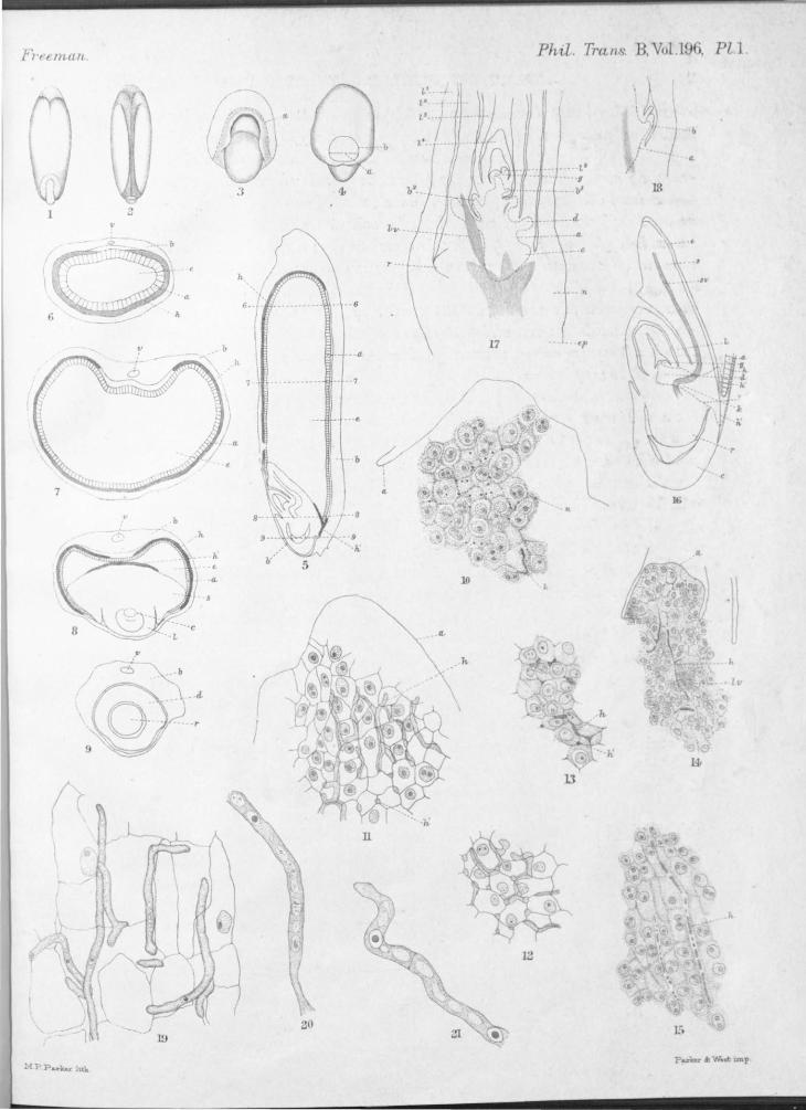
PLATE 1.
Fig. 1 ( X 6). A normal grain of Lolium, temulentum, outer smface, i.e., the convex or embryo side of the grain.
Fig. 2 ( X 6 ). ame from opposite side.
Fig. 3 ( X 11 ). Dis ected embryo seen from the front of the cotyledon ; '' a" marks the end of the aleurone layer.
Fig. 4 ( X 11 ). ame from back of cotyledon ; a as before, b = general limits of infection area.
I~ figmes 5- 8 the area in which the fungus is found is shaded. Fig. 5 ( X 12). Median longitudinal section of a grain, h = fungus in nucellus;
h' = ~ungus of. infection layer (for areas in embryo see fig. ] 6); q, = aleurone; b = fused pencarp pale and integuments; 6 . . . . G, 7 .... 7, &c., mark position of sections in figures 6-9.
Figs. 6-9 ( X 22). Cross-sections at levels indicated in fig. 5 ; letters as iu fig. 5

OF LOLIUM TEMULENTUM, L., THE DARNEL. 25
and v = vascular bundle of inner pale; s = scutellum; d = cole rhiza, r = root; l = leaf-sheath; c = area of hyphre at base of the growing point ; e = starch endosperm.
Fig. 10 ( X 425). Detail of the growing point of the embryo, showing nucleated cells with delicate walls, stained with HAIDENHAIN's hrematoxylin. n = nucleoli of intercellular fungus hyphre; h = heavily stained byphre at surface; a = outline of growing point.
Fig. 11 ( X 425). Same as :fig. 12, but stained with aniline-water-safranin; h = hyphre in longitudinal view; h' = hyphre in cross-section.
Fig. 12 ( X 425). Same as :fig. 11, but from base of hyphal cone; shows abundance of cross-sections of hyphre.
Fig. 13 ( X 425). Cross-section of embryo at lower part of th growing point (c, fig. 8) hrematoxylin.
Fig. 14 ( X 205). Longitudinal section of the growing point, showing dir ction ot the hyphre toward the infection layer; h = hypha; lv = vascular bundle of sheath (hrematoxylin).
Fig. 15 (425). Detail from a longitudinal section of an embryo throuO'h area k of fio-. 16 (hrematoxylin).
Fig. 16 ( + 425). Median longitudinal section of the embryo with the al uron layer of the grain in the region of the infection area still attached ; i = aleurone ; b = outline of hyphal area ; r = root ; c = coleorhiza ; cl = base of hyphal cone in the growing point ; e = epithelium ; s = scutellum ; sv = vascular bundle of the scutellum ; l = leaf- heath ; g = growin T
point; h = area of hyphre of the grain nucellus ; h' = infi cti n lay r. The area occupied by the fungu has a dott d outlin .
Fig. 17 ( X 45). Median longitudinal section through the growing p int of a 17-day seedling; l' = 1st leaf (sheath); l'-V = 2nd to 4th leav l5 = inception of 5th leaf; g = growing point ; b2 and bs = buds in axil of 2nd and 3rd leaves; (for b' see fig. 18); a = outline (dott d line) f hyphal area, at cit evaginates toward b', :fig. 1 ; d = network ofhyphre in leaf base ; lv = vascular bundle of leaf; r = lateral root ; n = 1st n le above the scutellum ; ep = top of epicotyl (hrematoxylin).
Fig. 18 ( X 45). Axil of 1st leaf of same series as fig. 17 : b' = bud; a = hyphal area ( dottyd line).
Figs. 19-21 (19 = x 755, 20 and 21 = x 1400). Detail of hyphre from the leaf base network. Host cells are somewhat shrunken in :ficr. 19. (FLEIDIIN ' weaker fluid without staining.)
PLATE 2.
(Figs. 22-27, inclusive, are all overstained in hrematoxylin, the hyphre staining homogeneously and very dark. They are similarly lettered.)
VOL. CXCVI.-B. E

26 MR. E. M. FREEMAN ON THE SEED-FUNGUS
Fig. 22 ( X 452 ). A longitudinal section through the infection area; c = epid.ermis of the scutellum; a = aleurone layer, which is here doubled by bendmg back, and a' = inside layer ; h = hyphre in grain nucellus ; h' = byphre of infection layer (at h" a hypha penetrates the aleurone layer); p = hyphre of infection layer penetrating between the epidermal cells of the scutell~m; e = remnants of cells of endosperm; at b the grain nucellus comes mto direct contact with the embryo.
Fig. 23 ( X 425). Longitudinal section of an infection area with a single aleurone layer.
Fig. 24 ( X 455). Cross-section of embryo through the infection area (in same region as fig. 8).
Fig. 25 ( X 755). Detail of hyphal region of infection layer ; hy.Phre are penetrat-ing on both sides of one cell of the scutellum epidermis. .
Fig · 26 and 27 ( X 425). Detail of consecutive sections showing the penetration and continuance of an infecting hypha. .
(Figs. 28-32 are from material fixed in chromic acid, 1 per cent., cut in the freezmg microtome and mounted in glycerine unstained ; they are similarly lettered.)
Fig. 28 ( X 425). Detail of cross-section of outer part of a grain which has been · · · h · · ushed ma germmatmg c amber 24 hours. l = pale; p = pencarp; i =er integuments; o = outer rows of nucellar cells; b = cavities in the nucellus (probably old cell lumina); h = hyphre; a = aleurone; e = starch-endosperm.
Fig. 29 ( X 755). Detail of cross-section of a grain in which no fungus was present.
Figs. 30, 31 and 32 (30 and 32 = x 425, 31 = X 7 55). Detail of cross-sections f · · h · the 0 grams mt e germinating chamber about 9 days, showing stages 1Il
disintegration of the nucellar hyphre; h' = cavities left by the disappearance of hyphre.
F~g. 33 ( X 1400). I:Iypha from a 6-day grain (hrematoxylin). . d Fig. 34 ( X 530). Dissected hyphre from the nucellus of the grain to show metho
of branching.
Fig. 35 ( X 530). Dissected hyphre which have been in a hancring drop of beer-. wort gelatine, 2 per cent., for 3 days. 0
Fia. 36 ( X 755). Hypha from a 24-hour grain, abundance of septation.
PLATE !l.
Figs. 37- 39 ( X 7 55). Cross-section of grains with hyphre penetrating the cell walls of the aleurone layer and invading the starch endosperm (swollen with potassium hydrate and stained with iodine), fig. 37 = through t_he aleurone; fig. 38 = in the starch region, and 39 shows the peculiar

OF LOLIUM TE~IULE1 TUM, L., THE DARNEL. '27
branching in the latter region (cell walls not shown), h = hyphal layer of grain nucellus, al = aleurone layer ; st = starch cell ; w = wall of tarch cell ; ci = knot formation in :m intercellular space.
Fig. 40 ( X 22). A longitudinal section of a very young inflorescence (L. temulentum, variety arvense). The entire system is permeated with hyphre.
Fig. 41 ( X 45 ). Longitudinal section of the last two nodes of a spikelet. a = last and sterile node ; m = internode; h = (dotted line) hyphal area · ip = inner pale; carp = carpel; v = ovule; st = stamen; op = outer pale.
Figs. 42--16 ( 42-44 = X 45 ; 45 and 46 = X 12). Illustrate the development of the ovule and the grain. Letters as in 41. The areas of denser aggregations of hyphre are shaded ; int = integuments ; ant = group of antipodal cells; i, t = infection layer; ov = egg cell; emb = embryo; emb. ac = embryo sac; endo = endosperm; f = funicle.
Figs. 42 = tangential, and 43 = median longitudinal section from the ame ovary. 43 contains an embryo-sac with eight cell ; in 45 the embryo is still an undifferentiated ovoid body, and the walls of the endosperm cell hav not all been formed; in 46 only the densely compacted hyphre can be seen ; the embryo is here about 300µ in length and has rudiments of th scutellum and growing point.
Fig. 47 ( X 755). Detail of cross-section through the ovary of fig. 43; p eri = inner wall of the carpel; i. int. = inner integument; o. int. = outer integument ; ep = epidermis of the nucellus ; h = hyphre in denser external region; h' = hypha in cross-section; cent. = central large-celled region.
Fig. 48 ( X 7 55). Longitudinal section of the egg cell end of the ernbr o sac with the nucellus on the inner ( = axis) side of the ovule, from stage of £er. 43 ·
h = hypba (future infection layer); e. s. = wall of embryo sac; ov = egg cell nucleus.
Fig. 49 ( X 755). Longitudinal section of an ovule, the upper end of £ . 45.
hows central region with the embryo sac crowding the byphre into an accumulating layer; a= wall of the embryo sac.
Fig. 50 ( X 7 5 5). Hyphre from section of fig. 45. Nuclei and eptation abundant. Occasionally a hypha is much swollen.
Fig. 51 ( X 272). Detail of the embryo region f fig. 46. hows bypha which has just reached the growing point, and its origin in he infection area ; g. pt. = growing point; sc. = scutellum; endo =starch end perm; al= the aleurone, which is commencing to differentiate; i. int. = inner integument ; nuc = nucellus ; h = hypba of infection layer.
E 2
Fruman . Ph,,,l. Tran . B, Vol l , Pl.1
b IL
J 4 IS
2
" a.
- c
.. •• n
.,,_
- b 17 . <f'
It
..
I ~ - -o
~~':s~ 9~-- · r
Lv
( 1J
12
20

Fr-nman. PU~
h
c •.. -..
,,.
Jl
33
MP.Pa .. ML• lu.h

Freema,n,. Phil. Tra-n.s. B, Vol.I , Pl.3.
a. .
48
MP.Pa.rkerli.tli..