phenylhydrazines in the cultivated mushroom
TRANSCRIPT
Phenylhydrazines in the Cultivated Mushroom (Agaricus bisporus) - occurrence, biological properties, risk
assessment and recommendations

By Andersson, H.C. and Gry, J.
TemaNord 2004:558
Phenylhydrazines in the Cultivated Mushroom (Agaricus bisporus) - occurrence, biological properties, risk
assessment and recommendations
Phenylhydrazines in the Cultivated Mushroom (Agaricus bisporus) - occurrence, biological properties, risk assessment and recommendations TemaNord 2004:558 © Nordic Council of Ministers, Copenhagen 2004 ISBN 92-893-1080-4 ISSN 0908-6692 Print: Ekspressen Tryk & Kopicenter Copies: 300
Printed on paper approved by the Nordic Environmental Labelling. This publication may be purchased from any of the sales agents listed on the last page.
Nordic Council of Ministers Nordic Council Store Strandstræde 18 Store Strandstræde 18 DK-1255 Copenhagen K DK-1255 Copenhagen K Phone (+45) 3396 0200 Phone (+45) 3396 0400 Fax (+45) 3396 0202 Fax (+45) 3311 1870
www.norden.org
The Nordic Food Policy Co-operation The Nordic Committee of Senior Officials for Food Issues is concerned with basic Food Policy issues relating to food and nutrition, food toxicology and food microbiology, risk evaluation, food control and food legislation. The co-operation aims at protection of the health of the consumer, common utilisation of professional and administrative resources and at Nordic and international developments in this field.
The Nordic Council of Ministers was established in 1971. It submits proposals on co-operation between the governments of the five Nordic countries to the Nordic Council, implements the Council's recommendations and reports on results, while directing the work carried out in the targeted areas. The Prime Ministers of the five Nordic countries assume overall responsibility for the co-operation measures, which are co-ordinated by the ministers for co-operation and the Nordic Co-operation committee. The composition of the Council of Ministers varies, depending on the nature of the issue to be treated.
The Nordic Council was formed in 1952 to promote co-operation between the parliaments and governments of Denmark, Iceland, Norway and Sweden. Finland joined in 1955. At the sessions held by the Council, representatives from the Faroe Islands and Greenland form part of the Danish delegation, while Åland is represented on the Finnish delegation. The Council consists of 87 elected members - all of whom are members of parliament. The Nordic Council takes initiatives, acts in a consultative capacity and monitors co-operation measures. The Council operates via its institutions: the Plenary Assembly, the Presidium and standing committees.

Table of contents
Table of contents ............................................................................................................... 5 Preface............................................................................................................................... 7 1. Sammanfattning............................................................................................................ 9 1. Summary .................................................................................................................... 13 2. Introduction ................................................................................................................ 17 3. Identity, physical and chemical properties, and analytical methods .......................... 19
3.1. Identity .............................................................................................................. 19 3.2. Physical and chemical properties...................................................................... 21 3.3. Chemical synthesis............................................................................................ 26 3.4. Analytical methods ........................................................................................... 26
4. Biosynthesis ............................................................................................................... 27 5. Occurrence.................................................................................................................. 31
5.1. The content of agaritine and related compounds in fresh mushrooms ............ 31 5.2. Influence of storage and processing.................................................................. 36
5.2.1. Refrigerating ........................................................................................... 36 5.2.2. Freezing and thawing.............................................................................. 39 5.2.3. Freeze-drying. ......................................................................................... 39 5.2.4. Drying. .................................................................................................... 41 5.2.5. Dry baking. ............................................................................................. 41 5.2.6. Boiling..................................................................................................... 41 5.2.7. Canning. .................................................................................................. 42 5.2.8. Pan-frying ............................................................................................... 43 5.2.9. Deep-frying ............................................................................................. 44 5.2.10. Microwave heating ............................................................................... 44
5.3. Other products containing the cultivated mushroom........................................ 44 5.4. Influence of cultivation, including genetic modification.................................. 45 5.5. Conclusion on occurrence................................................................................. 47
6. Production and consumption ...................................................................................... 49 6.1. Production ......................................................................................................... 49 6.2. Consumption ..................................................................................................... 49 6.3. Conclusion on production and consumption .................................................... 49
7. Toxicokinetics ............................................................................................................ 53 7.1. Absorption, distribution and excretion ............................................................. 53 7.2. Biotransformation ............................................................................................. 55 7.3. Conclusion on toxicokinetics............................................................................ 57
8. Effects in short-term tests........................................................................................... 59 8.1. In vitro studies on phenylhydrazine metabolism in the mushroom ................. 59 8.2. Studies on DNA binding................................................................................... 59 8.3. Tests in microorganisms ................................................................................... 63 8.4. Tests in cultured mammalian cells.................................................................... 70 8.5. In vivo tests ....................................................................................................... 71 8.6. Antimutagenic effects of A. bisporus ............................................................... 73
5

8.7. Conclusions on effects in short-term tests ........................................................ 73 9. Effects on experimental animals ................................................................................ 75
9.1. Acute and subchronic toxicity of mushroom hydrazines and HMBD............. 75 9.2. Long-term carcinogenicity studies. .................................................................. 77
9.2.1. Studies on the cultivated mushroom (Agaricus bisporus). ..................... 78 9.2.2. Studies on phenylhydrazines in the mushroom and related compounds 84
9.3. Non-conventional carcinogenicity studies........................................................ 92 9.4. Antineoplastic effects of phenylhydrazines...................................................... 93 9.5. Reproduction, embryotoxicity and teratogenicity ............................................ 93 9.6. Biological effects of A. bisporus ...................................................................... 94 9.7. Conclusion on effects on experimental animals ............................................... 95
10. Human implications .................................................................................................. 97 11. Previous estimations of risk ...................................................................................... 99 12. Present nordic risk assessment................................................................................ 101
12.1. Hazard identification. ...................................................................................... 101 12.2. Hazard characterization................................................................................... 102 12.3. Exposure characterization. .............................................................................. 103 12.4. Risk characterization. ...................................................................................... 104
13. Recommendations................................................................................................... 109 14. Reference ................................................................................................................ 111
6
Preface
The Nordic Committee of Senior Officials for Food Issues is an advisory body of the Nordic Council of Ministers which co-ordinates Nordic work in the field of food and nutrition. The Committee has given the Nordic Working Group on Food Toxicology and Risk Evaluation (NNT) the responsibility to promote co-operation and co-ordination among Nordic countries in matters relating to food toxicology and risk as-sessment.
Assessment of health risks connected with exposure to naturally occurring toxicants in foodstuffs has become an important area for NNT in the recent years. A series of Nordic reports based on the work performed by the Nordic project group on inherent natural toxicants in food plants and mushrooms has been published:
Gry, J. and Pilegaard, K. (1991) Hydrazines in the Cultivated Mushroom (Agaricus bisporus). Vår Föda 43;Supplement 1.
Uggla, A. and Busk, L. (1992) Ethyl carbamate (urethane) in alcoholic beverages and foodstuffs - A Nordic View. Nordiske Seminar- og Arbejdsrapporter 1992:570.
Størmer, F.C., Reistad, R. and Alexander, J. (1993) Adverse health effects of glycyr-rhizic acid in licorice. A risk assessment. Nordiske Seminar- og Arbejdsrapporter 1993:526.
Andersson, C., Slanina, P. And Koponen, A. (1995) Hydrazones in the false morel. TemaNord 1995:561.
Søborg, I., Andersson, C. and Gry, J. (1996) Furocoumarins in Plant Food - expo-sure, biological properties, risk assessment and recommendations. TemaNord 1996:600.
Gry, J. and Andersson, H.C. (1998) Nordic seminar on phenylhydrazines in the Cul-tivated Mushroom (Agaricus bisporus). TemaNord 1998:539.
Andersson, C. (1999) Glycoalkaloids in tomatoes, eggplants, pepper and two So-lanum species growing wild in the Nordic countries. TemaNord 1999:599.
Andersson, H.C. (2002) Calystegine alkaloids in Solanaceous food plants. Te-maNord 2002:513.
Andersson, C., Wennström, P. and Gry, J. (2003) Nicotine in Solanaceous food plants. TemaNord 2003:531.
There is a large and still increasing consumption of the cultivated mushroom in the Nor-dic countries. The Nordic project group on inherent natural toxicants in food plants and mushrooms has followed the scientific information on the toxicity of Agaricus bisporus and the phenylhydrazine derivatives occurring in the mushroom since the end of the 1980s. In 1991 the project group reviewed the available data and found it not to be of adequate standard to evaluate the human risk at the time. In 1996 the project group organised a Nordic Seminar on Phenylhydrazines in the Cultivated Mushroom (Agari-
7

cus bisporus) in Kolding, Denmark, to promote the exchange of information between chemists and toxicologists active in the field of Agaricus bisporus research.
The present report is an updated review of the phenylhydrazines in the cultivated mush-room (Agaricus bisporus) - occurrence, biological properties, risk assessment and rec-ommendations – (Databases searched: TOXLINGE, BIOSIS, MEDLINE and FSTA, June 2003 included) prepared by the Nordic project group on inherent natural toxicants in food plants and mushrooms.
The Project Group consisted of the following members:
Jørn Gry (co-ordinator) Danish Veterinary and Food Administration Denmark Henrik Frandsen Danish Veterinary and Food
Administration Denmark Christer Andersson National Food Administration Sweden Tore Aune University of Veterinary Medicine Norway Jan Alexander National Institute of Public Health Norway Anja Hallikainen National Food Agency Finland Torkell Johannesson Department of Pharmacology,
University of Iceland Iceland
The present report was initiated in 1996 and has been prepared by Christer Andersson1, Sweden, and Jørn Gry2, Denmark, after very thorough discussions in the project group and finally adopted by NNT.
The NNT acknowledges Dr. S. Clausen, Chemistry Department, Royal Veterinary and Agricultural University, Denmark, and Dr. H. Frandsen, Institute of Food Safety and Nutrition, Danish Veterinary and Food Administration, Denmark, for the chemical syn-thesis of agaritine and related phenylhydrazine derivatives. The synthesis was financed by a grant from the Nordic Council of Ministers. 1 National Food Administration, Box 622, SE-751 26 Uppsala, Sweden 2 Danish Veterinary and Food Administration, Mørkhøj Bygade 19,
DK-2860 Søborg, Denmark
8

1. Sammanfattning
Under perioden 1961 till 1985 påvisades fyra olika fenylhydraziner eller närbesläktade föreningar i den odlade champinjonen, Agaricus bisporus. Ett av dessa ämnen, agaritin (N2-[γ-L-glutamyl]-4-hydroxymetylfenylhydrazin) förekommer i störst mängd än de övriga fenylhydrazinerna. Halter mellan 80 och 1 700 mg/kg har återfunnits i den färska svampen. Vanligen ligger dock nivån i intervallet 200 till 500 mg/kg färskvikt. Utöver agaritin, innehåller den odlade champinjonen små mängder 4-karboxyfenylhydrazin (10-11 mg/kg färskvikt), β-N-[γ-L-(+)-glutamyl]-4-karboxyfenylhydrazin (42 mg/kg färskvikt) och 4-hydroxymetylbenzendiazonium joner (HMBD) (0.6-4 mg/kg färskvikt).
Studier av försöksdjur har visat att agaritin och/eller dess nedbrytningsprodukter absor-beras snabbt och i påtaglig omfattning från mag-tarmkanalen. Däremot kan ingen oför-ändrad agaritin påvisas i blodet. Enzymet γ-glutamyl transpeptidas förekommer i cell-membranen i flera av kroppens vävnader och tros vara det enzym som klyver agaritin till glutaminsyra och 4-hydroxymetylfenylhydrazin. Fenylhydrazinet omvandlas däref-ter till HMBD, som antas vara det ämne som ger upphov till reaktiva radikaler i vävna-derna. Radikalerna kan sedan orsaka skador på makromolekyler. Måttliga mängder DNA-skador har påvisats i olika vävnader från försöksdjur som exponerats oralt för agaritin och HMBD, vilket skulle kunna tyda på att fenylhydrazinerna är genotoxiska carcinogener. Denna tolkning stöds av observationen att såväl svampextrakt, som agari-tin och de andra fenylhydrazinerna i A. bisporus, inducerar mutationer i bakterier testa-de in vitro, om än i liten omfattning. Dessutom har in vivo studier på möss visat att aga-ritin inducerar genmutationer i transgena försöksdjur och 4-hydroxymetylbenzendiazonium jonen mikrokärnor i perifera lymfocyter.
Endast ett fåtal hydrazinderivat är kända från naturen. Industrin har emellertid syntetise-rat ett större antal hydrazinföreningar för egna ändamål. Eftersom flertalet av de nära ett hundra hydraziner som studerats (cirka 85%) är giftiga och/eller cancerframkallande i försöksdjur, inledde Toth och medarbetare en serie undersökningar för att utvärdera huruvida fenylhydrazinerna i den odlade champinjonen, till dessa närbesläktade ämnen och svampen i sig uppvisade carcinogen aktivitet. I tre av fyra långtidstudier utveckla-des tumörer i möss som utfordrades med rå, ungsbakad eller frystorkad A. bisporus. I den fjärde studien, med ungsbakade champinjoner, men med en annan (mer balanserad) utfodring av svampen, erhölls ingen ökning i tumörincidens. Inte heller erhölls någon tumörinduktion i två bristfälliga långtidsstudier utförda på råtta. Antalet råttor var allt-för lågt för att en mindre ökningar i tumörincidens skulle kunna påvisas. Dessutom är det tänkbart att behandlingen av svampfodret (ordinarie torkning och malning, alterna-tivt tvättning kombinerat med tryckkokning, homogenisering och blandning med kran-vatten) kan ha resulterat i en signifikant nedbrytning av de potentiellt aktiva fenylhyd-razinerna. Tre av de fyra fenylhydraziner eller närbesläktade ämnen som förekommer i A. bisporus - 4-karboxyfenylhydrazin, β-N-[γ-L-(+)-glutamyl]-4-karboxyfenylhydrazin and HMBD - framkallade tumörer när höga doser gavs till möss (Swiss) oralt (tvångs-matning eller i dricksvattnet), eller när ämnena injicerades under huden. Den fjärde fe-nylhydrazinen, agaritin, visade sig inte ge upphov till tumörer när ämnet gavs i dricks-vattnet eller injicerades under huden. Denna oväntade upptäckt har påtagligt försvårat
9
tolkningen av de data som erhållits vid studierna med fenylhydraziner. Emellertid skall påpekas att inga försiktighetsåtgärder vidtogs i cancerstudierna för att skydda agaritinet mot en eventuell oxidativ nedbrytning i vattenlösningen. Det har nämligen nyligen vi-sats att agaritin lätt bryts ned i väl syresatta vattenlösningar.
Det måste understrykas att de långtidsstudier av cancerinduktion som denna riskbedöm-ning bygger på, kan kritiseras av flera skäl. En och samma musstam (Swiss) har använts i samtliga studier. Musstammen skiljer sig från flertalet musstammar som används i cancerstudier genom att inte vara inavlad. Det faktum att mössen får para sig fritt, resul-terar i en större variation i känslighet mellan de olika mössen än vad som skulle vara fallet för djuren i inavlade stammar. Dessutom har studierna ofta använt sig av olämpli-ga kontrollgrupper - antingen historiska kontroller eller kontrollgrupper som inte star-tats samma dag som de behandlade grupperna. I stället för att genomföra cancerstudier-na på vanligt sätt under en förbestämd tidsperiod (vanligen 2 år), fick studien pågå till dess att djuren dog av hög ålder eller allvarlig sjukdom (inklusive tumörsjukdom). Can-cerstudier använder sig vanligen av tre dosgrupper. De aktuella studierna har oftast haft endast en dosgrupp. Några av studierna har kritiserats för att fodertillförseln inte varit balanserad. Vanligen studeras ett större antal biokemiska och toxikologiska parametrar parallellt med huvudvariabeln (tumörinduktion) för att bättre kunna utvärdera erhållna resultat. Sådana observationer har endast bristfälligt rapporterats i de publicerade studi-erna med odlad champinjon och dess fenylhydraziner.
Trots att flertalet cancerstudier på mus gav positivt utslag finns inga tecken på att vare sig A. bisporus eller fenylhydrazinerna i svampen är embryotoxiska eller teratogena i däggdjur vid biologiskt relevanta doser. Det skall dock påpekas att försöksuppläggning-en på de få studier som finns tillgängliga inte uppfyller de krav som ställs i moderna riktlinjer för hur sådana studier skall utföras. Det skall också påpekas att ytterst få toxi-citetstuder utförts med fenylhydrazinerna i den odlade champinjonen vid sidan av can-cerstudierna på försöksdjur.
Den uppskattade årskonsumtionen av odlad champinjon (Agaricus bisporus) per capita varierar mellan de nordiska länderna - från ungefär 0.6 kg per person i Finland till 2.4 kg per person i Sverige. Även marknadsandelen färsk champinjon varierar mellan de olika länderna, från 33% till 70%. Resten utgörs av konserverad champinjon.
Eftersom per capita intaget är en uppskattning av medelintaget och inte tar hänsyn till det faktum av svamp endast konsumeras av en del av populationen, blir per capita inta-get för lågt för att beskriva intaget hos dem som verkligen konsumerar svamp. En mind-re dansk studie tyder på att endast hälften av befolkningen konsumerar odlad champin-jon och att 5% av befolkning konsumerar fem gånger så mycket som medelintaget och 0.1% av befolkningen trettio gånger mer än medelintaget.
Med undantag för agaritin är vår kunskap dålig vad gäller hur lagring och tillagning av champinjon påverkar halten av fenylhydraziner i den konsumerade svampen. För agari-tin vet vi att halten reduceras i odlad champinjon både under lagring och tillredning. Hur mycket agaritin som försvinner beror på sättet svampen hanterats. Vanliga tillag-ningsmetoder halverar mängden agaritin. Konserverad champinjon innehåller högst 10% av den mängd som återfinns i färsk svamp.
Med utgångspunkt från angivna data kan per capita exponeringen för agaritin (som en markör för fenylhydrazinerna i odlad champinjon) i de nordiska länderna uppskattas. Exponeringen ligger någonstans i intervallet mellan 48 och 788 mg agaritin/år, vilket
10

motsvarar ett dagsintag på mellan 2.1 och 36 µg/kg kroppsvikt. Exponeringen påverkas av mängden svamp som konsumeras (nationalitet), halten av agaritin i den färska svam-pen (200-500 mg/kg) och huruvida man föredrar färsk (33-70%) eller konserverad svamp. En enstaka svamprätt med 75 g färsk A. bisporus resulterar i en exponering på cirka 15-33 mg agaritin, vilket motsvarar 250-550 µg/kg kroppsvikt.
Trots att cancerstudien med agaritin på (Swiss) möss var negativ, används sex andra cancerstudier baserade på oral tillförsel (tvångsmatning, i dricksvattnet eller i fodret) av odlad champinjon eller enskilda ämnen (tvångsmatning, i dricksvattnet eller i fodret) till Swiss möss i kombination med det uppskattade intaget av A. bisporus i de nordiska län-derna för att göra en kvantitativ riskbedömning av den odlade champinjonen. Tre av långtidsstudierna utfördes med A. bisporus per se, medan de andra tre genomfördes med de fenylhydraziner och närbesläktade ämnen som förekommer i svampen. Vid beräk-ningen av cancerrisken användes en linjär extrapolering från mus till människa som baserades på flera antagen redovisade i rapporten, men där de viktigaste var att metabo-lismen och känsligheten hos mus och människa är jämförbar.
Sammantaget tyder de svaga mutagena effekterna in vitro och in vivo (agaritin och HMBD) och de carcinogena effekterna hos (1) tre av de fyra fenylhydrazinerna i A. bis-porus och (2) den färska, ungsbakade och frystorkade svampen per se i Swiss albino möss, att A. bisporus är svagt cancerframkallande i försöksdjur och följaktligen kan innebära en risk för konsumenter. Den uppskattade livstidsrisken för en nordisk medel-konsument av färsk (eller frystorkad) A. bisporus är cirka 200 x 10-6(50 x 10-6 för finska konsumenter), medan intaget av en motsvarande mängd tillagad champinjon resulterar i en hälften så stor cancerrisk. I realiteten kommer medelkonsumenten att förtära en blandning av färsk och tillagad svamp. Vissa matlagningsmetoder kommer att reducera fenylhydrazininnehållet i svampen påtagligt, medan andra former för matberedning har mindre uttalad effekt. Kunskaperna om hur vanlig tillagning påverkar halten av andra fenylhydraziner än agaritin i champinjon är ännu dåligt känd. Av de fenylhydraziner och besläktade ämnen som förekommer i champinjonen och studerats i cancerstuder har HMBD högst carcinogen potential. Detta är också vad man kan vänta sig mot bakgrund av den förväntade verkningsmekanismen.
Det bör ånyo påpekas att den vanligaste fenylhydrazinen i A. bisporus, agaritin, inte framkallade tumörer i försöksdjuren när ämnet gavs i dricksvatten. Av den anledningen har ingen riskbedömning gjorts för agaritin. Emellertid kan man förvänta sig att den studieuppläggning som använts i åtminstone en av cancerstudierna riskerat att en signi-fikant mängd av agaritinet i dricksvattnet brutits ned.
11

12

1. Summary
During the period 1961 to 1985 four different phenylhydrazines or related compounds were detected in the cultivated mushroom, Agaricus bisporus. Of these, β-N-[γ-L-(+)-glutamyl]-4-hydroxymethylfenylhydrazine, also called agaritine, occurs in high amounts. A level between 80 and 1 700 mg/kg has been reported in fresh mushrooms. The average value found in mushrooms on the market is between 200 and 500 mg/kg fresh weight. In addition to agaritine, the cultivated mushroom also contains small quantities of 4-(carboxy)phenylhydrazine (10-11 mg/kg fresh weight), β-N-[γ-L-(+)-glutamyl]-4-(carboxy)phenylhydrazine (42 mg/kg fresh weight) and 4-(hydroxymethyl)benzene diazonium ions (HMBD) (0.6-4 mg/kg fresh weight).
Studies in experimental animals have shown that agaritine, and/or its degradation prod-ucts, are to a significant degree and rapidly absorbed from the gastro-intestinal tract, but in blood no unchanged agaritine can be detected. The enzyme γ-glutamyl transpepti-dase, present in various tissues of the body, is believed to be the enzyme responsible for cleaving agaritine into L-glutamic acid and 4-hydroxymethylphenylhydrazine. The lat-ter compound is subsequently transformed to HMBD, which is the substance believed to give rise to reactive radical species able to damage macromolecules. A low frequency of DNA damage has been observed in various tissues of experimental animals exposed orally to agaritine and HMBD, an observation indicating that these compounds may be genotoxic carcinogens. This interpretation is supported by the observation that mush-room extracts, as well as agaritine and the other phenylhydrazines occurring in A. bis-porus, induce a low frequency of mutations in bacterial test systems in vitro. Further-more, in vivo studies on mice have revealed that agaritine induces gene mutations in transgenic animals and that the 4-(hydroxymethyl)benzendiazonium ion induces micro-nuclei in peripheral lymphocytes.
In addition to the limited number of naturally occurring hydrazine derivatives, of which the phenylhydrazines in the cultivated mushroom is one example, the industry has pro-duced a number of synthetic compounds for industrial use. Since most (around 85%) hydrazines studied (about one hundred) are toxic and/or carcinogenic in experimental animals, Toth and co-workers undertook the work to assess the possible carcinogenic activity of the phenylhydrazines and related compounds in A. bisporus, as well as of the mushroom itself. In three, out of four, long-term carcinogenicity studies tumours were induced in various tissues of mice fed raw, baked or freeze-dried A. bisporus. In the fourth study, with baked mushrooms but with another (more balanced) feeding sched-ule, the increase in tumour incidence was not significant. No tumours were induced in two long-term studies in rats. But in these rather inadequate studies the number of ani-mals was too low to detect a relatively small increase in tumour incidence. Furthermore, the processing (pre-treatment) of the mushrooms (ordinary drying and milling, or wash-ing, combined with pressure cooking, homogenising and mixing with tap water) might well have resulted in a significant degradation of the potentially active phenylhydrazi-nes. Three of the four phenylhydrazines and related compounds known to occur in A. bisporus - 4-(carboxy)phenylhydrazine, β-N-[γ-L-(+)-glutamyl]-4-(carboxy)phenyl-hydrazine and HMBD - were carcinogenic at high doses when administered orally (by
13

gavage or in drinking water) and also when injected subcutaneously in Swiss mice. The fourth - and major - phenylhydrazine, agaritine, was found not to be carcinogenic when given in drinking water or by subcutaneous injection, a finding that has significantly muddled the interpretation of the carcinogenicity data. However, no precautions were taken in these studies to protect against a possible oxidative degradation of agaritine in water. Agaritine has recently been shown to be vulnerable to such degradation in aque-ous solution.
It must be underpinned that the long-term carcinogenicity studies on which the risk as-sessment has been made, might be criticised for several reasons. Only a single mouse strain, the Swiss mouse, has been used in the studies. In contrast to most mouse strains, which are inbred, the Swiss mouse is a randomly bred strain. This results in a heteroge-neity in sensitivity between animals. The studies have often used improper controls – either a historical control group, or a control group that was not started at the same time as the exposed group. In stead of carrying out the carcinogenicity studies in the normal way of exposing the animals for a predetermined time period (usually 2 years), the stud-ies were continued until the mice died or were found in poor condition. Standard car-cinogenicity studies have three dose groups but these studies have usually used only a single dose group. Furthermore, some of the studies have been criticised for resulting in an unbalanced diet. In general a number of biochemical and toxicological endpoints are studied besides the main end-point (tumours) in order to be able to interpret the data. Such studies have rarely been reported in the carcinogenicity studies with the cultivated mushroom and its phenylhydrazines.
In contrast to the many positive carcinogenicity studies, there are no indications that A. bisporus or the phenylhydrazines occurring in the mushroom are embryotoxic or terato-genic in mammals at biologically relevant doses. However, it should be stressed that the design of the few studies available also in this case did not follow modern guidelines for these types of study. It should also be stressed that very few toxicity studies other than carcinogenicity studies have been performed with the phenylhydrazines in the mush-room.
The estimated annual per capita intake of the cultivated mushroom (Agaricus bisporus) varies between the Nordic countries - being 0.6 kg in Finland having the lowest intake among Nordic countries and 2.4 kg in Sweden with the highest intake. Also the habit of using fresh mushrooms for dietary purposes varies between countries. The market share of fresh mushrooms was between 33% and 70%. The rest being preserved mushrooms, mainly sold in cans.
Since the per capita intake is an estimation of average intake and does not take into con-sideration the fact that mushrooms usually are consumed only by a part of the popula-tion, the per capita consumption is too low to express the consumption among mush-room eating consumers. Danish data, based on a limited consumer study, indicate that only around 50% of the population consume the cultivated mushroom, and that 5% of the population consume five times the median intake and 0.1% thirty times the median intake.
Except for agaritine, the data on the influence of storage and food processing on the amount of phenylhydrazines in the consumed mushroom are scanty. The agaritine level in the cultivated mushroom is reduced both during storage and processing. The magni-tude of the reduction depends on conditions during storage and food preparation. Nor-
14

mal cooking procedures about halves the agaritine content. Especially canned mush-rooms contain less than 10% of the agaritine level in fresh mushrooms.
Taken together, these data allows an estimation of the per capita exposure to agaritine (as a marker for the phenylhydrazines in the mushroom) in the Nordic countries. The estimated exposure in the Nordic countries is somewhere between 48 and 788 mg agari-tine/year, corresponding to a daily exposure of between 2.1 and 36 µg/kg body weight. The exposure being influenced by the quantity of mushrooms consumed (nationality), the amount of agaritine in the fresh mushroom (200-500 mg/kg) and the preference of using fresh instead of processed mushrooms (33-70%). A single mushroom dish with 75 g fresh A. bisporus would result in an exposure to 15-33 mg agaritine, that is, 250-550 µg/kg body weight.
Despite the carcinogenicity study with agaritine in mice being negative, six other car-cinogenicity studies in the same species, with oral application (by gavage, in drinking water or in the diet), were considered in combination with an estimated human intake to A. bisporus in the Nordic countries for the sake of performing a quantitative risk as-sessment of the cultivated mushroom. Of these studies, three were long-term studies with A. bisporus per se, and three were long-term studies with the phenylhydrazines and related compounds occurring in the mushroom. The calculation of the cancer risk used a linear extrapolation from mice to man, based on a number of assumptions described in the main text, of which the most important being the metabolism and sensitivity of the two species being identical.
Taken together the weak mutagenic effect in vitro and in vivo (agaritine and HMBD) and the carcinogenic effects of three of the four phenylhydrazines occurring in A. bis-porus, as well as of the fresh, baked and freeze-dried mushroom per se in Swiss albino mice all suggest that A. bisporus is weakly carcinogenic in animals and accordingly might impose a risk for the consumer. The estimated lifetime human cancer risk of an “average” Nordic consumer of fresh (or freeze-dried) A. bisporus is around 200 x 10-
6(50 x 10-6 for Finnish consumers), whereas intake of a corresponding amount of house-hold processed mushrooms results in a cancer risk only about half as high. In reality an average consumer will consume a mixture of fresh and processed mushrooms. Some types of processing (i.e. canning) will reduce the phenylhydrazine content of the mush-room substantially, whereas other types of processing seem to have a much less pro-nounced effect. However, the influence of standard cooking procedures on the content of other phenylhydrazines of the cultivated mushroom than agaritine is not very well studied. Of the phenylhydrazines and related compounds investigated in carcinogenicity studies and occurring in the mushroom, HMBD had the highest carcinogenic potency. This finding agrees with what is expected based on the anticipated mechanism of action.
It should also be noticed that the major phenylhydrazine in A. bisporus, agaritine, did not induce tumours when administered in the drinking water. Therefore, no risk assess-ment has been made for agaritine. However, it may be anticipated that due to the study design, agaritine could have been significantly degraded in the drinking water in one of these studies.
15

16

2. Introduction
In 1991 The Nordic Working Group on Food Toxicology and Risk Evaluation (NNT) reviewed the available data on phenylhydrazines naturally occurring in the cultivated mushroom (Agaricus bisporus L.), as well as data on the potential toxicity of these con-stituents and of the mushroom itself (Gry and Pilegaard, 1991).
The conclusion of this review was that the cultivated mushroom, the most consumed commercial mushroom in the world, contains substantial amounts of phenylhydrazines, about 500 mg per kg raw mushroom. Agaritine is the major phenylhydrazine in A. bis-porus (of many mycologists anticipated to be the same species as A. hortensis and A. brunnescens) but the mushroom also contains other phenylhydrazine derivatives. The review further concluded that “the concentration of these hydrazines may change during storage and processing and may vary depending on the strain and cultivation conditions. The degradation products have not been identified”. It continued: ”The published toxi-cological studies give rise to serious concern as to a possible human health risk from consumption of the cultivated mushroom. As no epidemiological data are available and as the long-term carcinogenicity studies with the mushroom and its hydrazine constitu-ents are not of an adequate standard, it is not possible to evaluate the human risk at pre-sent” (Gry and Pilegaard, 1991). In agreement with the Nordic project group Subrama-nian (1995) in a subsequent review concluded: ”We certainly need more data on the agaritine content in mushroom species and their possible biological effects”.
Based on the recommendations given in the Nordic report from 1991, a series of toxico-logical and chemical studies on the cultivated mushroom and its phenylhydrazine con-stituents were recently performed in the Nordic countries. The aim was to provide up-dated data for an assessment of whether the consumption of the cultivated mushroom constitutes a human risk.
To promote the exchange of information between chemists and toxicologists active in the field of Agaricus bisporus research, especially American, English, Czech and Nor-dic researchers, the “Nordic project group on inherent natural toxicants in food plants and mushrooms” organised a seminar in Kolding, Denmark, August 1996 (Gry and Andersson, 1998). The seminar was the first step in updating the review on ”Phenylhy-drazines in the Cultivated Mushroom (Agaricus bisporus)”.
The present report builds on all the scientific information that has been published on the toxicity of Agaricus bisporus and the phenylhydrazine derivatives occurring in the mushroom. It also draws on the studies performed within this project and the scientific information and recommendations given at the Nordic seminar on phenylhydazines in the Cultivated mushroom (Gry and Andersson, 1998).
17

18

3. Identity, physical and chemical properties, and analytical methods
The commonly eaten cultivated mushroom of commerce in the Western hemisphere, Agaricus bisporus, belongs to the Agaricaceae family, the meadow mushrooms. The species-determining name bisporus was derived from a latin word meaning "with two spores". A. bisporus was in reality derived from several different species and its contin-ual propagation over many centuries has resulted in several (close to 200) commercial strains which have no exact counterpart in nature (Toth, 2000a). Agaricus campestris (Field Mushroom or Meadow Mushroom) may be the most closely related of any of the common species encountered in North America (Toth, 1983).
3.1. Identity During the course of an investigation into the metabolism of α-keto acids in soluble extracts of basidiomycetes, Levenberg (1961) isolated a glutamin-containing compound from the press-juice of A. bisporus. He characterized the compound physically and chemically as β-N-[γ-L-(+)-glutamyl]-4-hydroxymethylphenyl-hydrazine and gave it the trivial name agaritine. This was an important observation, since it was the first re-ported occurrence of a phenylhydrazine derivative in natural products. Subsequently other investigators isolated agaritine from the mushroom using a improved method and confirmed its structure by synthesis (Daniels et al., 1961). The early investigators also established the level of agaritine to be fairly high in the cultivated mushroom (Kelly et al., 1962). The chemical structure of agaritine is given in Figure 1.
Agaritine is not the only nitrogen-nitrogen bond-containing substance that has been found in mushrooms. It is not even the only phenylhydrazine derivative. Already 1962 Levenberg demonstrated the occurrence of the 4-(hydroxymethyl)benzene-diazonium
HN
CH2OH
NH γ glutamyl
Fig. 1 The molecular structure of agaritine ( β-N-[γ-L-(+)-glutamyl]-4 hydroxy-
methylfenylhydrazine).
19

ion (Figure 2) in the basal-stalk section of the stipe of A. bisporus. This compound was not detected in the cap and upper portion of the stipe.
Already in connection with the identification of agaritine in the cultivated mushroom a highly active enzyme that catalyses the cleavage of agaritine to 4-(hydroxymethyl)-phenylhydrazine (Figure 2) and L-glutamic acid was identified (Levenberg, 1961). The enzyme was partially purified (26-fold) from soluble A. bisporus extracts and was shown to irreversibly catalyse hydrolysis of the γ-L-glutamyl-hydrazide bond of agari-tine. Hydrolysis and γ-glutamyl transfer were shown to be common functions of the same enzyme protein (Gigliotti and Levenberg, 1964). This degradation of agaritine to 4-(hydroxymethyl)phenylhydrazine and L-glutamic acid by the enzyme which subse-quently was given the name agaritine γ-glutamyl transferase has been confirmed by Baumgartner et al. (1998). The early observations lead to the speculation that a link exists between agaritine and the 4-(hydroxymethyl)benzenediazonium ion.
Properties that differentiates this agaritine degrading enzyme from other glutamotrans-ferases and glutamyltranspeptidases are: a) its inability to effect transfer to amino acids such as glycine, phenylalanine, leucine and aspartic acid, b) its ability to transfer to wa-ter, and c) its ability to utilize aromatic hydrazine and their γ-L-glutamyl derivatives as acceptor and donor substrates, respectively. The product of the enzymatic agaritine hy-drolysis, 4-(hydroxymethyl)phenylhydrazine is very unstable and has never been de-tected in A. bisporus.
Subsequently it was demonstrated that two enzyme systems capable of generating the 4-(hydroxymethyl)benzenediazonium ion from agaritine occur in the mushroom. One system is the one mentioned above, in which agaritine is oxidised to the diazonium ion via the unstable 4-(hydroxymethyl)phenylhydrazine. In the other system agaritine is oxidised to the diazonium ion without intermediate formation of 4-(hydroxymethyl)phenylhydrazine (Ross et al., 1982a). These systems (together with others) may be responsible for producing the low level (0.6-4 ppm) of 4-(hydroxymethyl)benzenediazonium ion that has been detected in A. bisporus.
A B C D
HN NH2
COOH
HN
COOH
NH γ glutamyl
HN
CH2OH
NH2
N
CH2OH
N+
Fig. 2 Other phenylhydrazine derivatives than agaritine, or related compounds oc-curring in Agaricus bisporus. (A) 4-(carboxy)phenylhydrazine; (B) β-N- (γ-L(+)-glutamyl)4-(carboxy)phenylhydrazine; (C) 4-(hydroxymethyl)phenyl-hydrazine; and (D) 4-(hydroxymethyl)benzenediazonium ion.
20
LaRue (1977) postulated 4-(carboxy)phenylhydrazine and β-N-[γ-L-(+)-glutamyl]-4-carboxyphenylhydrazine (Figure 2) to be possible biosynthetic precursors of agaritine. Both these substances were subsequently identified in A. bisporus by Chauhan and co-workers (1984, 1985).
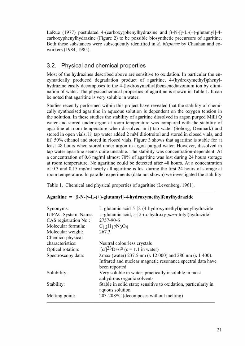
3.2. Physical and chemical properties Most of the hydrazines described above are sensitive to oxidation. In particular the en-zymatically produced degradation product of agaritine, 4-(hydroxymethyl)phenyl-hydrazine easily decomposes to the 4-(hydroxymethyl)benzenediazonium ion by elimi-nation of water. The physicochemical properties of agaritine is shown in Table 1. It can be noted that agaritine is very soluble in water.
Studies recently performed within this project have revealed that the stability of chemi-cally synthesised agaritine in aqueous solution is dependent on the oxygen tension in the solution. In these studies the stability of agaritine dissolved in argon purged Milli Q water and stored under argon at room temperature was compared with the stability of agaritine at room temperature when dissolved in i) tap water (Søborg, Denmark) and stored in open vials, ii) tap water added 2 mM ditiotreitol and stored in closed vials, and iii) 50% ethanol and stored in closed vials. Figure 3 shows that agaritine is stable for at least 48 hours when stored under argon in argon purged water. However, dissolved in tap water agaritine seems quite unstable. The stability was concentration-dependent. At a concentration of 0.6 mg/ml almost 70% of agaritine was lost during 24 hours storage at room temperature. No agaritine could be detected after 48 hours. At a concentration of 0.3 and 0.15 mg/ml nearly all agaritine is lost during the first 24 hours of storage at room temperature. In parallel experiments (data not shown) we investigated the stability
Table 1. Chemical and physical properties of agaritine (Levenberg, 1961). ____________________________________________________________________ Agaritine = β-N-[γ-L-(+)-glutamyl]-4-hydroxymethylfenylhydrazide Synonyms: L-glutamic acid-5-[2-(4-hydroxymethyl)phenylhydrazide IUPAC System. Name: L-glutamic acid, 5-[2-(α-hydroxy-para-tolyl)hydrazide] CAS registration No.: 2757-90-6 Molecular formula: C12H17N3O4 Molecular weight: 267.3 Chemico-physical characteristics: Neutral colourless crystals Optical rotation: [α]23D+6o (c = 1.1 in water) Spectroscopy data: λmax (water) 237.5 nm (ε 12 000) and 280 nm (ε 1 400). Infrared and nuclear magnetic resonance spectral data have been reported Solubility: Very soluble in water; practically insoluble in most anhydrous organic solvents Stability: Stable in solid state; sensitive to oxidation, particularly in aquous solution Melting point: 203-208oC (decomposes without melting) ____________________________________________________________________
21
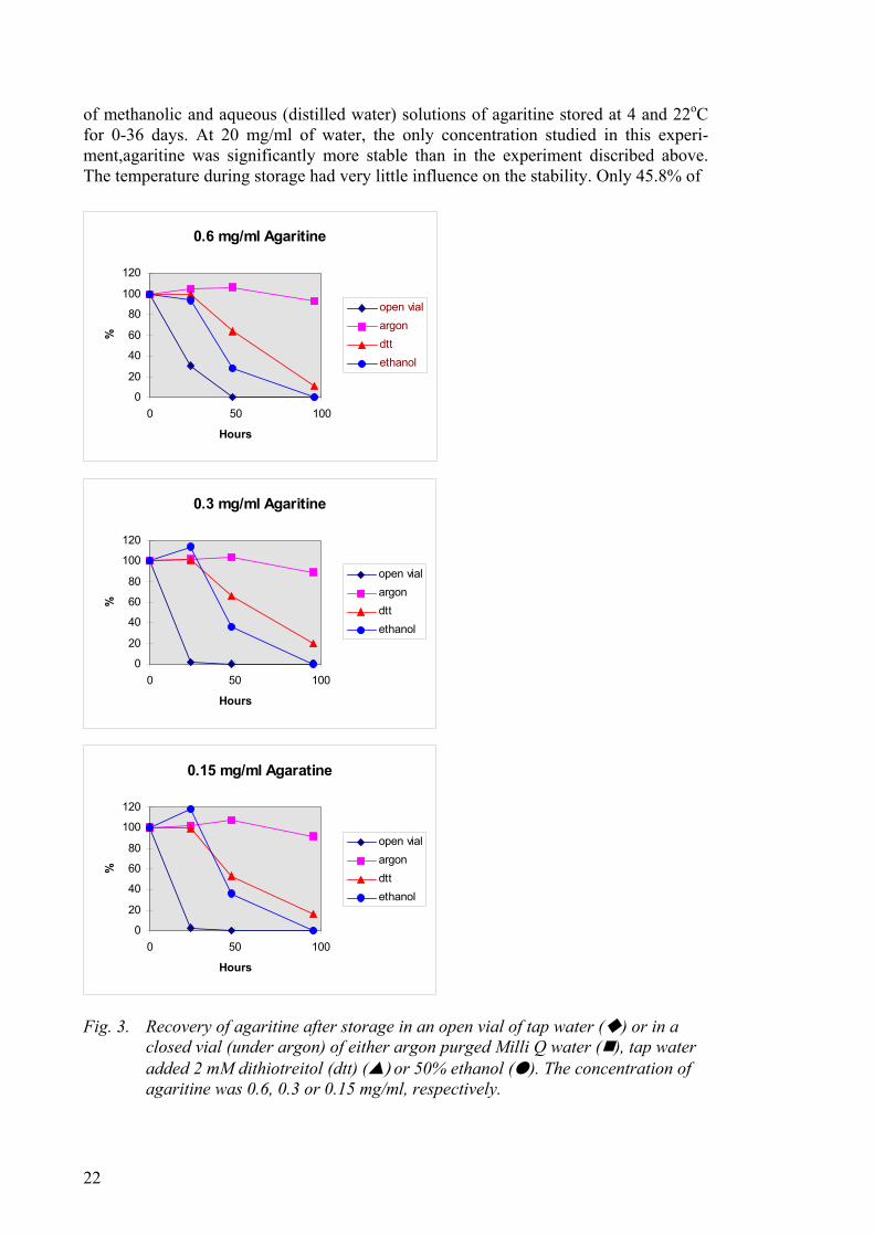
of methanolic and aqueous (distilled water) solutions of agaritine stored at 4 and 22oC for 0-36 days. At 20 mg/ml of water, the only concentration studied in this experi-ment,agaritine was significantly more stable than in the experiment discribed above. The temperature during storage had very little influence on the stability. Only 45.8% of
0.6 mg/ml Agaritine
0
20
40
60
80
100
120
0 50 100
Hours
%
open vialargondttethanol
0.3 mg/ml Agaritine
0
20
40
60
80
100
120
0 50 100
Hours
%
open vialargondttethanol
0.15 mg/ml Agaratine
0
20
40
60
80
100
120
0 50 100
Hours
%
open vialargondttethanol
Fig. 3. Recovery of agaritine after storage in an open vial of tap water ( ) or in a
closed vial (under argon) of either argon purged Milli Q water ( ), tap water added 2 mM dithiotreitol (dtt) (▲) or 50% ethanol (●). The concentration of agaritine was 0.6, 0.3 or 0.15 mg/ml, respectively.
22

the compound had been degraded during the first 14 days of storage in distilled water. The degradation in methanolic solution was less, only 7.8% being degraded during this time period. It is not known whether the difference in stability of agaritine observed in these two studies is due to different concentrations of agaritine being used or that the different types of water contained different amounts of oxygen or other compounds of importance for the degradation. The observation on stability described above may have bearing on the interpretation of many experimental studies (Frandsen and Gry, unpub-lished results).
In order to study the stability of agaritine in more details the “Nordic project group on inherent natural toxicants in food plants and mushrooms” in co-operation with Czech colleagues carried out a series of experiments. It was confirmed that the stability of aga-ritine solutions is highly influenced by the oxygen tension and the type/purity of solvent (Hajŝlová et al., 2002). In Figure 4 is shown the stability of agaritine (0.3 mg/ml) over 5 days when dissolved in tap water a 2 mM solution of dithiotreitol in tap water or in
a) closed vials
0
20
40
60
80
100
0 20 40 60 80 100 120hours
%
TW TW+dtt MeOH
b) open vials
0
20
40
60
80
100
0 20 40 60 80 100 12
hours
%
TW TW+dtt MeOH
0
Fig. 4. Degradation of agaritine (0.3 mg/ml) in various media (TW, tap water; dtt, 2
mM dithiotreitol in TW; MeOH, methanol) in (a) closed vials and (b) open vi-als.
23

methanol and kept in closed (Figure 4a) and open (Figure 4b) vials at ambient tempera-ture, respectively. The data in Figure 4 are the means from two parallel experiments. As can be seen, a successive drop in agaritine content occurred during storage in all tested media. However, the stability of aqueous solutions was significantly lower than that of methanolic solutions. Agaritine solutions were more stable in closed vials (Figure 4a).
The quickest degradation of agaritine occurred in tap water. Already after 48 h. no aga-ritine could be detected in tap water solutions stored in open vials. In closed vials, slightly < 50% of the compound remained after 120 h. Addition of dtt to the tap water improved the stability of the analyte, particularly when the solution was stored in closed vials. However, also in the presence of dithiotreitol nearly all agaritine was degraded after 120 h in open vials (around 11% remaining). Agaritine was most stable in metha-nol, around 87-88% of the compound remained after 120 h both when stored in open and closed vials. The increased stability of agaritine (0.3 mg/ml) in Milli Q water and Milli Q water purged with oxygen free gas, N2, was also confirmed (data not shown). In both solutions, agaritine was degraded to a similar degree as in methanol, > 75% re-maining after 120 h. The stability was slightly higher in Milli Q water purged with N2. (Hajŝlová et al., 2002).
The influence of pH on the degradation rate of agaritine in aqueous extracts of A. bi-torquis at ambient temperature was also studied as shown in Figure 5. Agaritine was degraded more quickly at pH 1.5 than at pH 4.5 especially during the first days of incu-bation. It was relatively more stable at pH 6.8. After an incubation of 24 h > 50% of the compound was degraded at pH 1.5 but only around 18% was at pH 6.8. The reduced stability of agaritine at low pH was confirmed in a study on agaritine in simulated gas-tric fluid (pH 1.2). In Table 2 it is shown that the breakdown of agaritine was faster both in extracts of fresh and of cooked mushrooms, compared to the pure solution of agari-tine in simulated gastric fluid. Rather surprisingly, the rate of degradation of agaritine was faster in the two mushroom extracts than in the pure solution of agaritine,
0
20
40
60
80
100
0 5 10 15 20
days
%
pH 1.5 pH 4.5 pH 6.8
Fig. 5. Degradation of agaritine at various pHs (the agaritine content in fresh mush-
rooms was 197.7 mg/kg).
24

Table 2. Degradation of agaritine in simulated gastric fluid (% of original content).
Time (days)
Pure solution of agaritine
Agaritine extract from fresh mush-
rooms
Agaritine extract from cooked mushrooms
0 100.0 100.0 100.0 1 83.4 50.1 82.1 2 73.6 36.9 56.7 5 60.7 27.2 38.6 8 53.8 11.7 21.4
16 32.1 n.d. n.d. 22 14.2 n.d. n.d.
Original content of agaritine in standard solution (100%) was 0.02 mg/ml, whereas it was 230.2 mg/kg in the “fresh mushrooms” and 183.9 mg/kg in the “cooked mushrooms”, respectively.
although the degradation was slower in the extract of boiled mushrooms compared to the extract of the fresh mushrooms. It was speculated whether a mushroom constituent, coextracted with agaritine, could have catalysed the degradations (Hajŝlová et al., 2002).
In assessing the possible risks associated with the consumption of the cultivated mush-room it is important to include an estimation of the degradation of agaritine, especially at the low pH in the stomach. The different stability of agaritine at various pH, as dem-onstrated above, is also of importance when extrapolating data obtained in experimental animals to man. The pH of the human stomach varies considerably, but it is commonly around pH 2. The pH of the rodent stomach, on the other hand, is higher, usually around pH 5 (Hajŝlová et al., 2002).
Finally, the breakdown of agaritine in tap water in closed vials was studied for five days. At the end of the experiment only 37% of agaritine remained. The gradual drop in concentration of agaritine with storage time was accompanied by the simultaneous for-mation of at least four unknown degradation products. At the end of the incubation (five days) the solution contained one of the four degradation products in higher concentra-tion (43%) than the remaining agaritine (37%) (Hajŝlová et al., 2002).
It can be concluded that agaritine is very unstable in aqueous solutions, especially if oxygen has not been carefully excluded, and at low pH.
Many toxicological studies performed with extracts of the cultivated mushroom and with agaritine per se set out to explore whether this constituent can give rise to adverse effects on living systems and they have given variable and not easily interpretable re-sults. Since few, if any, studies have carefully taken care of protecting agaritine solu-tions from oxidative degradation, it is highly likely that agaritine degradation has con-tributed to the variable results. Thus, in som experimental studies the exposure to agari-tine might have been much lower than intended. Further, the experimental material may have contained unidentified degradation products of agaritine with unknown toxic po-tential.
25
3.3. Chemical synthesis Methods to chemically synthesise agaritine, β-N-[γ-L-(+)-glutamyl]-4-carboxy-phenyl-hydrazine, and 4-(hydroxymethyl)benzenediazonium ion in the form of the tetrafluor-oborate have been published (Wallcave et al., 1979; Kelly et al., 1982; Ross et al., 1982a; Chauhan and Toth, 1984; Datta and Hoesch, 1987). The method to synthesise agaritine (Wallcave et al., 1979) have been found to be insufficient and the method has, therefore, subsequently been modified by others (Walker et al., 1997; Frandsen, 1998).
3.4. Analytical methods The first methods for agaritine analysis, from the early 1960's, were based on gravimet-ric and enzymatic assays. For example, Kelly et al. (1982), after a lengthy purification of agaritine from the mushroom found 360 mg agaritine per kg fresh weight. Levenberg (1961, 1964) used an enzymatic assay where agaritine was cleaved to 4-(hydroxy-methyl)phenylhydrazine and L-glutamic acid. In the presence of excess glyoxylic acid, the 4-(hydroxymethyl)phenylhydrazine was converted to a phenylhydrazone which was subsequently detected spectrophotometrically at 325 nm.
In 1978 Issenberg developed a non-published high performance liquid chromatographic (HPLC) procedure for the analysis of agaritine (Speroni and Beelman, 1982). This pro-cedure was modified and employed by Liu and others (1979, 1982) at the Pennsylvania State University, USA, to determine agaritine in fresh and processed mushrooms. Sev-eral difficulties were experienced with the procedure, which was based on methanol extracts of mushroom tissue being directly injected into the chromatograph. Below baseline resolution occurred between the agaritine peak and an unidentified peak imme-diately preceeding it, making quantification of agaritine difficult. The phenolic material present in the methanol extracts also had the potential to influence the chromatography.
Subsequently workers from the same Institute described a sensitive HPLC for analysis of agaritine (Speroni and Beelman, 1982). Freeze-dried mushrooms were extracted with methanol, extracts evaporated to dryness, and the residue resuspended in the mobile phase (0.005N NaH2PO4, pH 4.25) and subsequently passed through a C18 reverse-phase SepPak. The mobile phase was pumped through a cation-exchange column at a fixed speed and agaritine monitored at 237 nm. Recoveries of agaritine standards were high (>90 %) and the limit of detection 0.006 µg. To maintain the reliability of this pro-cedure frequent calibration with agaritine standards are needed. Free amino acids that strongly absorb in the UV was shown not to interfere with the agaritine peak.
26
4. Biosynthesis
Only a few studies have investigated the biosynthesis of agaritine experimentally, and these have come to partly different conclusions. The first investigators to study agaritine biosynthesis in Agaricus bisporus were Schütte et al. (1972) who analysed the incorpo-ration of radiolabelled compounds into agaritine. No incorporation of 3-14C-labelled tyrosine and phenylalanine was observed. However, incorporation rates of 0.14 %, 0.68 %, and 4.1 % were obtained with [U-14C]shikimic acid, [2-14C]glutamic acid, and p-aminobenzoic acid-[3,5-3H], respectively. Thus, p-aminobenzoic acid seems to a pre-cursor to agaritine. However, the conclusion has lately been critisized by Baumgartner et al. (1998), who believes that Schütte et al. (1972) actually studied the synthesis of γ-glutaminyl-4-hydroxybenzene (see below).
Schütte et al. (1972) also tried to determine the origin of the hydrazine part of the mole-cule by growing mushrooms supplied with 15N-labelled glutamic acid and glutamin-(CO15NH2). These studies showed that both the α-amino group and the amid-nitrogen from both labelled compounds were incorporated into agaritine.
Based on the information presented by Schütte et al. (1972) on the biosynthesis of aga-ritine and the chemical analytical data showning several phenylhydrazine-derivatives and arylbenzenediazonium ions to occur in A. bisporus and related species, La Rue (1977) proposed that agaritine biosynthesis and degradation occur in the genus Agaricus as shown in Figure 6. As the biosynthetic part of this scheme is based on the data of Schütte et al. (1972), the scheme has been questioned by Baumgartner et al. (1998).
The intermediary compound in Figure 6, 4-(carboxy)phenylhydrazine, has been de-tected in Agaricus bisporus at much lower levels than agaritine (Chuan et al., 1984). This observation indicates that either the biosynthesis of agaritine proceeds through alternate pathways not involving 4-(carboxy)phenylhydrazine or that a rapid conversion of this compound to other intermediates involved in agaritine biosynthesis occurs (Chauhan et al., 1984). Turner (1983) proposed agaritinal (Figure 7) as a possible in-termediate in agaritine biosynthesis. This aldehyde has not been found in Agaricus bis-porus, but it has been isolated from several wild-growing Agaricus species that also containing agaritine (Chulia et al., 1988; Stijve and Pittet, 2000).
It seems likely, according to Baumgartner et al. (1998), that the biosynthetic scheme suggested by Schütte et al. (1972) is for γ-glutaminyl-4-hydroxybenzene and not agari-tine (β-N-(γ-glutamyl)-4-hydroxymethylphenylhydrazine), and that there is no de novo synthesis of the aromatic residue of agaritine in the Agaricus bisporus fruit body. In their hands, 4-aminobenzoic acid was no precursor of agaritine in fruit bodies detached from the mycelium but an excellent precursor for γ-glutaminyl-4-hydroxybenzene. In the discussion on their findings, Baumgarter and co-workers refers to personal commu-nications with K. Sasaoka that had made a similar observation. Instead of the desgluta-myl moiety of agaritine being produced in the fruit body, these investigators show data supporting the view that the agaritine present in the fruit body is transported to this lo-cation from the vegetative hyphae.
27

COOH
NH
4-aminobenzoic acid
COOH
HN
4-(carboxy)phenyl- hydrazine
NH2
COOH
HN NH γ glutamyl
β-N-(γ-L(+)-glutamyl-4-(carboxy)phenylhydrazine
CH2OH
HN NH γ glutamyl
CH2OH
HN NH2
CH2OH
N N
β-N-(γ-L(+)-glutamyl-4-(hydroxymethyl)phenyl-hydrazine [agaritine]
4-(hydroxymethyl)-phenylhydrazine
4-(hydroxymethyl)-benzenediazoniumion
Fig. 6. A suggested but questioned biosynthetic route for agaritine in Agaricus mush-
rooms, and a presumed route of degradation.
Baumgartner et al. (1998) speculate that the assembly of agaritine takes place in the vegetative hyphae in contact with the wheat straw compost, using specific metabolic activities of representatives from three biologically different groups of organisms. At this location the assembly of agaritine requires both the aryl and the hydrazide moieties from exogenous sources (e.g. resulting from the breakdown of lignin by the fungus and
CHO
HN NH γ glutamyl Fig. 7. Chemical structure of agaritinal.
28

the diazotrophic activity of a bacterial commensal in the substratum). The investigators reported that rhizomorph contained 2.1 and 2.6 mg/g dry weight of agaritine and γ-glutaminyl-4-hydroxybenzene, respectively, when A. bisporus was grown on substra-tum, but only 1 mg/g dry weight when they tried to decontaminate hyphae from soil. As mentioned in the introduction, the agaritine catabolite 4-(hydroxymethyl)benzene-diazonium ion seems to be formed by two routes. One system, whereby the diazonium ion is generated enzymatically via the unstable compound 4-(hydroxymethyl)phenyl-hydrazine, and another in which the diazonium ion is generated directly from agaritine (Gigliotti and Levenberg, 1964; Ross et al., 1982a). Since leakage of agaritine into wa-ter was found to be negligible by Baumgartner et al. (1998), these authors suggest that its decrease in harvested mushrooms must be due to catabolism. Most probably, this catabolism embodies an oxidative transformation of the desglutamyl moiety of agari-tine, occurring either on the intact molecule, or following removal of the glutamate residue (Baumgartner et al., 1998).
Why Agaricus mushrooms produce agaritine is not known. Since agaritine has been shown to inhibit mono- and diphenolase activity, Espin et al. (1998) have suggested that agaritine could play a role in vivo as an endogenous regulator of the mushroom poly-phenol oxidase activity and thereby of the amounts of o-quinone formed. However, this does not seem to be a likely explanation.
On the other hand, it could very well be that a continuous degradation of agaritine to 4-(hydroxymethyl)phenylhydrazine occurs in A. bisporus, and results in a concomitant production of 4-(hydroxymethyl)benzenediazonium ions, which in vivo inhibits com-petitive fungi, since the production apparently increases with the age of the mushroom, making up for its growing vulnerability to attack by moulds. A few findings supports this hypothesis (Stijve et al., 1986):
a) unlike other higher fungi, Agaricus species are seldom if ever found covered with mould growth;
b) wild-growing species and strains contain much more agaritine than cultivated ones. The former may need more protection than the latter which grow in a pro-tected environment;
c) Agaricus species without agaritine produce other fungistatic compounds: the xanthodermi subgroup contains appreciable quantities of phenol, and the ni-troamino acids in members of the sylvaticus subgroup may have a similar func-tion.
29

30

5. Occurrence
5.1. The content of agaritine and related compounds in fresh mushrooms
Despite the evidence that agaritine is not anticipated to be the ultimate biologically ac-tive component in the cultivated mushroom, analytical work has nevertheless tended to concentrate on monitoring this compound, rather than its precursors, metabolites or de-composition products, presumably because of the relatively large amount of agaritine present in the mushroom, and the relative ease with which the analysis can be carried out. Agaritine could therefore be regarded as an indicator of the likely presence of po-tentially toxic phenylhydrazines. The natural occurrence of hydrazines and related com-pounds in mushrooms and plants has been reviewed by Toth (2000a).
Several factors, including strain of A. bisporus studied, cultural practice used in produc-tion, stage of cropping-cycle (flush), maturity and harvest may all interact to influence agaritine content. Without carefully controlling each of these variables, it is difficult to determine their individual influence on agaritine production. In the studies mentioned below , in which the agaritine content has been analyzed of the cultivated mushroom, this control has generally not been done.
Table 3 shows the amount of agaritine in fresh samples of A. bisporus, either obtained directly from mushroom growers or purchased in stores on the local market. Since the level of agaritine determined in A. bisporus is dependent both on the storage time and the conditions during storage (see next section), it is not surprising that slightly higher agaritine contents have been found in mushrooms obtained directly from the growers.
The overall range of agaritine levels that have been found in fresh A. bisporus are be-tween 80 and 1 730 mg/kg fresh weight, with average values between 200 and 500 mg/kg. The three studies that have presented data only on a mg/kg dry weight basis (Levenberg, 1964; Speroni and Beelman, 1982; Speroni et al., 1983) are in agreement with the rest of the studies. The latter method of presenting the content of agaritine gives figures that are approximately 10 times higher than when giving the content in mg/kg fresh weight (Liu et al., 1982; Fischer et al., 1984).
According to some authors, the difference in agaritine content reported can not be ex-plained by different parts of the fruiting body being studied in different investigations as there is no specific localisation of agaritine in the sporophores (Foret and Arpin, 1991). On the other hand, although Soulier and co-workers (1993) detected agaritine through-out the whole fruit body at fairly similar levels, the stipe base and hymenium contained significantly deviating levels of agaritine, lower levels in the stipe base and higher lev-els in the hymenium. This observation was recently confirmed within this project. We analysed various parts of the fruit body of cultivated A. bitorquis for the presence of agaritine and found the skin of the cap to contain 352 mg/kg (16% of the whole mush-room), the cap with its skin 254 mg/kg (56% of the whole mushroom), gills 254 mg/kg (12% of the whole mushroom), and the stem 194 mg/kg (16% of the whole mushroom),
31

respectively. The agaritine content of the whole mushroom was 249 mg/kg fresh weight (Schulzová et al., 2002).
One of the studies in Table 3 demonstrated a 4-fold difference in agaritine content among 14 lots of fresh mushrooms from 10 different growers, the range being 330-1 730 mg/kg fresh weight (Liu et al., 1982). Included in the analysis were five different strain types: white, off-white, golden white, cream, and brown. No indication of strain influence on mean values of agaritine concentration was apparent in the data, an obser-vation that subsequently has been confirmed by others (Speroni et al., 1983; Fischer et al., 1984; Stijve et al., 1986). In the study of Speroni and co-workers (1983), one strain (PSU-351 with brown colour phenotype) had significantly higher agaritine levels than the remaining seven strains, 5 100 mg agaritine per kg mushroom (on a dry weight ba-sis) as compared to 1 700-2 800 mg/kg. It was hypothesised that this strain, which was only recently isolated from nature, contains higher agaritine levels because it had not yet lost its inherited ability to inhibit growth of certain fungi. It is possible agaritine functions in vivo as an antimycotic agent. Also no relationship could be found between mushroom colour and agaritine level.
The observations referred to above were partly confirmed in our project. We purchased 28 different samples of fresh cultivated mushrooms with a cup diameter of 4-6 cm from the retail market in Prague, Czech Republic, 25 samples being A. hortensis (A. bis-porus) and the other three A. bitorquis (Schulzová et al., 2002). There was no signifi-cant difference in agaritine content between species. The agaritine content of the 28 samples varied between 165 and 457 mg/kg, the average being 272±69 mg/kg fresh weight (10th percentile 201 mg/kg, median 267mg/kg and 90th percentile 356 mg/kg). However, some samples from early flushes obtained directly from growers contained significantly more agaritine (700 mg/kg fresh weight – cap diameter 4 cm) (Schulzová et al., 2002). This observation has also been made by Sharman et al. (1990).
According to Levenberg (personal communication, Kelly et al., 1962), agaritine is pre-dominantly found in the fruiting bodies of young mushrooms - the concentration dimin-ishing with age and increase in size of the fruiting bodies. This observation has been confirmed by several investigators (Chiarlo et al., 1979; Fischer et al., 1984; Schulzová et al., 2002), whereas others have been unable to confirm the finding (Sharman et al., 1990, Foret and Arpin, 1991). For example, Fisher et al. (1984) compared the agaritine level in young (∅ cap 1.5-2 cm), medium age (∅ cap 5-6 cm) and old (∅ cap 7.5- cm) mushrooms of a white strain. They found the level in young mushrooms to be 629 mg/kg, in medium age mushrooms 438 mg/kg, and in old mushrooms 271 mg/kg (Fischer et al., 1984). It should be noted that in many of the studies presented in Table 3 it has not been tried to standardise the measurements of agaritine by specifying the size of the mushrooms analysed. No indication of a seasonal trend in agaritine content has been observed in A. bisporus (Liu et al., 1982).
The observation that smaller mushrooms contain higher amounts of agaritine than big-ger ones was, however, confirmed by Stijve and co-workers (1986) who analysed the agaritine content of two wild-life Agaricus species, A. arvensis and A. augustus, in vari-ous stages of development. Fruiting bodies belonging to the same mycelium were picked, lyophilised within 24 hours, and analysed. The results indicated indeed a dra-matic decrease in agaritine content with the age of the mushroom (measured as weight of the mushroom and diameter of the cap). It was suggested that the breakdown is due
32

33
Table 3. Agaritine content of fresh Agaricus bisporus determined in methanol extracts of the mushroom. The mushrooms were either
obtained fresh from growers (g) or purchased at the local market (m). If the samples were not freeze-dried shortly after being obtained (which does not influence the agaritine level), they were analysed within 24 hours.
Mushroom sample (g = grower; m = market)
Diameter of cap (cm)
Analytical method Content (mg/kg fresh weight) Average Range
Reference
2-3 days old mushrooms (g) not stated gravimetric ~ 400 - Kelly et al., 1962 2-3 days old mushrooms (g) not stated gravimetric ~ 220 - Daniels et al., 1961 1 fresh sample (g) not stated gravimetric ~ 3 300* - Levenberg, 1964 2 fresh samples (m) not stated HPLC 440; 720 170-1 170 Ross et al., 1982b 14 fresh samples of different strains (g)
3.0-4.5 HPLC 880 330-1 730 Liu et al., 1982
1 fresh sample (g) not stated HPLC 2 190* - Speroni and Beelman, 1982 8 fresh samples of different strains (g)
2.5-5.0 HPLC - 1 700-5 100* Speroni et al., 1983
2 fresh samples of different strains (g)
5.0-6.0 HPLC" 304±6.0; 438±2.5
- -
Fischer et al., 1984
11 fresh samples (m) 2.0-5.5 HPLC" 368±45 94-629 Fischer et al., 1984 1 fresh sample not stated ? 228 - Hashida et al., 1990 2 fresh samples of different strains (g)
not stated HPLC"" ~ 180 80-250 Sharman et al., 1990
5 fresh samples of different strains (m)
not stated HPLC TLC
160-650 Stijve et al., 1986
1 fresh sample within a Nordic project (g)
not stated HPLC 340 - Andersson et al., 1994
2 fresh sample (m) not stated HPLC 212; 229 - Andersson et al., 1999 5 fresh sample not stated HPLC 820 630-984 Burini et al., 1999 * given as mg/kg dry weight; " agaritine standard 94.5 % pure; "" agaritine standard 95 % pure.
to a constant enzymatic degradation of agaritine to 4-(hydroxymethyl)phenylhydrazine in the mushroom.
Table 4 shows which other phenylhydrazine derivatives or arylbenzenediazonium ions than agaritine that have been detected in A. bisporus. The table has tabulated all obser-vations of these compounds available in the literature. It is obvious that the occurrence of 4-(carboxy)phenylhydrazine, β-N-[γ-L-(+)-glutamyl]-4-carboxy-phenylhydrazine and 4-(hydroxymethyl)benzenediazonium ion in A. bisporus requires confirmation. Czech chemists recently extracted A. bisporus with methanol and analysed the extract for β-N-[γ-L-glutamyl]-4-carboxyphenylhydrazine, β-N-[γ-L-glutamyl]-4-formylphenylhydrazine, β-N-[γ-L-glutamyl]-4-methylphenylhydrazine, and 4-methylphenylhydrazine (Schulzová and Hajslová, personal communication). Low quan-tities of β-N-[γ-L-glutamyl]-4-carboxyphenylhydrazine, and β-N-[γ-L-glutamyl]-4-methylphenylhydrazine, and traces of β-N-[γ-L-glutamyl]-4-formylphenylhydrazine was detected in the fresh extract but the amounts were reduced upon storage of the ex-tract at room temperature. After 18 days levels were very low, but at this point in time small quantities of 4-methylphenylhydrazine was detected for the first time. There are substantial evidence that the 4-(hydroxymethyl)benzenediazonium ion exists in the fun-gus, although the investigators could not be certain whether it came through the metabo-lism of agaritine or if it exists as an independent entity (Levenberg, 1962). In addition to the compounds mentioned in Table 4, the presumed precursor to agaritine, 4-aminobenzoic acid, has been detected in A. bisporus at a level of 10 mg/kg fresh weight (Toth et al., 1997a).
In addition to the observations presented in Table 4, Stijve et al. (1986) analyzed for 4-(carboxy)phenylhydrazine in fresh samples of the cultivated mushroom, but could not detect any. This was perhaps not surprising since the detection limit was high in this study, around 100 mg/kg dry weight. In the same study the investigators were also un-able to detect 4-aminobenzoic acid and N-[γ-L-glutamyl]-4-(carboxy)phenylhydrazine with similar limit of detection.
Shortly after having detected agaritine in A. bisporus, Levenberg (1964) examined boiled press-juice extracts from as many as 45 representative genera of basidiomycetes. As part of this survey boiled press-juice preparations from the fruit bodies of 15 differ-ent species belonging to the genus Agaricus were tested. Table 5 summarises the infor-mation available on which mushroom species do, and which do not contain agaritine. Of the 15 Agaricus species tested by Levenberg (1964), 10 species (A. pattersonii, A. xanthodermus, A. argentatus, A. campestris, A. comtulus, A. crocodilinus, A. edulis, A. hortensis, A. micromegathus and A. perrarus) contained agaritine in quantities compa-rable to those found in A. bisporus. Five other species, all belonging to the sylvaticus subgroup (A. benesii, A. sterlingii, A. subrutilescens, A. sylvaticus, and A. sylvicola) were devoid of the compound. Three members of the Agaricus genus (A. campestris, A. diminutivus and A. hortensis) were found to contain additional substances active in the enzymatic assay. However, the products formed in these cases had different spectral characteristics than those resulting from agaritine itself. These latter species were be-lieved to contain other aromatic hydrazine derivatives of L-glutamic acid of as yet un-determined structure (Levenberg, 1964). The above scientific names for the different Agaricus species studied are cited from the corresponding publications. The nomencla-ture for a large number of Agaricus species has been dealt with in a study on the occur-rence
34
35
Table 4. Content of arylbenzenediazonium ions and other phenylhydrazines than agaritine in Agaricus bisporus determined in
methanol extracts of the mushroom. The mushrooms were either obtained fresh from growers (g) or purchased at the local market (m). If the samples were not freeze-dried shortly after being obtained (which does not influence the agaritine level), they were analysed within 24 hours.
Compound Mushroom sample
(g = grower; m = market) Diameter of cap (cm)
Content (mg/kg fresh weight)
Reference
4-(carboxy)phenylhydrazine 1-2 days old mushrooms (g) not stated 10.7±2.0
Chauhan et al., 1984
β-N-(γ-L(+)-glutamyl)4-(carboxy)phenylhydrazine
1-2 days old mushrooms (g) not stated 42±3 16
Chauhan et al., 1985 Toth et al., 1997a
4-(hydroxymethyl)benzene diazonium ion not stated not stated identified but not quantified
Levenberg, 1962
4-(hydroxymethyl)benzene diazonium ion 1-2 days old mushrooms (m) not stated 0.6 4
Ross et al., 1982a Toth et al., 1997a

of agaritine in wild Agaricus species (Schulzova et al., manuscript submitted for publi-cation).
Outside the genus Agaricus, agaritine has hitherto only been detected in two species, shiitake and the oyster mushroom. Very low levels, 0.82 mg/kg were found in shiitake (Lentinula edodes) by Hashida et al. (1990), whereas other investigators were unable to detect agaritine in fresh as well as canned shiitake (Stijve et al., 1986; Hajšlova et al., 1998). Italian investigators claim that fresh oyster mushroom (Pleurotus ostreatus) con-tains between 501 and 867 mg/kg (Burini et al., 1999). However, Czech investigators were unable to confirm this finding (Schulzová, personal communications).
Besides A. bisporus, four other Agaricus species known to harbour agaritine, have also been shown to have enzymatic γ-glutamyltransferase activity comparable to the activity in A. bisporus. These species are A. edulis, A. pattersonii, A. perrarus, and A. xantho-dermus. No enzyme with this activity could be detected in A. sterlingii and A. subrutilescens, species that do not contain agaritine, and in a number of mushroom spe-cies belonging to other genera (Gigliotti and Levenberg, 1964).
Three wild-growing Agaricus species have been shown to contain nitrogen-nitrogen bond-containing compounds other than agaritine. The poisonous mushroom A. xantho-dermus has been shown to contain leucoagaricone, γ-glutamyl-N´-4-(hydroxy)phenyl-hydrazine, and 4-(hydroxymethyl)benzenediazonium ions, the latter isolated in the form of its stable sulfonate (Hilbig et al., 1985; Dornberger et al., 1986), the edible A. silvati-cus small quantities of β-nitraminoalanine and its decarboxylation product N-nitroethylenediamine (Chilton and Hsu, 1975), and the edible A. arvensis, A. campes-tris, A. macrosporus, A. perrarus and A. subperonatus β-N-(γ-glutamyl)-4-formylphenylhydrazine (agaritinal, Figure 7) (Chulia et al., 1988; Stijve and Pittet, 2000). According to Stijve and Pittet (2000) the level of agaritinal could be substantial in some of these mushrooms (above 1 g/kg). In addition A. hortensis has been found to contain the γ-(p-hydroxy)anilide of glutamic acid (Jadot et al., 1960).
5.2. Influence of storage and processing The post-harvest storage and processing of A. bisporus may strongly influence the aga-ritine content of the mushroom. The influence of storage and processing on the level of other phenylhydrazines in the mushroom has been dealt with in one study (see section 5.2.5. Dry baking).
The most recent data on the influence of storage and processing on the content of agari-tine in cultivated Agaricus mushrooms, based on joint studies between a Czech group of food scientists and the “Nordic project group on inherent natural toxicants in food plants and mushrooms” are summarized in Table 6 (Hajŝlová et al., 1998; Andersson et al., 1999; Schulzová et al., 2002).
5.2.1. Refrigerating In one study, post-harvest storage of mushrooms for 5 days at 2oC or 12oC reduced agaritine levels by 68 %. There was no loss of agaritine during the first three days; the largest decrease occurred between day 4 and 5 (Liu et al., 1982). In a similar experi-ment,
36
Table 5A. Mushrooms reported to contain agaritine.
Species Reference Species ReferenceAgaricus abruptibulbus Stijve and Pittet, 2000 Agaricus macrosporus Stijve et al., 1986; Stijve and Pittet, 2000 Agaricus argentatus Levenberg, 1964 Agaricus micromegathus Levenberg, 1964 Agaricus arvensis Stijve et al., 1986; Stijve and Pittet, 2000 Agaricus niveolutescens Stijve et al., 1986; Stijve and Pittet, 2000 Agaricus augustus Stijve et al., 1986; Stijve and Pittet, 2000 Agaricus nivescens Stijve and Pittet, 2000 Agaricus bisporus (incl. var.) Stijve and Pittet, 2000 Agaricus pattersonii Levenberg, 1964 Agaricus bitorquis Stijve et al., 1986; Stijve and Pittet, 2000 Agaricus perrarus Levenberg, 1964; Stijve and Pittet, 2000* Agaricus blazei Stijve and Pittet, 2000; Stijve et al., 2003 Agaricus porphyrocephalus Stijve and Pittet, 2000 Agaricus campestris (incl. var.) Levenberg, 1964; Stijve and Pittet, 2000* Agaricus semotus Stijve and Pittet, 2000 Agaricus comtulus Levenberg, 1964; Stijve and Pittet, 2000 Agaricus silvicola Stijve et al., 1986; Stijve and Pittet, 2000 Agaricus crocodilinus Levenberg, 1964 Agaricus subperonatus Stijve et al., 1986; Stijve and Pittet, 2000 Agaricus edulis Levenberg, 1964; Stijve et al., 1986 Agaricus vaporarius Stijve et al., 1986; Stijve and Pittet, 2000 Agaricus excellens Stijve et al., 1986 Agaricus xanthodermus Levenberg, 1964; Stijve et al., 1986 Agaricus hortensis Stijve et al., 1986; Stijve and Pittet, 2000 Lentinus edodes Hashida et al., 1990 Agaricus lilaceps Stijve and Pittet, 2000 *both in Stijve et al., 1986 and in Stijve and Pittet, 2000
Table 5B. Mushrooms reported not to contain agaritine.
Species Reference Species ReferenceAgaricus benesii Levenberg, 1964 Agaricus placomyces Stijve and Pittet, 2000 Agaricus bernardii Stijve and Pittet, 2000 Agaricus sterlingii Levenberg, 1964 Agaricus cupreobrunneus Stijve and Pittet, 2000 Agaricus subrutilescens Levenberg, 1964 Agaricus fuscofibrillosus Stijve and Pittet, 2000 Agaricus sylvaticus Levenberg, 1964; Stijve and Pittet, 2000* Agaricus geesterani Stijve and Pittet, 2000 Agaricus xanthoderma Stijve and Pittet, 2000 Agaricus haemorrhoidarius Stijve et al., 1986; Stijve and Pittet, 2000 Agrocybe aegerita Stijve et al., 1986; Stijve and Pittet, 2000 Agaricus hortensis Stijve and Pittet, 2000 Amanita caesarea Stijve et al., 1986 Agaricus langei Stijve et al., 1986; Stijve and Pittet, 2000 Amanita rubescens Stijve et al., 1986 Agaricus meleagris Stijve et al., 1986 Armillaria edodes Hashida et al,1990 Agaricus phaeolepidotus Stijve and Pittet, 2000 Armillaria mellea Stijve et al., 1986 *both in Stijve et al., 1986 and in Stijve and Pittet, 2000

Table 5B. Mushrooms reported not to contain agaritine.
Species Reference Species ReferenceAuricularia auriculajudae Stijve and Pittet, 2000 Lepista nuda Stijve et al., 1986; Stijve and Pittet, 2000 Auricularia polytricha Stijve and Pittet, 2000 Leucoagaricus pudicus Stijve et al., 1986; Stijve and Pittet, 2000 Boletus edulis Stijve et al., 1986 Lycoperdon gemmatum Stijve et al., 1986 Calocybe gambosa Stijve et al., 1986 Macrolepiota procera Stijve et al., 1986
Macrolepiota rhacodes Stijve et al., 1986 Cantharellus cibarius Stijve et al., 1986 Marasmius oreades Stijve et al., 1986
Cantharellus tubaeformis Stijve et al., 1986 Micropsalliota brunneosperma
Stijve and Pittet, 2000
Clitopilus prunulus Stijve et al., 1986 Morchella conica Stijve et al., 1986 Coprinus comatus Stijve et al., 1986; Stijve and Pittet, 2000 Morchella vulgaris Stijve et al., 1986 Coprinus atramentarius Stijve et al., 1986 Pholiota nameko Stijve and Pittet, 2000 Craterellus cornucopioides Stijve et al., 1986 Pleurotus citrinopileatus Stijve and Pittet, 2000 Fistulina hepatica Stijve and Pittet, 2000 Pleurotus eryngii Stijve and Pittet, 2000 Flammulina velutipes Stijve et al., 1986; Stijve and Pittet, 2000 Pleurotus ostreatus Stijve et al., 1986; Stijve and Pittet, 2000 Grifola frondosa Stijve and Pittet, 2000 Pleurotus
salmoneostramineus Stijve and Pittet, 2000
Hericium erinaceus Stijve and Pittet, 2000 Russula cyanoxantha Stijve et al., 1986 Hirneola auricula-judae Stijve et al., 1986 Russula virescens Stijve et al., 1986 Hydnum repandum Stijve et al., 1986 Sarcodon imbricatum Stijve et al., 1986 Hygrophorus marzuolus Stijve et al., 1986 Sparassis crispa Stijve and Pittet, 2000 Hypholoma capnoides Stijve and Pittet, 2000 Sparassis laminata Stijve and Pittet, 2000 Hypholoma sublateritium Stijve and Pittet, 2000 Stropharia rugosaannulata Stijve et al., 1986; Stijve and Pittet, 2000 Hypsizigus tessulatus Stijve and Pittet, 2000 Suillus granulatus Stijve et al., 1986 Hypsizigus ulmarius Stijve and Pittet, 2000 Suillus luteus Stijve et al., 1986 Kuehneromyces mutabilis Stijve et al., 1986; Stijve and Pittet, 2000 Tricholoma equestre Stijve et al., 1986 Lactarius deliciosus Stijve et al., 1986 Tricholoma portentosum Stijve et al., 1986 Laetiporus sulphureus Stijve and Pittet, 2000 Tricholoma terreum Stijve et al., 1986 Langermannia gigantea Stijve et al., 1986 Tuber melanosporum Stijve et al., 1986 Leccinum griseum Stijve et al., 1986 Volvariella volvacea Stijve et al., 1986; Stijve and Pittet, 2000 Lepista nebularis Stijve et al., 1986 Xerocomus chrysenteron Stijve et al., 1986 *both in Stijve et al., 1986 and in Stijve and Pittet, 2000

two batches of Agaricus bisporus purchased from a local store with average agaritine contents of 440 and 720 mg/kg, respectively, were stored in the refrigerator (4oC) in polyethylene bags for 2 weeks. After one week one of the batches had lost 2 % of its agaritine content, whereas the other batch had lost 47 %. The corresponding figures after two weeks were 36 and 76 %, respectively (Ross et al., 1982b).
Since it is normal for mushrooms to be stored for periods equivalent to a week at 2oC in fresh-market channels, it is probable that consumers are exposed to lower agaritine lev-els than are found in freshly harvested mushrooms. From a practical standpoint, the use of extended post-harvest storage as a means to reduce agaritine in fresh mushrooms would not be desirable since deterioration in quality would most likely occur (Liu et al., 1982).
A similar reduction in the agaritine content during storage at low temperature was dem-onstrated also in Agaricus hortensis (Schulzová et al., 2002). The fresh mushroom put into the refrigerator contained 393 mg agaritine per kg fresh weight. After 6 days in the refrigerator (5 ºC) the agaritine content was reduced by 25%, after 10 days by 40% and after 14 days by 50% (Table 6).
5.2.2. Freezing and thawing. Freezing and thawing A. bisporus results in a significant reduction in agaritine content of the mushroom. Liu et al. (1982) observed about 20 % of the original agaritine content in thawed mushrooms, whereas Stijve et al. (1986) only observed 2 % of the original agaritine content. The method of freezing - quickly in liquid nitrogen or slowly at -20oC - did not influence the end result. The agaritine levels were 23-74 % lower in A. bis-porus that had been rapidly frozen i liquid nitrogen than in mushrooms that had been immediately freeze-dried (equivalent to fresh mushrooms) (Sharman et al., 1990). Liu and colleagues suggested that the majority of the loss in agaritine occurred during thaw-ing, when disruption of the cellular structures separating the degrading enzymes from the substrate would allow the enzymatic breakdown of agaritine not otherwisepossible (Liu et al., 1982). It is not known whether any breakdown of agaritine occurred during the 30 days of storage at -25oC.
When A. bitorquis, containing 249 mg agaritine/kg, was stored for one week at -18ºC, the agaritine content was reduced by 25% when analysed after extraction with methanol without thawing the sample and by 48% after thawing for one hour at room temperature (or by 33% after thawing 0.5 minutes in microwave oven). After storage of A. bitorquis, containing 261 mg agaritine/kg, at -18 ºC for one month without thawing, the agaritine content was reduced by 41% and after thawing for one hour at room temperature by 77%, as shown in Table 6 (Schulzová et al., 2002).
5.2.3. Freeze-drying. Freeze-drying or lyophilisation seems to have no influence on the agaritine content of A. bisporus (Liu et al., 1982). In accordance with this observation, two Swiss studies found high levels of agaritine in lyophilised samples of the cultivated mushroom, the levels being between 2 110 and 7 800 mg/kg dry weight (Fischer et al., 1984; Stijve et al., 1986).
39

40
Table 6. Influence of storage and household processing on the agaritine content of cultivated Agaricus mushrooms (Hajŝlová et al., 1998; Andersson et al., 1999; Schulzová et al., 2002). Process Conditions (ºC) Time Amount of agaritine remaining in
the mushroom (%)1 Storage: Refrigerating 5 6 days 75 Refrigerating 5 14 days 50 Drying 25 24 hours 82 Drying 50 7.5 hours 76 Drying 40-602 7 hours 81 Freezing without thawing -18 7 days 75 Freezing with thawing -18 7 days 52 Freezing without thawing -18 30 days 59 Freezing with thawing -18 30 days 23 Freeze drying -51 18 hours ~100 Household processing: Boiling ~100 5 minutes 44 Boiling ~100 60 minutes 12 Dry baking 200 10 minutes 77 Dry baking 200 20 minutes 57 Dry baking 200 30 minutes 24 Deep frying 150 10 minutes 50 Deep frying 170 5 minutes 52 Frying (whole mushroom caps, oil) 150 10 minutes 43 Frying (sliced, oil) ~100 5 minutes 34 Frying (sliced, butter) ~100 5 minutes 69 Microwave heating 1000 W, 2450 MHz3 15 seconds 59 Microwave heating 1000 W, 2450 MHz3 30 seconds 49 Microwave heating 1000 W, 2450 MHz3 60 seconds 35 Canned mushrooms - - < 10
1 100% = agaritine content in fresh mushrooms before processing 2 40 ºC for two hours, then 60 ºC for five hours 3 20 gram sliced mushrooms
Freeze-drying of A. hortensis, containing 410 mg agaritine/kg, at -51 ºC for 18 hours did not influence the content of agaritine. The content in the dried lyophilised mush-room was around 4000 mg/kg dry weight (Schulzová et al., 2002).
5.2.4. Drying. Very low levels of agaritine have been reported in dried Italian A. bisporus (pumizia), whereas extremely high amounts have been detected in dried sliced mushrooms on the market in UK, 6 520 mg/kg dry weight (Sharman et al., 1990). Unfortunately, it is not known how these two samples have been produced. As mentioned above, freeze-drying seems to have negligible effect on the agaritine content of the cultivated mushroom.
Schulzová et al. (2002) followed the agaritine content of A. hortensis during drying at ambient temperature (25 ºC), at 50 ºC and at programmed elevated temperature (40 ºC for two hours followed by 60 ºC). The drying of the sliced mushrooms was continued until the material obtained constant weight. The agaritine content was reduced by 18-24% (Table 6).
5.2.5. Dry baking. Because in some countries humans consume A. bisporus mostly in baked form, e.g. in the USA, the amounts of agaritine in baked mushrooms have been investigated. Gannett and Toth (1991) analysed for agaritine and other hydrazine derivatives in extracts from fresh or baked (10 minutes at 225oC) mushroom. Baking reduced the level of agaritine by 27 %, 4-(carboxy)phenylhydrazine by 10 % and β-N-[γ-L-(+)-glutamyl]-4-carboxy-phenylhydrazine by 23 % when compared to the fresh extract. In another experiment, the fresh or baked homogenate of A. bisporus was analysed for 4-(hydroxymethyl)-benzenediazonium ion. Baking reduced the content by 41 %. Ganett and Toth (1991) concluded that dry baking reduced the content of the four nitrogen-nitrogen bond-containing compounds by about 25 % only.
Dry-baking was also studied by Schulzová et al. (2002). A. bitorquis, containing 340 mg agaritine/kg, was heated on baking paper for 0 to 45 minutes at 200 ºC. After 10 minutes baking, the agaritine content was reduced by 23%, after 20 minutes by 43%, after 30 minutes by 76 % and after 45 minutes by 89%, but in the latter case, after 45 minutes baking at 200 ºC, the organoleptic quality of the mushrooms was unsatisfactory (Table 6).
5.2.6. Boiling. The loss of water-soluble compounds from mushroom tissue is a result of shrinkage and extraction, and has been carefully studied by Biekman et al. (1996), using mannitol as an example of such compounds.
In an American investigation blanching of two different strains of A. bisporus in boiling water for 5 minutes reduced the agaritine content by 57 % and 75 %, respectively. At the same time these two strains lost 20 % and 24 % in fresh weight, usually referred to as shrinkage (Liu et al., 1982). Similarly, when two fresh samples of A. bisporus con-taining 290 and 295 mg/kg agaritine, respectively, were blanched for 5-7 min, the agari-tine content in the mushrooms were reduced to 181 and 169 mg/kg, respectively. After the blanching the brine contained 116 and 134 mg/kg agaritine, respectively, giving a total amount of 297 and 303 mg/kg i the two samples (Fischer et al., 1984). In agree-
41

ment with these observations Japanese investigators have reported a significant decrease in agaritine content of A. bisporus boiled at 100oC for 10 minutes (Hashida et al., 1990).
Also, Schulzová et al. (2002) studied the effect of boiling on the content of agaritine in the cultivated mushroom. Sliced fruit bodies of A. bitorquis, containing 360 mg agari-tine/kg, were boiled in closed jars in steam. Similar results as shown in Figure 8 were obtained, when whole mushrooms were used instead of sliced mushrooms. As can be seen in Figure 8, boiling resulted in a significant progressive decrease of agaritine in solid mushrooms, whereas the concentration in the aqueous solution increased during the first 15 minutes. No further increase in the agaritine content of the boiling water was observed thereafter. The most pronounced changes occurred during the first minute of boiling. The solid mushrooms had lost 55% of their agaritine after 5 minutes of boiling and about 90% after 60-120 minutes of boiling. The total amount of agaritine in the system was also reduced during cooking, being about 75% of the original amount after 5 minutes and about 60% of the original amount after 60 minutes of boiling.
5.2.7. Canning. In the case of low-fat foods such as brine-packed mushrooms, destruction or prevention of the germination of the spores of Clostridium botulinum and some thermophilic bacte-ria causing economic spoilage is of primary importance during the canning process. Speroni and co-workers (1985) have studied the thermal degradation kinetics of agari-tine in model systems and agaritine retention in canned mushrooms, and Sastry et al. (1985) the agaritine retention in mushrooms during the canning process. The former investigators studied the effect of pH and heating on agaritine degradation in buffer, mushroom puree and canned mushrooms. This was done by warming up agaritine with citric-phosphate buffer (in ampoules) or agaritine and mushroom puree buffered with the same salts to specified temperatures (101-133oC) during a predetermined time in-tervals. Additionally, cans containing quartered mushrooms with brine and buffer were stillretored using five thermal process schedules with equivalent lethalities for
0
4
8
12
16
0 20 40 60 80 100 120min
mg/mushrooms mg/broth mg/systém
Fig. 8. Amount of agaritine (mg) remaining in mushrooms (A. bitorquis), cooking broth and the whole system (showing the extent of agaritine degradation dur-ing cooking) after boiling in water for up to 120 minutes.
42
Clostridium botulinum. In both the buffer and puree systems, agaritine degradation could be modelled by first-order kinetics. The results of both investigations indicated that high temperature-short time treatment during the canning process resulted in the greatest agaritine retention in the mushroom part (Sastry et al., 1985). The removal of agaritine by blanching would be especially desirable because any breakdown products of agaritine formed during the thermal process would not accumulate in the can. Fur-thermore, would any of the breakdown products be carcinogenic, it may be more desir-able to optimise the blanching operation to remove agaritine from the mushrooms be-fore retorting and then use high temperature-short time processing to actually minimise the destruction of agaritine and formation of hazardous breakdown products in the can. One of the first measurements of the agaritine level in mushrooms per se as well as in mushroom products was performed by American investigators (Ross et al., 1982b). They found no agaritine in canned A. bisporus, but 440 mg/kg in fresh mushrooms. It was suggested by these investigators that agaritine is decomposed by the temperatures and pressures that are used during food processing.
Recently, the Nordic project group on inherent natural toxicants in food plants and mushrooms” in collaboration with food chemists in the Czech Republic analysed 35 different trademarks of canned mushroom products for agaritine, 25 of which were based on cut mushrooms and 10 on whole mushrooms (Andersson et al., 1999). These cans were produced in the Netherlands (18 products), China (6 products), France (4 products), India (3 products), Spain (2 products), the Czech Republic (1 product) and Sweden (1 product). On average whole mushrooms contained 14.9±6.7 mg agaritine per kg product whereas cut mushrooms contained 18.1±7.8 mg/kg. There was no statisti-cally significant difference between these two values. Agaritine levels in brine were generally slightly less than the amount detected in the canned mushrooms.
Table 7 summarises published measurements on agaritine content of canned Agaricus bisporus. It is evident that relatively low agaritine levels are found in canned products, usually less than 10 % of the agaritine level detected in fresh mushrooms (Liu et al., 1982; Stijve et al., 1986; Hajslŏvá et al., 1998; Andersson et al., 1999). A comparison of concentration values in mushrooms and brine indicates a near-equilibrium situation be-tween solid and liquid phases. This equilibration is probably established during can storage.
5.2.8. Pan-frying In the Nordic countries frying of the cultivated mushrooms in butter or vegetable oils is a much more common cooking procedure than dry baking. Therefore, the influence of frying sliced A. hortensis (298 mg agaritine/kg) in these fats for 5 minutes on the agari-tine content of the mushroom was investigated by the joint Czech-Nordic research group (Schulzová et al., 2002). The temperature of the vegetable oil and butter, respec-tively, was around 100 ºC during the entire frying period, the mushroom slices being stirred continuously during the process. After frying for 5 minutes in vegetable oil, the agaritine content had been reduced to 34% of the original agaritine content in fresh mushrooms, but only 0.6% of the original agaritine content could be recovered in the oil. Frying, sliced mushrooms in butter for 5 minutes reduced the agaritine content to 69% of the agaritine content in fresh mushrooms before processing. Only 2.8% of the original agari-
43

Table 7. Reported levels of agaritine in canned Agaricus bisporus.
Agaritine content (mg/kg fresh weight) mushrooms brine
Number of samples
Reference
31 (1-64) 57 (3-104) 11 Fischer et al., 1984 28 (20-33) 9 (6-11) 12 Sharman et al., 1990 undetectable undetectable 3 Ross et al., 1982 29 (23-35) 34 (25-43) 2 Stijve et al., 1986 65 (40-90) 115 (80-150) 2 Liu et al., 1982 undetectable not determined not given Hashida et al., 1990 17.2 (<3-30) 11.0 (<3-30) 35 Andersson et al., 1999
tine content was recovered in the butter. In another experiment whole mushrooms caps of A. bitorquis was fried at around 150 ºC in a pan with vegetable oil for 10 minutes (5 minutes on each side). The mushrooms originally contained 338 mg/kg agaritine. Ten minutes of frying reduced the agaritine content by 57% (Table 6) (Schulzová et al. 2002).
5.2.9. Deep-frying Schulzová et al. (2002) studied the influence of deep-frying at 150 and 170 ºC in vege-table oil on the agaritine content of A. hortensis (the original amount was 429 mg/kg). The agaritine content was reduced to approximately 50%, either after 10 minutes of deep-frying at 150 ºC or after 5-7 minutes at 170 ºC (the optimal cooking time in both cases with respect to organoleptic properties) (Table 6).
5.2.10. Microwave heating The influence of microwave heating on the agaritine content of A. bitorquis has been investigated. Twenty gram of sliced mushroom (original content of agaritine 276 mg/kg) was heated in the microwave oven (1000W, 2450 MHz). After 15 seconds the agaritine content was reduced by 41%, after 30 seconds by 51%, after 45 seconds by 61% and after 60 seconds by 65% (Schulzová et al., 2002).
5.3. Other products containing the cultivated mushroom Table 8 shows the reported levels of agaritine in miscellaneous types of A. bisporus products on the market - some products contain high levels and others low. In one of the first measurements of agaritine content of mushrooms and mushroom products, the compound was undetectable in mushroom soup (Ross et al., 1982b). Low agaritine lev-els in ready-to-eat mushroom soup have been confirmed by whereas Andersson and co-workers (1999) observed somewhat higher agaritine levels in some mushroom soups based on a dry powder. However, in the later study the amount of agaritine in the ready-to-eat product never exceeded 30 mg per portion. On the other hand, there are products that contain higher agaritine levels than others, but such products can not easily be iden-tified by the consumers.
44

Table 8. Reported levels of agaritine in miscellaneous types of Agaricus bisporus proucts.
Type of product Agaritine content
(mg/kg fresh weight) Reference
Frozen mushrooms 330 Ross et al., 1982b Mushrooms sautéed in olive oil at 300oC (7 min.)
300 Ross et al., 1982b
Sliced mushrooms and mushroom powder used as ingredients for dehydrated soups
100 - 250* Stijve et al., 1986
Pasta sauce (n=4) 1.6-15.3 Andersson et al., 1999 Mushroom soup 0 Ross et al., 1982b Mushroom soup <5 Sharman et al., 1990 Mushroom soup (n=3) 1.8-62.8 Andersson et al., 1999 Mushroom sauce 3.9 Andersson et al., 1999
* recalculated, assuming 90% water in mushroom Sharman and co-workers (1990),
As fresh mushrooms in general have a short shelf life compared to most vegetables, the industry have tried to develop mushroom products, with longer shelf lives, that could be exported to distant destinations. This could be done by removing water and adding in-hibitors such as salt to the mushrooms. Agaricus bisporus has recently been explored for snack development by using osmotic dehydration in concentrated salt solution (Brennan and Salmier, 1998). The laboratory studies have been promising, but nothing is known about the safety of the product. For example, it is not known to what extent the processing influence the phenylhydrazines in the mushroom.
5.4. Influence of cultivation, including genetic modification Commercial mushroom growing entails several production steps, which may affect aga-ritine concentration in the harvested mushroom. Mushroom composting involves a type of fermentation whereby the original starting material is chemically and biologically altered to provide the mushroom mycelium with the proper nutrients needed for growth. Mushroom compost types are commonly classified according to the starter material used. "Natural composts" consist mainly of wheat straw-bedded horse manure. They are generally the most widely used and least expensive compost types. "Synthetic com-posts" consist largely of hay and crushed corn cobs and are generally any compost where horse manure is not the major ingredient. "Blended composts" are made from mixtures of "natural" and "synthetic" composts. Most type of composts, in addition, also requires nitrogen supplements, Gypsum to aid in pH control, and often many other ingredients.
Mushrooms grown in synthetic compost had higher agaritine levels than mushrooms obtained from natural or blended compost (Speroni et al., 1983). On a dry weight basis two samples of mushroom grown on synthetic compost contained approximately 4 100 and 7 500 mg/kg agaritine, respectively, whereas mushrooms grown on blended com-
45

post contained 2 700 and 3 100 mg/kg and mushrooms grown on natural compost 2 400 and 2 700 mg/kg, respectively. Within a given flush, the mushrooms harvested from synthetic composts were always higher in agaritine concentration than mushrooms from natural or blended composts. Actual isolation of the compost constituents affecting the agaritine levels will require additional research.
Spawning refers to the inoculation of the compost with the mycelium of a selected strain of A. bisporus. Earlier it was believed that mycelium contains substantially less agaritine than the fruiting bodies (Speroni et al., 1983), but later studies have shown this not to be the case. Soulier and coworkers (1993) detected agaritine in 17 out of 20 sam-ples of distal mycelia and 19 out of 20 samples of proximal ones, with a mean agaritine content (depending on strain type) around 135 mg per litre cell solution. These observa-tions indicate that the mycelium can be considered as an active site of synthesis of aga-ritine and its presence within this tissue cannot result from a translocation from fruiting bodies. On the contrary, Baumgartner et al. (1998) have suggested that agaritine is transported from the vegetative hyphae of the mycelium to the fruit body.
During the cropping or harvesting period, rhythmic 3- to 5-d harvests, which constitute a single flush or break, are followed by periods when few or no mushrooms are avail-able for harvest. Growers usually harvest from at least three but generally no greater than eight flushes per crop before aborting the mushroom bed. Later flushes produce smaller yields than earlier flushes. At the same time there is a strong tendency for agari-tine concentrations of mushrooms to increase with the later flushes (Speroni et al., 1983).
A few pesticides, for example chlorfenovinphos, chlorpyrifos, cyromazine, diflu-benzuron, methoprene and prochoroz are used to control competing insects and fungi during Agaricus cultivation. It is not known whether the use of these pesticides influ-ence the agaritine content of the mushrooms.
Commercially grown strains of A. bisporus on occasion produce sectors that display undesirable phenotypes. Such sections show altered compost colonisation, have reduced yield and produce inferior quality mushrooms. Genetic studies of such inbreeding de-pression sectors have revealed loss of heterozygocity at specific loci, and changes in deheterokaryotization, alteration in the frequency of somatic recombination, chromo-some loss, chromosomal length polymorphisms and possible chromosome transloca-tions, as well as changes in copy number of the ribosomal DNA repeats (Horgen et al., 1996). Inbreeding depression, as defined by eight different fitness components, have been reported in A. bisporus (Xu, 1995), but the relevance of inbreeding depression to A. bisporus breeding in general, and the agaritine content of the mushroom in particular, is not yet known.
Little genetic variation exists between cultivated strains of A. bisporus. During the last decade new prospects for A. bisporus strain improvement has been anticipated (Kerri-gan, 1993), and considerable efforts have been devoted to development of molecular biological techniques, especially a transformation system for the mushroom. The un-availability of a practical gene transfer system has been the single largest obstacle pre-cluding the use of molecular approaches for the genetic improvement of mushrooms. Although several investigators have shown considerable interest in developing useful transformation technologies for mushrooms (Royer and Horgen, 1991; Challen et al., 1991; Li and Horgen, 1993; Challen and Elliott, 1994; van der Rhee et al. 1996a,
46

1996b), no methods have been generally used due to low transformation efficiency or lack of utility and convenience. The most marked obstacles for developing a satisfac-tory transformation system has been attributed to factors such as the generation of false poitives, low level of integration of transforming DNA, poor expression of foreign se-quences, DNA modifications after integration or low competence of certain mushroom strains (Mikosch et al., 2000). An important step to a more successful transformation system was taken when De Groot et al. (1998) succeeded in transforming Agaricus bis-porus and other mushrooms with an Agrobacterium-mediated fungal transformation system. The advantage with this system was that it was more convenient than the previ-ous protoplast-based schemes (van der Rhee et al., 1996a, 1996b). Its disadvantage was a low efficiency. The situation was substantially improved when Chen et al. (2000) re-cently reported successful transformation of fruiting body tissue by Agrobacterium tu-mefaciens-mediated methods. The choice of promoter, strain of A. tumefaciens, and type of fruiting body tissue were critical for optimal transformation efficiency. Transgenic vegetative cultures were obtained in less than 2 weeks, and fruiting bodies 8 weeks later under controlled environmental conditions. A similar system for transformation of vege-tative mycelium of A. bisporus has also been reported (Mikosch et al., 2001).
A reduction in the endogenously regulated browning reaction (Wichers and van Leeu-wen, 1996) is an obvious goal for research on transformation of A. bisporus, but a re-duction in agaritine content could also be a task for future investigations. It is clear that turnover of glutamate/glutamine has a central position in fungal nitrogen metabolism. Whether the nitrogen metabolism in A. bisporus has any bearing on the agaritine level or turnover (biosynthesis/degradation) of agaritine is not known (Baars et al., 1995). As quite a large number of genes have been isolated, sequenced and mapped on all the 13 chromosome pairs of Agaricus bisporus (a total of 41 genes in year 2000) (Whiteford and Thurston, 2000), there is now a more promising future for the transformation of this species.
5.5. Conclusion on occurrence Commercial mushroom growing entails several production steps (factors) which may influence the agaritine concentration in the harvested mushroom. Factors having influ-ence include strain-type of A. bisporus, cultural practice employed in production, stage of cropping-cycle (flush), and maturity of mushrooms at harvest. Since the amount of agaritine measured in A. bisporus, in addition, depends on how long and under which conditions the mushrooms are stored after harvest, it should be kept in mind that the agaritine level in ”fresh” mushrooms purchased in local stores may be lower than the levels in fresh mushrooms obtained directly from mushroom producers.
Table 9 summarises available data on estimated average agaritine content of fresh A. bisporus and marketed products containing this mushroom. High amounts of agaritine are found in fresh and freeze-dried mushrooms, and possibly also in frozen A. bisporus. However, there are indications that the levels are reduced upon thawing of the mush-rooms. The level of agaritine in mushrooms dried at room temperature is insufficiently investigated.
47

Table 9. Estimated average agaritine content of A. bisporus, fresh and processed mush-rooms. References are given in the main text.
Type of mushroom product Estimated agaritine content
(% of fresh weight) - in relation to content in fresh mushroom
Fresh 200-500 mg/kg Refrigerated after the first few days about 75-100% Refrigerated after one week about 50-75% Refrigerated after two weeks about 50% Frozen without thawing about 50-75% Frozen with thawing about 25-50% Freeze-dried no influence on the agaritine content Dried about 75-80% Dry-baked about 25-75% (depending on time) Boiled for 5 minutes extracts about 50%1) Boiled for 60 minutes extracts about 90%2) Pan-fried about 50% Deep-fried about 50% Microwave-heated for up to 60 seconds about 50% Canned less than 10%
1) Total amount in boiled mushroom and boiling water about 75% of original amount in the fresh mushroom; 2) Total amount in boiled mushroom and boiling water about 60% of original amount in the fresh mushroom.
The information on how the agaritine content of A. bisporus is influenced by food pro-duction is limited. However, research devoted to these questions is ongoing. Dry baking of the mushroom, as it is done during pizza baking, reduces the agaritine content by about 25%. Wet processing of mushrooms, like boiling, reduces the agaritine levels by about 50%. Low quantities of agaritine are present in canned A. bisporus, usually less than 10% of the quantity in fresh mushrooms. Also microwave-heating may reduce the agaritine content by more than 50% within one minute.
In addition to the cultivated mushroom (A. bisporus), 10 wild Agaricus species (A. pa-tersonii, A. xanthodermus, A. argentatus, A. campestris, A. comptulis, A. crocodilinus, A. edulis, A. hortensis, A. micromegathus and A. perrarus) contain agaritine in quanti-ties comparable to those found in A. bisporus.
Since molecular biological techniques to transform A. bisporus were recently devel-oped, many different types of modification of the mushroom strains can be foreseen in the future. One such modification could possibly lead to reduced levels of phenylhydra-zines.
48

6. Production and consumption
6.1. Production The cultivated mushroom, Agaricus bisporus, is the most extensively consumed species of mushroom worldwide (Royse, 1997). In 1991 37 % of the world production of cultivated edible mushrooms was Agaricus bisporus - the quantity produced being 1.6 million tonnes compared with 1.2 million tonnes in 1986 - an increase in production of 30 % during these five years. The most consumed mushroom species/genera and their production in 1986 and 1991 are listed in table 10. The major producers of the cultivated mushroom are the EU-countries, China and the USA. The European mushroom production and processing totalled 1 022 million kg in 1995 (Toth, 2000a).
6.2. Consumption According to Moss and Mitchell (1994) eight Western countries account for 85 % of the worldwide cultivated mushroom consumption. The USA is the largest mushroom consumer with 30 %, Germany is second with almost 20 % , United Kingdom, France and Italy are each responsible for approximately 10 % of the worldwide consumption.
The annual per capita intake of the cultivated mushroom (fresh, canned and total) in eight Western countries is shown in table 11. In 1998-1999 the United States annual consumption of this fungus totalled 455 million kg (Toth, 2000a).
In the Nordic countries the total annual production of Agaricus bisporus is about 12 000 tonnes. Data on the production and estimated per capita intake of this mushroom in the five Nordic countries are given in table 12. As shown in this table the estimated annual per capita intake of the "fresh" (or frozen) mushroom varies between countries from 0.4 to 1.6 kg; for "preserved" (e.g. canned) mushroom from 0.2 to 1.6 kg and the "total" quantity of mushroom from 0.6 to 2.4 kg.
It should be underpinned that the estimated per capita intakes are based on information on production, export and import but do not take into consideration the vary different eating habits with respect to edible mushroom within the populations. This is for instance reflected in a recent Danish study on dietary habits in Denmark (Danskernes Kostvaner 1996, Andersen, 1998) where according to the replies to a questionnaire more than half of the adults had not eaten cultivated mushrooms, 5 % did eat more than five times the median intake and for instance 1 0/00 had eaten thirty times more than the median intake.
6.3. Conclusion on production and consumption The cultivated mushroom is the most consumed mushroom worldwide, with the USA having the largest consumption. However, the annual per capita intakes are highest in Germany, Netherlands and Canada: 3.2, 2.9 and 2.7 kg respectively (1990).
49
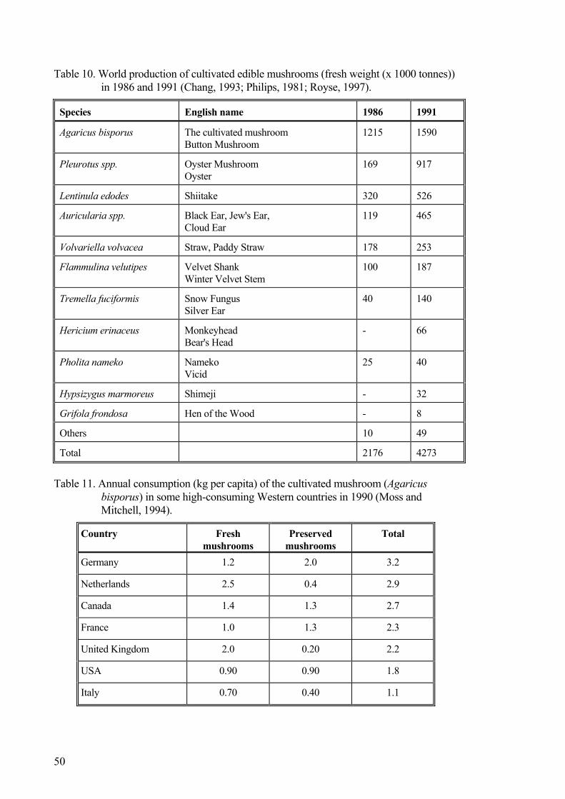
Table 10. World production of cultivated edible mushrooms (fresh weight (x 1000 tonnes)) in 1986 and 1991 (Chang, 1993; Philips, 1981; Royse, 1997).
Species English name 1986 1991
Agaricus bisporus The cultivated mushroom Button Mushroom
1215 1590
Pleurotus spp. Oyster Mushroom Oyster
169 917
Lentinula edodes Shiitake 320 526
Auricularia spp. Black Ear, Jew's Ear, Cloud Ear
119 465
Volvariella volvacea Straw, Paddy Straw 178 253
Flammulina velutipes Velvet Shank Winter Velvet Stem
100 187
Tremella fuciformis Snow Fungus Silver Ear
40 140
Hericium erinaceus Monkeyhead Bear's Head
- 66
Pholita nameko Nameko Vicid
25 40
Hypsizygus marmoreus Shimeji - 32
Grifola frondosa Hen of the Wood - 8
Others 10 49
Total 2176 4273
Table 11. Annual consumption (kg per capita) of the cultivated mushroom (Agaricus
bisporus) in some high-consuming Western countries in 1990 (Moss and Mitchell, 1994).
Country Fresh mushrooms
Preserved mushrooms
Total
Germany 1.2 2.0 3.2
Netherlands 2.5 0.4 2.9
Canada 1.4 1.3 2.7
France 1.0 1.3 2.3
United Kingdom 2.0 0.20 2.2
USA 0.90 0.90 1.8
Italy 0.70 0.40 1.1
50

Table 12. Annual Nordic production and consumption (per capita intake) of the culti-vated mushroom (Agaricus bisporus) in 1996 or 19971)
Denmark (1997) Fresh (tonnes) Preserved (ton-
nes)4) Total (tonnes)
Production 8 5103) 96 8 6063) Imported 2 051 4 020 6 071 Total 10 561 4 116 14 677 Exported 2 371 818 3 189 Available for consumption 8 190 3 298 11 488 Estimated per capita intake2) 1.6 kg 0.63 kg 2.2 kg Finland (1997) Production 1 500 - 1 500 Imported 0 300 1 100 1 400 Total 1 800 1 100 2 900 Exported 0 030 - 0 030 Available for consumption 1 770 1 100 2 870 Estimated per capita intake2) 0.35 kg 0.22 kg 0.56 kg Iceland (1996) Production negligible negligible negligible Imported 302 262 564 Total 302 262 564 Exported negligible negligible negligible Available for consumption 302 262 564 Estimated per capita intake2) 1.1 kg 0.98 kg 2.1 kg Norway (1996) Production 350 - 350 Imported 2 700 4 387 7 087 Total 3 050 4 387 7 437 Exported 0 - - Available for consumption 3 050 4 387 7 437 Estimated per capita intake2) 0 .70 kg 1.0 kg 1.7 kg Sweden (1996/1997) Production 1 700 661 2 361 Imported 5 915 13 790 19 705 Total 7 615 14 451 22 066 Exported 0 661 661 Available for consumption 7 615 13 790 21 405 Estimated per capita intake2) 0.86 kg 1.6 kg 2.4 kg
1) Production, import and export figures are provided from: Denmark: Danmarks Statistik, 1998; Finland: Hal-likainen, pers. comminication, 1998; Iceland: Johannesson, pers. communication, 1998; Norway: Aune, pers. communication, 1998; Sweden: Andersson, pers. communication, 1998; 2) Danish population: 1996: 5.216 x 10⁶, Finnish population: 1996: 5.099 x 10⁶, Icelandic population: 1996: 0.267 x 10⁶, Norwegian population: 1996: 4.348 x 10⁶, Swedish population: 1996: 8.816 x 10⁶ (Source: Norden i tal, 1997) 3) Corrected for cultivated mushrooms not sold by the producer. 4) The weight of dried, cultivated mushrooms are included in “Preserved (tonnes)” and is calculated as ten times the dried weight.
51

In the Nordic countries the annual per capita intakes are somewhat lower (1996/97): Sweden (2.4 kg), Denmark (2.2 kg), Iceland (2.1 kg), Norway (1.7 kg) and Finland (0.6 kg).
52
7. Toxicokinetics
7.1. Absorption, distribution and excretion There are two investigations available on the absorption of agaritine in experimental animals. In one of these, albino Swiss mice from a randomly bred colony, weighing about 30 g, were given a single dose of 3 mg agaritine in 0.1 ml water by gavage (Ross et al., 1982b). The animals were anaesthetised and killed at various times after treat-ment. The blood and five sections of the stomach and gut were examined for agaritine content. Agaritine was found in all parts of the gastrointestinal tract (stomach, upper and lower small intestine, caecum and large intestine) from 15 minutes to 2 hours after treatment. The amount of dosed agaritine recovered was high (70 % or more) during the first 15 minutes after dosing but decreased thereafter, being 53 % at 30 minutes, and only 15-17 % at 1 h and 2 h after administration. No agaritine was detected in any part of the gut 3 hours after administration. Agaritine was not detected in the blood at any time (the detection limit was 30 µg/ml). The authors assumed that agaritine was not detected in the blood due to an inadequacy of the analytical techniques, but it could of course also be due to metabolism of the compound.
The other investigation was comprised of a series of separate experiments. In single dose balance studies agaritine was administered by gavage to male Sprague-Dawley rats and Swiss Hsd/Ola:HIN/s mice; both species received a total dose of 25 mg agaritine/kg body weight with 14C-agaritine added at levels of 3 µCi and 0.6 µCi per animal for rats and mice, respectively (Walton et al., 2000). Urine and faeces were collected at 24 hour intervals for up to 96 hours (rats) and 120 hours (mice).
In rats similar amounts of radioactivity were excreted in urine and faeces (43.3% and 44.2% of the dose, respectively) mainly in the first 24 hours, and excretion was essen-tially complete after 48 hours. In mice, excretion of radioactivity occurred to a much greater extent in urine (75.8%) than in faeces (23.2%) and was more protracted than in the rat with significant amounts of radioactivity being excreted up to 96-120 hours. In particular, the faecal excretion appeared to be biphasic and prolonged, possibly suggest-ing some biliary excretion and even enterohepatic circulation (accounting for the more prolonged urinary excretion than in the rat). However, this remains to be investigated and would be somewhat unusual in that biliary excretion of xenobiotics is normally greater in rat than mouse (Walker et al., 1998; Walton et al., 2000). No unchanged aga-ritine, no N`-acetyl-4-(hydroxymethyl)phenylhydrazine, and no 4-(hydroxymethyl)-benzene diazonium ion was detected in the urine. There was also no evidence that glu-curonide and sulfate conjugates were present. All radioactivity was linked to unidenti-fied metabolites, mainly three different compounds. Although qualitative differences in the nature of the agartine metabolites in the two animal species were not evident, quan-titative differences could be discerned (Walton et al., 2000).
At termination of the single dose studies, 96 hours (rats) and 120 hours (mice) after dos-ing, significant residual activity was found in liver and kidney of the rat; the radiolabel being covalently bound to protein. When expressed in terms of protein, in the rat the
53

extent of binding was 1.1 and 1.0 pg/mg protein in the liver and kidney, respectively; the corresponding values for the mouse were 0.9 and 0.8 pg/mg prtoein. Electrophoresis indicated that this was non-specific binding and not associated with specific protein(s). Less radioactivity was determined in the intestine, spleen, heart and lungs (Walker et al., 1998; Walton et al., 2000).
Blood levels of radioactivity were determined in the rats and mice of this experiment. Radioactivity was very rapidly absorbed in the mouse, blood levels peaking after 30 minutes at about 8.5 µg agaritine equivalent/ml blood; excretion was similarly rapid and blood levels had fallen to 0.02 µg by 24 hours after dosing. This resulted in a half-life of 1 hour in mice. In the rat, maximum blood levels were achieved 3-4 hours after dosing and the peak concentration (2.3 µg agaritine equivalent/ml) was about a third of that in mouse. The half-life was 3 hours in the rat. The area under the curve (AUC) values were similar in both animal species, indicating that the extent of absorption was the same. The results also indicate that none of the radioactivity in blood is associated with unchanged agaritine, N`-acetyl-4-(hydroxymethyl)phenylhydrazine, or 4-(hydroxymethyl)benzene diazonium ion. The area under the curve (AUC) values, were similar in both animal species, indicating that the extent of absorption was the same (Walker et al., 1998; Walton et al., 2000).
In subsequent multiple dose studies animals received doses of unlabelled agaritine (25 mg/kg body weight) on three consecutive days prior to a fourth dose containing 14C-agaritine (3µCi for rats and 0.6µCi for mice). Following this protocol in the rat, urinary excretion of radioactivity was similar to that observed after a single dose (40.0%) but a greater proportion (55.7%) of radioactivity was excreted in faeces and it was more pro-tracted, significant amounts being observed up to 96 hours after dosing. In the mouse, as in the single dose study, urinary excretion predominated (61.4%) but was proportion-ately somewhat less with more excreted in faeces (31.8%). In these studies, preliminary TLC analysis indicated the presence of three significant metabolites in urine, none of which retained the glutamate moiety; no unchanged agaritine was detected. Thus, re-peated administration of agaritine to rats and mice did not alter the urinary metabolic profile and excretion of radioactivity (Walker et al., 1998; Walton et al., 2000).
The fact that no agaritine could be detected in the plasma and urine of rats and mice to which agaritine was administered orally by gastric gavage, could either indicate that agaritine is quickly degraded chemically or microbiologically in the stomach, or subject to an extensive first-pass effect during its passage through the gastrointestinal tract (Walton et al., 1997a). As γ-glutamyl transpeptidase activity in the gut is only about 1% of that present in the kidney (Orlowski and Meister, 1970), it is likely that other micro-bial or intestinal enzymes are involved, or that agaritine is chemically degraded in the gastrointestinal tract (Walton et al., 1997a). As high γ-glutamyl transpeptidase activity is present in the serum, at least in humans, it is conceivable that the hydrazine is rapidly metabolised in the blood.
Walton and co-workers (2000) also feed male Hsd/Ola:NIH/S mice Agaricus bisporus according to the feeding schedule used by Toth and Erickson (1986) when they demon-strated the carcinogenicity of the cultivated mushroom (see section 9.2.1). The mice were fed a normal balanced diet for 4 days/week and during the three remaining days were fed a diet consisting solely of locally obtained, finely diced A. bisporus; both diets were provided ad libitum. After four weeks each mouse was given by gastric gavage a single dose of [14C]agaritine (25 mg/kg body weight). The animals were then transferred
54
to metabolic cages, and urine and feces were collected every 24 hours. A group of five mice served as controls and were maintained on the control diet throughout the experi-mental period. Studies of the body weight during the experiment showed that the mice lost body weight during the three days they were given the mushroom feed, and quickly recovered it when they were given normal mice pellets. The average daily intake of mushrooms over the entire feeding period was 6.6 g per day per mouse. No difference in the excretion profile of [14C]agaritine was seen between the animals fed the mush-room diet and those that were maintained on a balanced semisynthetic diet. Qualita-tively and quantitatively, also the metabolic profile of [14C]agaritine was not altered by the feeding of the mushroom diet.
7.2. Biotransformation In vitro studies on the metabolism of agaritine have been performed with both purified enzymes, crude enzyme preparations and tissue slices. The metabolism of agaritine and the closely related arylhydrazines N'-acetyl-4-(hydroxymethyl)phenylhydrazine and 4-methylphenylhydrazine by mixed-function oxidases obtained from uninduced livers of Swiss mice was studied in vitro by Lawson and Chauhan (1985). 4-Methylphenylhydrazine hydrochloride and the N-acetyl-derivatives of 4-methylphenyl- and 4-(hydroxymethyl)phenylhydrazine, were all readily metabolised to diazonium ions that were stable enough to interact with a trapping agent, 2-naphthol, to give a coloured azo product. Their metabolism was inhibited by metyrapone and carbon monoxide. The glutamylated compound agaritine was poorly metabolised in this study, and the metabo-lism was not inhibited by metyrapone or carbon monoxide, suggesting that the minor breakdown in this case represented nonspecific decomposition, not metabolism.
The first step in the metabolism of agaritine instead seems to be the removal of the glu-tamyl group. Studies performed at pH 9.0 have demonstrated that pig´s kidney γ-glutamyl-transpeptidase is capable of hydrolysing agaritine to 4-(hydroxymethyl)-phenylhydrazine and L-glutamic acid (Ross et al., 1982b). In these studies sodium gly-oxylate was used as a trapping agent for 4-(hydroxymethyl)phenylhydrazine, and the produced chromophore quantified by measuring the absorbance at 325 nm. The concen-tration of glyoxylate 4-(hydroxymethyl)phenylhydrazine increased with time due to generation of 4-(hydroxymethyl)phenylhydrazine from agaritine. The hog kidney en-zyme has properties that are very similar to the properties of γ-glutamyltranspeptidase from the cultivated mushroom, but the activity in vitro of the mammalian enzyme at room temperature was 7-9 times higher than that of the mushroom enzyme.
γ-Glutamyl transpeptidase is a cell surface glycoprotein that cleaves γ-glutamyl amide bonds. The most abundant physiologic substrates for the enzyme are glutathione and glutathione-conjugated compounds. γ-Glutamyl transpeptidase initiates the cleavage of extracellular glutathione into its constituent amino acids, which can then be transported into the cell, and initiates the initial step in the conversion of glutathione-conjugated compounds to mercapturic acid (Hanigan, 1998; Taniguchi and Ikeda, 1998). Appar-ently it also removes the glutamyl moiety from agaritine. Purified γ-glutamyl transpep-tidase from bovine kidney has been shown to rapidly convert agaritine into two metabo-lites; only 10% of agaritine remained following a 2 h incubation (Walton et al., 1997a). The same metabolites were generated when the purified enzyme was replaced with kid-ney homogenates from rats or mice (the former was more active than the second). The
55
major metabolite has been tentatively identified as 4-(hydroxymethyl)phenylhydrazine, whereas the minor metabolite is an unknown degradation product(s) of agaritine and possibly also of 4-(hydroxymethyl)phenylhydrazine. The minor metabolite was also formed in small amounts in the absence of the enzyme.
When the γ-glutamyl transpeptidase activity was determined in various tissues of rat and mice, using γ-glutamyl-7-aminomethylcoumarin as substrate in pooled tissue from 5 animals, rat kidney were shown to have higher activity than mouse kidney, 168 and 82 nmol/min per mg protein, respectively (Walton et al., 1997a). Hepatic preparations, namely homogenates, microsomes and cytosol, and lung homogenates from either rat or mouse failed to metabolise agaritine even after a 3 h incubation. γ-Glutamyl transpepti-dase activity in homogenates from rat liver and lung was 1 and 11 pmol/min per mg protein respectively, and in mouse liver and lung 1 and 14 pmol/min per mg protein, i.e. orders of magnitude lower than the activity encountered in the kidney (Walton et al., 1997a).
In agreement with these observations, kidney preparations metabolically converted aga-ritine to product(s), which bound covalently to protein. Preparations from the rat were about three times more effective than preparations from the mouse (Walton et al., 1997a). Liver homogenates, cytosol or microsomes, from rats and mice, were unable to metabolise agaritine.
Thus, it seems likely that agaritine is able to produce reactive metabolites after first hav-ing been deglutamylated to the arylhydrazine 4-hydroxymethylphenylhydrazine by γ-glutamyl transpeptidase, and then further metabolised to the corresponding diazonium ion. Gannett et al. (1997) trapped aryl radicals during the metabolism of arylhydrazines by rat liver microsomes and microsomes from C5O cells (a mouse keratinocyte cell line). This observation show that arylhydrazines have the potential to give rise to aryl radicals, which subsequently may give rise to C8-arylguanine adducts, and could, at least partly, explain the genotoxicity of arylhydrazines and related compounds that are oxidatively metabolised to arenediazonium ions, the precursor to aryl radicals.
Other in vitro studies showed ram seminal vesicle prostaglandin(H)synthase to be able to metabolise agaritine, N'-acetyl-4-(hydroxymethyl)phenylhydrazine and 4-methyl-phenylhydrazine. However, since N'-acetyl-4-(hydroxymethyl)phenylhydrazine was much more efficiently metabolised than the two other compounds, acetyl groups seems to promote the enzyme-substrate interaction in this case (Lawson, 1987). Agaritine dif-fers from 4-(hydroxymethyl)phenylhydrazine in the nature of the substituent on the hy-drazine moiety and was poorly metabolised – agaritine has a glutamyl group instead of an active acetyl group. Also mouse lung prostaglandin(H)synthase and mouse lung cy-tochrome P-450 were shown to metabolise N'-acetyl-4-(hydroxymethyl)phenylhydra-zine and 4-methylphenylhydrazine to reactive metabolites.
Price et al. (1996) used precision-cut rat, mouse and human liver slices and precision-cut rat and mouse lung slices to investigate the in vitro metabolism of [ring-U-14C]agaritine. Liver and lung slices were examined because these two tissues were sites of tumour formation in mice fed fresh Agaricus bisporus (Toth and Erickson, 1986). The protein content of liver slice tissue was greater than the protein content of lung slice tissue. Levels of cytochromo P-450 were greater in rat and mouse than in human liver slices. The cytochrome P-450 content of lung slices was not determined.
56

In the metabolism studies, tissue slices were incubated with 25 µM radiolabeled agari-tine for 0.5-4 hours. A time-dependent covalent binding of [14C]agaritine metabolites to tissue slice proteins was observed in both liver and lung slices from all three species (Price et al., 1996). Covalent binding to liver slice proteins was much greater in mouse than in rat and human liver slices, whereas covalent binding was greater in mouse than in rat lung slices. Based on these in vitro studies the authors speculate whether the rat might be a better model for studying Agaricus toxicity in man than the mouse. γ-Glutamyl transpeptidase activity was greatest in human liver slices and lowest in mouse liver slices. However, no relationship was observed between tissue slice γ-glutamyl transpeptidase activity and [14C]agaritine metabolite covalent binding. HPLC analysis of media samples from the mouse liver slice culture (which had the greatest rate of co-valent binding), revealed agaritine and at least one major and two minor unidentified metabolites. The metabolism of [14C]agaritine to polar products by mouse liver slices was time dependent.
7.3. Conclusion on toxicokinetics No agaritine could be found in the gastro-intestinal tract of mice 3 h after administration of the compound by the oral route. When the toxicokinetics of agaritine was studied with radiolabelled compounds, radioactivity was rapidly absorbed and blood levels peaked within a few hours, the time point being dependent on the species investigated. The rate of clearance of the activity from blood was also dependent on the species. No unchanged agaritine was detected in blood.
Agaritine metabolites were excreted in urine and faeces. In the urine, three unidentified metabolites, all without the glutamate moiety, have been detected. Some days after an oral administration of radiolabeled agaritine, a significant residual activity was detected covalently bound to tissue proteins in internal organs such as the liver and the kidneys. In vitro studies with liver and lung tissue slices exposed to agaritine confirmed these observations. Since the covalent binding to liver slice proteins was much greater in mouse tissue than in rat and human tissue and the binding to proteins in the later two tissues were of similar magnitude, it has been suggested that rats might be a better ex-perimental model system than the mouse for studying Agaricus toxicity in man. Further studies are required to confirm this suggestion.
Other in vitro studies, focusing on biotransformation of agaritine, have shown that puri-fied γ-glutamyl transpeptidase, isolated from various tissues of many species, or kidney homogenate from rats and mice (which are rich in this enzyme) converts agaritine to two metabolites, of which the major have been tentatively identified as 4-(hydroxy-methyl)phenylhydrazine. The γ-glutamyl transpeptidases used in these studies have very similar properties to the γ-glutamyl transpeptidase occurring in the cultivated mush-room.
The fact that no unchanged agaritine could be detected in the plasma and urine of rats and mice administered this compound orally or by gastric gavage, could indicate that agaritine is chemically or microbiologically degraded in the stomach, or subject to an extensive first-pass effect during its passage through the gastrointestinal tract. As γ-glutamyl transpeptidase activity in the gut is only about 1% of that present in the kidney it is likely that microbial or other intestinal enzymes are involved, or that agaritine is chemically degraded in the gastrointestinal tract. As high γ-glutamyl transpeptidase ac-
57

tivity is present in the serum, at least in humans, it is also conceivable that the hydrazine is rapidly metabolised in the blood.
58

8. Effects in short-term tests
8.1. In vitro studies on phenylhydrazine metabolism in the mushroom
Extracts of Agaricus bisporus metabolised 14C-agaritine, producing three main unidenti-fied metabolites. The metabolism was strongly stimulated by 2-mercaptoethanol (which restores metabolic activity of the mushroom γ-glutamyl transpeptidase). All three me-tabolites produced a weak mutagenic response in Salmonella typhimurium TA 104. By incubating agaritine with purified hog kidney γ-glutamyl transpeptidase, which converts agaritine to 4-(hydroxymethyl)phenylhydrazine, it could be demonstrated that none of the three metabolites of agaritine produced by the mushroom extract was identical to this compound. Addition of iodoacetic acid (an inhibitor of γ-glutamyl transpeptidase) to the incubation mixture of agaritine and mushroom extract, impaired the metabolism of agaritine to 4-(hydroxymethyl)phenylhydrazine by the mushroom homogenate. There were some indications that one of the three main metabolites could have been a hydra-zone formed by reaction of 4-(hydroxymethyl)phenylhydrazine with an organic acid present in the mushroom. This could explain why 4-(hydroxymethyl)phenylhydrazine was not detected among the metabolites, but at present this is only a speculation (Walton et al., 2001).
Since tyrosinase (Seo et al., 2003) occur in Agaricus bisporus and this enzyme metabo-lise agaritine and N´-acetyl-4-(hydroxymethyl)phenylhydrazine in vitro, it has been suggested that agaritine to some extent is metabolised by tyrosinase in the mushroom in vivo (Walton et al., 1997b, 2001). This suggestion is compatible with recent reports that agaritine inhibits mushroom tyrosinase in vitro (Espin et al., 1998). Since commercial tyrosinase has been shown to be contaminated with laccase, the possibility that the me-tabolism of agaritine and N´-acetyl-4-(hydroxymethyl)phenylhydrazine was catalysed, at least in part, by laccase can not be ruled out (Walton et al., 2001). The metabolites produced by tyrosinase had a weak mutagenic mutagenic effect in a bacterial test sys-tem (Walton et al., 2001).
8.2. Studies on DNA binding To study the potential for generation of free radicals during food consumption, Good-man et al. (2002) used electron paramagnetic resonance spectroscopy to investigate free radical generation on maceration (simulating mouth chewing) of a range of vegetable products commonly consumed in an uncooked state. The radicals formed as a result of physical damage were trapped in ‘spin-trapping’ experiments using several different diamagnetic molecules. Various varieties of Agaricus bisporus were tested together with the vegetables. Clear signals were observed when the mushroom was macerated in the presence of 4-POBN (α-(pyridyl-1-oxide)-N-t-butyl-nitrone) and PBN (phenyl-t-butyl-nitrone), indicating the formation of large quantities of adducts of the radical from 4-hydroxymethylbenzene diazonium salts. Parallel studies on the vegetables showed
59

that saliva caused a major reduction in the intensity of the spin-trapped signal, suggest-ing the presence of radical scavengers in saliva (Goodman et al., 2002).
The 4-(hydroxymethyl)benzenediazonium ion (HMBD) is a highly reactive compound that is believed to be produced from agaritine in the mushroom or when agaritine is me-tabolised in mammals. It has chemical potential to interact with macromolecules such as DNA.
The first study showing a covalent binding of HMBD to DNA dates back to 1986 (Shepard et al., 1986). In this study tritium-labelled and unlabelled 4-hydroxy[methyl-3H]benzenediazonium ion were synthesised by the investigators and used to expose five female Crl:CD-1 (ICR)BR mice orally to 450-879 mg/kg tritium-labelled HMBD. After an exposure period of 6 h, mice were killed and DNA extracted from homogenated stomach, small intestine, large intestine/appendix and liver tissue. Much higher specific activity was found in stomach and small intestine DNA compared to the activity in large intestine/appendix or liver DNA. This observation might indicate that the compound acts as an activation-independent genotoxic carcinogen producing most DNA damage at the site of application. DNA from the studied tissues was enzymatically digested and nucleotides separated with HPLC. In the stomach 44 % of the eluted radioactivity was due to covalent adducts, whereas the corresponding figure in the small intestine was 16-38 %. This finding was interpreted as a demonstration that HMBD undergoes covalent binding to DNA in vivo, which could be the mechanism for 4-(hydroxymethyl)-benzenediazonium ion-induced carcinogenicity (Shepard et al., 1986).
Subsequently, Japanese investigators studied in vitro the ability of HMBD to modify bases and sugar moieties occurring in DNA (Hiramoto et al., 1995a, 1995b). When the four deoxyribonucleosides were incubated together with HMBD at 37oC, pH 7.4, elec-tron spin resonance spin-trapping techniques revealed the formation of 4-(hydroxymethyl)phenyl radicals and a reduction in the levels of all four nucleosides. The reduction in nucleoside levels was inhibited by ethanol and Cys. Major products of the reaction of deoxyguanosine (dGuo) and deoxyadenosine (dAdo) with HMBD were isolated and their structures established as 8-[4-(hydroxymethyl)phenyl]dGuo (8-HMP-dGuo) and 8-[4-(hydroxymethyl)phenyl]dAdo (8-HMP-dAdo), respectively. The same types of DNA adducts were isolated from calf thymus DNA treated with HMBD. These DNA damages could be demonstrated with 32P-postlabeling. In addition to these major DNA adducts, other DNA adducts were formed at lower frequencies. These were not characterised further. Malonaldehyde was released from DNA treated with HMBD, which indicate that also the deoxyribose moieties of DNA were damaged. The forma-tion of adducts in DNA was inhibited by ethanol or 2-mercaptoethanol. Taken together these results indicate that the carbon centred 4-(hydroxymethyl)phenyl radical gener-ated from HMBD after removal of a nitrogen molecule can directly modify the base and the sugar moieties of DNA without generating oxygen-derived radicals (Figure 9). The inhibitory effect of ethanol was ascribed its scavenging activity for the carbon-centred radical. The inhibitory effect of Cys and 2-mercaptoethanol was found to be due to the formation of reversible adducts between HMBD and the SH compounds.
When plasmid supercoiled DNA was incubated with HMBD, the supercoiled DNA was converted into a nicked circular relaxed form and subsequently also into a linear form. Sequence analysis indicated that the reactive compound produced from HMBD cleaved
60

O
HN
H2N N N
NCH2OH
N
N
NH2
N
NCH2OH
Purine base damage
Pyrimidine base damage
Deoxyribose damage
Strand breakingCH2OH
CH3CH2OH
CH3CHOH.
Adduct
Cys2-Mercaptoethanol
N N
CH2OH
N2
CH2OH
.+
DNA
Fig. 9. Mechanism of 4-(hydroxymethyl)benzenediazonium ion (HMBD)-induced DNA
damage. the plasmid DNA strand non-specifically. HMBD also fragmented the intracellular DNA of Escherichia coli, and induced micronuclei in peripheral reticulocytes of mice exposed to this substance (Hiramoto et al., 1995a). These are probably lesions secon-dary to the damage of bases and sugar moieties in DNA.
Hiramoto and co-workers (1998) have followed up their in vitro studies by administrat-ing male ICR mice intraperitoneally with HMBD and searching for DNA adducts formed in the liver in vivo by the 32P-postlabeling technique. As adducts could be dem-onstrated 24 h after administration but not 48 h after administration, the investigators suggested that the DNA lesion was repaired to a large extent.
The observations made by the Japanese group and referred to above, have been con-firmed by American investigators (Lawson et al., 1995; Gannett et al., 1996). The American investigators also noted that HMBD had other mechanisms of action. The specific adducts formed between HMBD and DNA appears to depend on the structure of the reactive intermediate. Reduction of HMBD first produces an aryl diazenyl radical and, following elimination of nitrogen, aryl radicals. The two radical species, aryl di-azenyl radicals and aryl radicals, can react at the C8 position of guanine and form C8-azo or C8-arylguanine adducts, respectively. In addition to the electron spin resonance signal identifying the 4-(hydroxymethyl)phenyl radicals, the American investigators also identified spin trapped hydroxyl radicals, indicating that hydroxyl radicals and aryl radicals are generated simultaneously. Furthermore, the American group early recog-nised that HMBD also appears to react with NH2 groups in bases to form triazene de-rivatives (Gannett et al., 1987). For example, the reaction of HMBD with adenine re-sulted in a 1:1.8 mixture of triazene adducts where the terminal nitrogen of the 4-(hydroxymethyl)benzenediazonium ion and the 4-(formyl)benzenediazonium ion, re-
61

spectively, were bound to the N-2 position of adenine. The mechanism of the latter compound being formed is believed to be an oxidation in situ (Gannett et al., 1987). Together these observations suggests that either HMBD itself or the 4-(hydroxymethyl)phenyl radical may serve as the ultimate genotoxicant. C8-4(Hydroxymethyl)phenylguanine has also been identified in C50 cells (an immortalised non-differentiated murine keratinocyte cell line) and primary mouse keratinocytes ex-posed to 10-100 µM HMBD (Lawson et al., 1995). Similar exposures of cultured Chi-nese hamster V79 cells to HMBD resulted in DNA single-strand breaks and DNA cross-links, which probably are secondary lesions in DNA.
The consequences of C8-arylguanine and other DNA adducts being formed are un-known. Gannett and co-workers (1996) have suggested that induction of C8-arylguanine may cause a change in conformation about the glycosidic bond from anti to syn, and therefore disrupt the normal guanine-cytosine base pair and give rise to muta-tions. In turn, the potential mutagenic lesion may be readily and rapidly repaired. Simi-larly C8-adenine adducts and triazene formation at N6 of adenine might be mutagenic lesions. If HMBD is continuously produced from agaritine in the cultivated mushroom or by the internal metabolism in a consumer of Agaricus bisporus, any of the lesions formed by the reaction of HMBD and DNA might be genotoxic, and possibly promote the induction of cancer.
Also agaritine and/or its metabolites have been shown to bind covalently to DNA in vivo. In these studies 94% pure synthesised 14C-U-ring-labelled agaritine was gavaged to four male and four female C57BL/6J/zur mice that were sacrificed 24 h later (Shepard and Schlatter, 1998). DNA was isolated from the liver, kidneys and stomach of the treated animals and the radioactivity determined. The exposure to 14C-ring-labelled agaritine resulted in measurable levels of radioactivity covalently associated with the DNA from all three sources. The highest specific activity was observed in the stomach, the first site of contact between agaritine and the tissues. The binding was sub-stantially less to DNA of the other tissues. As the activity was five times higher in males than in females, the authors conclude that enzymatic processes are involved in either the activation of agaritine or the detoxification of the ultimate genotoxic agent. As DNA-binding is only one step in the complex process of carcinogenesis, it is not surprising that this sex difference did not occur in the mushroom carcinogenicity studies of Toth and co-workers reported in section 9.2.1. These DNA-binding studies, together with earlier studies on in vivo mutagenicity in transgenic mice by the same research group, identified agaritine to be a very weak genotoxic agent.
The ability of agaritine to damage the DNA of pancreatic acinar cells have been investi-gated both after in vivo and in vitro exposure (Curphey et al., 1987). The damage looked for were primary and secondary lesions detectable with the DNA elution technique of Kohn at 1 hour after exposure to agaritine. The ability of agaritine to induce lesions at other time points after exposure were not investigated. Doses to the experimental ani-mals in the range 0.1-1 mmol/kg, which are doses at which established carcinogens are effective at the selected time point, failed to induce any observable DNA breaks after exposure of rats and hamsters in vivo as well as rat and hamster pancreatic tissue in vi-tro. According to the authors, the absence of DNA damage in the pancreases of either hamsters or rats receiving agaritine at moderately high doses suggests that this com-pound is not likely to be a pancreatic carcinogen.
62
In addition to the DNA-binding studies of agaritine or agaritine metabolites reported above, Price and co-workers (1996) have detected covalent protein binding after expo-sure to radiolabled agaritine (see, section biotransformation).
8.3. Tests in microorganisms As mentioned above, the ultimate carcinogenic metabolite of agaritine is believed to be the highly reactive and mutagenic 4-(hydroxymethyl)benzene diazonium ion. For this metabolite to be formed, the glutamyl moiety must be removed from the molecule to release the free phenylhydrazine that subsequently, presumably, is oxidised to the dia-zonium ion, most likely by the cytochrome P450-dependent mixed function oxidases (Ioannides, 1996).
No less than 20 different studies have investigated the mutagenic activity of either ex-tracts of the cultivated mushroom (Agaricus bisporus) or of the purified hydrazine de-rivatives occurring in the mushroom in various strains of Salmonella typhimurium in the absence or presence of a metabolising system. Usually, for historical reasons, micro-somes from livers of aroclor-treated rats have been used as metabolising systems in these assays, but hepatic activation systems from mice and hamster have also been used with similar results (Papaparaskeva et al., 1991). These hepatic activation systems con-tain to little γ-glutamyl transpeptidase activity to efficiently remove the glutamyl moiety from agaritine.
In all cases, except one, the Salmonella strains used in the tests have been designed to detect reverse mutations from histidine auxotrophy (his-) to histidine prototrophy (his+). The strains designated TA97, TA98, TA1537, TA1538 and TA2637 are particu-larly suitable to detect frame-shift mutations, whereas strains TA100 and TA1535 are good at detecting base-pair substitutions. Strains TA102, TA104 and TA1530 detects various types of mutations. Since the end-point is histidine-prototrophy (his+), tests with all these strains are vulnerable to impurities with histidine in the test solution.
Fifteen out of the 20 mutagenicity studies in S. typhimurium have been performed with mushroom extracts (Table 13). Of these 15 studies, twelve have found extracts to have a weak mutagenic activity (approximately corresponding to a doubling of the spontaneous mutation frequency). Three studies were unable to detect any mutagenic activity in Salmonella strains TA98 and TA100 (De Flora et al., 1979; Morales et al., 1990b; Uejima et al., 1986). One of the negative studies were performed on canned Agaricus bisporus which are known to contain only low levels of agaritine (Morales et al., 1990b).
Of the twelve positive studies in the Salmonella assay, five used water extracts and six alcohol extracts of the mushroom. Extracts of Agaricus bisporus are expected to contain histidine since around 50 % of the amino acids in the mushroom occurs in free form. Measurements of the amino acid contents of ethanol extracts of fruiting bodies have revealed 5.38 µmol histidine per g fruiting body in one study (Oka et al., 1981), 2.46 µmol histidine per g dry material in another (Abe et al., 1980), and 16 mg histidine per kg fresh weight in a third (Minamide et al., 1980). The amount of free histidine was reduced during storage of mushrooms at room temperature, but slightly increased dur-ing storage at 1oC (Minamide et al., 1980). Thus, since mushroom extracts may contain quite high concentrations of histidine it is important to control for this artefact-creating
63
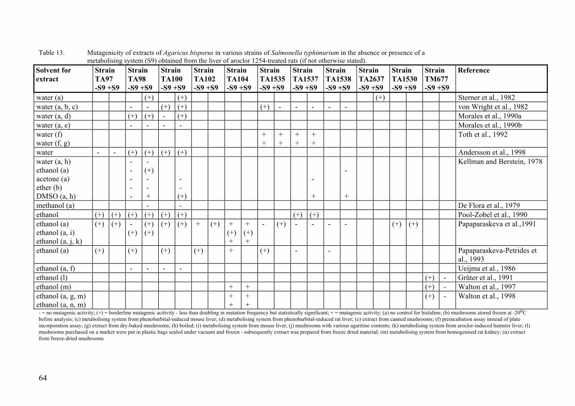
64
Table 13. Mutagenicity of extracts of Agaricus bisporus in various strains of Salmonella typhimurium in the absence or presence of a metabolising system (S9) obtained from the liver of aroclor 1254-treated rats (if not otherwise stated).
Solvent for extract
Strain TA97 -S9 +S9
Strain TA98 -S9 +S9
Strain TA100 -S9 +S9
Strain TA102 -S9 +S9
Strain TA104 -S9 +S9
Strain TA1535 -S9 +S9
Strain TA1537 -S9 +S9
Strain TA1538 -S9 +S9
Strain TA2637 -S9 +S9
Strain TA1530 -S9 +S9
Strain TM677 -S9 +S9
Reference
water (a) (+) (+) (+) Sterner et al., 1982 water (a, b, c) - - (+) (+) (+) - - - - - von Wright et al., 1982 water (a, d) (+) (+) - (+) Morales et al., 1990a water (a, e) - - - - Morales et al., 1990b water (f) water (f, g)
+ +
+ +
+ +
+ +
Toth et al., 1992
water - - (+) (+) (+) (+) Andersson et al., 1998 water (a, h) ethanol (a) acetone (a) ether (b) DMSO (a, h)
- - - - -
- (+) - - +
- - (+)
- +
- +
Kellman and Berstein, 1978
methanol (a) - - De Flora et al., 1979 ethanol (+) (+) (+) (+) (+) (+) (+) (+) Pool-Zobel et al., 1990 ethanol (a) ethanol (a, i) ethanol (a, j, k)
(+) (+) - (+)
(+) (+)
(+) (+) + (+) + (+) +
+ (+) +
- (+) - - - - (+) (+) Papaparaskeva et al.,1991
ethanol (a) (+) (+) (+) (+) + (+) - - Papaparaskeva-Petrides et al., 1993
ethanol (a, f) - - - - Ueijma et al., 1986 ethanol (l) (+) - Grüter et al., 1991 ethanol (m) + + (+) - Walton et al., 1997 ethanol (a, g, m) ethanol (a, n, m)
+ +
+ +
(+) - Walton et al., 1998
- = no mutagenic activity; (+) = borderline mutagenic acitivity - less than doubling in mutation frequency but statistically significant; + = mutagenic activity; (a) no control for histidine; (b) mushrooms stored frozen at -20oC before analysis; (c) metabolising system from phenobarbital-induced mouse liver; (d) metabolising system from phenobarbital-induced rat liver; (e) extract from canned mushrooms; (f) preincubation assay instead of plate incorporation assay; (g) extract from dry-baked mushrooms; (h) boiled; (i) metabolising system from mouse liver; (j) mushrooms with various agaritine contents; (k) metabolising system from aroclor-induced hamster liver; (l) mushrooms purchased on a market were put in plastic bags sealed under vacuum and frozen - subsequently extract was prepared from freeze dried material; (m) metabolising system from homogenised rat kidney; (n) extract from freeze-dried mushrooms

factor when testing such extracts for mutagenic activity. Although higher histidine con-centrations are found in water extracts than in alcohol extracts, Grüter and co-workers (1991) showed that the histidine level in ethanol extracts were high enough to more than double the spontaneous frequency of revertant colonies. The same conclusion has been drawn by Andersson et al. (1998) and suggested by Pool-Zobel and co-workers (1990). Of the twelve positive studies eight had not controlled for the histidine content of the extract (Kellman and Berstein, 1978; Sterner et al., 1982; von Wright et al., 1982; Morales et al., 1990a; Papaparaskeva et al., 1991; Papaparaskeva-Petrides et al., 1993; Walton et al., 1997a; Walton et al., 1998), and in four cases the detected mutagenic ac-tivity could very well be explained solely as an artefact due to histidine (Kellman et al., 1978; Sterner et al., 1982; von Wright et al., 1982; Morales et al., 1990a). Thus, there are a number of studies with Salmonella typhimurium in which the positive response of Agaricus bisporus extracts can not easily be explained as artefacts resulting from impu-rities of histidine in the mushroom extract.
Nearly all studies with mushroom extracts have used freshly prepared extracts, and these have seldom induced mutation frequencies higher than three times the control. Unexpectedly, the study that used the oldest extract (approximately 24 h old) and that accordingly had the highest risk that agaritine could be degraded, was the study that observed the strongest mutagenic activity: extracts from raw mushrooms increased the mutation frequency over the background approximately 19 times in strain TA1535 in the absence of a metabolising system and approximately five times in strain TA1537 (Toth et al., 1992). Even larger increases were observed in the presence of a metabolis-ing cytosolic system from aroclor-induced rat liver.
A mutagenic activity comparable to that of extracts from fresh A. bisporus was found in extracts from dry-baked (10 min 225oC) mushrooms (Toth et al., 1992). Amino acid analysis showed the extracts from unbaked mushrooms to contain 0.54 mg histidine per ml, while the equivalent value of the baked mushroom was 0.37. According to the au-thors, the addition of these amounts of free histidine is insignificant in the Ames assay (Toth et al., 1992).
The observation that extracts from dry-baked (10 min 225oC) mushrooms harbours a mutagenic activity of similar magnitude as extracts from fresh A. bisporus has recently been confirmed by Walton et al. (1998). A more prolonged baking at a lower tempera-ture, that is at 100oC for 4 hr, suppressed the mutagenic response of extracts. However, since these investigators evaporated the filtrate extract down to about 10% of its origi-nal volume, they not only concentrated the presumed mutagenic activity but also the co-extracted histidine, increasing the likelihood that the observed direct mutagenicity were an artefact due to the presence of histidine (Grüter et al., 1991). Inclusion of an activa-tion system comprising hepatic cytosol from Aroclor 1254-induced rats clearly induced a mutagenic response of extracts from raw as well as dry-baked (10 min at 225oC and 4 hr at 100oC) mushrooms (Walton et al., 1998). Also extracts from freeze-dried mush-rooms were reported to contain mutagenic activity but again the observed activity might be an artefact due to the presence of histidine.
Cooking Agaricus bisporus before preparation of mushroom extracts reduced the mutagenic activity of extracts more efficiently than dry-baking - boiling a water extract for 10 minutes reduced the volume of the extract by less than 10 % but the mutagenic activity by 50 % (von Wright et al., 1982).
65

Extracts of course contain a magnitude of different compounds. Their number and their quantity being dependent on the solvent used to prepare the extract. The contribution of agaritine to the mutagenic activity of ethanol extracts from Agaricus bisporus has been questioned by some investigators (Papaparaskeva et al., 1991; Papaparaskeva-Petrides et al., 1993). They have observed a lower mutagenic activity of agaritine than of mush-room extracts containing equivalent amounts of this compound. Furthermore, and in contrast to most other investigators, they have obtained a potentiation of the mutagenic-ity of mushroom extracts in the presence of rat metabolising systems (hepatic cytosolic fractions). However, they found no effect of the metabolising system on the mutagenic-ity of agaritine. The cytosol-induced increase in mutagenic activity was impaired by dicoumarol and menadione, an excellent substrate and a potent inhibitor, respectively, of the cytosolic enzyme DT-diaphorase. DT-Diaphorase is largely linked with the deac-tivation of quinones. The most susceptible Salmonella strain to the mutagenicity of mushroom extracts in this study was TA104, which is a strain particularly sensitive to reactive oxygen-generating species such as hydroperoxides and quinones. In agreement with the indication that the extract might contain oxidasing compounds, the investiga-tors found the mutagenicity of mushroom extracts to be reduced when the incubation mixture was supplemented with superoxide reductase, catalase, reduced glutathione or DMSO. To explain these results the investigators suggested that some of the mushroom phenol quinones are activated to genotoxic species by endogenous tyrosinase during the extraction procedure (Papaparaskeva-Petrides et al., 1993).
One explanation to these deviating observations might be that the mushroom extract contain chitin/chitosan that may chelate metal ions having a hydrogen peroxide (H2O2)-generating ability (Chung et al., 1998). If this is the case, a part of the mutagenic activ-ity of mushroom extracts could very well be due to reactive oxygen species.
Morales et al. (1990a) reported a slightly higher mutagenic activity in aqueous extracts prepared from previously frozen mushrooms (maintained at -20oC for 3 months) than in extracts prepared from fresh mushroom. This observation may suggest that chemical changes during freezing affect the structure and activity of some of the many com-pounds present in the mushroom flesh. Since freezing and thawing the cultivated mush-room have been reported to reduce the level of agaritine, also the observation of Morales et al. (1990a) could be interpreted as other compounds than the mushroom phenylhydrazines being responsible for a part of the mutagenic activity of mushroom extracts.
Grüter and co-workers (1991) are the only investigators that have used a Salmonella strain (TM677) which detects forward mutations and not reverse mutations to histidine prototrophy, to determine a mutagenic activity in Agaricus bisporus extracts. This strain detects mutations to 8-azaguanine resistance and is not sensitive to the presence of his-tidine in the tested extract. Studies with this strain revealed that extracts of fresh mush-rooms contained low but significant mutagenic activity in the absence of a metabolising system. No mutagenic activity was observed in the presence of a metabolising system or in extracts from canned mushrooms.
It should be mentioned that also extracts of A. silvaticus and A. arvensis, both growing in the Nordic countries and picked by mushroom hunters, have been reported to contain a mutagenic activity (Table 14). Extracts from another popular species among mush-room hunters, A. campestris, were void of such an activity. Neither of these studies have
66

67
Table 14. Mutagenicity of extracts of Agaricus species growing in the nature in various strains of Salmonella typhimurium in the absence or presence of a metabolising system (S9) obtained from the liver of aroclor 1254-treated rats (if not otherwise stated).
Solvent for extract
Strain TA97 -S9 +S9
Strain TA98 -S9 +S9
Strain TA100 -S9 +S9
Strain TA102 -S9 +S9
Strain TA104 -S9 +S9
Strain TA1535 -S9 +S9
Strain TA1537 -S9 +S9
Strain TA1538 -S9 +S9
Strain TA2637 -S9 +S9
Strain TA1530 -S9 +S9
Strain TM677 -S9 +S9
Reference
ethanol (a) ethylacetate (a) A. silvaticus
(+) -
+ -
(+) +
Sterner et al., 1982
ethanol (a) ethylacetate (a) A. arvensis
(+) -
+ -
- -
Sterner et al., 1982
water (a, b) A. campestris
- - - - Morales et al., 1990a
(a) no control for histidine; (b) metabolizing system from phenobarbital-induced rat liver.

controlled for the presence of histidine in the extracts (Sterner et al., 1982; Morales et al., 1990a).
Perhaps the strongest indication that mushroom extracts might contain mutagenic activ-ity comes from studies with purified compounds. When these were tested in the Salmo-nella mutagenicity assay, all hydrazine derivatives that have been shown to occur in Agaricus bisporus have been identified as mutagenic in at least one strain of S. typhi-murium (Table 15). In general, the mutagenic activity of these compounds were not influenced, or at least not increased, by a metabolising system prepared from livers of aroclor 1254-exposed rats. Furthermore, strains TA97 and TA1537 were most sensitive to these compounds, indicating a frameshift activity. 4-(Carboxy)-phenylhydrazine (CPH), a suggested precursor to agaritine, was tested at doses over 1 mg per plate but had the lowest mutagenic activity. Perhaps due to the high concentrations, CPH was very toxic in the absence of a metabolising system. Agaritine (100-2000 µg/plate) was weakly mutagenic in most Salmonella strains, whereas the 4-(hydroxymethyl)benzene-diazonium ion (HMBD; 5-135 µg/plate) was the most mutagenic compound of the Aga-ricus-hydrazines tested. When the bacteria were incubated with agaritine in the presence of γ-glutamyl transferase from bovine kidney, a pronounced increase in the mutagenic activity was obtained (Friederich et al., 1986; Walton et al., 1997a). A doubling in the mutation frequency induced by agaritine was also obtained in the presence of a kidney homogenate (Walton et al., 1997a). The increased mutagenic efficiency of agaritine in the presence of γ-glutamyl transferase (purified or in a kidney homogenate) could pos-sibly be a result of a continous production of HMBD when the glutamyl moiety is en-zymatically removed from agaritine. Incorporation of an incubation system consisting of rat hepatic microsomes fortified with an NADPH-generating system further potenti-ated the mutagenic respons of agaritine elicited in the presence of rat kidney homoge-nate (Walton et al., 1997a). The liver homogenate had no effect in the absence of the kidney homogenate, presumably because it is devoid of significant γ-glutamyl transpep-tidase activity.
These observations after co-incubation of Salmonella bacteria with agaritine and γ-glutamyl transferase contradict observations made in studies where bacteria were incu-bated with mushroom extracts and either γ-glutamyl transferase or a kidney homogenate containing this enzyme activity. In these later studied neither γ-glutamyl transferase, nor a kidney homogenate had any influence on the mutagenic activity of mushroom extracts (Pool-Zobel et al., 1990; Papaparaskeva et al., 1991; Walton et al., 1997a).
An increased mutagenic activity of agaritine in S. typhimurium (TA1537) was not only observed in the presence of γ-glutamyl transferase, but also after incubating the agari-tine-exposed bacteria at 25oC for 16 h in the presence of alkaline pH (pH 8.2 and 9.0), as compared to the directly determined agaritine mutagenicity at neutral and slightly acidic pH - pH 5 and 7 (Friedrich et al., 1986).
Besides agaritine, a weak mutagenic activity was also induced by the unstable 4-(hydroxymethyl)phenylhydrazine (HMPD), which have not yet been identified in Aga-ricus bisporus, and its synthetically stabilised form N'-acetyl-4-(hydroxymethyl)-phenylhydrazine (AHMPH; 100-1000 µg/plate), Table 10 (Friederich et al., 1986; Ro-gan et al., 1982).
68

69
Table 15. Comparative mutagenicity of the hydrazine derivatives occurring in Agaricus bisporus in various strains of Salmonella typhimurium in
the absence or presence of a metabolising system (S9) obtained from the liver of aroclor 1254-treated rats. Tested hydrazine derivatives were β-N-(γ-L(+)glutamyl)-4-hydroxymethyl)phenylhydrazine (agaritine), 4-(carboxy)phenylhydrazine (CPH), 4-(hydroxymethyl)phenylhydrazine (HMPH), N'-acetyl-4-(hydroxymethyl)phenylhydrazine (AHMPH) and 4-(hydroxymethyl)benzenediazonium ion (HMBD).
Compound tested Strain
TA97 -S9 +S9
Strain TA98 -S9 +S9
Strain TA100 -S9 +S9
Strain TA102 -S9 +S9
Strain TA104 -S9 +S9
Strain TA1535 -S9 +S9
Strain TA1537 -S9 +S9
Strain TA1538 -S9 +S9
Strain TA2637 -S9 +S9
Type of assay Reference
Agaritine (a) - - (+) (+) spottest De Flora et al., 1979 Agaritine (a) (+) (+) + (+) preincubation Rogan et al., 1982 Agaritine (a) (+) - - (+) (+) (+) - (+) - plate incorporation Papaparaskeva-Petrides et al.,
1993 Agaritine (b) - - - - (+) - plate incorporation Sterner et al., 1982 Agaritine (c) (+) (+) (+) (+) preincubation Ueijma et al., 1986 Agaritine (d) + - - - - (+) - preincubation Friederich et al., 1986 Agaritine (i) (+) plate incorporation Walton et al., 1997b Agaritine (d) + γ-GT
+ (+) + + - + + preincubation Friederich et al., 1986
CPH - - preincubation Malca-Mor and Stark, 1982 CPH + - - - - - - preincubation Friederich et al., 1986 CPH +* + -* (+) -* (+) plate incorporation Andersson et al., 1998 CPH (+) plate incorporation Walton et al., 1997b HMPH (e) - (+) + (+) preincubation Friederich et al., 1986 AHMPH (f) - - + - preincubation Rogan et al., 1982 AHMPH (j) (+) plate incorporation Walton et al., 1997b HMBD (g) (+)* + preincubation Rogan et al., 1982 HMBD (h) + + + + - + + preincubation Friederich et al., 1986 HMBD + plate incorporation Lawson et al., 1995 HMBD (i) + plate incorporation Walton et al., 1997b - = no mutagenic activity; (+) = borderline mutagenic acitivity - less than doubling in mutation frequency but statistically significant; + = mutagenic activity(a) agaritine synthesised by Toth and colleagues (>98 % pure); (b) agaritine purified from mushrooms; (c) origin of agaritine unknown; (d) isolated from A. bisporus, 76.5 % pure; (e) gift from Hoechst; (f) AHMPH synthesised by Toth and colleagues (>98 % pure); (g) HMBD synthesised by Toth and colleagues (>98 % pure); (h) synthesized; (i) agaritine synthesised by Walton and colleagues (91 % pure); (j) AHMPH synthesised by Walton and colleagues (97 % pure); * pronounced cytotoxicity

Urine collected from rats and mice during a single dose balance study with agaritine, and which contained agaritine metabolites, was assayed for mutagenicity in the Ames test using Salmonella typhimurium strains TA98, TA100 and TA104, with and without metabolic activation. No increase in mutation frequency above background was de-tected in any sample (Walker et al., 1997).
Only one short-term study have used other bacteria than Salmonella, and this study was negative. In the bioluminescence test 4-(carboxy)phenylhydrazine was unable to induce any revere mutations of Photobacterium leiognathi SD18 from dark variant to lumines-cent state (Levi et al., 1986).
It should be stressed that the observations reported in chapter 3 on differential stability of agaritine solutions under various conditions of storage may have a bearing on the interpretation of the mutagenicity data of agaritine reported above, and possibly also on the interpretation of the data obtained with mushroom extracts. Generally, investigators have not stated under which conditions and for how long agaritine solutions and mush-room-extracts have been stored. Thus, it is possible that in some studies the concentra-tion of agaritine present during treatment, if present at all, is substantially less than the concentrations reported.
8.4. Tests in cultured mammalian cells Cancer cells often become resistant to cytostatic drugs. One mechanism whereby resis-tance may develop is through amplification of genes important for cell proliferation. When tested at concentrations up to 25 µl per incubation mixture, ethanol- and ethy-lacetate-extracts of A. bisporus did not induce adeno-associated virus amplification in Syrian hamster embryo cells in vitro (Pool-Zobel et al., 1990). At higher concentrations the solvent (a 1:1 mixture of ethanol and DMSO) were toxic to the cells.
Similarly, no DNA single-strand breaks were observed in rat hepatocytes exposed for 1 h in vitro to extracts (ethanol/ethylacetate) derived from cooked or raw A. bisporus (Pool-Zobel et al., 1990). Neither was any genotoxic effect detected in cells cultured in the presence of mushroom extracts for longer periods of time (8-24 h).
In an investigation reported only in the form of an unpublished abstract, test substances in the form of fresh or canned A. bisporus were tested for their ability to induce gene mutations (6-thioguanine resistance) and sister chromatid exchanges in Chinese hamster ovary cells (Santa Maria and Caballo, 1989). No mutations were induced by canned mushrooms. Fresh mushrooms were not tested in this assay. Both fresh and canned mushrooms stimulated the induction of sister chromatid exchanges. However, it is ques-tionable whether the increase in sister chromatid exchange frequency observed in this study is of any biological significance. Furthermore, canned mushrooms were more efficient than fresh mushrooms to induce sister chromatid exchanges.
In a study exploring the possibility to use cell culture techniques to determine toxicity and antitoxicity of test compounds, water- and methanol-extracts of A. bisporus were found to be moderately or weakly active in three assays (inhibition of protein synthesis, MMT conversion to formazan and Kenacid blue staining), and without effect in a fourth (release of lactate dehydrogenase into the culture media) (Phillips, 1996).
Very few short-term studies with pure phenylhydrazines or the 4-(hydroxymethyl)-benzenediazonium ion have been performed in cultured mammalian cells. In one study,
70

the 4-(hydroxymethyl)benzenediazonium ion was found to induce gene mutations (thioguanine resistance) in V79 cells (a lung fibroblast cell line derived from the Chi-nese hamster) in a concentration-dependent manner (the concentrations varied between 10 and 100 µM) (Lawson et al., 1995). In another study, only reported in the form of a short abstract, agaritine, 4-(hydroxymethyl)benzenediazonium tetrafluoroborate and N'-acetyl-4-(hydroxymethyl)phenylhydrazine did not induce any mutations to ouabain re-sistance in V79 cells in the absence or presence of metabolically competent liver cells (Kuszynski et al., 1981). Since the concentrations of the various hydrazine derivatives used in these experiments were not stated, it is possible that the contradictory results obtained with the diazonium ion in the studies of Lawson et al. (1995) and Kuszynski et al. (1981) either is due to the slightly different experimental conditions of the two stud-ies or to different concentrations of the diazonium ion being used by the two groups of investigators.
The hepatocyte/DNA repair test, which measures unscheduled DNA synthesis, is known to be sensitive to various classes of DNA-reactive carcinogens, and is regarded as a suitable short-term test for in vitro detection of chemical carcinogens. The test combines intact cell metabolism with the broadest capability for xenobiotic biotrans-formation (isolated primary hepatocytes), together with a biochemical endpoint for genotoxicity. Whereas agaritine (10-1000 µM) neither elicited unscheduled DNA syn-thesis in rat hepatocytes nor in mouse hepatocytes, N'-acetyl-4-(hydroxymethyl)phenylhydrazine was positive in both types of hepatocytes (Mori et al., 1988). However, the magnitude of the positive response was low compared to that of other types of carcinogenic chemicals.
8.5. In vivo tests Recently, Big Blue transgenic mice were used to study the mutagenic activity of A. bis-porus in vivo (Shepard et al., 1995). These mice (a strain derived from C57Bl6 mice) bear the bacterial lacI gene, incorporated into a lambda bacteriophage shuttle vector, stably inserted into the DNA of every cell. Following dietary exposure to fresh mush-rooms, dried mushrooms and mushroom extracts enriched in agaritine (30 %, w/w), the shuttle vector was specifically isolated from genomic DNA of forestomach, liver, lung, kidney and glandular stomach tissue, and the marker gene expressed in E. coli bacteria. Material from forestomach, liver and lung were selected for analysis because they were target organs in the fresh mushroom carcinogenicity study (Toth and Erickson, 1986). Kidney was examined because the tissue level of γ-glutamyl transferase, the first en-zyme in the postulated bioactivation pathway of agaritine, is highest in this organ. The glandular stomach was examined in some studies for comparison with forestomach re-sults. Mutations in the lacI gene of the five tissues were detected with a colour test. On the basis of previous work with other compounds correlating carcinogenic potency with results of the lacI in the transgenic mouse, a 15-week exposure period was predicted to produce a significant increase in mutation frequency in target organs, if the tumours observed in Toths study (Toth and Ericson, 1986) were indeed due to a genotoxic sub-stance(s).
An obvious weaknesses of the study was the low number of animals used, only 2 mice per treatment. Dietary administration of crude agaritine (75 % pure) in the feed, result-ing in an exposure of 120 mg/kg body weight/day, increased the mutation frequency by
71

100 % in the kidney and by 50 % in the forestomach. The agaritine-feed did not influ-ence the mutation frequency in the liver and lungs, whereas a significant decrease in mutation was observed in the glandular stomach. The possible antimutagenic effect of agaritine in the glandular stomach was not pursued further. Mice receiving fresh or dried mushrooms in the diet (30 and 80 mg agaritine/kg body weight/day, respectively) showed no increase in mutant frequency in any of the target organs.
It is not easy to compare the carcinogenic potency of different carcinogens in experi-mental systems. One method used in studies on transgenic rodents is based on the TD50 doses (the daily dose rate that produced a 50 % increase in tumours over controls after lifelong exposure). When the increased mutation frequency induced by feeding trans-genic mice 120 mg agaritine/kg body weight/day were compared with the mutation fre-quency induced by TD50 doses of various organ-specific carcinogens in mice, the TD50 for agaritine was estimated to approximately 250 mg/kg body weight/day, putting agari-tine into the class of weak genotoxic carcinogens (Shepard et al., 1995). It should be emphasised that doses above 100 mg agaritine/kg body weight/day were precluded in a 2-year feeding study due to toxicity (Toth et al., 1981a), see section 9.2.2.1. The im-provement in sensitivity offered by the transgenic methods in comparison with tradi-tional carcinogenicity studies lies in the fact that a gene, rather than the animal itself, is the unit of observation, and that mutations in the target gene can accumulate over the whole treatment period (Shepard et al., 1995). However, a gene mutation is not equiva-lent to the induction of a tumour.
Pool-Zobel and co-workers (1990) have tested mushroom extracts in two other in vivo assays. In one of these they investigated the induction of DNA single-strand breaks in liver cells of male Sprague Dawley rats at 1h after an oral exposure to ethanol extracts of fresh A. bisporus purchased at the local market. This endpoint could be viewed as a secondary effect to primary DNA damage. No single-strand DNA breaks were detected in this experiment. In the other study male C57BL/JC mice (5 animals per group) were dosed with the same type of extracts (corresponding to 13.25 g mushroom/kg body weight) and killed 24 hours later. The bone marrow was prepared and the incidence of micronucleated polychromatic erythrocytes determined. The extract had no influence on the frequency of micronuclei in bone marrow cells of. The latter experiment had two weaknesses which makes it difficult to interpret the results. Firstly, the extract was an ethanol solution, whereas the control solution was distilled water. Secondly, only one (and a very early) fixation time was used, which increases the likelihood of missing a potential effect.
However, in agreement with the German study, investigators at the National Food Ad-ministration i Sweden found water extracts of A. bisporus (corresponding to approxi-mately 14.5 mg agaritine/kg body weight) intraperitoneally injected into 6 weeks old NMRI male mice not to induce micronuclei in polychromatic bone marrow erythrocytes (Andersson et al., 1998). In the same study 200 mg/kg body weight 4-(carboxy)phenylhydrazine, dissolved in DMSO, had no influence on the frequency of micronucleated polychromatic erythrocytes, although there was an indication of an in-creased micronucleus incidence in this experiment. It was concluded that the potential of 4-(carboxy)phenylhydrazine to induce micronuclei in vivo should be investigated further.
Japanese investigators have studied the influence of the antioxidant status of the mouse feed on the efficiency of micronuclei induction in peripheral reticulocytes of male ICR
72

mice intraperitoneally injected with the 4-(hydroxymethyl)benzene-diazonum ion, HMBD (Kato et al., 1995). The antioxidant status of the feed was regulated by its vita-min E content. The vitamin E level in the blood of mice fed vitamin E-deficient diet for a week was significantly lower than that in mice fed a diet adequate in vitamin E. Inde-pendently on the type of mouse feed given to the animals (10 animals per group), a sin-gle administration of 90 mg HMBD/kg body weight resulted in a significant induction of micronuclei in peripheral reticulocytes, particularly at 48 h after injection of the test-compound (Kato et al., 1995; Hiramoto et al., 1995a). At this HMBD concentration, feeding a vitamin E deficient diet resulted in a significantly (p<0.05) enhanced micro-nucleus frequency as compared to the frequencies observed in animals fed vitamin E adequate diet. These studies indicate that the HMBD ion gives rise to chromosome damage and that vitamin E deficiency leads to a reduction in protective ability against chromosome damage.
8.6. Antimutagenic effects of A. bisporus Agaricus bisporus does not only contain mutagens, but also antimutagens. For example, there is one report claiming that ethanol extracts of A. bisporus and A. abruptibulvus inhibits the mutagenesis of 2-nitrofluorene in strain TM677 of Salmonella typhimurium (Grüter et al., 1990). To what extent these antimutagenic compounds in mushroom ex-tracts masks an effect of mutagenic compounds is not known.
8.7. Conclusions on effects in short-term tests The 4-(hydroxymethyl)benzenediazonium ion (HMBD) is a reactive compound be-lieved to be produced from agaritine in the mushroom or when agaritine is metabolised by γ-glutamyl transpeptidase in mammals. The HMBD is metabolically converted to two types of radical species, aryl diazenyl radicals and aryl radicals, that may react with carbon atoms of deoxyribose units in DNA, and nitrogen atoms and the C-8 position of purines to create specific types of DNA damage (see, Figure 9). DNA damage has been observed in various tissues of mice orally exposed to HMBD. Also in vitro studies have revealed that HMBD may induce DNA damage in deoxyribonucleotides, isolated calf thymus DNA, and cultured mammalian cells.
Also agaritine and/or its metabolites have been shown to bind covalently to liver, kid-neys and stomach DNA of mice exposed in vivo. The binding was five times higher in males than in females.The highest specific activity was observed in the stomach, the first site of contact between agaritine and the tissues.
In vitro studies have been performed with the bacterium Salmonella typhimurium and with cultured mammalian cells. The bacterial tests (Ames assay) have revealed a weak mutagenic activity of water- and alcohol-extracts of A. bisporus. Available information indicate that the mutagenic activity could be due to the phenylhydrazines (4-(carboxy)phenylhydrazine, agaritine, 4-(hydroxymethyl)phenylhydrazine) and the 4-(hydroxymethyl)benzenediazonium ion, and/or to other hitherto unidentified com-pounds occurring in the mushroom. The latter compounds could possibly be quinone derivatives or reactive oxygen species.
The interpretation that mushroom hydrazines may partly be responsible for the genotoxic effect of mushroom extracts is strengthened by the observation that purified
73

mushroom hydrazines, including agaritine and the 4-(hydroxymethyl)benzenediazonium ion, give rise to mutations in the Salmonella assay. It should also be noted that the mutagenic activity of agaritine was increased upon removal of the glutamyl moiety by γ-glutamyl transpeptidase.
The few in vitro studies that have been performed with cultured mammalian cells are generally of poor quality and can not be used independently to establish whether ex-tracts of A. bisporus or the purified mushroom hydrazines are mutagenic.
The in vivo studies on mice have produced contradictory results. Whereas extracts of A. bisporus had no influence on the frequency of micronuclei in bone marrow cells of male mice in two separate experiments, other in vivo studies on mice revealed that agaritine induced gene mutations and the 4-(hydroxymethyl)benzenediazonium ion micronuclei in peripheral reticulocytes. In addition, one study indicated that intraperitoneally in-jected 4-(carboxy)phenylhydrazine might induce micronuclei in polychromatic bone marrow erythrocytes, but this possibility has to be investigated further.
If HMBD is continuously produced from agaritine in the cultivated mushroom or by the internal metabolism in humans consuming Agaricus bisporus, any of the lesions formed by the reaction of HMBD with DNA might be genotoxic, and possibly stimulate cancer induction.
74
9. Effects on experimental animals
9.1. Acute and subchronic toxicity of mushroom hydrazines and HMBD
The first toxicity study with the phenylhydrazines and the 4-(hydroxymethyl)-benzenediazonium ion (HMBD) occurring in Agaricus bisporus dates back to 1924. In these early studies Bodansky compared the severity of anemia and liver damage in-duced by hydrazine with that induced by various hydrazine derivatives in the dog and the rabbit. Unlike hydrazine and phenylhydrazine, 4-(carboxy)phenylhydrazine had marginal effects on both endpoints. No serious liver damage as determined by histologi-cal examination and measurement of blood suger levels could be detected in a female dog injected twice, two days apart, with 0.3 g 4-(carboxy)phenylhydrazine. Also a sub-sequent administration of 2.2 g of this compound did not produce any detectable effects (Bodansky, 1924a). The weight of the dog was not given, but the total dose would be around 280 mg/kg body weight for a dog weighing 10 kg. Similarly, four subcutaneous injections with 0.025, 0.1, 0.3 and 1.0 g 4-(carboxy)phenylhydrazine in a rabbit (total dose 790 mg/kg body weight) only resulted in a marginal reduction in red blood cell count and blood hemoglobin level (Bodansky, 1924b). The author suggested that 4-(carboxy)phenylhydrazine is easily detoxified in the animal body.
When it some years later was observed that compounds with hydrazine groups may hin-der the formation of tuberculous lesions, Minami (1933) studied the effect of exposing experimental animals to 4-(carboxy)phenylhydrazine and more than ten other hydrazine derivatives regarding the degree of anemia produced by the treatment and what influ-ence it had on the oxygen levels in blood. In these studies male guinea pigs (350 g) were subcutaneously injected every second day, for two weeks, with aqueous solutions of 4-(carboxy)phenylhydrazine, the cumulative dose for the two dose groups being around 410 and 820 mg per animal, respectively. The body weight of animals injected with both doses increased during the experiment, but the magnitude of the increase was higher for those that received the lower dose. No toxicity was registered by visual in-spection. No splenic enlargement was found at biopsy, neither was anemia found in other organs, although such autopsy findings were recorded in animals exposed to other hydrazine derivatives. The exposure had no influence on the number of blood corpus-cles, red blood cells and amount of hemoglobin. No control animals were used in this study. Instead, the values registered in the animals before the beginning of the treatment was used as control values. The conclusion was that 4-(carboxy)phenylhydrazine nei-ther induced anemia, nor any other observable toxicity in male guinea pigs. When it comes to predicting the risk for humans consuming A. bisporus, the relevance of these toxicity studies that administered the test compounds by subcutaneous injection is un-known.
However, toxicity studies (28 days long) have also been performed in connection with carcinogenicity studies, mainly to determine the maximum tolerated dose of a chemical, which could then be given for life. In these tests, four parameters for toxicity; survival
75

rates, body weights, chemical consumption levels (in drinking water or feed), and histo-logical changes, were used to assess toxicity (Toth et al., 1975). Table 16 summarises the toxicity data obtained in such dose-setting experiments. In the studies with subcuta-neously injected agaritine (isolated from A. bisporus) the highest doses were 200 and 400 mg/kg body weight. Mice treated with these doses clinically showed excitation, uncoordinated runs, convulsions, paralysed lower backs and ultimately died. Histopa-thological findings included vacuolated cells in the liver and kidney tubules, edema and desquamation of bladder epithelium and hyperemia and haemorrhages of the lungs (Toth and Sornson, 1984). The highest dose of N2-[γ-L(+)-glutamyl]-4-carboxyphenyl-hydrazine tested in the dose-setting experiment, 2 400 mg/kg body weight, was non-toxic in Swiss mice when given as a single intragastric instillation. A toxic dose level can, therefore, not be given for this compound. In addition to the observations from dose-setting experiments reported for Swiss mice in Table 16, Pilegaard and co-workers (1997) have found 1 550 mg 4-(carboxy)phenylhydrazine/kg drinking water to be toxic in A/J mice (a mouse strain easily developing lung tumours).
Table 16. Toxic doses of various phenylhydrazines and the 4-(hydroxymethyl)-
benzenediazonium ions occurring in A. bisporus, as determined by a set of criteria in Swiss albino mice (Toth et al., 1975).
Compound Administration Toxic dose level Reference 4-(carboxy)phenyl-hydrazine
In drinking water for 35 days
2.5 g/l McManus et al., 1987
Agaritine A single intragas-tric instillation
625 mg/l; single administration
Toth et al., 1981a
Agaritine Subcutaneous injec-tion
200 mg/g body weight for females; 100 mg/g body weight for males; 28 days
Toth and Sornson, 1984
4-(hydroxymethyl)-benzenediazonium ion, sulfate form
A single subcutane-ous injection
100 µg/g body weight
Toth, 1987
4-(hydroxymethyl)-benzenediazonium ion, tetrafluorobo-rate form
A single subcutane-ous injection
50 µg/g body weight
Toth et al, 1981b
4-(hydroxymethyl)-benzenediazonium ion, tetrafluoroborate form
A single intragatric instillation
450 µg/g body weight
Toth et al, 1982
76
9.2. Long-term carcinogenicity studies. The observation that chemotherapeutic agents containing a nitrogen-nitrogen bond in-duce tumours in experimental animals, has stimulated a systematic search for tumouri-genic hydrazine derivatives. This search did not focus only on the synthetic hydrazine derivatives used in industry, agriculture, and medicine, but also included hydrazine de-rivatives occurring naturally in tobacco, ”edible” mushrooms and bay leaves. Today alkyl-, aryl-, and acyl-substituted hydrazines have been identified as tumour-inducing substances in many different types of tissue in mice, hamsters and rats.
In the middle of the seventies Toth and his co-workers at the Eppley Institute for Re-search on Cancer, University of Nebraska Medical Center, initiated a concentrated ef-fort to reveal the possible significance of hydrazines in the causation of dietary cancer (Toth, 1975). Toth and his co-workers have concentrated on the substances present in the False Morel (Gyromitra esculenta) and the cultivated mushroom (Agaricus bis-porus).
The compounds of interest in Agaricus bisporus are agaritine, its presumed precursors 4-(carboxy)phenylhydrazine and β-N-(γ-L(+)-glutamyl)4-(carboxy)phenylhydrazine, as well as the presumed degradation products 4-(hydroxymethyl)phenylhydrazine and the 4-(hydroxymethyl)benzenediazonium ion.
Table 17A and 17B shows the experimental results obtained when these compounds, and the fresh and dry-baked cultivated mushroom have been tested for their ability to induce tumours in mice: the tumour types that were found in statistically increased fre-quencies (indicated by *, ** or ***) or that were most common are given, as well as the number of tumour-bearing animals and the tumour incidences. In many cases one ani-mal carried more than one tumour, but the multiplicity of tumours is not given in the table. If an animal were found to have both benign and malignant tumours, it is only counted in the group of animals bearing malignant tumours. The malignant tumours are identified by italics in Fig. 17A and 17B. In addition to the 13 published long-term stud-ies on mice (all of which are presented in Table 17A and 17B), two rather poor studies with oral administration of cooked and dried cultivated mushroom in rats have been published.
In the standard design of the carcinogenicity studies performed by Toth and co-workers at the Eppley Institute for Research on Cancer, the tested compound was dissolved in the drinking water at concentrations that had been determined from toxicity studies per-formed prior to the chronic experiment. The solution was prepared trice weekly and protected from light, and the total consumption of drinking water measured at the same intervals during the treatment period. Each concentration tested was given for the life-span of 50 males and 50 females randomly bred Swiss albino mice that were 6 weeks old at the beginning of the experiment. Thus, in contrast to many other long-term car-cinogenicity studies, the animals were not killed after a definite treatment period or at a definite percentage of survivors in the experiment. If not otherwise stated, the control group was made up of the same number of males and females as the exposed groups, and the animals were of the same age at the beginning of the experiment as the animals in the exposed groups. However, in a substantial number of experiments the control group was not started at the same time as the treated groups. A control group with a particular route of administration and a fixed number of animals were used as control group for all long-term studies that were started with the same type of dosing regimen
77

as long as at least 50% of the original number of animals in the control group was still living. When 50% of the animals in the control group had died a new control group was started and all studies that were started from this point in time would have the new group as the control group. As a consequence, the time span between starting the con-trol group and the treated groups could be as long as around 1.5 years (Gry and Anders-son, 1998).
The treated and the control animals were carefully checked and weighed at weekly in-tervals, and gross pathologic changes recorded. The animals were either allowed to live until they died by themselves, or were killed with ether when found in poor condition. Complete necropsy and histological examinations were performed on all animals. Sta-tistical analysis on tumour induction was carried out by using Fisher´s exact test for 2x2 tables (Armitage, 1971) on the sum of animals with benign and malignant tumours. In the tables, the symbols *, **, and ***, represents the statistical significance, e.g. the probability that chance alone had produced a difference in tumour incidence between treated and control groups larger than 0.05, 0.01 and 0.001, respectively. Usually, no statistical analysis was carried out on animals with specific benign (normal letters in Table 17A and 17B) and malignant (italics in Table 17A and 17B) tumours.
Though the essential information is included in Tables 17A and 17B, the following text will summarise the findings in the separate experiments for the interested reader. The general experimental design in studies by Toth and co-workers will not be reported again when the respective studies are discussed. The reader is referred to the original paper to get information on the histological characterisation of the neoplastic altera-tions.
9.2.1. Studies on the cultivated mushroom (Agaricus bisporus). In 1986, Toth and Erickson published the results of a study in which groups of 50 fe-male and 50 male Swiss albino mice were fed uncooked Agaricus bisporus ad libitum for 3 days, followed by semisynthetic diet for 4 days each week for their life span. Con-trol animals were fed the semisynthetic diet. The study lasted until all mice had died or had been killed with ether when found in poor condition; that is, between 150 and 160 weeks after the start of the experiment.
The estimated average daily mushroom consumption was 11 g per mouse for both sexes, which seems rather high. The authors emphasised that the figure takes into ac-count the fact that, on the average, 48% of the fresh mushroom is dehydrated in 24 hours at room temperature. Since no specific information was given, this intake presu-maly was an average over the entire experimental period. The mushrooms used were purchased twice weekly locally. There was no indication that the contents of agaritine or other phenylhydrazine derivatives were measured in the mushrooms during the study.
The treatment had no substantial effect on the survival rate as compared with that of the untreated controls. However, the average weekly weight curves of the animals given mushroom supplementation were substantially lower throughout the study than that of the untreated animals. The dose of A. bisporus given to the animals in the chronic ex-periment had been selected from an initial toxicity study in which mice were fed the mushroom ad libitum for 4.5, 4, 3.5, 3 and 2.5 days each week followed by semisyn-thetic diet for the rest of the week for a total of 70 days. The feeding did not result in a weight gain for all dose groups in this toxicity study. Because the body weight of mice
78

treated with A. bisporus for 3 days increased steadily, with the exception of an initial short decrease, this dose regimen was selected for the chronic experiment.
As shown in Table 17A, feeding of uncooked mushrooms to mice induced a statistically significant increase in the total number of tumours of bone and forestomach in both sexes and of lung tumours in the males (Toth and Erickson, 1986). An increase in tu-mours of the liver was suggestive but not statistically significant (p<0.07 for the females and p<0.06 for the males).
Recently, Toth and co-workers have presented data from two carcinogenicity studies with dry-baked cultivated mushrooms. The A. bisporus mushroom is mainly eaten in baked form (pizza) in the United States. The baking was done at 220-230oC for 10 min-utes. In one of the studies the baked A. bisporus mushroom was fed to Swiss mice (50 females, 50 males) for 3 days followed by a semisynthetic diet for 4 days each week for life. The mushroom consumption was 14.7 g per day per female and 13.0 g per day per male when the age of the mice were 57-60 weeks. The same number of animals in the untreated control group were fed the semisynthetic diet.
Though the survival were good in both groups (information on weight gain of animals missing), mice that had received the interrupted lifelong oral administration of baked A. bisporus feed developed statistically increased incidences of tumours in the forestom-ach, glandular stomach, duodenum and ovary (Table 17A). In the females, the inci-dences were 20, 12, 14 and 12 %, while in males they were 16, 20 and 4% in the rele-vant tissues (the last two non-significant). In the untreated control only an ovarian tu-mour (incidence 2%) was detected (Toth et al., 1997a).
In another experiment with baked A. bisporus mice were fed the mushroom for 12 hours each day for 5 days each week for life (Toth et al., 1997b). After each feeding cycle, the animals receive a well-balanced semisynthetic diet for 12 hours each day for 5 days, plus the remaining 2 days each week. Consumption data were generated during one month when the mice were 57-60 weeks old. The females consumed 4.8 g baked A. bis-porus per day and the males 4.2 g per day. In this experiment with baked A. bisporus, as well as in the experiment reported above (Toth et al., 1997a), treatment did not influ-ence the survival negatively. Information on weight gain of the animals is missing. The tumour incidences in lungs, blood vessels, cecum, and colon were increased in the group fed the baked mushroom as compared to the incidence in the untreated control group. However, none of the increases were statistically significant (Table 17A).
Toth and co-workers (1998) have reported that Swiss mice fed for life with a semisyn-thetic diet mixed with 10%, 5% or 2.5% lyophilised A. bisporus powder had similar survival rates as animals fed a semisynthetic diet non-substituted with lyophilised mushrooms. However, no information on weight gain of the animals was given. The mice given the mushroom-containing feed developed tumours in the lungs, forestom-ach, glandular stomach, and ovaries in certain exposure groups. Some of the tumour incidences in mushroom-fed mice were found to be significantly increased (10%-dose: females-ovaries 22%, males-lungs 60%; 5%-dose: females-glandular stomach 12% and ovaries 8%; 2.5%-dose: males and females-forestomach 10% and 10%, respectively), although no dose-response relationship was established (Table 17A). According to some estimates, less than 10% of the A. bisporus is consumed in lyophilised form in the United States.
79

80
Table 17A. Tumourigenicity of the cultivated mushroom (Agaricus bisporus) in randomly bred Swiss albino mice. Compound tested
Route of admini-stration
Dose Exposure Effect of treatment treated animals control animals males females males females (50/group) (50/group) (100/group)(100/group)
Reference
Agaricus bisporus uncooked
p.o. 3 days per week
lifelong 31 (62%)** 18 13 6 (12%) 6 14 (28%)** 11 3 8 (16%)** 5 3
20 (40%) 9 11 4 (8%) 4 19 (38%)*** 16 3 8 (16%)** 6 2
lung adenomas adenocarcinomas liver benign hepatomas forestomach squamous cell papillomas squamous cell carcinomas bone osteomas osteosarcomas
17 (38%) 8 9 1 (2%) 1 2 (4%) 2 0 0 (0%) 0 0
13 (26%) 6 7 0 (0%) 0 0 (0%) 0 0 0 (0%) 0 0
Toth and Erickson, 1986
Agaricus bisporus dry-baked
p.o. 3 days per week
lifelong 8 (16%)*** 7 1 0 10 (20%)*** 0 10 2 (4%) 2
10 (20%)** 9 0 1 6 (12%)* 1 5 7 (14%)* 7 6 (12%)* 5 1
forestomach squamous cell papillomas leiomyocarcomas squamous cell carcinomas glandular stomach polypoid adenomas adenocarcinomas duodenum adenocarcinomas ovaries adenomas adenocarcinomas
0 (0%) 0 0 0 0 (0%) 0 0 0 (0%) 0
0 (0%) 0 0 0 0 (0%) 0 0 0 (0%) 0 1 (2%) 0 1
Toth et al., 1997a
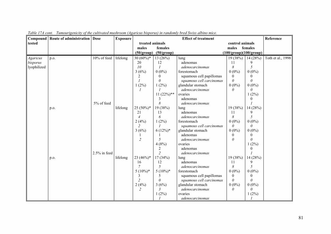
81
Table 17A cont. Tumourigenicity of the cultivated mushroom (Agaricus bisporus) in randomly bred Swiss albino mice. Compound tested
Route of administration Dose Exposure Effect of treatment treated animals control animals males females males females (50/group) (50/group) (100/group)(100/group)
Reference
Agaricus bisporus lyophilized
p.o. p.o. p.o.
10% of feed 5% of feed 2.5% in feed
lifelong lifelong lifelong
30 (60%)* 20 10 3 (6%) 2 1 1 (2%) 1 25 (50%)* 21 4 2 (4%) 2 3 (6%) 1 2 23 (46%)* 16 7 5 (10%)* 3 2 2 (4%) 2
13 (26%) 12 1 0 (0%) 0 0 1 (2%) 1 11 (22%)** 3 8 19 (38%) 13 6 1 (2%) 1 6 (12%)* 1 5 4 (8%) 2 2 17 (34%) 12 5 5 (10%)* 5 0 3 (6%) 3 1 (2%) 1
lung adenomas adenocarcinomas forestomach squamous cell papillomas squamous cell carcinomas glandular stomach adenocarcinomas ovaries adenomas adenocarcinomas lung adenomas adenocarcinomas forestomach squamous cell carcinomas glandular stomach adenomas adenocarcinomas ovaries adenomas adenocarcinomas lung adenomas adenocarcinomas forestomach squamous cell papillomas squamous cell carcinomas glandular stomach adenocarcinomas ovaries adenocarcinomas
19 (38%) 11 8 0 (0%) 0 0 0 (0%) 0 19 (38%) 11 8 0 (0%) 0 0 (0%) 0 0 19 (38%) 11 8 0 (0%) 0 0 0 (0%) 0
14 (28%) 9 5 0 (0%) 0 0 0 (0%) 0 1 (2%) 0 1 14 (28%) 9 5 0 (0%) 0 0 (0%) 0 0 1 (2%) 0 1 14 (28%) 9 5 0 (0%) 0 0 0 (0%) 0 1 (2%) 1
Toth et al., 1998

82
Table 17A cont. Tumourigenicity of the cultivated mushroom (Agaricus bisporus) in randomly bred Swiss albino mice. Compound tested
Route of admini-stration
Dose Exposure Effect of treatment treated animals control animals males females males females (50/group) (50/group) (100/group)(100/group)
Reference
Agaricus bisporus dry-baked
p.o. 12 h a day for 5 days per week
lifelong 26 (52%) 17 9 4 (8%) 2 2 9 (18%) 9 3 (6%) 2 1
17 (34%) 8 9 12 (24%) 3 12 6 (12%) 6 1 (2%) 1 0
lung adenomas adenocarcinomas blood vessels hemangiomas hemangiosarcomas cecum polypoid adenomas colon polypoid adenomas adenocarcinoma
19 (38%) 11 8 3 (6%) 2 1 4 (8%) ? 0 (0%) 0 0
14 (28%) 9 5 7 (14%) 5 2 4 (8%) ? 0 (0%) 0 0
Toth et al., 1997b
In addition to these more or less criticised long-term studies on mice, two rather poor long-term studies on rats have been published. The first of these were intended to com-pare the wholesomeness of irradiated Agaricus bisporus with that of non-irradiated Agaricus bisporus, but data from the control groups in this experiment gives some in-formation on the effect of consuming cooked Agaricus bisporus (van Logten et al., 1971). In this study 4 groups of 10 female and 10 male Wistar rats, weanlings of treated parents or controls, were fed on a standard diet (control group), on a diet containing 25% cooked non-irradiated mushrooms (a second control group) or on a diet containing 25% cooked mushrooms previously irradiated with γ-rays (350 krad) or with electrons (500 krad). The mushroom diet was prepared by cooking washed mushrooms (1600 g) for 2.5 minutes in a domestic pressure cooker, after which the mushrooms were ho-mogenised in the cooking liquid, made up to 2 litre with tap water, mixed with the nor-mal standard diet, pelletised and oven-dried (60oC). Food and drinking water were given ad libitum.
The study lasted for up to 112 weeks, at which time over 50 % of the animals were still alive. There was no difference in survival between experimental and control groups. The food intake and the average daily intake of mushrooms were not recorded, and the phenylhydrazine content in the feed not determined. The method of producing the feed should have reduced the phenylhydrazine level substantially in comparison to the level that would have been expected in a feed produced in a similar way from uncooked cul-tivated mushroom. No evidence was found that a feed containing 25% irradiated or 25 % non-irradiated mushrooms had any effect on the growth of the rats. Neither did the treatment induce tumours.
The value of this study when it comes to evaluating the effect of A. bisporus consump-tion is questionable. The authors had originally planned the experiment as a reproduc-tion study, but as the animals had already been on the mushroom diet for 1 year when the breeding was finished they were kept for another year. The female rats were used for breeding five litters during the study and the parents of the treated group had been treated as well. Comparison of the results of the first 90-day test with those of the sec-ond, carried out with animals of the F3 generation, indicates that the feeding of irradi-ated mushrooms during four generations was without effect (van Logten et al., 1971). The experimental groups (treated as well as control groups) consisted of only 10 male and 10 female rats, and the diets in the respective groups were not isocaloric.
The second long-term study on rats were performed with twenty 50-day-old female Sprague-Dawley rats (from Charles River) that for 500 days (1.37 years) - until the ter-mination of the experiment - were fed a pelletised diet containing 30 % A. bisporus (Matsumoto et al., 1991). The diet was prepared by mixing milled cultivated mushroom that had been dried at room temperature with a powdered basal diet. Twenty-four con-trol animals were feed the basal diet only. Water and feed were given ad libitum. Al-though all animals survived until the end of the experiment, the average body weight gain of rats receiving cultivated mushroom in the diet were not as pronounced from week 30 and onwards as that of the control rats. The difference in body weight between the two groups 40 weeks after the start of the experiment were statistically significant. In the 20 rats fed the A. bisporus diet, mammary tumours developed in 6 rats, thymoma adenoma in 1 rat, adrenal adenomas in 3 rats and pituitary adenoma in 1 rat. In the con-trol group of 24 rats the corresponding tumour numbers were 12, 0, 4 and 1, respec-tively. Thus, no significant difference was observed in the incidence of these tumours
83

between the A. bisporus fed group and the control group. The study was performed with only one sex (female rats), was based on to few animals, and was to short to make it possible to evaluate A. bisporus for carcinogenicity. Furthermore, the investigators did not determine the influence of the drying procedure of the prepared feed on the agaritine level in the consumed product.
I addition to the described long-term carcinogenicity studies, Danish investigators have studied whether A. bisporus is able to induce lung adenomas in A/J mouse, a short-term carcinogenicity model for lung tumours (Pilegaard et al., 1997). Only female mice were used in the study and the exposure period was 26 weeks. And only 15 animals were used per treatment group. The mice were fed lyophilised A. bisporus in the semisyn-thetic diet; 11% and 22% of the diet was replaced by freeze-dried mushrooms. The in-take of the mushroom diets were equivalent to an intake of 92 or 166 mg agaritine per kg body weight per day. The exposure did not influence the frequency of lung adeno-mas in this study. The specificity of the assay is good, but false-negative results are not uncommon because of weak sensitivity (Griesemer, 1991).
It is well known that certain lectins derived from beans belonging to genus Phaseolus are toxic to animals and human beings fed with either raw bean or the purified lecin. The bean lectins impair protein digestion and cause inadequate absorption of nutrients, particularly amino acids, thus resulting in protein malnutrition and, occasionally, death. The fact that also A. bisporus contain lectins may influence the interpretation of the carcinogenicity studies where A. bisporus has been fed to experimental animals. In a study aiming to explore the possible mechanism of lection-induced malabsorption, Ortiz and coworkers (1992) fed young Sprague-Dawley rats with a rat chow containing 25-75% dried A. bisporus mushroom or 100 mg of purified A. bisporus lectin for 5 days. Animals fed mushroom concentrations of >50% or purified lectin showed a significant decrease in body weight and protein digestibility. Their small intestine showed a flat-tened mucosa with fused villi as well as alterations from the columnar to the cuboidal type of absorptive surface cells. Some rats were fed with a 75% supplemented diet con-sisting of A. bisporus fruitbodies heated at 100oC for 1 h before they were incorporated into the chow. No histological lesions were found in the small intestine of these ani-mals. Unfortunately, it is not known to what extent the lectins occurring in A. bisporus influence the outcome of carcinogenicity studies with uncooked mushrooms. It is clear, however, that the control animals have not been exposed to these compounds.
9.2.2. Studies on phenylhydrazines in the mushroom and related compounds 9.2.2.1. Agaritine.
Toth and colleagues have performed two studies with agaritine. In the first of these studies, Swiss mice received chemically synthesised agaritine (purity 98%) as a 0.0625% solution in the drinking water for their lifetime (Toth et al., 1981a). This dose was, however, too toxic for the males and an additional group of 50 males receiving a 0.0313% agaritine-solution in the drinking water was started separately. In this study, the untreated control group was made up of 100 animals of each sex, and these were 5 weeks of age at the beginning of the study.
The study lasted 120-130 weeks (until all animals had died or were killed with ether when moribund). The average daily intake of agaritine in the groups receiving a 0.0625% solution was 3.15 mg for females and 3.94 mg for males. The average daily
84

intake of agaritine for males receiving the lover dose agaritine in drinking water (0.0313% solution) was 2.68 mg per male. Agaritine at the 0.0625% level substantially reduced the survival rates in males compared to the survival rates in untreated control males, whereas this dose when given to the females and the lower dose level when given to the males did not effect the survival rates.
As shown in Table 17B, none of the exposures to agaritine in drinking water resulted in a detectable carcinogenic effect. However during the course of the study, a substantial number of animals developed convulsions and eventually died of the seizures.
In the second long-term carcinogenicity study with agaritine on mice, the compound was isolated from commercially purchased cultivated mushroom and administered as subcutaneous injections (Toth and Sornson, 1984). One of the two agaritine-exposed groups received five subcutaneous injections of 100 µg agaritine (purity 98%) in 0.01 ml physiological saline per gram body weight at weekly intervals. The other exposed group of 50 Swiss mice of each sex received a single subcutaneous injection of 100 µg (females) or 50 µg (males) agaritine in 0.01 ml physiological saline per gram body weight. Also in this experiment, the untreated control group of 100 female and 100 male Swiss mice were observed from 5 weeks of age. This experiment apparently used the same control group as the experiment which had administered agaritine orally in drink-ing water (Toth et al., 1981a). Thus, the animals in the control group were neither in-jected with the solute, nor kept concurrently with the treated animals. The study lasted 140-150 weeks (until all mice had diet or had been killed when found in poor condi-tion). Toth and Sornson (1984) concluded that subcutaneous injection with agaritine at the two dose levels tested, neither had an effect on the survival rates, nor on the cancer incidences as compared with that of the untreated control (Table 17B).
The observations reported in chapter 3 on the differential stability of agaritine solutions under various conditions of storage may be important for the interpretation of the car-cinogenicity studies with agaritine. In the first study with agaritine reported above (Toth et al., 1981), agaritine was dissolved in drinking water and the animals consumed the solution ad libitum. The solution was prepared three times weekly, and the total con-sumption of water containing agaritine was measured at the same intervals during the treatment period. The investigators reported that the drinking solution was contained in brown bottles because of the possible light sensitivity of the chemical. When an agari-tine solution (625 mg/l) for the chronic experiment was analysed by HPLC chromatog-raphy after standing for 48 hours at room temperature, it was found to contain 90% of the original compound unchanged. However, since bubbles of air will pass through the solution every time the mice drinks from the bottle, the agaritine solution in the car-cinogenicity study will continuously be supplied fresh oxygen that may contribute to the degradation of agaritine. Thus, it is possible that the mice in this study have been ex-posed to much lower agaritine levels than were reported.
The agaritine used in the other carcinogenicity study with this compound was isolated from the commercial cultivated mushroom, A. bisporus. Neither was the purity of the isolate stated, nor the time period given for how long the agaritine solutions (in physio-logical saline) were left standing before being injected into the mice. Because this in-formation is lacking it is difficult to estimate whether there is a possibility that the aga-ritine exposure could be less than stated also in this experiment.
85

86
Table 17B. Tumourigenicity of the phenylhydrazines and related compounds occurring in the mushroom in randomly bred Swiss albino mice.
Compound tested
Route of administration Dose Exposure Effect of treatment treated animals control animals males females males females (50/group) (50/group) (100/group)(100/group)
Reference
Agaritine p.o. p.o.
0.0625 % 0.03125% (males only)
lifelong lifelong
6 (12%) 4 2 2 (4%) 0 2 1 (2%) 8 (16%) 7 1 5 (10%) 2 3 1 (2%)
13 (26%) 9 4 2 (4%) 0 2 6 (12%)
lungs adenomas adenocarcinomas blood vessels angiomas angiosarcomas Malignant lymphomas lungs adenomas adenocarcinomas blood vessels angiomas angiosarcomas Malignant lymphomas
19 (19%) 10 9 5 (5%) 2 3 10 (10%) 19 (19%) 10 9 5 (5%) 2 3 10 (10%)
29 (29%) 15 14 7 (7%) 4 3 15 (15%)
Toth et al., 1981a
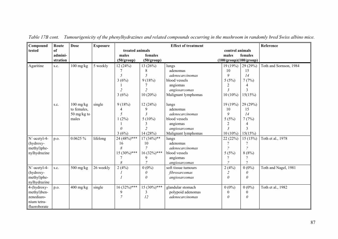
87
Table 17B cont. Tumourigenicity of the phenylhydrazines and related compounds occurring in the mushroom in randomly bred Swiss albino mice.
Compound tested
Route of admini-stration
Dose Exposure Effect of treatment treated animals control animals males females males females (50/group) (50/group) (100/group)(100/group)
Reference
Agaritine s.c. s.c.
100 mg/kg 100 mg/kg to females, 50 mg/kg to males
5 weekly single
12 (24%) 7 5 3 (6%) 1 2 3 (6%) 9 (18%) 4 5 1 (2%) 1 0 3 (6%)
13 (26%) 8 5 9 (18%) 7 2 10 (20%) 12 (24%) 9 3 5 (10%) 3 2 14 (28%)
lungs adenomas adenocarcinomas blood vessels angiomas angiosarcomas Malignant lymphomas lungs adenomas adenocarcinomas blood vessels angiomas angiosarcomas Malignant lymphomas
19 (19%) 10 9 5 (5%) 2 3 10 (10%) 19 (19%) 10 9 5 (5%) 2 3 10 (10%)
29 (29%) 15 14 7 (7%) 4 3 15(15%) 29 (29%) 15 14 7 (7%) 4 3 15(15%)
Toth and Sornson, 1984
N´-acetyl-4-(hydroxy-methyl)phe-nylhydrazine
p.o. 0.0625 %
lifelong 24 (48%)*** 16 8 15 (30%)*** 7 8
17 (34%)** 10 7 16 (32%)*** 9 7
lungs adenomas adenocarcinomas blood vessels angiomas angiosarcomas
22 (22%) ? ? 5 (5%) ? ?
15 (15%) ? ? 8 (8%) ? ?
Toth et al., 1978
N´-acetyl-4-(hydroxy-methyl)phe-nylhydrazine
s.c. 500 mg/kg
26 weekly 2 (4%) 1 1
0 (0%) 0 0
soft tissue tumours fibrosarcomas angiosarcomas
2 (4%) 2 0
0 (0%) 0 0
Toth and Nagel, 1981
4-(hydroxy-methyl)ben-zenediazo-nium tetra-fluoroborate
p.o. 400 mg/kg single 16 (32%)*** 9 7
15 (30%)*** 3 12
glandular stomach polypoid adenomas adenocarcinomas
0 (0%) 0 0
0 (0%) 0 0
Toth et al., 1982

88
Table 17B cont. Tumourigenicity of the phenylhydrazines and related compounds occurring in the mushroom in randomly bred Swiss albino mice.
Compound tested
Route of admini-stration
Dose Exposure Effect of treatment treated animals control animals males females males females (50/group) (50/group) (100/group)(100/group)
Reference
4-(hydroxy-methyl)ben-zenediazo-nium tetra-fluoroborate
s.c. 50 mg/kg 26 weekly 9 (18%)*** 0 5 2 2 3 (6%) 2 1
11 (22%)* 0 10 0 1 9 (18%)** 4 5
subcutaneous tissue fibroma fibrosarcomas rhabdomyosarcomas angiosarcomas skin squamous cell papillomas squamous cell carcinomas
3 (6%) 1 2 0 0 0 (0%) 0 0
3 (6%) 0 3 0 0 0 (0%) 0 0
Toth, 1987
4-(hydroxy-methyl)ben-zenediazo-nium sulfate
s.c. 50 mg/kg 26 weekly 20 (40%)*** 19 0 1 2 (4%) 0 2
16 (32%)*** 13 2 1 7 (14%)* 2 5
subcutaneous tissue fibrosarcomas rhabdomyosarcomas angiosarcomas skin squamous cell papillomas squamous cell carcinomas
2 (4%) 2 0 0 0 (0%) 0 0
0 (0%) 0 0 0 1 (2%) 1 0
Toth, 1987
4-(carboxy)-phenylhydra-zine
p.o. 0.125% lifelong 21 (42%)*** 6 15
7 (14%)* 3 4
aorta and large arteries leiomyomas leiomyosarcomas
2 (4%) 0 2
0 (0%) 0 0
McManus et al., 1987
N2-[γ-L(+)-glutamyl]-4-(carboxy)-phenylhydra-zine
p.o. 1.4 g/kg
52 weekly 13 (26%)*** 0 13 0
1 (2%) 0 0 1
subcutaneous tissue fibroma fibrosarcomas myxosarcomas
0 (0%) 0 0 0
3 (6%) 1 2 0
Toth, 1986a

9.2.2.2. N’-acetyl-4-(hydroxymethyl)phenylhydrazine
The American investigators at University of Nebraska Medical Centre have performed two studies with N’-acetyl-4-(hydroxymethyl)phenylhydrazine, a compound that does not occur in the cultivated mushroom. In A. bisporus agaritine is believed to be enzy-matically cleaved to L-glutamate and 4-(hydroxymethyl)-phenylhydrazine. Since 4-(hydroxymethyl)phenylhydrazine is a relatively unstable compound the investigators attached an acetyl group to it to render the compound feasible for biological experi-ments.
In the first study with N´-acetyl-4 (hydroxymethyl)phenylhydrazine (purity >97%), the compound was given as a 0.0625% solution in drinking water for the lifetime of Swiss albino mice (Toth et al., 1978). The average daily intake of N´-acetyl-4-(hydroxymethyl)phenylhydrazine was 6.4 mg for female mice and 6.6 mg for male mice. The experiment lasted for up to 120-130 weeks. The control group of 100 mice of each sex was 5 weeks at the beginning of the study.
There was no significant difference in survival between the N´-acetyl-4 (hydroxy-methyl)phenylhydrazine-treated mice and the mice fed the control diet. As shown in Table 17B the exposure gave rise to statistically increased frequencies of lung tumours and blood vessel tumours in both sexes. The authors did not indicate the number of be-nign and malignant tumours for the control group.
In the second study with N’-acetyl-4-(hydroxymethyl)phenylhydrazine Swiss albino mice received 26 separate injections of 500 µg of the compound in 0.01 ml physiologi-cal saline per gram body weight at weekly intervals (Toth and Nagel, 1981). The control animals received 26 subcutaneous injections 0.01 ml physiological saline per gram body weight at weekly intervals. The experiment lasted for 120-130 weeks. The survival rate and tumour incidence among the treated animals were not different from that in control animals (Table 17B).
9.2.2.3. 4-(Hydroxymethyl)benzenediazonium ions
Two forms of the 4-(hydroxymethyl)benezenediazonium ion have been tested in long-term carcinogenicity studies - the tetrafluoroborate and the sulfate form. Both com-pounds were chemically synthesised by the investigators. The purity of the 4-(hydroxymethyl)benezenediazonium tetrafluoroborate was 95% and that of the 4-(hydroxymethyl)benezenediazonium sulfate 98%.
The 4-(hydroxymethyl)benezenediazonium tetrafluoroborate was tested both after oral application and after subcutaneous injections. In one study with the 4-(hydroxy-methyl)benezenediazonium tetrafluoroborate, the compound was given as a single in-tragastric dose by gavage at a concentration of 400 µg in 0.01 ml 0.9% NaCl solution per gram body weight, which was close to the maximum tolerated dose (Toth et al., 1982). The control groups were inadequately characterised in this study. As a solvent-instilled control, 50 mice of each sex, received 41 weekly intragastric doses of 100 µg sodium tetrafluoroborate in 0.01 ml NaCl solution per gram body weight. A third group consisted of untreated mice. The study lasted for 120-130 weeks. Only data on the 4-(hydroxymethyl)benezenediazonium tetrafluoroborate-treated group was presented.
The investigators claim that the 4-(hydroxymethyl)benezenediazonium tetrafluorobo-rate-treatment hand no significant effect on the survival when compared with the sur-vival of the solvent-injected control animals or the untreated controls. This seems to be
89

a rash statement, since none of the 50 exposed males survived longer than 80-90 weeks. Whereas the 4-(hydroxymethyl)benezenediazonium tetrafluoroborate-treatment induced glandular stomach tumours, no such tumours were found in the untreated controls (Ta-ble 17B). Toth et al. (1982) mentioned that some of the adenocarcinomas probably started to grow as polypoid adenomas whereas others developed into malignancy with-out the transitory benign stage.
When Swiss albino mice were given 26 subcutaneous injections at weekly intervals with 50 µg 4-(hydroxymethyl)benezenediazonium tetrafluoroborate in 0.01 ml 0.9% sodium chloride solution per gram body weight, the treatment shortened the survival when compared with the solvent-injected controls (Toth et al., 1981b). The control re-ceived 26 subcutaneous weekly injections with sodium tetrafluoroborate (the dose of tetrafluoroborate corresponded to the dose given to the treated group). Whereas all treated animals had died within 70-80 weeks after the beginning of the treatment, some of the control animal lived for up to 110-120 weeks. As shown in Table 17B, the subcu-taneous injections with 4-(hydroxymethyl)benezenediazonium tetrafluoroborate in-duced a statistically significant increase in tumours of the subcutis and skin of female Swiss mice.
In the study with the sulfate form of the 4-(hydroxymethyl)benezenediazonium ion, Swiss albino mice received 26 subcutaneous weekly injections at 50 µg in 0.01 ml 0.9% NaCl solution per gram body weight (Toth, 1987). The control animals received the same number of subcutaneous injections with 31 µg sodium sulfate in 0.01 ml 0.9% NaCl solution per gram body weight. As was the case after treatment with the tetra-fluoroborate of the 4-(hydroxymethyl)benezenediazonium ion, treatment with the sul-fate form of the compound resulted in shortened survival in comparison with the control group. Furthermore, the treatment resulted in statistically increased frequencies of tu-mours of cubcutis (both sexes) and skin (only females) - Table 17B.
9.2.2.4. 4-(Carboxy)phenylhydrazine
Commercially available 4-(carboxy)phenylhydrazine hydrochloride (p-hydra-zinobenzoic acid; purity >97%) was administered to Swiss albino mice at a dose of 0.125% in drinking water for lifetime; that is for 120-130 weeks for the treated mice and 150-160 weeks for the control animals (McManus et al., 1987). The average daily intake of 4-(carboxy)phenylhydrazine per animal was 4.7 mg for females and 5.1 mg for males.
Treated mice showed an increased mortality rate. Particularly at the early stages of the study, a substantial numbers of mice died suddenly with ruptured aortae (32% of fe-males and 50% of males). As shown in Table 17B the treatment also induced a statisti-cally significant increase in smooth muscle cell tumours of the aorta and the large arter-ies.
Two major histopathological changes observed could contribute to the explanation of both the ruptures and the tumours. First the aortic walls had undergone a diffuse intimal and inner medial fibrinoid necrosis, accompanied by widespread medical elastinolysis. Secondly, cells from the media proliferated.
Already twenty years earlier, Roe and his coworkers (1967) had tested 4-(carboxy)phenylhydrazine in Swiss mice. Only 25 female mice were used in this ex-periment, and only one dose level was tested. Virgin animals were treated 5 days each
90

week for 40 weeks with 1 mg 4-(carboxy)phenylhydrazine in 0.2 ml distilled water by gavage. 85 untreated female Swiss mice served as controls.
The authors studied the incidence of lung tumours in treated and control animals 40-60 weeks after treatment. At 40-50 weeks after treatment, one out of the 7 exposed mice still living had developed lung tumours, whereas the corresponding incidence among the control animals were 2 out of 37. At 50-60 weeks after the beginning of the treat-ment one out of 5 survivors had developed lung tumours. In the controls 6 out of 42 survivors had developed lung tumours. The authors concluded that 4-(carboxy)phenylhydrazine was without apparent carcinogenic effect in the lung.
I addition to the carcinogenicity studies with 4-(carboxy)phenylhydrazine described above, Danish investigators have studied whether supplementation of this compound (1000 ppm) to the drinking water for 26 weeks could give rise to lung adenomas in fe-male A/J mouse, a short-term carcinogenicity model for lung tumours (Pilegaard et al., 1997). The average intake of 4-(carboxy)phenylhydrazine in this study was 106 mg/kg body weight/day. No lung adenomas were induced. However, the data was based on only 15 animals and the exposure to 4-(carboxy)phenylhydrazine was lower in this ex-periment than in the positive long-term carcinogenicity study of McManus et al. (1987).
9.2.2.5. β-N-(γ-L-(+)-Glutamyl)-4-(carboxy)phenylhydrazine
Swiss mice received chemically synthesised β-N-(γ-L (+)-glutamyl)-4-(carboxy)phenylhydrazine (purity: 95%) as 52 weekly intragastric installations of 1.4 mg in 0.01 physiological saline per gram body weight (Toth, 1986). The experiment lasted for 150-160 weeks (until all mice had died or were killed when found in poor condition).
The treatment had no substantial effect on the survival time. Although β-N-(γ-L-(+)-glutamyl)-4 (carboxy)phenylhydrazine was administered by the oral route, the treated males developed a statistically significant increase in subcutaneous tumours which were located on the shoulder areas and backs of animals. The number of tumour bearing ani-mals is shown in table 17B. Toth (1986) proposed that some of the chemicals might have been regurgitated after the application and might have been rubbed and dispersed on the skin of the animal. If the subcutaneous tumours of the males are induced by re-gurgitation, then the authors information on the toxicity by oral administration is sparse. Instead of giving the control animals water (the solute) weekly by gavage, they were given water ad libitum in ordinary drinking bottles.
9.2.2.6. Other phenylhydrazines
A compound closely related to the agaritine degradation product 4-(hydroxymethyl)-benzenediazonium ion has been identified in A. xanthodermus, a wild nonedible relative to A. bisporus (Dornberger et al., 1986). This compound was the 4-(hydroxy)-benzenediazonium ion (Figure 10A). When the 4-(hydroxy)benzenediazonium sulfate was subcutaneously injected 36 times at weekly intervals in Swiss mice at 2 µg per gram body weight, tumours were induced in the subcutis (Toth et al., 1989a). In parallel experiments two injections with 10 µg 4-(hydroxy)benzenediazonium sulfate per gram body weight, one week apart, had no tumourigenic activity.
91

en-
Fig. 1
The tis theidentfrom benzevals 1and s
Anot(the dWhentaneointragexpotragabloodphenmice injecfound
9.3.In onporusplantof moof A.times% in
92
N N , HSO4
OH
+ -
A) 4-Hydroxybenzen- diazonium ion
0. One phenylhydrazine and bisporus but have been teshydroxybenzenediazonium(C) 4-methylphenylhydraz
hird benzenediazonium ion th 4-(methyl)benzenediazonium
ified in any Agaricus speciesthe 4-(hydroxymehtyl)
nediazonium sulfate was adm0 or 16 times at 50 µg per gr
kin were induced (Toth et al.,
her compound speculated to birect degradation product of this compound in hydrochlous injection at 140 µg per graastic instillations of 250 µg
sed animals. The subcutaneoustric treatment, in addition to vessels in both sexes (Tot
ylhydrazine unexpectedly indadministered the hydrochlorid
tions (Toth and Nagel, 1981) in these two studies is not kn
Non-conventional care study, the activity of synth were tested for their ability
ed with these compounds in puse urinary bladder carcinom
bisporus. Although the agar lower than the agaritine con animals administered extract
N
CH3
N , HSO4+ -
B) 4-Methylbenz diazonium ion
two benzenediazonium ionsted for carcinogenicity in S sulfate; (B) 4-(methyl)benzine hydrochloride.
at has been shown to indu ion (Figure 10B). This c
, but it has been speculatebenzenediazonium ion.inistered by subcutaneous am body weight, tumours o1989b).
e produced from 4-hydroxagaritine) is 4-methylphenyride form was administeredm body weight weekly for per gram body weight, tums injection resulted in lung lung tumours in the femalh et al., 1977). In subsequced soft-tissue tumours ae form of the compound by. The reason behind the down.
cinogenicity studies esised agaritine and metha
to induce tumours in mouaraffin wax (Hashida et al.,a was 30.8% in animals expitine content of shiitake extent of A. bisporus-extract, s from fresh shiitake (Lent
CH3
NH NH3 , Cl-+
C) 4-Methyl hydrazine hydrochlorid
that do not occur in A. wiss albino mice; (A) 4-enediazonium sulfate;
ce tumours in Swiss mice ompound has never been d that it could be formed When 4-(methyl)-injection at weekly inter-f the subcutaneous tissue
ymethylphenyl-hydrazine lhydrazine (Figure 10C). to Swiss mice by subcu-10 weeks, or as 7 weekly ours were induced in the tumours, whereas the in-e, induced tumours in the uent studies 4-(methyl)-t injection sites of Swiss 26 weekly subcutaneous
ifferent types of tumours
nol extracts from A. bis-se bladder epithelium in- 1990). The observed rate osed to methanol extracts tracts was more than 200 the tumour rate was 23.5
inus edodes). The tumour

rates in animals exposed to extracts of dry shiitake and synthesised agaritine was 9.8 and 50%, respectively. The tumour incidence in the negative control was 5.4%. A statistically significant increase in the tumour incidence, at the p<0.01 level, were observed for animals exposed to agaritine and methanol extracts of fresh A. bisporus.
ivo situation.
9.4. Antineoplastic effects of phenylhydrazines Today 96 hydrazine derivatives and related compounds have been studied for carcino-genic action in long-term experiments in laboratory animals and 82 have given a posi-tive response (Toth, 2000). In addition, case-control and cohort studies of one common hydrazine in industrial settings and four hydrazines used in medicine indicate that they play a role in the development of certain types of human cancer (Toth, 1994). However, hydrazines and hydrazine-containing natural products have also been reported to have antineoplastic activity (Toth, 1996, 2000).
In one of these studies 5-6 weeks old male C3H and CDF1 mice were inplanted with MM-46 carcinoma cell or IMC carcinoma cells and given a diet including 10, 20 or 30% powdered A. bisporus (Nanba and Koruda, 1988). On the 30th day after tumour transplantation, the experiment was terminated. The tumour inhibition ratio in the 10, 20 and 30% mushroom-fed groups were 59, 67 and 76% in C3H mice and 33, 45 and 55% in the CDF1 mice. The experiment also included studies on phagocytic activity of macrophages, and measurement of superoxide anion production and activities of lym-phokine-activated killer cells and cytotoxic T cells. It was concluded that a mushroom-supplemented diet suppressed the growth of tumours.
As estrogens have a role in breast cancer development, and estorgen production by aromatase/estrogen synthetase in breast cancer plays a dominant role in tumour proliferation, it is interesting to note that extracts of Agaricus bisporus inhibits the aromatase activity dose dependently in vitro (Grube et al., 2001). It is not known whether these observations have any bearing on the in v
9.5. Reproduction, embryotoxicity and teratogenicity There are only three reports on embryotoxicity and teratogenicity of phenylhydrazines in A. bisporus.
In an old report describing the teratogenic effects of semicarbazide hydrochloride in-jected (2-3 mg) into 4-6 days old embryonated White Leghorn eggs, the authors state that similar effects to semicarbazide is produced by 4-(carboxy)phenylhydrazine (Neu-man et al., 1956). The effect is characterised by production of lower beak and skeletal malformations (bent tarsometatarsal and tibiotarsal bones).
An embryotoxicity and teratogenicity study with water extracts of A. bisporus have been performed in rats within this Nordic project. In this study pregnant Sprague-Dawley rats were administered a freshly prepared water extract of the mushroom by gastric intubation daily on days 6-15 of pregnancy and killed on day 19 of gestation (Andersson et al., 1994). The extracts were prepared from freeze-dried A. bisporus, con-taining 340 mg agaritine per kg mushroom, and the amounts administered corresponded to a human intake of 3 kg mushrooms per day. When treated and control dams were examined for embryotoxicity (preimplantation loss, number of resorptions) and fetuses
93

for teratogenicity (external and skeletal malformations), using standard teratological techniques, no significant differences could be detected between the groups. Neither could any influence on the body weights of the mothers and the fetuses be detected.
In an investigation on the effect of gamma irradiation on the storage characteristics and nutritional quality of mushrooms, dried and ground unirradiated, organically grown A. bisporus were incorporated in feed and given to pregnant albino CF1 mice (2-5 days before term) as a control exposure when exploring the effects of irradiated mushrooms on female animals and their offspring (Campbell et al., 1968). The agaritine content of the mouse feed was not determined and the feeding period of the pregnant dams was to late for any teratogenic effects to be expected in the offspring. No exceptional findings were noted in this experiment, but very little information was given on the results.
9.6. Biological effects of A. bisporus In this section some studies on physiological effects related to A. bisporus consumption are reported. Some of the physiological effects are adverse, others of a protective na-ture. It should be kept in mind that it generally is unknown which compounds in the mushroom are responsible for the observed effects. There is no direct reason to suspect the phenylhydrazines to be responsible for the effects.
Although Agaricus bisporus is not generally recognised as having medicinal value (Chang, 1996), some investigators have explored the cultivated mushroom also in this respect. Consumption of various types of mushroom have been shown to lower the plasma cholesterol concentration. The mechanism for this effect is not known. Recently, Dutch investigators studied the hypocholesterolemic properties of A. bisporus by feed-ing groups of female rats for 6 weeks with diets where parts of the corn starch and dex-trose of the commercial diet had been exchanged for a small quantity of cholesterol and in total 2.5, 5 or 10 % mushroom (Beynen et al., 1996). Inclusion of A. bisporus in the rat diet had no effect on the plasma and liver cholesterol concentrations of the rats.
The cultivated mushroom has been used in folklore medicine for treatment of diabetes in northern Europe, although scientific evidence for its efficiency have been lacking. To explore a possible hypoglycaemic effect of mushroom consumption, normal and strep-tozotozin diabetic adult male lean mice (a colony breed at Aston University, UK) were fed a pelleted diet containing 6.25 % dried fruit bodies of A. bisporus during 28 days (Swanston-Flatt et al., 1989). The glucose homeostasis was not affected in non-diabetic mice, but in mice with induced streptozotocin diabetes (reduction in hyperphagia, polydipsia and body weight loss) hyperglycaemia was reduced already during the first week after induction of the disease. The percentage glycated haemoglobin two weeks after streptozotocin administration was also reduced and the initial reduction in plasma insulin and pancreatic insulin counteracted. Thus, the mushroom improved insulin-induced hypoglycaemia in streptozotocin diabetic mice.
Since consumption of A. bisporus reduced the initial insulinopenic effect of streptozoto-cin and countered the depletion of pancreatic insulin, the mechanism for the ameliora-tion of diabetes by this mushroom may be connected to a protective effect against islet B-cell destruction. Extracts of the cultivated mushroom have been reported to affect islet B-cells by stimulating calcium uptake and insulin release (Ahmad et al., 1984a). This has been attributed to a lectine activity in the mushroom (Ahmad et al., 1984b; Ewart et al., 1975).
94

In addition to the phenylhydrazines shown in Figures 1 and 2, A. bisporus contains an-other nitrogen-containing compound that also containing a glutamyl moiety. That com-pound is γ-L-glutaminyl-4-hydroxybenzene which is believed to be used in a tyrosinase-catalyzed metabolic pathway that produces inhibitors ostensibly responsible for the in-duction of dormancy in this species (Weaver et al., 1971a). As a first step γ-L-glutaminyl-4-hydroxybenzene is hydrolysed to γ-L-glutaminyl-3,4-dihydroxybenzene, which is oxidised to γ-L-glutaminyl-3,4-benzoquinone, both reactions being catalysed by tyrosinase (Weaver et al., 1971b). The quinone compound undergoes a base-catalyzsed non-enzymatic reaction to yield 2-hydroxy-4-iminoquinone. Both quinones may inhibit sensitive enzymes, for example enzymes involved in energy production and DNA synthesis, and give rise to cytostatic and cytocidal activity. Studies with isolated enzymes and permeabilised L1210 lymphocytic leukemia cells have revealed that the mechanism by which DNA synthesis is affected by γ-L-glutaminyl-4-hydroxybenzene is by an inhibition of ribonucleotide reductase and not DNA polymerase (FitzGerald et al., 1984). The inhibition occurs in the range at which γ-L-glutaminyl-4-hydroxybenzene is found in gill tissue, close to 1 g/kg tissue. Histological evidence of severe kidney and pancreas damage (necrosis) was observed in neonatal C57Bl/6J mice injected with γ-L-glutaminyl-3,4-dihydroxybenzene, but not in mice injected with N2-methyl-γ-L-glutaminyl-3,4-dihydroxybenzene (Boekelheide et al., 1980). The LD50 value for the active compound was 100 to 200 mg/kg. Since γ-L-glutaminyl-3,4-dihydroxybenzene, but not the methylated derivative, was an excellent substrate for γ-glutaminyl-transpeptidase, it was believed that the transpeptidase produces glutamic acid and 4-aminocatechol from γ-L-glutaminyl-3,4-dihydroxybenzene, and that air oxi-dises 4-aminocatechol to the ultimate cytotoxic compound 2-hydroxy-4-iminoquinone. The toxic effect was registered in the two tissues having the highest γ-glutaminyl-transpeptidase activity.
From Agaricus campestris a similar precursor for γ-L-glutaminyl-3,4-benzoquinone has been found in the form of the amino acid agaridoxin, 3,4-dihydroxy-(γ-L-glutamyl)-anilide (Szent-Gyorgyi et al., 1976).
9.7. Conclusion on effects on experimental animals Only a few and poorly performed studies have dealt with the subchronic toxicity of mushroom hydrazines. The only observation of importance coming from these studies is the comparatively low toxicity of 4-(carboxy)phenylhydrazine in dogs, rabbit and guinea pigs.
Since also the few published studies on embryotoxicity and teratogenicity are of poor quality, very little information can be extracted from these reports of reproductive toxic-ity of phenylhydrazines in A. bisporus, water extracts of the mushroom and dried and ground mushroom. Whereas orally supplied water extracts of A. bisporus had no terato-genic activity in Sprague Dawley rats, high concentrations of 4-(carboxy)phenyl-hydrazine injected into eggs, much higher concentrations than man can ever be exposed to through consumption of the cultivated mushroom, produced lower beak and skeletal malformations in embryonated White Leghorn chickens.
None of the 13 long-term carcinogenicity studies with A. bisporus or the phenylhydra-zines in the mushroom in mice, or the 2 long-term carcinogenicity studies with A. bis-
95

porus in rats have been performed according to approved standard protocols for car-cinogenicity studies. All cancers studies on mice have been performed in a single labo-ratory and with only one strain of mice, the outbred Swiss albino mice. Factors that did not follow standard procedures and that might have had profound influence on the end results were that mice were exposed to the tested compounds from the sixth week of age until they died or were found in poor condition instead of for a predetermined length of time, and that the control group were usually not started at the same time as the treated groups.
Of the mushroom hydrazines, 4-(carboxy)phenylhydrazine, N2-[γ-L(+)-glutamyl]-4-
(carboxy)phenylhydrazine and 4-(hydroxymethyl)benzendiazonium ion (as tetra-fluoroborate and sulfate) were identified as carcinogens at high doses in Swiss albino mice. Furthermore, a stabilised derivative of 4-(hydroxymethyl)phenyl-hydrazine, N´-acetyl-4-(hydroxymethyl)phenylhydrazine, was carcinogenic in the same system. On the other hand agaritine was found not to be carcinogenic when administered by the oral and subcutaneous route. Since agaritine was administered in drinking water changed only three times a week and the concentration of agaritine in the drinking water was not analysed, it is possible that the compound was partly degraded in the drinking water.
However, tumours were induced in several tissues when fresh or dry-baked A. bisporus were fed to mice ad libitum for 3 days, followed by semisynthetic diet for 4 days each week for life. Whereas the fresh mushroom induced tumours in the lung, forestomach and bone, the dry-baked mushroom induced tumours in the forestomach, glandular stomach, duodenum and ovaries. Since the average weekly weight curves of the animals given fresh A. bisporus were substantially lower than that of untreated animals it has been speculated that the carcinogenic response of the treatment was induced by the treatment protocol and not the cultivated mushroom. Although feeding mice with dry-baked mushrooms for 12 hours a day for 5 days each week for life and a well-balanced semisynthetic diet for the rest of the time (periodic alterations in nutrient levels) in-creased the number of tumours in some tissues, none of the increases in tumour fre-quency were statistically significant. It is impossible to determine whether the lower carcinogenicity registered in this study was due to a lower intake of mushrooms (and phenylhydrazines) or a more balanced feeding protocol. Both studies in which rats were fed A. bisporus were of poor quality. Thus, if A. bisporus per se is carcinogenic in ex-perimental animals, its activity should be low.
96

10. Human implications
There are two aspects of human intoxication after consumption of A. bisporus or its wild-growing relatives. One possibility, although unlikely, is that the cultivated mush-room itself could give rise to acute intoxications of the consumer. The second and more likely possibility is that consumers of what is believed to be the cultivated mushroom or edible wild-growing Agaricus species might be exposed to toxic look-alike contaminat-ing mushrooms. The risk for such incidents is of course highest when eating the wild-growing Agaricus species.
Consumers are exposed to wild mushrooms from many sources. They can harvest wild mushrooms for their own personal use, buy mushrooms from businesses that collect and sell mushrooms to the public and retail distributors, or order dishes that contain wild mushrooms in restaurants. Wild mushrooms from commersial sources may be contami-nated by toxic look-alikes, which are collected with edible mushrooms through either ignorance or negligence. Mushroom pickers are often paid by the weight for their wild harvest, a practice that discourage selective harvesting and culling for the elimination of undesirable species and that encourages fraud. Field drying the wild harvest before sale to cooperative processing and packaging operations makes effective culling more diffi-cult because the appearance of the dried products has changed (Gecan and Cichowicz, 1993). Consequences of poisoning due to contamination with toxic look-alikes range from unpleasant gastrointestinal disturbances to major organ failure and death.
The risk for intoxications occurring after consumption of products sold on the market has been studied in the United States.Ten Food and Drug Administration Districts each were directed to collect 30 samples of canned and dried imported mushrooms during one year (October 1988-October 1989), and 20 samples of canned, dried, and fresh im-ported and domestic mushrooms for 1 year (October 1989-October 1990) (Gecan and Cichowicz, 1993). Among the 344 mushroom samples collected and analyzed during the 2-year survey, only 3 samples were supposed to be Agaricus species (Meadow Mushroom; Agaricus campestris). The toxic look-alikes to Agaricus species are mainly Death Angel (Amanita bisporigera), Spring Amanita (Amanita verna), Destroying An-gel (Amanita virosa), and Death Cap (Amanita phalloides). None of these toxic look-alikes were found in the 3 Agaricus-samples.
97

98

11. Previous estimations of risk
In 1983 the International Agency for Research on Cancer evaluated agaritine for car-cinogenicity based on data available up until then (see chapter 9). No evidence was found for the carcinogenicity of agaritine in experimental animals. However, there was limited evidence of the metabolites of agaritine being carcinogenic in experimental ani-mals. In the absence of epidemiological data, no evaluation of the carcinogenicity of agaritine to humans could be made. No carcinogenicity study of the cultivated mush-room per se was available at the time of the IARC evaluation.
Schlatter (1986) approached the task of performing a risk assessment of phenylhydrazi-nes in A. bisporus in several ways. One approach was a rough linear extrapolation of the cancer risk from mouse to man using as experimental data the carcinogenicity study of Toth and Erickson (1986) in which mice were fed A. bisporus 3 days a week for their life-span. In that study the intake of mushrooms lead to a tumour incidence of 0.4. As-suming agaritine and/or its metabolites to be responsible for the carcinogenic effect and the mushroom consumption to correspond to an intake of 50 mg agaritine/kg body weight/day, as well as the metabolism of Swiss mice being comparable to that of man, Schlatter (1986) calculated that a human cancer risk of 1/106 would correspond to a daily intake of approximately 0.1 µg agaritine/kg body weight/day (corresponding to about 6 µg daily for a human). Another approach by Schlatter (1986) to attempt a risk assessment of agaritine and its hydrolysis products was drawn upon the DNA binding and in vivo mutagenesis studies of Shepard et al. (1986). In this study the DNA binding of the 4-(hydroxymethyl)benzenediazonium ion (HMBD), the anticipated ultimate car-cinogen arising from agaritine metabolism, was measured in mice orally supplied with this compound. The HMBD bound weakly to stomach DNA, but made very few DNA adducts to liver DNA. To be able to compare the quantity of DNA adducts produced by HMBD with that produced by well known animal carcinogens the author calculated the Covalent Binding Index, CBI (Lutz, 1979). The HMBD had a CBI value of 13 in the stomach and below one in the liver. For comparison, the strong carcinogen nitrosodi-methylamine has a CBI value of 6000 and aflatoxin B1 a value of 12000, both measured in rat liver (Lutz, 1979). Since there was no DNA binding data available for agaritine, but this mushroom compound according to other investigators (Friedrich et al., 1986) was 10-100 times less mutagenic than HMBD in bacterial test systems, a CBI value of 0.3 for all tissues was estimated and assumed for agaritine. By making a number of somewhat speculative assumptions related to DNA adducts and to which risks are toler-able and which are not, Schlatter (1986) compared the CBI values and concluded that an exposure to about 200 µg agaritine/person/day or 0.5 g fresh A. bisporus/person/day would be tolerable. This exposure corresponds to one mushroom meal every three to four months.
In 1993 Toth and Gannett attempted an assessment of the carcinogenic potency of Aga-ricus bisporus based on their long-term carcinogenicity studies in the Swiss mouse with the mushroom itself, the phenylhydrazines and benzenediazonium ion occurring in the mushroom, and N´-acetyl-4-(hydroxymethyl)-phenylhydrazine. The latter compound does not occur in the mushroom. The N-acetylation of 4-(hydroxymethyl)phenyl-
99
hydrazine (the unstable substance formed by hydrolysis of agaritine) was made in order to stabilise the molecule. Although treatment with agaritine itself resulted in no detect-able carcinogenic action in Swiss mice, treatments with the other test compounds and the raw mushroom (A. bisporus) induced tumours in various tissues of the animals. By taking into account the daily or weekly dose levels of individual phenylhydrazines or the mushroom per se, as well as the length of the exposure period, the following total amounts were calculated to induce a significant increase of tumours in Swiss mice (25 g mouse, average life span: 70 weeks): 10 mg 4-(hydroxymethyl)benzenediazonium sul-fate, 1 800 mg N2-[γ-L(+)-glutamyl]-4-carboxyphenylhydrazine, 2 400 mg 4-(carboxy)phenylhydrazine and 23 kg A. bisporus. Assuming that the susceptibility of the Swiss mouse to the carcinogenic stimulus is similar to humans, and that an average person has a weight of 70 kg and a life span of 50 years, Toth and Gannett (1993) calcu-lated that the following amounts of these compounds would be required to induce tu-mours in man: 28 g 4-(hydroxymethyl)benzenediazonium sulfate (corresponding to 2.6 kg mushroom/day for 50 years, with mushrooms containing 0.6 mg/kg of the com-pound), 5.04 g N2-[γ-L(+)-glutamyl]-4-carboxyphenylhydrazine (corresponding to 6.7 kg mushroom/day for 50 years, with mushrooms containing 42 mg/kg of the com-pound), 6.72 g 4-(carboxy)phenylhydrazine (corresponding to 36 kg mushroom/day for 50 years, with mushrooms containing 10 mg/kg of the compound) and 350g A. bisporus per day for 50 years. The authors estimated that the phenylhydrazines studied only ac-counted for a fraction of the carcinogenicity demonstrated for raw Agaricus bisporus. It should be stressed that no dose-response studies were performed with the phenylhydra-zines of the mushroom or with the mushroom itself. At the single dose used the most sensitive tumour type was induced in approximately one third of the exposed animals. Therefore, it is very likely that the induction of tumours could be demonstrated at lower dose levels as well. The calculated exposure level that would give rise to tumours in man is related to that high tumour incidence.
More recently, Swiss investigators updated their risk assessment of agaritine in the cul-tivated mushroom, now basing it on in vivo mutagenicity studies in transgenic Big Blue mice (Shepard et al., 1995). Their studies revealed that the mutation frequencies were increased by 100% in the kidney and by 50% in the forestomach of mice fed during 105 days a powdered chow containing a mushroom extract with 30% agaritine (120 mg aga-ritine/kg body weight/day). Based on the assumption that fresh, cooked and canned mushroom on average contain 150 mg agaritine/kg mushroom and that the per capita consumption of the cultivated mushroom is 4 g a day for a Swiss consumer, the investi-gators estimated a daily agaritine exposure of 0.6 mg/person. This would correspond to approximately 10 µg/kg body weight for a person weighing 60 kg. Using the TD50 value (see section on mutagenicity) of agaritine derived from the in vivo mutagenicity data in Big Blue mice, a rough estimate of the cancer risk at the calculated human exposure level of agaritine was obtained by a linear extrapolation. The lifetime cancer risk from an average daily A. bisporus consumption was estimated to 20x10-6, based solely on the genotoxic properties of the mushroom. Substances in the mushroom that might posi-tively or negatively influence the carcinogenicity through non-genotoxic mechanisms are not detected in this in vivo assay and have not been included in the analysis (Shepard et al., 1995). This estimate of cancer risk is based on mutagenicity data from a very low number of animals.
100
12. Present nordic risk assessment
Due to the high content of biologically active phenylhydrazines in Agaricus bisporus, coupled with the popularity of this mushroom in the European kitchen, several attempts have been made to estimate the potential risk related to consumption of Agaricus bis-porus in man as discussed in the previous chapter.
In 1991 the Nordic Working Group on Food Toxicology and Risk Evaluation (NNT) expressed the opinion that published toxicological studies give rise to serious concern as to a possible human health risk from consumption of the cultivated mushroom. As no epidemiological data were available and the long term carcinogenicity studies with the mushroom and its hydrazine constituents had an inadequate standard or apparently were contradictory, it was not possible to evaluate the human risk at that time (Gry and Pile-gaard, 1991).
Since then several experimental studies have become available that could contribute to the present risk assessment:
12.1. Hazard identification. Agaritine (β-N-[γ-L-(+)-glutamyl]-4-hydroxymethylphenylhydrazine) was identified in A. bisporus already in the early 1960s. The level of agaritine found in fresh mushrooms is between 80 and 1 700 mg/kg, with average values between 200 and 400 mg/kg. In addition to agaritine, the cultivated mushroom also contains small quantities of 4-(carboxy)phenylhydrazine (10-11 mg/kg fresh weight), β-N-[γ-L-(+)-glutamyl]-4-(carboxy)phenylhydrazine (42 mg/kg fresh weight) and 4-(hydroxymethyl)benzene dia-zonium ions (0.6-4 mg/kg fresh weight). In addition to a limited number of natural hy-drazine derivatives, the industry has produced a number of synthetic compounds for industrial use. Since most of the synthetic hydrazines have significant biological activ-ity, and many are toxic and/or carcinogenic, Toth and co-workers undertook the work to assess the possible carcinogenic activity of the phenylhydrazines and related com-pounds in A. bisporus. Their studies indicated that most of the phenylhydrazines and related compounds in the mushroom are carcinogenic in Swiss albino mice. The only compound that was tested negative was agaritine, a finding that significantly muddled the interpretation of the carcinogenicity data. The suspicion that the phenylhydrazines and the benzenediazonium ion in the mushroom are carcinogenic has subsequently been strengthened by biochemical and mutagenicity studies. Furthermore, the cultivated mushroom per se has been shown to possess mutagenic and carcinogenic activity.
101

12.2. Hazard characterization. Absorption and metabolism. Studies in experimental animals have shown that agaritine, and/or its degradation products, are to a significant degree and rapidly absorbed from the gastro-intestinal tract. Blood levels peak within a few hours, but no unchanged aga-ritine is detected. No major qualitative differences in the pharmacokinetic behaviour of agaritine in the rat and mouse have been observed, and, furthermore, the metabolic pro-file of this compound seems to be the same in both species. None of the metabolites have been identified. Nonetheless, the enzyme γ-glutamyl transpeptidase, present in various tissues of the body, is believed to be the enzyme responsible for cleaving agari-tine into L-glutamic acid and 4-hydroxymethylphenylhydrazine. The latter compound is subsequently transformed to HMBD. It is the HMBD that is believed to produce reac-tive radical species able to damage macromolecules.
Subchronic toxicity. Very little is known about the subchronic toxicity of mushroom phenylhydrazines. The only information of importance it the rather low toxicity found for 4-(carboxy)phenylhydrazine in dogs, rabbits and guinea pigs.
Teratogenicity. There are no indications that A. bisporus or the phenylhydrazines occur-ring in the mushroom are embryotoxic or teratogenic in mammals at biologically rele-vant doses. However, it should be stressed that the design of the few studies available did not follow modern guidelines for these types of study. It should also be stressed that very few toxicity studies other than carcinogenicity studies have been performed with the phenylhydrazines in the mushroom.
DNA-binding and mutagenicity in vitro and in vivo. Much more interest has been fo-cused on whether the phenylhydrazines may induce cancer or not. A low frequency of DNA damage has been observed in various tissues of experimental animals exposed orally to agaritine and HMBD, an observation indicating that these compounds may be genotoxic carcinogens. This interpretation is supported by the observation that mush-room extracts, as well as agaritine and the other phenylhydrazines occurring in A. bis-porus, induce a low frequency of mutations in bacterial test systems in vitro. Further-more, in vivo studies on mice have revealed that agaritine induces gene mutations in transgenic animals and that the 4-(hydroxymethyl)benzendiazonium ion induces micro-nuclei in peripheral lymphocytes.
Carcinogenicity. Several controversial carcinogenicity studies with A. bisporus and its phenylhydrazine constituents have shown carcinogenic effects in Swiss mice; the frac-tion of treated animals with the highest increase of a specific tumour compared to the controls is shown in Table 18. Thus, in three out of four long-term carcinogenicity stud-ies tumours were induced in various tissues of mice fed raw, baked or freeze-dried A. bisporus. In the fourth study, with baked mushrooms but with another (more balanced) feeding schedule, the increase in tumour incidence was not significant. No tumours were induced in two long-term studies in rats, but in these rather inadequate studies the number of animals were too low to detect a relatively small increase in tumour inci-dence. Furthermore, the processing (pre-treatment) of the mushrooms (ordinary drying and milling, or washing, combined with pressure cooking, homogenising and mixing with tap water) might well have resulted in a significant degradation of the potentially active phenylhydrazines in these studies. Three of the four phenylhydrazines and related compounds known to occur in A. bisporus - CPH, GCPH and HMBD - were carcino-
102

genic at high doses when administered orally (by gavage or in drinking water) and also when
Table 18. Fraction of treated animals with the most pronounced increase of a specific tumour.
Material or compound tested in the cancer study (reference) Parameter Fresh raw mushroom (Toth and Ericson, 1986)
Dry-baked mushroom (Toth et al., 1997a)
Freeze-dried mushroom (Toth et al., 1998)
CPH (McManus et al., 1987)
GCPH (Toth, 1986a)
HMBD (Toth et al., 1982)
Daily exposure per mouse
4.7 g 5.57 g 1.25 g* 1250 mg/l drinking water for life
1400 mg/kg body weight/1 day weekly for 52 weeks
Single dose 400 mg/kg body weight
Fraction of tumour-bearing animals with the highest increase of a specific tumour (type of tu-mour)
38% (fore-
stomach in males)
20% (fore-
stomach in females, glandular
stomach in males)
22% (lung in males)
38% (aorta and large arter-
ies in males)
26% (subcuta-neous tis-
sue in males)
32% (glandular stomach in
males)
* 1.25 g freeze-dried mushroom approximately corresponds to 12.5 g raw mushroom injected subcutaneously in Swiss mice. The fourth - and major - phenylhydrazine, agari-tine, was found not to be carcinogenic when given in drinking water or by subcutaneous injection - but no precautions were taken in these studies to protect against a possible oxidative degradation of agaritine in water. Agaritine has recently been shown to be vulnerable to such degradation in aqueous solution (Hajkova et al., submitted).
It should be stressed that dose-response effects were only investigated in one of the car-cinogenicity studies. In this study no significant dose-response effect could be demon-strated. Otherwise, the studies only contained two groups, one control group and one exposed group, with 50 animals of each sex in each group. It should also be noted that the tumour profiles induced by the various mushroom phenylhydrazines and by the mushroom per se differ.
12.3. Exposure characterization. The estimated annual per capita intake of the cultivated mushroom (Agaricus bisporus) varies between the Nordic countries - being 0.6 kg in Finland having the lowest intake among Nordic countries and 2.4 kg in Sweden with the highest intake. Also the habit of using fresh mushrooms for dietary purposes varies between countries. The market share of fresh mushrooms was between 33 and 70%. The rest being preserved mushrooms, mainly sold in cans.
103

Since the per capita intake is an estimation of average intake and does not take into con-sideration the fact that mushrooms usually are consumed only by a part of the popula-tion, the per capita consumption is too low to express the consumption among mush-room eating consumers. Danish data, based on a limited consumer study, indicate that only around 50% of the population consume the cultivated mushroom, and that 5% of the population consume five times the median intake and 0.1% thirty times the median intake.
The potentially toxic compounds in the cultivated mushroom are the phenylhydrazines. The most abundant compound being β-N-[γ-L-(+)-glutamyl]-4-hydroxymethylphenyl-hydrazine (agaritine) which usually occurs in quantities between 200 and 400 mg/kg in the fresh mushroom. Sometimes levels up to 1700 mg/kg have been found. An antici-pated metabolite of agaritine, the 4-hydroxymethylbenzenediazonium ion (HMBD), is believed to be the most potent phenylhydrazine but it occurs at low levels, between 0.6 and 4 mg/kg fresh weight. The other phenylhydrazine derivatives in the mushroom are 4-(carboxy)phenylhydrazine (CPH, 10-11 mg/kg fresh weight) and γ-glytamyl-4-(carboxy)phenylhydrazine (GCPH, 16-42 mg/kg fresh weight).
The data on the influence of food processing on the amount of phenylhydrazines in the consumed mushroom are scanty. Frozen and freeze-dried mushrooms seems to contain similar agaritine levels as fresh mushrooms, whereas air-dried, cooked and fried mush-rooms contain reduced levels, but to what extent is not well known. Canned mushrooms only contain up to 10% of the agaritine level in fresh mushrooms.
Taken together, these data allows an estimation of the per capita exposure to agaritine in the Nordic countries. Agaritine was used as a representative of the phenylhydrazines in the mushroom. The estimated exposure in the Nordic countries is somewhere between 48 and 788 mg agaritine/year, corresponding to a daily exposure of between 2.1 and 36 µg/kg body weight. The exposure being influenced by the quantity of mushrooms con-sumed (nationality), the amount of agaritine in the fresh mushroom (200-400 mg/kg) and the preference of using fresh instead of processed mushrooms (33-70%). The effect of household processing of the mushroom on the agaritine content was otherwise not taken into consideration. A single mushroom dish with 75 g fresh A. bisporus would result in an exposure to 15-33 mg agaritine, that is, 250-550 µg/kg body weight.
12.4. Risk characterization. Six carcinogenicity studies in mice with oral application (by gavage, in drinking water or in the diet) were considered in combination with an estimated human intake to A. bisporus in the Nordic countries for the sake of performing a quantitative risk assess-ment of the cultivated mushroom. Of these studies, three were long-term studies with A. bisporus per se, and three were long-term studies with the phenylhydrazines and related compounds occurring in the mushroom. The calculation of the human lifetime cancer risk at average intake of Agaricus bisporus (or the corresponding amounts of the phenylhydrazines in the mushroom) in the Nordic countries are presented in Table 19. The calculations were done by linear extrapolation from high to low dosages using one-dose carcinogenicity studies in Swiss mice (assuming man and mouse having identical metabolism and sensitivity). The exercise was based on a number of additional assump-tions: the weight of a mouse on average being 25 g and that of man 60 kg; the average life span of a mouse being 70 weeks; there is no reduction in weight during dry-baking
104

of mushrooms for 10 minutes; freeze-dried mushrooms weighing 10% of raw mush-rooms; and the influence of processing on phenylhydrazine content of mushrooms not being taken into consideration.
Table 19. Estimated life-time human cancer risk from the intake of Agaricus bisporus
in the Nordic countries (average of intakes in Denmark, Iceland, Norway and Sweden). (Finnish figures within brackets.)
Material or compound tested in the cancer study Parameter Fresh raw mushroom (Toth and Erickson, 1986)
Dry-baked mushroom (Toth et al., 1997a)
Freeze-dried mushroom (Toth et al., 1998)
CPH (Mc-Manus et al.,
1987)
GCPH (Toth, 1986a)
HMBD (Toth et al.,
1982)
Daily ex-posure per mouse
4.7 g 5.57 g (males)
1.25 g* 1250 mg/l drinking water for
life
1400 mg/kg body
weight/1 day weekly
for 52 weeks
Single dose 400 mg/kg
body weight
Daily ex-posure per kg body weight (mouse)
189 g 229 g 50 g 204 mg 149 mg 0.82 mg
Daily hu-man intake of mush-room per kg body weight
0.1 g (0.025 g)
0.1 g (0.025 g)
0.1 g (0.025 g)
0.1 g (0.025 g)
(containing 10-11 mg CPH/kg)
0.1 g (0.025 g)
(containing 16-42 mg CCPH/kg)
0.1 g (0.025 g) (containing 0.6-4 mg
HMBD/kg)
Human cancer risk due to life-long expo-sure
193 x 10-6 (52x10-6)
86 x 10-6 (23x10-6)
211 x 10-6 (56x10-6)
1.8-2.0 x 10⁻⁶
(0.5x10⁻⁶)
2.7-7.0 x 10⁻⁶
(0.7-1.9 x10⁻⁶)
23-150 x 10-6
(6-40x10-6)
* 1.25 g freeze-dried mushroom approximately corresponds to 12.5 g raw mushroom Thus, with all the uncertainties mentioned above in mind the calculations result in an estimated lifetime human cancer risk for an “average“ Nordic consumer of fresh (or freeze-dried) A. bisporus up to 200 x 10-6. In reality an average consumer will consume a mixture of fresh and processed mushrooms. Some types of processing (i.e. canning) will reduce the phenylhydrazine content of the mushroom substantially, whereas other types of processing seem to have a much less pronounced effect. However, the influ-ence of standard cooking procedures on the phenylhydrazine content of the cultivated mushroom is not very well studied. Of the phenylhydrazines and related compounds investigated in carcinogenicity studies and occurring in the mushroom, HMBD had the highest carcinogenic potency. This finding agrees with what is expected based on the anticipated mechanism of action.
105

The human exposure estimation (per capita intake) is based on production volume in-formation which suffers from a number of shortcomings. In Denmark, for example, around half of the adult population claim not to consume the cultivated mushroom, 5% eat more than five times the median intake and 0.1% thirty times more than the median intake. Non-consumers of the cultivated mushroom would of course not have an addi-tional cancer risk from the phenylhydrazines in the mushroom, whereas sub-populations of “high“ consumers would have correspondingly higher cancer risks. It should be no-ticed that the Finnish population only consumes one quarter of what is consumed of the cultivated mushroom in the other four Nordic countries. The estimated lifetime cancer risk for the average Finnish consumer should accordingly be lower than in the other Nordic countries.
Clearly, the carcinogenic potential of A. bisporus and the possible contribution of agari-tine are difficult to assess in the face of contradictory experimental data, which may be due to the different exposure protocols employed in the carcinogenicity studies. In con-trast to A. bisporus, its major phenylhydrazine, agaritine, did not induce tumours when administered in the drinking water to mice. Therefore, no risk assessment has been made for this compound. However, it may be anticipated that due to the study design, agaritine could have been significantly degraded in the drinking water.
It must be underpinned that the long-term carcinogenicity studies on which the risk as-sessment has been made, might be criticised for several reasons. Only a single mouse strain, the Swiss mouse, has been used in the studies. In contrast to most mouse strains, which are inbred, the Swiss mouse is a randomly bred strain. This results in a heteroge-neity in sensitivity between various animals. The studies have often used improper con-trols – either a historical control group, or a control group that was not started at the same time as the exposed group. In stead of carrying out the carcinogenicity studies in the normal way of exposing the animals for a predetermined time period (usually 2 years), the studies were continued until the mice died or were found in poor condition. Standard carcinogenicity studies have three dose groups but these studies have only used a single dose group. Some of the studies have been criticised for resulting in an unbalanced diet. In general a number of biochemical and toxicological endpoints are studied besides the main end-point (tumours) in order to be able to interpret the data. Such studies have rarely been reported in the carcinogenicity studies with the cultivated mushroom and its phenylhydrazines.
Conclusion: The carcinogenic effect of three of the four phenylhydrazines (4-(carboxy)phenylhydrazine (CPH), 4-(hydroxymethyl)benzenediazonium ion (HMDB), β-N-(γ-L-glutamyl)-4-carboxyphenylhydrazine (GCPH)) or related compounds occur-ring in the “Cultivated Mushroom” (Agaricus bisporus), as well as the fresh, baked and freeze-dried mushroom per se in mice is taken as the critical effect.
The carcinogenicity has been investigated only in an out-bred mice strain, the Swiss albino mice; not in two animal species as usually required for carcinogenicity testing.
Various in vitro and in vivo studies have shown that A. bisporus extracts contain mutagenic activity, and that the mushroom constituents agaritine and HMBD are mutagenic. Except for the latter compound (HMBD), the mutagenic activity is very weak. These findings suggest that consumption of A. bisporus might impose a risk for the consumer. The magnitude of the risk is difficult to estimate. However, using data obtained in the mouse studies and extrapolating to mean human consumption in the
106
Nordic countries by the use of a linear model, indicate that in the worst case life-time risk might be in the order of 50-200x10-6. For most consumers the actual risk is proba-bly lower than this estimate both because of the conservative risk model used and be-cause processing (e.g. boiling, frying, microwave heating or drying) will reduce the agaritine content in A. bisporus by up to 50% or even more. Further, it is anticipated that the content of the other phenylhydrazine derivatives also will be reduced during processing.
107

108

13. Recommendations
In order to perform a more well founded risk assessment of the phenylhydrazines in Agaricus bisporus the present report has revealed that more studies are required particu-larly in relation to the following subjects: Carcinogenicity study • A carcinogenicity study of fresh Agaricus bisporus with an appropriate feeding pro-
tocol using several doses on rats should be performed. Other toxicity studies • The metabolites/degradation products of Agaricus bisporus phenylhydrazines occur-
ring in blood of consumers of the mushroom should be identified. • The role of γ-glutamyl transferase in vitro and in vivo should be further studied. • The DNA-binding of agaritine should be investigated further after appropriate condi-
tions of treatment. • The mutagenic activity of agaritine, 4-(carboxy)phenylhydrazine, β-N-(γ-L(+)-gluta-
myl)-4-(carboxy)phenylhydrazine and 4-(hydroxymethyl)benzenediazonium ion in mammalian cells in vitro should be investigated.
Chemical studies • The occurrence of 4-(carboxy)phenylhydrazine, β-N-(γ-L(+)-glutamyl)4-(carboxy)-
phenylhydrazine and 4-(hydroxymethyl)benzenediazonium ion in Agaricus bisporus should be confirmed.
• The content of agaritine and other phenylhydrazines in other (wild and cultivated) Agaricus species should be studied.
• The influence of storage and household processing conditions on the levels of 4-(carboxy)phenylhydrazine, β-N-(γ-L(+)-glutamyl)4-(carboxy)phenylhydrazine and 4-(hydroxymethyl)benzenediazonium ion in Agaricus bisporus should be investigated.
• The degradation products of agaritine and other phenylhydrazines formed in Agari-cus bisporus under household conditions should be identified.
109

110

14. Reference
Abe, H., Gotoh, S. and Aoyama, M. (1980) Distribution of free and combined amino acids in ethanolic extracts of spontaneous edible mushrooms. Eiyo to Shokuryo 33:169-176.
Ahmad, N., Khan, M.M., Rastogi, A.K. and Kidwai, J.R. (1984a) Effects of age on Aga-ricus bisporus PHA-B stimulated insulin release and 45Ca2+ uptake in vitro by islets of Langerhans. Acta Diab. Lat., 21:349-355.
Ahmad, N., Bansal, R., Rastogi, A.K. and Kidwai, J.R. (1984b) Effects of PHA-B frac-tion of Agaricus bisporus lectin on insulin release and 45Ca2+ uptake by islets of Langerhans. Acta Diab. Lat., 21:63-70.
Albert, Z., Orlowska, J., Orlowski, M. and Szewczuk, A. (1964) Histochemical and biochemical investigations of gamma-glutamyl transpeptidase in the tissues of man and laboratory rodents. Acta Histochem. (Jena) 18:78-89.
Andersson, H.C., Slanina, P. and Cekan, E. (1994) Lack of embryotoxicity and terato-genicity in rats after per oral application of extracts from the cultivated mushroom (Agaricus bisporus): A pilot study. In: Proc. Int. Euro Food Tox IV Conf., H. Kozlowska, J. Fornal and Z. Zdu´nczyk (Eds.), Polish Acad. Sci., Olsztyn, pp. 492-495.
Andersson, H.C., Hajslova, J., Schulzova, V., Panovská, Z., Hajkova, L. and Gry, J. (1999) Agaritine content in processed foods containing the cultivated mushroom (Agaricus bisporus) on the Nordic and Czech market. Food Add. Contam., 16:439-446.
Baars, J.J., Op den Camp, H.J.M., van der Drift, C., Van Griensven, L.J.L.D. and Vo-gels, G.D. (1995) Regulation of nitrogen-metabolizing enzymes in the commercial mushroom Agaricus bisporus. Cur. Microbiol., 31:345-350.
Baumgartner, D., Hoesch, L. and Rast, D.M. (1998) The biogenesis of β-N-(γ-glutamyl)-4-hydroxymethylphenylhydrazine (agaritine) in Agaricus bisporus. Phytochemistry 49:465-474.
Beynen, A.C., Fielmich, A.M., Lemmens, A.G. and Terpstra, A.H.M. (1996) Farm-grown mushrooms (Agaricus campestris) in the diet of rats do not affect plasma and liver cholesterol concentrations. Nahrung 40:343-345.
Biekman, E.S.A., Kroese-Hoedeman, H.I. and Schijvens, E.P.H.M. (1996) Loss of sol-utes during blanching of mushrooms (Agaricus bisporus) as a result of shrinkage and extraction. J. Food Enginering 28:139-152.
Bodansky, M. (1924a) The action of hydrazine and some of its derivatives in producing liver injury as measured by the effect on levulose tolerance. J. Biol. Chem., 58:799-811.
Bodansky, M. (1924b) The effect of compounds related to hydrazine in producing an-hydremia and experimental anemia. Pharm. Exp. Ther., 23:127-133.
Boekelheide, K., Graham, D.G., Mize, P.D. and Vogel, F.S. (1980) The role of γ-glutamyl transpeptidase in the nephrotoxicity of an Agaricus bisporus metabolite. Am. J. Pathol., 100:651-662.
Breene, W.M. (1990) Nutritional and medicinal value of specialty mushrooms. J. Food Protec., 53:883-894.
111

Burini, G., Curini, M., Marcotullio, M.C. and Policicchio, P. (1999) Determination of agaritine in cultivated mushrooms using high performance liquid chromatography with fluorometric detection. Ital. J. Food Sci., 11:39-46.
Campbell, J.D., Stothers, S., Vaisey, M. and Berck, B. (1968) Gamma irradiation influ-ence on the storage and nutritional quality of mushrooms. J. Food Science 33:540-542.
Challen, M.P. and Elliott, T.J. (1994) Evaluation of the 5-fluoroindole resistance maker for mushroom transformation. Cult. Mush. Newsl., 2:13-20.
Challen, M.P., Rao, B.G., and Elliott, T.J. (1991) Transformation strategies for Agari-cus. In: Genetics and Breeding of Agaricus, L.J.L.D. van Griensven (Ed.), Pudoc, Wageningen, pp. 129-134.
Chang, S.T. (1993) Mushroom biology: The impact on mushroom production and mushroom products. In: Chang, S.T. et al. (Eds.): Mushroom biology and mush-room products. Chinese Univ. Press, Hong Kong.
Chang, R. (1996) Functional properties of edible mushrooms. Nutrition Rev., 54:S91-S93.
Chauhan, Y.S. and Toth, B. (1984) Synthesis of N2-[γ-L-(+)-glutamyl]-4-carboxyphenylhydrazine, a postulated precursor of agaritine of Agaricus bisporus. J. Agric. Food Chem., 32:676-678.
Chauhan, Y., Nagel, D., Issenberg, P. and Toth, B. (1984) Identification of p-hydrazinobenzoic acid in the commercial mushroom Agaricus bisporus. J. Agric. Food Chem., 32:1067-1069.
Chauhan, Y., Nagel, D., Gross, M., Cerny, R. and Toth, B. (1985) Isolation of N2-[γ-L-(+)-glutamyl]-4-carboxyphenylhydrazine in the cultivated mushroom Agaricus bisporus. J. Agric. Food Chem., 33:817-820.
Chen, X., Stone, M., Schlagnhaufer, C. and Romaine, C.P. (2000) A fruiting body tissue method for efficient Agrobacterium-mediated transformation of Agaricus bis-porus. Appl. Environm. Microbiol., 66:4510-4513.
Chiarlo, B., Cajelli, E. and Acerbo, C. (1979) The presence of agaritine in a mushroom (Agaricus bisporus) commonly cultivated in Italy. Fitoterapia 50:111-113.
Chilton, W.S. and Hsu, C.P. (1975) N-Nitrosamines of Agaricus silvaticus. Phytochem., 14:2291-2292.
Chulia, A.J., Bernillon, J., Favre-Bonvin, J., Kaouadji, M. and Arpin, N. (1988) Isola-tion of β-N-[γ-glutamyl]-4-formylphenylhydrazin (agaritinal) from Agaricus campestris. Phytochem., 27:929-930.
Chung, L.Y., Schmidt, R.J., Hamlyn, P.F., Sagar, B.F., Andrews, A.M. and Turner, T.D. (1998) Biocompatibility of potential wound management products: Hydrogen peroxide generation by fungal chitin/chitosans and their effects on the prolifera-tion of murine L929 fibroblasts in culture. J. Biomed. Mater. Res., 39:300-307.
Clayson, D.B., Biancifiori, C., Milia, U. and Santilli, F.E.G. (1966) The induction of pulmonary tumours in BALB/c/Cb/Se mice by derivatives of hydrazine. Proc. 3rd Quadren. Conf. Cancer, L. Severi (Ed.), p. 869-880.
Clemencon, H. (1987) Toxikologische Literatur-Rückschau: Agaritin. Mycologia Hel-vetica 2:267-275.
Curphey, T.J., Coon, C.I., Schaeffer, B.K. and Longnecker, D.S. (1987) In vivo and in vitro genotoxicity of selected compounds toward rodent pancreas. Carcinogenesis 8:1033-1037.
112

Daniels, E.G., Kelly, R.B. and Hinman, J.W. (1961) Agaritine: an improved isolation procedure and confirmation of structure by synthesis. J. Am. Chem. Soc., 83:3333-3334.
Danmarks Statistik (1998) Landbrugsafdelingen. Danskernes kostvaner 1995 (1996) Hovedresultater, Levnedsmiddelstyrelsen,
Sundhetsministeriet. Datta, S. and Hoesch, L. (1987) Novel synthesis of agaritine, a 4-hydrazinobenzyl-
alcohol derivative occurring in Agaricaceae. Helvetica Chimica Acta 70:1261-1267.
De Flora, S. (1981) Study of 106 organic and inorganic compounds in the Salmo-nella/microsome test. Carcinogenesis 2:283-298.
De Flora, S., Cajelli, E. and Brambilla, G. (1979) Mutagenicity assay of an Agaricus bisporus extract. IRCS Medical Science: Cancer; Drug Metabolism and Toxicol-ogy; Environmental Biology and Medicine; Metabolism and Nutrition 7:185.
De Groot, M.J.A., Bundock, P., Hooykaas, P.J.J. and Beijersbergen, A.G.M. (1998) Agrobacterium tumefaciens-mediated transformation of filamentous fungi. Nat. Biotechnol., 16:839-842.
Dornberger, K., Ihn, W., Schade, W., Tresselt, D., Zureck, A. and L. Radice (1986) An-tibiotics from basidiomycetes. Evidence for the occurrence of the 4-hydroxybenzenediazonium ion in the extracts of Agaricus xanthodermus Genevier (Agaricales). Tetrahedron Letters 27:559-560.
Espin, J.C., Jolivet, S. and Wichers, J. (1998) Inhibition of mushroom polyphenol oxi-dase by agaritine. J. Agric. Food Chem., 46:2976-2980.
Ewart, R.B.L., Kornfeld, S. and Kipnis, D.M. (1975) Effect of lections on hormone re-lease from isolated rat islets of Langerhans. Diabetes 24:705-714.
Fischer, B. (1987) Vorkommen und Genotoxizitaet von Agaritin und seinen Metaboli-ten, einer Gruppe von Phenylhydrazin-derivaten im kultivierten Champignon (Agaricus bisporus). Abhandlung. Eidgenössischen Technischen Hochschule, Zü-rich.
Fischer, B., Lüthy, J. and Schlatter, C. (1984) Gehaltbestimmung von Agaritin im Zuchtchampignon (Agaricus bisporus) mittels Hochleistungsflüssigchromato-graphie (HPLC). Z. Lebensm. Unters. Forsch., 179:218-223.
FitzGerald, G.B., Rosowsky, A. and Wick, M.M. (1984) Inhibition of ribonucleotide reductase by naturally occurring quinoles from spores of Agaricus bisporus. Bio-chem. Biophys. Res. Commun., 120:1008-1014.
Foret, V. and Arpin, N. (1991) Space-time distribution of some soluble nitrogeneous compounds of the Agaricus bisporus sporophore. In: Science and Cultivation of Edible Fungi, M.J. Maher (Ed.), Balkerna, Rotterdam, pp. 729-735.
Frandsen, H. (1998) Synthesis of cultivad mushroom phenylhydrazine-derivatives. In: Nordic Seminar on Phenylhydrazines in the Cultivated mushroom (Agaricus bis-porus), TemaNord 1998:539 (Nordic Council of Ministers), p. 38.
Freese, E., Sklarow, S. and Freese, E.B. (1968) DNA damage caused by antidepressant hydrazines and related drugs. Mutation Research 5:343-348.
Friederich, U., Fischer, B., Lüthy, J., Hann, D., Schlatter, C. and Würgler, F.E. (1986) The mutagenic activity of agaritine - a constituent of the cultivated mushroom Agaricus bisporus - and its derivatives detected with the Salmonella/mammalian microsome assay (Ames test). Z. Lebensm. Unters. Forsch., 183:85-89.
Gannett, P.M. and Toth, B. (1991) Heat sensitivity of chemicals of the Agaricus bis-porus (AB) mushroom. Proc. Am. Assoc. Cancer Res., 32:116.
113
Gannett, P.M., Lawson, T. and Toth, B. (1987) Anomalous reaction of adenine and 4-(hydroxymethyl)benzenediazonium ion. Tetrahedron Letters 28:1015-1018.
Gannett, P.M., Lawson, T., Miller, M., Thakkar, D.D., Lord, J.W., Yau, W.-M. and Toth, B. (1996) 8-Arylguanine adducts from arenediazonium ions and DNA. Chemico-Biol. Interact., 101:149-164.
Gannett, P.M., Shi, X., Lawson, T., Kolar, C. and Toth, B. (1997) Aryl radical forma-tion during the metabolism of arylhydrazines by microsomes. Chem. Res. Toxi-col., 10:1372-1377.
Gecan, J.S. and Cichowicz, S.M. (1993) Toxic mushroom contamination of wild mush-rooms in commercial distribution. J. Food Protect., 56:730-734.
Gigliotti, H.J. and Levenberg, B. (1964) Studies on the γ-glutamyltransferase of Agari-cus bisporus. J. Biol. Chem., 239:2274-2284.
Goodman, B.A., Glidewell, S.M., Arbuckle, C.M., Bernardin, S., Cook, T.R. and Hill-man, J.R: (2002) An EPR study of free radical generation during maceration of uncooked vegetables. J. Sci. Food Agricul., 82:1208-1215.
Griesemer, R.A. (1991) Applications of mouse lung tumor models: Carcinogenesis studies. Exp. Lung Res., 17:403-404.
Grube, B.J., Eng, E.T., Kao, Y.-C., Kwon, A. and Chen, S. (2001) White button mush-room phytochemicals inhibit aromatase activity and breast cancer cell prolifera-tion. J. Nutr., 131:3288-3293.
Grüter, A., Friederich, U. and Würgler, F.E. (1990) Antimutagenic effects of mush-rooms. Mutation Research 231:243-249.
Grüter, A., Friederich, U. and Würgler, F.E. (1991) The mutagenicity of edible mush-rooms in a histidine-independent bacterial test system. Fd Chem. Toxic., 29:159-165.
Gry, J. and Pilegaard, K. (1991) Hydrazines in the cultivated mushroom (Agaricus bis-porus). Vår Föda 43:Suppl. 1: 1-38.
Hajšlová, J., Panowska, Z. and Guziur, J. (1998) Agaritine content of foods containing the cultivated mushroom (Agaricus bisporus) on the Czech and the Swedish mar-ket. In: Nordic Seminar on Phenylhydrazines in the Cultivated Mushroom, J. Gry and C. Andersson (Eds.), TemaNord 1998:539:37.
Hajšlová, J., Hájková, L., Schulzová, Frandsen, H., Gry, J. and Andersson, H.C. (2002) Stability of agaritine - a natural toxicant of Agaricus mushrooms. Food Add. Cont., 19:1028-1033.
Hanigan, M.H. (1998) γ-Glutamyl transpeptidase, a glutathionase: its expression and function in carcinogenesis. Chem.-Biol. Interact., 111-112:333-342.
Hashida, C., Hayashi, K., Jie, L., Haga, S., Sakurai, M. and Shimizu, H. (1990) Quanti-ties of agaritine in mushrooms (Agaricus bisporus) and the carcinogenicity of mushroom methanol extracts on the mouse bladder epithelium. Nippon-Koshu-Eisei-Zasshi 37:400-405.
Hilbig, S., Andries, T., Steglich, W. and Anke, T. (1985) The chemistry and antibiotic activity of the toadstool Agaricus xanthoderma (Agaricales). Angew. Chem. Int. Ed. Engl., 24:1063-1065.
Hiramoto, K., Kaku, M., Kato, T. and Kikugawa, K. (1995a) DNA strand breaking by the carbon-centered radical generated from 4-(hydroxymethyl) benzenediazonium salt, a carcinogen in mushroom Agaricus bisporus. Chemico-Biol. Interact., 94:21-36.
114

Hiramoto, K., Kaku, M., Sueyoshi, A., Fujise, M. and Kikugawa, K. (1995b) DNA base and deoxyribose modification by the carbon-centered radical generated from 4-(hydroxymethyl)benzenediazonium salt, a carcinogen in mushroom. Chem. Res. Toxicol., 8:356-362.
Hiramoto, K., Ando (nee Kaku), M., Kato, T. and Kikugawa, K. (1998) 32P-Postlabeling detection of DNA base adducts in mice induced by 4-(hydroxymethyl)benzenediazonium salt, a carcinogen in mushroom Agaricus bis-porus. Jpn. J. Toxicol. Environ. Health 44:300-304.
Hirono, I. (1987) Mushroom hydrazines. In: Naturally Occurring Carcinogens of Plant Origin. Toxicology, Pathology and Biochemistry. Bioactive molecules 2:127-137.
Horgan, P.A., Carvalho, D., Sonnenberg, A., Li, A. and Van Griensven, L.J.L.D. (1996) Chromosomal abnormalities associated with strain degeneration in the cultivated mushroom, Agaricus bisporus. Fungal Genet. Biol., 20:229-241.
Hughes, D.H., Lynch, D.L. and Somers, G.F. (1958) Chromatographic identification of the amino acids and carbohydrates in the cultivated mushroom Agaricus campes-tris L. exFries. Agricul. Food Chem., 6:850-853.
IARC (1983) Agaritine (L-glutamic acid-5-[2-(4-hydroxymethyl)-phenylhydrazide]). IARC Monographs 31:63-69.
Jadot, J., Casimir, J. and Renard, M. (1960) Séparation et caractérisation du L(+)-γ-(p-hydroxy)anilide de lácide glutamique à partir de Agaricus hortensis. Biochim. Biophys. Acta 43:322-328.
Jenner, A.M. and Timbrell, J.A. (1994) Influence of inducers and inhibitors of cyto-chrome P450 on the hepatotoxicity of hydrazine in vivo. Arch. Toxicol., 68:349-357.
Jung, R., Engelhart, G., Herbolt, B., Jäckh, R. and Müller, W. (1992) Collaborative study of mutagenicity with Salmonella typhimurium TA102. Mutation Research 278:265-270.
Kato, T., Hikage, S. and Kikugawa, K. (1995) Enhancement of 5-diazouracil and 4-(hydroxymethyl)benzenediazonium salt-induced micronuclei of mice peripheral reticulocytes by vitamin E-deficiency. (Japanese) Environ. Mut. Res. Commun., 17:187-190.
Kellman, A. and Berstein, R.L. (1978) A mutagen in extracts of Agaricus bisporus. Mi-crobial Genetics Bulletin 44:4-6.
Kelly, R.B., Daniels, E.G. and Hinman, J.W. (1962) Agaritine: Isolation, degradation and synthesis. J. Organ. Chem., 27:3229-3231.
Kerr, L.H., Wiley, R.C. and Sheu, M.-J. (1985) Solid-liquid extraction of mushroom solids and concentration by reverse osmosis. J. Food Science 50:1300-1305.
Kerrigan, R.W. (1993) New prospects for Agaricus bisporus strain improvement. Rept. Tottori Mycol. Inst., 31:188-200.
Koch, B., Regnat, W., Schedel, I., Hermanek, H., Leibold, W. and Kalden, J.R. (1983) The binding of different lectins on peripheral blood mononuclear cells from pa-tients with chronic inflammatory and malignant diseases. Immunobiol., 164:99-109.
Kuszynski, C., Langenbach, R. Malick, L., Tompa, A. and Toth, B. (1981) Liver cell-mediated mutagenesis of V-79 cells by hydrazine and related compounds. Envi-ron. Mutagens 3:323-324.
LaRue, T.A. (1977) Naturally occurring compounds containing a nitrogen-nitrogen bond. Lloydia 40:307-321.
115
Lawson, T. (1987) Metabolism of arylhydrazines by cytochrome P-450 mixed function oxidases and prostaglandin(H)synthase from mouse lungs. Cancer Letters 34:193-200.
Lawson, T. and Chauhan, Y. (1985) Metabolism of arylhydrazines by mouse liver mixed-function oxidases in vitro. J. Agric. Food Chem., 33:218-219.
Lawson, T., Gannett, P. and Toth, B. (19xx) DNA-adduct formation by an ingredient of Agaricus bisporus (AB) and carcinogenesis by the mushroom AB. Chem. Car-cinogenesis ?:697.
Lawson, T., Gannett, P. and Toth, B. (1988) Chemical and carcinogenesis studies with the diazonium ions related to the cultivated mushroom Agaricus bisporus (AB). Proc. Am. Assoc. Cancer Res. Annual Meet., 29:94.
Lawson, T., Gannett, P.M., Yau, W.-M., Dalal, N.S. and Toth, B. (1995) Different pat-terns of mutagenicity of arenediazonium ions in V79 cells and Salmonella typhi-murium TA102: Evidence for different mechanisms of action. J. Agric. Food Chem., 43:2627-2635.
Leakakos, T and Shank, R.C. (1994) Hydrazine genotoxicity in the neonatal rat. Toxi-col. Appl. Pharmacol., 126:295-300.
Levenberg, B. (1961) Structure and enzymatic cleavage of agaritine, a phenylhydrazine of L-glutamic acid isolated from Agaricaceae. J. Am. Chem. Soc., 83:503-504.
Levenberg, B. (1962) An aromatic diazonium compound in the mushroom Agaricus bisporus. Biochim. Biophys. Acta 63:212-214.
Levenberg, B. (1964) Isolation and structure of agaritine, a γ-glutamyl-substituted aryl-hydrazine derivative from Agaricaceae. J. Biol. Chem., 239:2267-2273.
Levi, B.Z., Kuhn, J.C. and Ulitzur, S. (1986) Determination of the activity of 16 hydra-zine derivatives in the bioluminescence test for genotoxic agents. Mutation Re-search 173:233-237.
Li, A. and Horgen, P. (1993) Attempts to develop a transformation system in Agaricus bisporus, using particle bombardment and several other novel approaches. Cult. Mush. Newsl., 1:11-16.
Liu, J.-W., Beelman, R.B., Lineback, D.R. and Speroni, J.J. (1982) Agaritine content of fresh and processed mushrooms [Agaricus bisporus (Lange) Imbach]. J. Food Sci-ence 47:1542-1544 and 1548.
van Logten, M.J., den Tonkelaar, E.M. and van Esch, G.T., van Steenis, G. and Kroes, R. (1971) The wholesomeness of irradiated mushrooms. Food Cosmet. Toxicol., 9:379-388.
Lutz, W.K. (1979) In vivo covalent binding of organic chemicals to DNA as a quantita-tive indicator in the process of chemical carcinogenesis. Mutation Research 65:289-356.
Malca-Mor, l. and Stark, A.-A. (1982) Mutagenicity and toxicity of carcinogenic and other hydrazine derivatives: correlation between toxic potency in animals and toxic potency in Salmonella typhimurium TA1538. Appl. Environ. Microbiol., 44:801-808.
Mathison, B.H., Murphy, S.E. and Shank, R.C. (1994) Hydralazine and other hydrazine derivatives and the formation of DNA adducts. Toxicol. Appl. Pharmacol., 127:91-98.
Matsumoto, K., Ito, M., Yagyu, S., Ogino, H. and Hirono, I. (1991) Carcinogenicity examination of Agaricus bisporus, edible mushroom, in rats. Cancer Letters 58:87-90.
116
Matsushita, H., Endo, O., Matsushita, H., Yamamoto, M. and Mochizuki, M. (1993) Mutagenicity of alkylhydrazine oxalates in Salmonella typhimurium TA100 and TA102 demonstrated by modifying the growth conditions of the bacteria. Muta-tion Research 301:213-222.
McManus, B.M., Toth, B. and Patil, K.D. (1987) Aortic rupture and aortic smooth mus-cle tumors in mice. Induction by p-hydrazinobenzoic acid hydrochloride of the cultivated mushroom Agaricus bisporus. Laboratory Invest., 57:78-85.
Mikosch, T.S.P., Lavrijssen, B., Sonnenberg, A.S.M. and van Griensven, L.J.L.D. (2001) Transformation of the cultivated mushroom Agaricus bisporus (Lange) us-ing T-DNA from Agrobacterium tumefaciens. Curr. Genet., 39:35-39.
Miller, A. (1987) Mushroom carcinogenicity. Food Chem. Toxic., 25:207. Minami, S. (1933) The anemia causing action of various hydrazine derivatives, their
influence on oxygen in the blood, and their relation to their chemical structure. Jap. J. Exp. Med., 11:253-299.
Minamide, T., Habu, T. and Ogata, K. (1980) Effect of storage temperature on keeping freshness of mushrooms after harvest. Nippon Shokuhin Kogyo Gakkaishi 27:281-287.
Morales, P., Bermúdez, E., Hernández, P.E. and Sanz, B. (1990a) The mutagenicity of some spanish edible mushrooms in the Ames test. Food Chem., 38:279-288.
Morales, P., Bermúdez, E., Sanz, B. and Hernández, P.E. (1990b) A study of the mutagenicity of some commercially canned spanish mushrooms. Fd Chem. Toxic., 28:607-611.
Mori, H., Sugie, S., Yoshimi, N., Iwata, H., Nishikawa, A., Matsukubo, K., Shimizu, H. and Hirono, I. (1988) Genotoxicity of a variety of hydrazine derivatives in the hepatocyte primary culture/DNA repair test using rat and mouse hepatocytes. Jpn. J. Cancer Res., 79:204-211.
Moss, S. and Mitchell, D. (1994) Market analysis of the mushroom industry. Mushroom News (October).
Nanba, H. and Kuroda, H. (1988) Potentiation of host-mediated antitumor activity by orally administered mushroom (Agaricus bisporus) fruit bodies. Chem. Pharm. Bull. 36:1437-1444.
Norden i tal 1997 (1997) Norisk Ministerråd, Kbenhavn. Neuman, R.E., Maxwell, N. and McCoy, T.A. (1956) Production of beak and skeletal
malformations of chick embryo by semicarbazide. Proc. Soc. Exp. Biol. Med., 92:578-581.
Nielsen, P.A., Lagersted, A., Danielsen, S., Jensen, A.A., Hart, J. and Larsen, J.C. (1992) Mutagenic activity of nine N,N-disubstituted hydrazines in the Salmo-nella/mammalian microsome assay. Mutation Research 278:215-226.
Oka, Y., Tsuji, H., Ogawa, T. and Sasaoka, K. (1981) Quantitative determination of the free amino acids and their derivatives in the common edible mushroom, Agaricus bisporus. J. Nutr. Sci. Vitaminol., 27:253-262.
Ortiz, R., Sanchez, R., Páez, A., Montaño, L.F. and Zenteno, E. (1992) Induction of intestinal malabsorption syndrome in rats fed with Agaricus bisporus mushroom lectin. J. Agric. Food Chem., 40:1375-1378.
Papaparaskeva, C., Ioannides, C. and Walker, R. (1991) Agaritine does not mediate the mutagenicity of the edible mushroom Agaricus bisporus. Mutagenesis 6:213-217.
Papaparaskeva-Petrides, C., Ioannides, C. and Walker, R. (1993) Contribution of pheno-lic and quinoid structures in the mutagenicity of the edible mushroom Agaricus bisporus. Food Chem. Toxic., 31:561-567.
117

Parodi, S., De Flora, S., Cavanna, M., Pino, A., Robbiano, L., Bennicelli, C. and Bram-billa, G. (1981) DNA-damaging activity in vivo and bacterial mutagenicity of six-teen hydrazine derivatives as related quantitatively to their carcinogenicity. Can-cer Research 41:1469-1482.
Philips, R. (1981) Mushrooms and other fungi of Great Britain and Europé. D. Reid and R. Rayner (Eds.), edition 1994, Macmillan Publishers Ltd.
Phillips, B.J. (1996) Development of cell culture techniques for assessment of the toxic-ity of plant products. Toxicol. in Vitro 10:69-76.
Pilegaard, K., Kristiansen, E., Meyer, O.A. and Gry, J. (1997) Failure of the cultivated mushroom (Agaricus bisporus) to induce tumors in the A/J mouse lung tumor model. Cancer Letters 120:79-85.
Pool-Zobel, B.L. (1990) Mutagene und Carcinogene in Lebensmitteln. Lebensmittel-chemie 44:133-139.
Pool-Zobel, B.L., Schmezer, P., Sinrachatanant, Y., Kliagasioglu, F., Reinhart, K., Mar-tin, R., Klein, P. and Tricker, A.R. (1990) Mutagenic and genotoxic activities of extracts derived from the cooked and raw edible mushroom Agaricus bisporus. J. Cancer Res. Clin. Oncol., 116:475-479.
Price, R.J., Walters, D.G., Hoff, C., Mistry, H., Renwick, A.B., Wield, P.T., Beamand, J.A. and Lake, B.G. (1996) Metabolism of [ring-U-14C] agaritine by precision-cut rat, mouse and human liver and lung slices. Food Chem. Toxicol., 34:603-609.
Roe, F.J.C., Grant, G.A. and Millican, D.M. (1967) Carcinogenicity of hydrazine and 1,1-dimethylhydrazine for mouse lung. Nature 216:375-376.
Rogan, E.G., Walker, B.A., Gingell, R., Nagel, D.L. and Toth, B. (1982) Microbial mutagenicity of selected hydrazines. Mutation Research 102:413-424.
Ross, A., Nagel, D. and Jae, H.-S. (1981) The identification of aromatic diazonium ions in mushroom extracts. Proc. Am. Assoc. Cancer Res. AACR Abstracts, 1981, p. 140
Ross, A.E., Nagel, D.L. and Toth, B. (1982a) Evidence for the occurrence and forma-tion of diazonium ions in the Agaricus bisporus mushroom and its extracts. J. Ag-ric. Food Chem., 30:521-525.
Ross, A.E., Nagel, D.L. and Toth, B. (1982b) Occurrence, stability and decomposition of β-N[γ-L-(+)-Glutamyl]-4-hydroxymethylphenylhydrazine (agaritine) from the mushroom Agaricus bisporus. Food Chem. Toxic., 20:903-907.
Royer, J.C. and Horgen, P.A. (1991) Towards a transformation system for Agaricus bisporus. In: Genetics and Breeding of Agaricus, L.J.L.D. Van Griensven (Ed.), Pudoc, Wageningen, p. 135-139.
Royse, D.J. (1997) Specially mushrooms and their cultivation. In: J. Janick (Ed.) Horti-cultural Rev., 19:19-97.
Runge-Morris, M., Wu, N. and Novak, R.F. (1994) Hydrazine-mediated DNA damage: Role of hemoprotein, electron transport, and organic free radicals. Toxicol. Appl. Pharmacol., 125:123-132.
Saito, M., Yasumoto, T. and Kaneda, T. (1975) Quantitative analyses of eritadenine in shiitake mushroom and other edible fungi. J. Jap. Soc. Food Nutr., 28:503-513.
Santa Maria, A. and Caballo, C. (1989) Mutagenic activity of edible mushrooms in cul-tured Chinese hamster cells. EEMS, Rhodes, Greece, 21-27 October 1989 (non-published).
Sastry, S.R., Bellman, R.B. and Speroni, J.J. (1985) A three-dimensional finite element model for thermally induced changes in foods: Application to degradation of aga-ritine in canned mushrooms. J. Food Sci., 50:1293-1299.
118

Schlatter, C. (1986) Toxicological significance of some hydrazines in food (cycasin, gyromitrin and agaritine). Proc. EuroFood II, October 15-18, Zürich, Switzerland, p. 333-342.
Schütte, H.R., Liebisch, H.W., Miersch, O. and Senf, L. (1972) Untersuchungen zur Biosynthese des Agaritins in Agaricus bisporus. Anales de Quimica 68:899-905.
Seo, S.-Y., Sharma, V.K. and Sharma, N. (2003) Mushroom tyrosinase: recent pros-pects. J. Agric. Food Chem., 51:2837-2853.
Sharman, M., Patey, A.L. and Gilbert, J. (1990) A survey of the occurrence of agaritine in U.K. cultivated mushrooms and processed mushroom products. Food Add. Contam., 7:649-656.
Shepard, S.E. and Schlatter, C. (1998) Covalent binding of agaritine to DNA in vivo. Food Chem. Toxicol., 36:971-974.
Shepard, S.E., Fischer, B. and Lutz, W.K. (1986) Covalent binding of 4-hydroxymethylbenzene diazonium ion to DNA in the mouse. Proc. EuroFood Tox II, October 15-18, 1986, Zürich, Switzerland, p. 258-263.
Shepard, S.E., Gunz, D. and Schlatter, C. (1995) Genotoxicity of agaritine in the lacI transgenic mouse mutation assay: Evaluation of the health risk of mushroom con-sumption. Food Chem. Toxic., 33:257-264.
Siegers, C.-P., Böre-Younes, H., Thies, E., Hoppenkamps, R. and Younes, M. (1984) Glutathione and GSH-dependent enzymes in the tumorous and nontumorous mu-cosa of the human colon and rectum. J. Cancer Res. Clin. Oncol., 107:238-241.
Soulier, L., Foret, V. And Arpin, N. (1993) Occurrence of agaritine and γ-glutaminyl-4-hydroxybenzene (GHB) in the fructifying mycelium of Agaricus bisporus. Mycol. Res., 97:529-532.
Speroni, J.J. and Bellman, R.B. (1982) High performance liquid chromatographic de-termination of agaritine in cultivated mushrooms. J. Food Sci., 47:1539-1541.
Speroni, J.J., Beelman, R.B. and Schisler, L.S. (1983) Factors influencing the agaritine content in cultivated mushrooms, Agaricus bisporus. J. Food Protec., 46:506-509.
Speroni, J.J., Sastry, S.K. and Beelman, R.B. (1985) Thermal degradation kinetics of agaritine in model systems and agaritine retention in canned mushrooms. J. Food Protec., 50:1306-1311.
Steglich, W. (1980) Pigments of higher fungi (Macromycetes). In: Pigments in Plants, F.-C. Czygan (Ed.), Stuttgart, p. 393-412.
Sterner, O., Bergman, R., Kesler, E., Magnusson, G., Nilsson, L., Wickberg, B., Zimer-son, E. and Zetterberg, G. (1982) Mutagens in larger fungi. I. Forty-eight species screened for mutagenic activity in the Salmonella/microsome assay. Mutation Re-search 101:269-281.
Stijve, T. and Pittet, A. (2000) Absence of agaritine in Pleurotus species and in other cultivated and wold-growing mushrooms not belonging to the genus Agaricus. Dtsch. Lebensm. Rdsch., 96:251-254.
Stijve, T., Fumeaux, R. and Philippossian, G. (1986) Agaritine, a p-hydroxymethyl-phenylhydrazine derivative in cultivated mushrooms (Agaricus bisporus), and in some of its wild-growing relatives. Dtsch. Lebensm. Rdsch., 82:243-248.
Stijve, T., Pittet, A., Andrey, D., Lopes de Almeida Amazonas, M.A. and Goessier, W. (2003) Potential toxic constituents of Agaricus Brasiliensis (A. Blazei ss. Heinem.), as compared to other cultivated and wild-growing edible mushrooms. Dtsch. Lebensm. Rdsch., 99:475-481.
Subramanian, C.V. (1995) Mushrooms: Beauty, diversity, relevance. Current Science 69:986-998.
119

Swanston-Flatt, S.K., Day, C., Flatt, P.R., Gould, B.J. and Bailey, C.J. (1989) Glycae-mic effects of traditional european plant treatments for diabetes. Studies in normal and streptozotocin diabetic mice. Diabetes Research 10:69-73.
Szent-Gyorgyi, A., Chung, R.H., Boyajian, M.J., Tishler, M., Arison, B.H., Schoenewaldt, E.F. and Wittick, J.J. (1976) Agaridoxin, a mushroom metabolite. Isolation, structure, and synthesis. J.Org. Chem., 41:1603-1606.
Taniguchi, N. and Ikeda, Y. (1998) γ-Glutamyl transpeptidase: catalytic mechanism and gene expression. Advances in Enzymology and Related Areas of Molecular Biol-ogy, Vol. 72: Amino acid Metabolism, Part A, D.L. Purich (Ed.), John Wiley & Sons, Inc., pp. 239-278.
Timbrell, J.A. (1994) Do in vivo and in vitro biochemical and toxic effects correlate? Studies with hydrazine. TEN 1:131-133.
Toth, B. (1975) Synthetic and naturally occurring hydrazines as possible cancer causa-tive agents. Cancer Research 35:3693-3697.
Toth, B. (1977) Tumorigenesis with 4-methylphenylhydrazine hydrochloride in mice. Fed. Proc., 36:1087.
Toth, B. (1979a) Hepatocarcinogenesis by hydrazine mycotoxins of edible mushrooms. J. Toxicol. Environ. Health 5:193-201.
Toth, B. (1979b) Mushroom hydrazines: Occurrence, metabolism, carcinogenesis, and environmental implications. In. Naturally Occurring Carcinogens-Mutagens and Modulators of Carcinogenesis, E.C. Miller et al. (Eds.), Jpn. Sci. Soc. Press, to-kyo, p. 57-65.
Toth, B. (1983) Carcinogens in edible mushrooms. In: Carcinogens and Mutagens in the Environment. Vol. III Naturally Occurring Compounds: Epidemiology and Distri-bution. H.F. Stich (Ed.), CRC Press, Inc., Boca Raton, Florida, pp. 99-108.
Toth, B. (1985) Hydrazines, edible mushrooms and cancer. Anticancer Research 5:611. Toth, B. (1986) Carcinogenesis by N2-[γ-L-(+)-glutamyl]-4-carboxyphenyl-hydrazine
of Agaricus bisporus in mice. Anticancer Research 6:917-920. Toth, B. (1987) Cancer induction by the sulfate form of 4-
(hydroxymethyl)benzenediazonium ion of Agaricus bisporus. in vivo 1:39-42. Toth, B. (1988) Toxicities of hydrazines: A review. in vivo 2:209-242. Toth, B. (1991) Carcinogenic fungal hydrazines. in vivo 5:95-100. Toth, B. (1993) Teratogenic hydrazines. A review. in vivo 7:101-110. Toth, B. (1994) A review of cancer risk associated with human exposure to hydrazines.
Int. J. Oncol., 4:231-239. Toth, B. (1995) Mushroom toxins and cancer (Review). Internat. J. Oncol., 6:137-145. Toth, B. (1996) A review of the antineoplastic action of certain hydrazines and hydra-
zine-containing natural products. in vivo 10:65-96. Toth, B. (2000) Hydrazines and Cancer. A guidebook on the carcinogenic activities of
hydrazines, related chemicals, and hydrazine-containing natural products. Har-wood Academic Publishers, Australia, pp.243.
Toth, B. and Chauhan, Y.P. (1985) Induction of arteriosclerosis in mice by p-hydrazinobenzoic acid hydrochloride (HBA) of Agaricus bisporus. Federation Proc., 44:523.
Toth, B. and Erickson, J. (1986) Cancer induction in mice feeding of the uncooked cul-tivated mushroom of commerce Agaricus bisporus. Cancer Reserach 46:4007-4011.
120

Toth, B. and Gannett, P. (1993) Agaricus bisporus: An assessment of its carcinogenic potency. Mycopathologia 124:73-77.
Toth, B. and Nagel, D. (1977) Tumorigenicity of the N´-acetyl derivative of 4-hydroxymethylphenylhydrazine, an ingredient of Agaricus bisporus. Proc. Am. Assoc. Cancer Research 18:15.
Toth, B. and Nagel, D. (1981) Studies of the tumorigenic potential of 4-substituted phenylhydrazines by the subcutaneous route. J. Toxicol. Environ. Health 8:1-9.
Toth, B. and Nagel, D. (1981) Carcinogenesis by mycotoxins of two edible mushrooms. Annual Meeting Abstracts 4:67A.
Toth, B. and Sornson, H. (1984) Lack of carcinogenicity of agaritine by subcutaneous administration in mice. Mycopathologia 85:75-79.
Toth, B., Shimizu, H., Sornson, H., Issenberg, P. and Erickson, J. (1975a) Sex depend-ent toxicity of four chemicals. Res. Commun. Chem. Pathol. Pharmacol., 10:577-580.
Toth, B., Nagel, D., Shimizu, H., Sornson, H., Issenberg, P. and Erickson, J. (1975b) Tumorigenicity of n propyl-, n amyl- and n allyl-hydrazines. Toxicity of agaritine. Proc. Am. Assoc. Cancer Research 16:61.
Toth, B., Tompa, A. and Patil, K. (1977) Tumorigenic effect of 4-methylphenylhydrazine hydrochloride in Swiss mice. Z. Krebsforsch., 89:245-252.
Toth, B., Nagel, D., Patil, K., Erickson, J. and Antonson, K. (1978) Tumor induction with the N´-acetyl derivative of 4-hydroxymethylphenylhydrazine, a metabolite of agaritine of Agaricus bisporus. Cancer Research 38:177-180.
Toth, B., Raha, C.R., Wallcave, L. and Nagel, D. (1981a) Attempted tumor induction with agaritine in mice. Anticancer Research 1:255-258.
Toth, B., Patil, K. and Jae, H.-S. (1981b) Carcinogenesis of 4-(hydroxymethyl)benzenediazonium ion (tetrafluoroborate) of Agaricus bisporus. Cancer Research 41:2444-2449.
Toth, B., Nagel, D. and Ross, A. (1982) Gastric tumorigenesis by a single dose of 4-(hydroxymethyl)benzenediazonium ion of Agaricus bisporus. Br. J. Cancer 46:417-422.
Toth, B., Gannett, P. and Lawson, T. (1987) Hydrazines and diazonium ions of mush-room origin and cancer. Eur. J. Cancer Clin. Oncol., 23:1805-1806.
Toth, B., Gannett, P. and Lawson, T. (1988) Agaricus bisporus mushroom and its tox-ins: biological actions and chemistry. Anticancer Research 8:1067.
Toth, B., Patil, K., Taylor, J., Stessman, C. and Gannett, P. (1989a) Cancer induction in mice by 4-hydroxybenzenediazonium sulfate of the Agaricus xanthodermus mushroom. in vivo 3:301-306.
Toth, B., Taylor, J., Mattson, B. and Gannett, P. (1989b) Tumor induction by 4-(methyl)benzenediazonium sulfate in mice. in vivo 3:17-22.
Toth, B., Gannett, P., Rogan, E. and Williamson, J. (1992) Bacterial mutagenicity of extracts of the baked and raw Agaricus bisporus mushroom. in vivo 6:487-490.
Toth, B. Erickson, J., Gannett, P.M. and Patil, K. (1997a) Carcinogenesis by the culti-vated baked Agaricus bisporus mushroom in mice. Oncology Rep., 4:931-936.
Toth, B., Erickson, J. And Gannett, P. (1997b) Lack of carcinogenesis by the baked mushroom Agaricus bisporus in mice: Different feeding regimen. in vivo 11:227-232.
Toth, B., Gannett, P., Visek, W.J. and Patil, K. (1998) Carcinogenesis studies with the lyophilized mushroom Agaricus bisporus in mice. in vivo 12:239-244.
121
Turner, W.B. and Aldridge, D.C. (1983) Fungal Metabolites II. Academic Press, Lon-don.
Ueijma, M., Konouchi, T., Ogawa, T. And Ohnishi, Y. (1986) Mutagenicity and an-timutagenicity of edible mushrooms. Mutation Research 164:284.
van de Rhee, M.D., Graca, P.M.A., Huizing, H.J. and Mooibrock, H. (1996a) Transfor-mation of the cultivated mushroom, Agaricus bisporus, to hygromycin B resis-tance. Mol. Gen. Genet., 250:252-258.
van de Rhee, M.D., Werten, M.O., Huizing, H.J. and Mooibrock, H. (1996b) Higly effi-cient homologous integration via tandem exo-beta-1,3-glucanase genes in the common mushroom Agaricus bisporus. Curr. Genet., 30:166-173.
Vogel, F.S., McGarry, S.J., Kemper, L.A.K. and Graham, D.G. (1974) Bacteriocidal properties of a class of quinoid compounds related to sporulation in the mush-room, Agaricus bisporus. Am. J. Pathol., 76:165-174.
Wallcave, L., Nagel, D.L., Raha, C.R., Jae, H.-S., Bronczyk, S., Kupper, R. and Toth, B. (1979) An improved synthesis of agaritine. J. Org. Chem., 44:3752-3755.
Walker, R., Papaparaskeva-Petrides, C. and Ioannides, C. (1993) Mutagens in edible fungi. Mutagenesis 8:476.
Walker, R., Walton, K., Coombs, M.M. and Ioannides, C. (1998) A comparison of the pharmacokinetics of agaritine in the rat and mouse: Preliminary studies. In: Nor-dic Seminar on Phenylhydrazines in the Cultivated Mushroom (Agaricus bis-porus), J. Gry and C. Andersson (Eds.), TemaNord 1998:539:26-31.
Walton, K., Coombs, M.M., Catterall, F.S., Walker, R. And Ioannides, C. (1997a) Bio-activation of the mushroom hydrazine, agaritine, to intermediates that bind cova-lently to proteins and induce mutations in the Ames test. Carcinogenesis 18:1603-1608.
Walton, K., Coombs, M.M., Walker, R. and Ioannides, C. (1997b) Bioactivation of mushroom hydrazines to mutagenic products by mammalian and fungal enzymes. Mutation Research 381:131-139.
Walton, K., Walker, R. and Ioannides, C. (1998) Effect of baking and freeze-drying on the direct and indirect mutagenicity of extracts from the edible mushroom Agari-cus bisporus. Fd Chem. Toxicol., 36:315-320.
Walton, K., Coombs, M.M., King, L.J., Walker, R. and Ioannides, C. (2000) Fate of the mushroom hydrazine agaritine in the rat and mouse. Nutr. Cancer 37:55-64.
Walton, K., Coombs, M.M., Walker, R. and Ioannides, C. (2001) The metabolism and bioactivation of agaritine and of other mushroom hydrazines by whole mushroom homogenate and by mushroom tyrosinase. Toxicology 161:165-177.
Weaver, R.F., Rajagopalan, K.V., Handler, P., Rosenthal, D. and Jeffs, P.W. (1971a) Isolation from the mushroom Agaricus bisporus and chemical synthesis of γ-L-glutaminyl-4-hydroxybenzene. J. Biol. Chem., 246:2010-2014.
Weaver, R.F., Rajagopalan, K.V., Handler, P. and Byrne; W.L. (1971b) γ-L-Glutaminyl-3,4-benzoquinone. Structural studies and enzymatic synthesis. J. Biol. Chem., 246:2015-2020.
Whiteford, J.R. and Thurston, C.F. (2000) The molecular genetics of cultivated mush-rooms. Adv. Microbial Physiol., 42:1-23.
Wichers, H.J. and van Leeuwen, J. (1996) Kinetics of mushroom discolouration. In: AgriFood Quality. An Interdisciplinary Approach, G.R. Fenwick et al. (Eds.), Royal Soc. Chem., pp. 275-278.
122

123
von Wright, A., Knuutinen, J., Lindroth, S. and Pellinen, M. (1982) The mutagenicity of some edible mushrooms in the Ames test. Food Chem. Toxic., 20:265-267.
von Wright, A., Knuutinen, J. and Widen, C.-G. (1982b) Mutagenic compounds in some wild and cultivated edible mushrooms. Mutation Research 97:235-236.
Xu, J. (1995) Analysis of inbreeding depression in Agaricus bisporus. Genetics 141:137-145.