phd thesis gábor varga md program leader:...
TRANSCRIPT
GASTROESOPHAGEAL REFLUX DISEASE: TREATMENT, OUTCOME AND SPECIAL
ASPECTS
PhD Thesis
(Summary)
Gábor Varga MD
Program leader: Professor Erzsébet Rőth MD, PhD, DSc
Program: A-327 Keringéspatológiai állapotok vizsgálata
in vivo sebészi modelleken és klinikai beteganyagon
Consultant: Professor Örs Péter Horváth MD, PhD, DSc
Department of Surgery, Medical Faculty
University of Pécs
Pécs, 2008
1
1. Introduction
Gastroesophagal reflux disease (GERD) is probably the most frequently occurring
functional foregut disorder and accounts for approximately 75% of esophageal pathology in the
industrial countries. Beside it’s frequency it is also very expensive in both primary and secondary
care. The annual direct cost for managing GERD in the USA is estimated to be more than $ 9
billion dollars. The precise prevalence of GERD is unknown, in western populations, 25% of people
report having heartburn at least once a month, 12% at least once a week and 5% describe daily
symptoms. In East Asia the prevalence of heartburn is lower with 11% reporting heartburn at least
once a month, 4% weekly and 2 % daily. There is a lack of information about the prevalence of
heartburn in other geographical regions, but symptoms of GERD are less common in non-western
populations. Recent population-based studies revealed a 15-31% overall prevalence of GERD in
Europe. The first ever antireflux operation was published by Rudolf Nissen in 1956 with the title: „a
simple surgical technique to influence reflux esophagitis”. Later this so called Nissen
fundoplication was performed on several hundred patients in Europe and in the USA for more than
two decades. In 1991 Dallemagne et al. published their initial experience with laparoscopic Nissen
fundoplication. The initial operative outcome of laparoscopic approach was similar to the open
procedure, but mortality and morbidity were less than 0,2% and 5% respectively. Since its
introduction in 1991 the number of performed laparoscopic Nissen fundoplication has rosen
significantly and become the most widely applied antireflux procedure.
2. Results and outcome of laparoscopic antireflux surgery
2.1. Patients and methods
Study population
From January 1998 to December 2006, 241 patients with GERD underwent a laparoscopic
Nissen fundoplication in the Department of Surgery Medical Faculty University of Pécs. There
were 140 women and 101 men with a mean age of 48,7 years (range18-80). Before the operation all
patients underwent our routine functional foregut investigations, which contain barium X-ray
esophagogram, endoscopy with biopsies from the distal esophagus, stationary water perfusion
manometry and 24-hour esophageal pH monitoring. In selected cases 24-hour Bilitec (bilirubin
monitoring) was also performed.

2
Esophageal Manometry
Manometry was performed using a water-perfused system (perfusion manometry and
portable data recording system, Medtronic, Sweden) with a multiple-lumen catheter with 5
measurement point with 0,8 mm opening, located 5 cm apart. A pneumohydraulic pump was used
for perfusion with distilled water at a rate of 0.5 mL/min. The mean end-expiratory LES pressure,
length (overall, intraabdominal) of the LES, and LES relaxation were determined. Intraluminal
esophageal pressures were recorded at 5, 10, and 15 cm above the upper margin of the LES. The
manometric response to 10 standardized wet swallows (5-mL water bolus) was recorded. Mean
amplitude and duration of the esophageal contractions in response to the wet swallows were
determined.
24-hour esophageal pH monitoring
pH monitoring was performed (portable pH recording system, Medtronic, Sweden) by
placing an antimony multi-use electrode 5 cm above the upper border of the manometrically
determined LES. After 24 hours of measurement the probe was removed and data was downloaded
into a computer and analyzed using a commercial software (Polygram, Medtronic, Sweden).
DeMeester score was used to define esophageal acid exposure.
24-hour Bilitec monitoring
In selected cases where duodenogastric or duodeno-gastroesophageal reflux was suspected
long term bilirubin monitoring (Bilitec 2000, Medtronic, Sweden) of the esophagus or stomach was
performed and analyzed by a commercial software (Polygram NET, Medtronic, Sweden). The
probe was placed in the stomach 5 cm below the lower border or in the esophagus 5 cm above the
upper border of the LES.
Surgical procedure
In all patients a floppy Nissen-DeMeester fundoplication was performed. Patient is placed in
the lithotomic position. Pneumoperitoneum of 13-15 mmHg is created inserting a Veres needle
above the umbilicus at approximately one third of the line connecting the navel and the xyphoid
cartilage. A 10 mm trocar in placed at this site for the 30o telescope. The other trocars are than
inserted under direct visual control. A 10 mm trocar for the liver retractor is placed in the right
anterior axillary line, a 5 mm trocar for an atraumatic grasper in the midclavicular line under the
costal margin and two further 10 mm trocars under the left costal margin, one in the midclavicular
line for dissection and one in the anterior axillary line for an additional atraumatic grasper or later,
at the changed position, for the camera. As soon as all instruments are in position, and the left liver
3
lobe is retracted upwards the hiatal hernia can be visualized. After identifying the hernia the upper
portion of the stomach is pulled back to the abdomen and the gastric fundus is mobilized by
dissecting the short gastric vessels and the detachment of the retrofundic area. After complete
dissection of the fundus, the left crus is identified. The right crus is visualized after dividing the
hepatogastric ligament above the hepatic branch of the vagal nerve. The peritoneal layer between
the right crus and the gastroesophageal (GE) junction is split and the division of the peritoneum
continues anteriorly, along the phrenoesophageal ligament to the left crus. The dissection is than
extended posteriorly behind the GE junction. Once the crura are dissected the distal esophagus is
mobilized in the lower mediastinum. The hiatus is than closed using non-absorbable single stitches,
starting at the preaortic membrane. Finally the fundus was pulled through behind the esophagus and
a 360 degree floppy fundoplication was performed with three single stitches. One of the stitches
was also sutured to the muscle of esophagus to anchorage the fundoplication. During the hiatoplasty
and the fundoplication a 60 Ch bougie was inserted through the esophagus to prevent dysphagia.
Postoperative management
All patients were started on regular doses of antiemetic for the first 24 hours. No
postoperative nasogatric decompression was used. A Gastrografin swallow was performed on
postoperative day 1 to check wrap integrity, rule out leakage, and assess esophageal clearance.
Patients were then started on a liquid diet. If they tolerated the liquids, solid food was allowed and
they were discharged home on the 3rd postoperative day. Patients were routinely examined in our
gastrointestinal surgery outpatient department 6 weeks postoperatively, then again at 6 months, and
thereafter at 1-year intervals. All patients were asked to undergo repeat barium esophagogram if
they were free of symptoms. If any symptoms of dysphagia or recidiv reflux appeared functional
testing, including endoscopy, 24-hour pH recording and esophageal manometry was performed.
2.2. Results
A total of 261 procedures were performed for 241 patients. The principal presenting
symptoms before the operation were heartburn (n = 180, 74,7%), regurgitation (n = 123, 51%),
epigastric pain (n = 106, 43,9%), dysphagia (n = 31, 12,9%), and respiratory symptoms (n = 41,
17%). The mean duration of symptoms was 59,4 months (range, 1–396 months). Endoscopic
findings showed that 58 (24,1%) of the 241 patients had no esophagitis, 65 (27%) had esophagitis
Savary-Miller grade 1, 61 (25,3%) had grade 2, 30 (12,4%) had grade 3, and 27 (11,2%) had grade
4 esophagitis. Barrett’s metaplasia was observed in 24 (9,9%) patients. The mean preoperative
DeMeester score was 50,8 (range, 8,2–222,4), average LES pressure was 9,7 mmHg (range, 3,1-35
mmHg).
4
Major intraoperative complications occurred in 15 cases (6,2%). There were 6 bleeding, 1
injury of the spleen, 3 stomach and 5 esophageal perforations. 3 of the 5 esophageal perforation was
identified only on the second, third and fifth postoperative day. These complications were
manifested by a rapid onset of tachycardia, pyrexia, and peritonism. Unfortunately Gastrografin
swallows showed no sign of leakage. The patients were returned to the theater, and the perforation
was repaired through a midline laparotomy. Two of the 3 delayed reoperated patients died (0,82%).
Conversion to laparotomy was performed in 13 (5,4%) of 241 patients. Indication for conversion
was 6 bleeding, one splenectomy, 3 stomach and two esophageal perforation and one
cardiopulmonary insufficiency due to pneumoperitoneum. Pneumothorax was observed in 11
patients, which was managed by a thoracic drainage. The average hospital stay was 6,4 days (range
3-16).
The mean postoperative follow-up was 48,5 months (range 2-107 months). From the 241
operated patients with GERD revisional surgery was necessary in 20 patient, which makes an
overall failure rate of 8,3%. Indication for redo was therapy resistant dysphagia or recurrent reflux
symptoms with objective evidence of failure with pathologic reflux on 24-hour esophageal pH
monitoring. Reason for failure was dysphagia in three (15%), slippage of the fundoplication on the
stomach (“telescope phenomenon”) in three (15%), disruption of the wrap in one (5%),
paraesophageal hiatal hernia and disruption of the wrap in two (10%) and recurrent hiatal hernia in
11 (55%) patients. In addition another 10 remedial operations were performed, in whom the primary
fundoplication was performed by other hospitals. In all but one of these cases the cause for failure
was a recurrent hiatal hernia. One patient had recurrent hiatal hernia with disruption of the wrap.
This makes a 67% of overall failure rate due to recurrent hiatal hernia.
In our series patients with revisional fundoplication were presented between 3 and 83 months
(mean 23,4) after their initial operation. 7 redo fundoplications were completed laparoscopically, 3
were converted to laparotomy, 7 were open fundoplication and 3 were performed through a
thoracolaparotomy. Intraoperative complication occurred in 2 cases (10%) cases. There were 1
stomach perforation and one delayed splenectomy due to left subdiaphragmatic abscess. From all 30
patients with remedial surgery the complication rate was 13,3% with two additional gastric
perforation. Both conversion rate and major intraoperative complications were significantly higher
in the remedial surgery group, compared to primary laparoscopic antireflux patients.
5
3. Factors predicting outcome of antireflux surgery
3.1. Introduction
The surgical management of gastroesophageal reflux disease (GERD) has improved with a
better understanding of the underlying pathophysiology of the disease and technical refinements of
the antireflux repair. However, the failure rate of all antireflux procedures, both open and
laparoscopic, is reported to be 10%, ranging from 3%–30%. The most common pattern of
fundoplication failure is anatomical; this includes fundoplication disruption, crus closure failure,
paraesophageal hernia formation, a slipped Nissen, and a too tight fundoplication. The purpose of
this analysis was to objectively identify factors that predispose to antireflux surgical failure. In
order to do so we investigated the correlation between various prognostic factors and failure of
antireflux procedure. Failure was defined symptomatically and functional testing basis and
requirement of remedial surgery. Those patients who required resumption of medical therapy
without redo surgery were not identified as failed antireflux operation cases.
3.2. Patients and methods
A retrospective case-control study was completed to determine the influence of different
factors on antireflux surgery failure by retrospectively analyzing the data accumulated prospectively
before the primary antireflux procedure, regarding to our standard preoperative investigations.
Twenty study cases that underwent reoperative antireflux surgery and 221 control group patients
without clinical evidence of failure after primary antireflux surgery were compared. Our
prospectively collected data was maintained on an Excel spreadsheet, and statistical analysis was
performed using SpSS Version 13.0. All predictors, such as symptoms (regurgitation, heartburn,
dysphagia, epigastric pain), presence of Barrett’s metaplasia, supraesophageal symptoms,
preoperative response to PPI, use of antidepressant medications and short esophagus on endoscopy
were recorded as ‘‘Yes’’ or ‘‘No’’, except age, sex, onset of symptoms and the objective assessors
of the disease severity, which included grade of esophagitis, size of hernia, lower esophageal
sphincter pressure and DeMeester score. The alpha level for the entire analysis was set at 0.05. The
univariate analysis was performed using the Student t test and the chi-square test when appropriate.
3.3. Results
Baseline characteristics
The two groups were comparable regarding to patients characteristics. In the study group the
average age was 45,2 years (range 22-70) compared to a mean age of 49,4 years (range18-80) in the
control group (p=0,40). There were 60% female in the case group, and 58% female in the control
6
group (p=0,85). The univariate analysis showed that age and gender have no role in the outcome of
antireflux surgery.
Symptoms
Of the preoperative registered symptoms investigated none of them were found to be
associated with failure of antireflux surgery. Although heartburn, regurgitation and epigastric pain
were more frequent in the study group (90 % vs. 73%, 65% vs 49% and 45% vs. 43,9%) univariate
analysis did not reached the level of significance (p=0,1; 0,19; 0,92). Dysphagia were recorded only
5% of redo patients compared to 13,5% of the control group (p=0,27). Respiratory symptoms were
almost similar (15% vs. 17,2%) between the two groups. Analysis of the length of the symptoms
also showed no significant difference (p=0,13).
Medications
From medications, patients used before the operation, the daily usage of antipsychotic drugs
(antidepressant or tranquillizer) and the response to PPI were investigated. We found that almost
one quarter of the patients with GERD (24,5%) uses some kind of antipsychotic drug daily. In the
study group this ratio was 40 % compared to 23 % in the control group, which was not a
statistically significant difference (p=0,092). Also the percentage of patients in the study group,
who did not respond well to PPI, was higher (30%) compared to the control group (9%). This
difference was statistically significant (p=0,004), which means that no response to PPI has influence
on the outcome of antireflux surgery.
Disease severity
Endoscopic severity of esophagitis showed no significant difference between the two groups
by univariate analysis. The only factor from the endoscopic parameters, which had prognostic value
by univariate analysis on the success of the antireflux surgery, was the esophageal shortening. Short
esophagus described by the endoscopist was present 60% of the patients who later had failed
antireflux procedure compared to 6,8 % of the patients who have no signs of failure (p=0,0001).
Functional tests
From the routinely used esophageal functional tests, DeMeester score, LES pressure and
motility disorders of the esophageal body were investigated. The latter was defined if any problem
with the amplitude, velocity or propagation of a swallow wave was observed on manometry.
Although more severe reflux on pH monitoring was recorded in the study group (mean DeMeester
score: 51,1 vs. 48,7) and the LES pressure was also lower (11,2 mmHg vs. 13,5), none of these
7
factors reached the statistical significance (p=0,94; p=0,60 respectively) to be related to surgical
failure. Esophageal body motility disturbances had no influence on outcome of laparoscopic
antireflux surgery (p=0,67).
Hiatal hernia
Because we found that the most common reason for failure is a recurrent hiatal hernia this
factor was examined in details. Regardless of hernia size, hiatal hernia is not a predictive factor on
outcome of the antireflux surgery (p=0,83). Although the percentage of patients with a hiatal hernia
preoperatively was almost the same in the two groups (57,5 % vs. 55%), the percentage of patients
with hernia of more than 3 cm in size was significantly higher in the study group (p=0,007). We
found that patients with preoperative hiatal hernias greater than 3 cm were significantly associated
with failure after antireflux surgery.
3.4. Conclusion
From the several factors investigated only three had effect on the outcome of laparoscopic
Nissen fundoplication by univariate analysis. We found that large hiatal hernia and short esophagus
can predict a possible failure after laparoscopic fundoplication and patients who had no response to
PPI are also at greater risk for failure. All other factors (i.e age, gender, symptoms, endoscopic
severity of esophagitis, functional tests results) had no influence on the outcome of laparoscopic
Nissen fundoplication.
4. Special aspects of gastroesophageal reflux disease
4.1. Hypertensive conditions of the lower esophageal sphincter
It is well known that one of the most important factors in the pathogenesis of
gastroesophageal reflux disease is the insufficient pressure of lower esophageal sphincter (LES).
This can be due to an inadequate overall or intraabdominal length of the sphincter and/or
hypotension of LES. Although the association between GERD and hypertensive conditions of lower
esophageal sphincter has always seemed paradoxical, several studies reported that such conditions
(i.e Hypertensive lower esophageal sphincter, Achalasia) can be accompanied with GERD. Both
achalasia and hypertensive lower esophageal sphincter (HLES) is characterized with elevated lower
esophageal sphincter pressure. The difference between the two diseases is the capability of the LES
to relax. In HLES there are no esophageal body motility disturbances and relaxation of the LES is
normal, while in achalasia the loss of inhibitory neurons of the LES resulting in an incomplete
relaxation of LES during swallow. The normal peristaltic activity of the esophageal body
8
disappears, or simultaneous wave forms are generated by the initiation of a swallow. The exact
cause of both achalasia and HLES is still unknown.
4.1.1. Hypertensive lower esophageal sphincter
HLES was first described by Code et al in 1960. It is classified as a primary esophageal
motility disorder and characterized by a high resting pressure of LES, which exceeds the upper limit
of LES pressure measured in normal population. It is distinguished from diffuse esophageal spasm
and achalasia, which also presented with elevated pressure of LES, by normal esophageal body
motility and LES relaxation. The most common symptoms in patients with HLES are dysphagia and
chest pain, and therefore, therapy was mostly focused on decreasing the pressure of LES by surgical
or medical means. Recent reports showed that HLES can also be associated with symptoms of
GERD and abnormal esophageal acid exposure, measured by 24-h pH monitoring. Therefore,
therapy recommendation for reducing sphincter pressure raises questions of worsening
gastroesophageal acid reflux. On the other hand, therapy for abnormal acid exposure with a
fundoplication carries a possible risk of more severe obstructive symptoms. A prospective study
was performed on patients with HLES associated with GERD to evaluate the effect of laparoscopic
fundoplication.
4.1.2. Patients and methods
Overall Patients’ Characteristics
Between January 1999 and 2006, a total of 241 patients underwent laparoscopic
fundoplication for GERD. Out of them, six patients had GERD associated with HLES. Inclusion
criteria were HLES detected by stationary manometry and typical symptoms of GERD. HLES was
defined if the pressure of LES was above 35 mmHg (>95 th percentile of normal population),
relaxation of LES was normal and no esophageal body motility disorder was present. Patients with
achalasia or other esophageal motility disorders were excluded. All patients underwent endoscopy,
24-h esophageal pH monitoring, stationary esophageal manometry, and barium swallow as well.
There was no disturbance in esophageal clearance on barium X-ray. Out of the six patients who
entered the study, there were five women and one man with a mean age of 40.5 years (range 19–
74). Four patients had endoscopic signs of esophagitis and 3 presented with a hiatal hernia. In all
patients, laparoscopic floppy Nissen fundoplication was performed. No intra- and perioperative
morbidity was observed. There was no mortality. Patients were first called back for manometry and
24-h pH monitoring 6 weeks after the operation. Then, they were yearly followed by symptom
questionnaire and barium swallows. At later follow-ups, patients who agreed underwent 24-h
9
esophageal pH monitoring and stationary esophageal manometry. The mean follow up time was 56
months (range 50–61). At late follow-up, only two patients agreed to undergo functional testing.
4.1.3. Results
24-hour esophageal pH monitoring
Before the operation the mean DeMeester score was 41,7 (range 16,7-86). Six weeks after
surgery the score returned to a normal value of 2,9 (range 0,3-4,1). At late follow-up only two
patients agreed to undergo 24-hour pH monitoring. The mean DeMeester score was 1,2.
Stationary esophageal manometry
The mean pressure of LES was 50,55 mmHg (range 35,6 - 81,3) before surgery. After the
operation the average LES pressure was 24,7 mmHg (range 23,2 - 26,6) at six weeks and 15,7
mmHg at late follow-up.
Symptoms
Besides major symptoms of reflux, two patients also had a slight dysphagia. Although the
pressure of LES was higher in the two patients who had dysphagia besides their reflux symptoms,
there was no significant difference regarding LES pressure (54.8 vs. 48.4) or DeMeester score (41.5
vs. 41.9) between the two patients with slight dysphagia and the other four without dysphagia. The
only difference was that the two patients with dysphagia had the more severe esophagitis on
endoscopy, and they both had a hiatal hernia. No chest pain was observed before the operation. Six
weeks after the operation, all patients were symptom-free. No new onset dysphagia or chest pain
developed in the first year of follow-up. The dysphagia, in the two patients who had minor
dysphagia before the operation, was also resolved. One patient developed dysphagia 2 years after
surgery. He needed a redo surgery. A laparoscopic exploration was performed. As a reason for
dysphagia, a too tight posterior hiatoplasty was diagnosed, which was managed laparoscopically.
No abnormality with the wrap was observed. After this remedial operation, the patient became
symptom free. The annually performed symptom assessments revealed no recurrence of any of the
three major reflux symptoms, and no further case of dysphagia was observed during the average of
56 months follow-up.
4.1.4. Conclusion
On the basis of our results, we must assume that there are two types of HLES. One is a
primary esophageal motor disorder with symptoms of dysphagia and chest pain and with good
response to myotomy. The other is the HLES, which is probably secondary to abnormal
10
gastroesophageal acid exposure. In these patients, primary symptoms include typical symptoms of
gastroesophageal reflux, and abnormal esophageal acid exposure can be observed on pH
monitoring. Therefore, we suggest that in this subgroup of HLES patients, a 360° fundoplication
should be performed instead of a cardiomyotomy, to control reflux and thereby eliminate the cause
of HLES.
4.2. Achalasia and gastroesophageal reflux disease
Achalasia is a primary motor disorder characterized by usually a hypertensive lower
esophageal sphincter, which fails to relax completely after swallowing and by aperistalsis of the
thoracic esophagus, due to a loss of Auerbach’s plexuses. Since the LES of achalasia patients is
normo- or hypertensive and relaxation is absent or impaired, there is controversy whether
gastroesophageal reflux can occur in these patients prior to treatment.
There are several prospective 24h pH studies which have shown, that untreated achalasia
patients are capable of demonstrating true acid reflux. Heartburn was described in one third of
patients with achalasia, furthermore esophagitis and Barrett’s esophagus were also found in some
patients and some reports showed that the prevalence of hiatal hernia in achalasia patients varies
between 4-10 % which can be as high as 25 % in the elderly population. And not even axial but
paraesophageal hiatal hernia is published with associated achalasia. It is still not clear weather two
coincidental diseases (i.e. achalasia and GERD) are present or one disease can transforms into the
other.
Since all treatment of achalasia is directed toward elimination of the outflow resistance to
obtain a good esophageal emptying into the stomach one can assume that patients with GERD
associated achalasia need different treatment because standard cardiomyotomy could worsen
gastroesophageal reflux. In order to rule out this possibility we changed our therapeutic strategy in
patients with GERD associated achalasia and instead of the routinely used laparoscopic Heller
myotomy with anterior Dor fundoplication, we performed laparoscopic myotomy with 360 degree
Nissen fundoplication. In the time interval 1998-2006 26 patients underwent laparoscopic surgery
for achalasia. From them 3 patients had suspected gastroesophageal reflux associated achalasia and
therefore they all underwent laparoscopic myotomy but instead of an anterior fundoplication, a 360
degree Nissen fundoplication was performed.
4.2.1. Case reports
Case 1.
A 44-year-old male, with a two year history of classic heartburn and dysphagia, presented in
February 1999. His heartburn and symptoms of gastroesophageal reflux started in 1997, heartburn
11
was decreased after meal. These symptoms were transformed into dysphagia within a year.
Symptoms were progressive over a 6-months period until dysphagia occurred with every meal when
he stopped to eat and drank cold water. 15 kg-s weight loss was observed in this two year period.
Esophagogastroscopy was performed in 1998, showing erosive esophagitis, inflammation was found
in the squamocolumnar junction by histology as well. Patient was treated with lansoprazole 30 mg
once a day and cisapride 10 mg three times daily resulting in improvement of the symptoms.
Clinically, dysphagia progressed rapidly when reendoscopy was performed which revealed a dilated
esophageal body with intact mucosa. Cardia was closed, but the endoscope could pass through the
cardia suspected to be fibrotic. Barium swallow showed dilated esophageal body with a short
smoothly tapered segment at the esophagogastric junction, decreased peristalsis and retention of
barium thought to be consistent with achalasia. Esophageal manometry demonstrated LES average
resting pressure of 34.4 mm Hg. LES did not relax properly (relaxation < 90%). Degluditory waves
were of low amplitude and simultaneous, „mirror image” wave forms were generated by the
initiation of a swallow. 24hr pH-metry showed acid reflux, with total DeMeester score of 94,9. Using
pH 3 as a discriminant threshold for GERD the reflux score was: 62,3. Hypertrophy of the LES was
found during operation, with the rigidity of the cardia. After a follow-up of 72 months the patient is
symptom free. On esophagogram no signs of dysphagia or reflux were seen.
Case 2.
42-year-old man presented in September 2000 with the chief complaint of dysphagia.
Symptoms were progressive over a four months period. He felt heartburn, regurgitation,
gastroesophageal reflux was experienced the same time that was inhibited by on demand taking of
H2 blocker, nizatidine (150 mg). Endoscopy revealed dilatation and atony of the esophageal body,
closed cardia that did not open during the procedure. Mild inflammation was found in the distal
portion of the esophagus, the instrument passed through the sphincter with an increased pressure.
Barium swallow showed a slightly dilated esophageal body with nonpropulsive contractions, tight,
nonrelaxing sphincter that allowed contrast material to escape in small quantities from the
esophagus. Sequentially propagated waves transversing the distal esophageal body were absent, low
amplitude simultaneous contraction waves were observed. Baseline pressure of the esophageal body
was elevated. The resting pressure of the LES was not increased (24,4 mm Hg) however incomplete
relaxation could be observed. 24 h pH-metry revealed nocturnal acid reflux that was not related to
meal. Patient was last seen on follow-up 58 months after the operation. He had no dysphagia and a
reflux symptom was also not recorded. Barium X-ray still showed some dilatated esophagus but no
outflow obstruction was detected. Gastroesophageal reflux was not seen by esophagogram.
12
Case 3.
A 65-year old woman was admitted to our clinic in 2003. She had a history of seven years of
typical reflux symptoms with heartburn, regurgitation. No dysphagia was observed at that time. The
first endoscopy was performed in 2001 where Savary-Miller II stage esophagitis was described with
Helicobacter pylori infection from the stomach. Patient was put on PPI therapy and eradication of
H.pylori was performed. Control endoscopy in 2002 showed no signs of esophagitis with normal
cardia and stomach. Before the admittance the patient’s reflux symptoms disappeared and
dysphagia progressed. Endoscopy found esophageal dilatation, with esophagitis and the cardia was
narrowed. Histology of biopsies from the distal esophagus showed chronic esophagitis and Barrett’s
metaplasia. Manometry and 24 hour pH monitoring was performed. The LES pressure was 34,3
mmHg with percent relaxation of 11,5. Esophageal motility showed that 25 % of the swallows was
simultaneous and 75 % were uncoordinated with the mean pressure of 23,4 mmHg. At that time
there was no abnormal acid reflux on pH monitoring. The first two follow-up of the patients was
uneventful. No dysphagia or reflux was observed. At three years follow-up the patient was still
symptom free, but esophagogram revealed a small amount of reflux in Trendelenburg position,
without any signs of obstruction of the distal cardia. The patient was very satisfied with the result of
the operation and she did not agreed to undergo 24-hour pH monitoring, so she was advised to take
PPI regularly.
4.2.2. Conclusion
If preoperative examinations produce a strong suspicion of achalasia, which developed on
the basis of GERD, we suppose that instead of an endoscopic dilatation a Heller’s myotomy should
be considered with a proper 360 degree fundoplication, as anterior hemifundoplication does not
protect fully against reflux.
4.3. Large hiatal hernias
4.3.1. Introduction
Once the diagnosis of type II or type III paraesophageal hiatal hernia is made, because they
imply a greater risk for the patient such as hemorrhage, strangulation, volvulus and perforation, it is
recommended to perform elective repair. Although laparoscopic repair of type I sliding hernia is
well established with a proven record of efficacy and safety, results regarding laparoscopic
paraesophageal hiatal hernia seems to be controversial. Short and intermediate results of feasibility
and efficacy were promising, but recently several series showed a variable incidence of recurrence
ranging between 0 and 40 % on midterm follow-up. Outcome results usually depend on how the
recurrence is defined, anatomically or subjectively. Although the dissection and posterior closure of
13
the crura, the so called posterior hiatoplasty is an essential step in any antireflux operation, the
breakdown of this repair and the recurrent hiatal hernia is the one of the most common reason for
failure of antireflux surgery. This is probably due to the constant movement of the diaphragm and
the pressure gradient between the negative intrathoracic and positive intraabdominal pressures
during coughing, sneezing or laughing. Another problem of hiatal repair is the lack of a strong
fascia adjacent to the hiatal aperture for which the closing sutures encompass predominantly the
muscle of the crura. In some cases, especially in large hiatal hernias, the hiatal closure can be
performed only with tension between the cruras. So it seems that the hiatal closure is the weakest
link in the otherwise well elaborated methods of antireflux surgery.
To overcome this problem several attempts were made to reinforce the closure. In order to
avoid using any foreign material around the cardia the employment of the teres ligament, as
described earlier for cardiopexy, seemed suitable. In 2003 a prospective study was introduced in our
department which was design to evaluate the safety and efficacy of a new laparoscopic procedure.
Midterm result of this study leaded us to start another prospective study from 2006 with a
modification of mesh hiatoplasty to prevent recurrence and mesh related complications. In order to
compare the results and outcome of the different procedures for large hiatal hernia retrospective
analysis of our prospectively collected data was performed. A historical control group was set up,
which contained patients without any reinforcement of the repair for large hiatal hernia. The further
two groups included patients from the above mentioned two prospective studies.
4.3.2. Patients and method
From the 241 patients in the time interval from 1998-2006 there were 60 patients presented
with large hiatal hernia. Another 10 patients, who had large hiatal hernia secondary to a failed
antireflux operation perfomed in other hospitals, were also enrolled in this study, which made a
total of 70 patients with large hiatal hernia. 31 patients underwent laparoscopic Nissen-DeMeester
fundoplication with posterior hiatoplasty between 1998-2002 (Group A). In 26 patients from 2003-
2006 reinforcement of the hiatal closure was performed with ligament teres hepatis (Group B) and
in 2006 a further 13 patients underwent a modified mesh repair (Group C). The mean age was 57
years (range 33-74) in Group A, 62,8 (range 39-80) in Group B and 59 (range 43-72) in Group C
respectively. Patients were followed with our standard follow-up procedure, to obtain objective data
and to discover any possible anatomic recurrence. On a regular basis (3 months, 6 months, 1 year
after the operation and then yearly) patients were called back for evaluation. All patients were
questioned for symptoms including heartburn, regurgitation, dysphagia, chest pain. Barium swallow
were also performed in all cases at all follow-up times. If any symptom of reflux was detectable
functional foregut testing was performed. Anatomic recurrence was defined if a recurrent sliding
14
hiatal hernia, without subjective symptoms was detected. Symptomatic recurrence was defined if
large axial or paraesophageal hiatal hernia was observed with subjective symptoms confirmed with
functional testing and if a remedial surgery was indicated.
Surgical procedure
In Group A our standard antireflux procedure was performed as described earlier. In patients in
Group B the surgical procedure was the same until the hiatal closure. After the hiatoplasty the
position of the camera is changed to the left lateral port and the liver retractor is changed to a
grasper. The teres ligament is stretched downward with a grasper and is dissected with ultrasonic
dissector from the parietal peritoneum. Great care has to be taken to preserve the blood supply of
the ligament and to avoid haematoma formation. In order to do so the dissection should proceed
between the two vascular arcades (liver and peritoneal attachment), which supplies the ligament,
and should stop at the hepatic branch, which feeds the hepatic vascular arcade. After mobilization
of the ligament teres the initial position of the instruments are restored and the ligament is pulled
through the GE window (between the closed crura and the posterior esophageal aspect). The
stronger part of the ligament, which is covered by peritoneum, is sutured to the left crus with two
non-absorbable stitches at the level of the esophagus. The right crus is than also sutured to the
ligament at the same level creating a U-shaped reinforcement of the crura around the posterior
aspect of the esophagus. Finally a Nissen-DeMeester fundoplication is performed.
In Group C after the hiatal closure a polypropylene “U” shape mesh is introduced. It is than
placed on the hiatoplasty at an onlay position and fixed with staples (Protac, Autosuture). To avoid
connection between the mesh and the esophagus or stomach, and therefore reduce the risk of mesh
erosion, the omentum of the dissected great curvature is used. It is brought behind the esophagus
and fixed to the right crus with non-absorbable stitches. In this position the omentum is between the
mesh and the cardia and covers the mesh completely. In all cases a Nissen fundoplication is also
added.
Statistical analysis
Statistical analysis was performed with Chi2 with Yates correction. P values < 0.05 were
considered significant.
4.3.3. Results
Perioperative data
In Group A the mean operative time was 93 minutes (range 78-110). The conversion rate
was 16%, 2 cases due to bleeding, 1 because of cardiopulmonary reasons and 2 for adhesions due to
15
previous operations. Mean hospital stay was 7,3 days (range 4-16). No mortality was observed,
perioperative morbidity was 9,6 % which included 2 bleedings and one pneuomothorax. In Group B
the mean operative time was 115 minutes (range 96-130). There was no perioperative mortality. 6
conversions (23%) to open surgery were performed all of them were redo operation. The morbidity
was 11,5% which included 1 intraoperative pneumothorax, 1 wound disruption after conversion and
1 late subdiaphragmatic abscess on the left side. Mean hospitalization time was 7,4 days (range 4-
30 days). In Group C the mean operative time was 110,5 minutes (range 90-145). There were no
conversions to open procedure. Mean hospital stay was 5,7 days (range 3-7). Mortality rate was
7,7%. One patient died due to the consequence of esophageal perforation. In this latter case we
think that the perforation was caused by the bougie, because it was at the level of tracheal
bifurcation. Unfortunatelly the routinely used esophagogram on the first day did not show any
leakage and the perforation was discovered only the fifth postoperative day. Reoperation was
performed with esophageal suture and drainage but the patient died on the 10 th postoperative day.
Follow-up data and recurrence rates
In Group A through an average of 34,6 months (until the beginning of the teres study)
follow-up there were 2 anatomic and 8 symptomatic recurrences with hiatal hernia on barium X-
ray. The latter 8 patients all underwent remedial surgery. Unfortunately the primary preoperative
data of this historical control group was not sufficient enough to further evaluate these patients in
order to determine what leaded to recurrence. The average time elapsed after the primary operation
was 25,6 months (range 6-42).
In Group B the follow-up was 13-46 months with an average of 30,9 months. 2 patients
reported reflux symptoms postoperatively one and two years respectively. They were evaluated
with esophagogastroscopy, stationer manometry and 24-hour esophageal pH monitoring. Neither
signs of abnormal reflux nor anatomic recurrence was detected by these patients. 1 patient had
severe pain after eating 18 months after the operation. Investigations revealed a 3 cm
paraesophageal recurrent hernia. This patient underwent remedial surgery. 1 patient had temporary
mild dysphagia and fullness after eating 2 years after surgery. Barium swallow showed slippage of
the fundoplication into the mediastinum. Barium swallows revealed anatomic recurrence with
slipped fundoplication in another two asymptomatic patients, 12 months after the operation. The
latter 3 patients are now under strict surveillance, repeated barium swallows showed no changes in
the size or type of recurrent hiatal hernia and no renewal of symptoms were recorded until this time.
Out of these three patients two were predictable for failure due to the very thin right crus observed
during the operation. From the 4 patients who had anatomic recurrence 3 had extreme large hiatal
hernia (diameter was ≥ 9 cm) at the primary operation and one was a redo. From the 26 patients
16
who entered the study 21 patients had a smaller hiatal hernia diameter than 9 cm. Among these 21
patients only one (4,7%) (who had recurrent hiatal hernia at primary operation) anatomical
recurrence was observed. From the 5 patients who had a hiatal hernia greater than 9 cm in diameter,
3 (60%) had anatomic recurrence which was statistically significant (p=0,02). Regarding to
symptomatic recurrence no recurrence was observed from the 21 patients with hiatal hernia size
between 6-9 cm and one recurrence was found in the 5 patients who had extreme large hiatal hernia.
This difference also reached statistically significance (p=0,037). Failed previous antireflux
operations did not affect the anatomic recurrence rate. Out of 6 patients who had the ligamentum
teres operation as a redo only one (16,6%) had recurrence, while in the remnant 20 patient without
previous fundoplication 3 (15%) had anatomic recurrence.
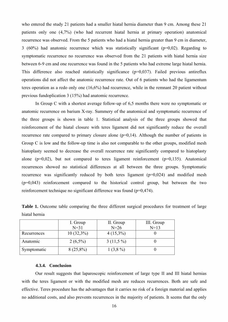
In Group C with a shortest average follow-up of 6,5 months there were no symptomatic or
anatomic recurrence on barium X-ray. Summery of the anatomical and symptomatic recurrence of
the three groups is shown in table 1. Statistical analysis of the three groups showed that
reinforcement of the hiatal closure with teres ligament did not significantly reduce the overall
recurrence rate compared to primary closure alone (p=0,14). Although the number of patients in
Group C is low and the follow-up time is also not comparable to the other groups, modified mesh
hiatoplasty seemed to decrease the overall recurrence rate significantly compared to hiatoplasty
alone (p=0,02), but not compared to teres ligament reinforcement (p=0,135). Anatomical
recurrences showed no statistical differences at all between the three groups. Symptomatic
recurrence was significantly reduced by both teres ligament (p=0,024) and modified mesh
(p=0,043) reinforcement compared to the historical control group, but between the two
reinforcement technique no significant difference was found (p=0,474).
Table 1. Outcome table comparing the three different surgical procedures for treatment of large
hiatal hernia
I. Group N=31
II. Group N=26
III. Group N=13
Recurrences 10 (32,3%) 4 (15,3%) 0
Anatomic 2 (6,5%) 3 (11,5 %) 0
Symptomatic 8 (25,8%) 1 (3,8 %) 0
4.3.4. Conclusion
Our result suggests that laparoscopic reinforcement of large type II and III hiatal hernias
with the teres ligament or with the modified mesh are reduces recurrences. Both are safe and
effective. Teres procedure has the advantages that it carries no risk of a foreign material and applies
no additional costs, and also prevents recurrences in the majority of patients. It seems that the only
17
limitation of this technique is the size of the hiatal hernia and possibly the weakness of the right
crus, therefore we presume that in such cases of extreme large hiatal hernia a prosthetic mesh would
be probably advisable. In order to avoid any mesh related complication we prefer to cover the mesh
with the omentum.
4.4. Duodeno-gastroesophageal reflux
4.4.1. Introduction
Pathologic duodenogastric (DGR) and duodeno-gastroesophageal reflux (DGERD) can be
detected in 10% to 50% of all reflux patients. Although duodenogastric reflux is a physiologic
phenomenon which occurs normally nights and postprandial, excessive or pathologic
duodenogastric reflux has been implied in the development of gastritis, gastric ulcer disease,
dyspepsia, and even carcinoma. When duodenogastric and duodeno-gastroesophageal reflux
present at the same time grade of the esophageal mucosa injury is strongly correlated with
the amount of bile and acid refluxated into the esophageal lumen. Studies showed that in
patients with more severe complication of GERD such as Barrett’s metaplasia or esophageal
strictures significantly more acid and bile was detected on esophageal pH and bilirubin
monitoring in the esophageal lumen, compared to healthy volunteers or in patients with less
severe grade of esophagitis. The effect of pathologic duodenogastric reflux and its toxic
components on the esophageal mucosa have been demonstrated also in several
experimental animal models.
Although many facts are known about the association of alkaline duodenogastric and
gastroesophageal reflux, the therapeutic implications are still controversial. One of the
reasons for that is, that the diagnosis of pathologic duodenogastric reflux, the accurate assessment
of this abnormal condition, is very difficult to establish. The detection of alkaline reflux and/or
the detection of its different components have been difficult and unreliable. Prior methodologies
employed for measuring DGER, including endoscopy, aspiration studies (both gastric and
esophageal), scintigraphy, and ambulatory pH monitoring, have technical difficulties and do not
accurately measure DGER. Currently, the most commonly used means of assessing DGER is the
ambulatory esophageal and gastric bilirubin monitoring system (Bilitec). Although measuring bile
reflux with Bilitec can help to ensure the diagnosis of abnormal duodenogastric and duodeno-
gastroesophageal reflux, indications for surgical intervention is still unanonymous. Furthermore the
precise surgical intervention is not yet widely agreed and accepted. The most often used technique
is a biliary diversion with Roux-en-Y suprapapillary duodenojejunostomy also called duodenal switch.
18
4.4.2. Patients and methods
In those patients where symptoms were suspectible for DGR or DGER beside our routine
preoperative workup (barium X-ray, endoscopy, manometry, 24-hour esophageal pH monitoring)
bilirubin monitoring with Bilitec 2000 was performed. Bilirubin exposure was analyzed above the
0,25 absorbance level in the stomach and 0,14 in the esophagus. Abnormal bile reflux was defined
when the exposure exceeded the 95th percentile of healthy controls, which was 24,8 in the stomach
and 11,8 in the esophagus. From 241 GERD patients who underwent a laparoscopic antireflux
procedure 45 patients had two-channel (esophagus and stomach) 24-hour bilirubin monitoring
before the operation because suspected duodeno-gastroesophageal reflux or because normal pH
monitoring results. A further 16 patients had Bilitec after their initial antireflux operation for
recurrent and atypical symptoms with normal 24-hour esophageal pH monitoring. From these 61
patients four (6,5%) underwent a duodenal switch procedure. They all were females with a mean
age of 41,8 years (range 32-53).
Surgical procedure
Our preference is to perform the biliary diversion procedure through a right subcostal upper
abdominal incision, but other authors prefer an upper midline incision. When the Roux-en-Y
suprapapillary duodenojejunostomy is begun, the duodenum is mobilized by a Kocher maneuver to
feel the head of the pancreas and especially the position of the papilla of Vater as accurately as
possible. Care must be taken to dissect an area around the duodenum well above the papilla and
without devascularizing the proximal duodenum. Using the thumb and index finger around the
duodenum, it is usually possible to feel the closest approximation at a point along the medial
duodenal border just proximal to the papilla at approximately 3 to 7 cm distal to the pylorus,
depending on the anatomic situation of the patient. At this point the duodenum is dissected free of
the head of the pancreas by carefully dividing the small vessels. It is essential to keep a minimal of
3 cm of postpyloric duodenum because only in this case remains enough receptor in this region for
duodenal inhibition of acid secretion. It is important to reduce the risk of jejunal ulcers. Precautions
must also be taken to avoid damage to the intrapancreatic common bile duct. When the channel
between pancreatic head and duodenum is completed, the duodenum can easily be divided and
closed with a linear stapling device. Since the dissection is carried down to the most distal point just
proximal of the papilla, it is not advisable to oversew the stapling suture to avoid obstruction of the
common bile duct. The first or second jejunal loop, depending on the vascular status of the
mesentery, is pulled supracolically into the area of the duodenal bulb through an opening in the
transverse mesocolon, and to the right of the midcolic vessels. A dissection point in the jejunal loop
is identified to prepare a limb that is long enough to complete the Roux-en-Y jejunojejunostomy
19
without tension. The distal jejunal loop is then sutured to the proximal duodenal stump in an end-to-
end anastomosis. The proximal jejunal limb is anastomosed end-to-side to the distal jejunal limb 40
to 50 cm distal to the duodenojejunal anastomosis. The opening in the mesocolon is closed.
4.4.3. Results
From the 45 patients who had Bilitec before their antireflux operation 18 (40%) patients had
abnormal duodeno-gastroesophageal reflux. After a successful laparoscopic fundoplication only one
(5,5%) patient had remnant symptoms suspectible for duodenogastric bile reflux. In this patient,
symptoms appeared after ERCP, EST and stone extraction from common bile duct followed by a
laparoscopic cholecystectomy. After the antireflux procedure, reflux symptoms disappeared and
only epigastric pain and loss of appetite remained. Conservative treatment was started with
sucralfate and prokinetic drugs with little effect. Because persistent symptoms of duodenogastric
reflux a duodenal switch was performed half year after the laparoscopic fundoplication. Functional
investigation before biliary diversion, showed no recurrent gastroesophageal reflux. At follow up
patient was symptom free, and she returned to her normal eating habit.
From the 16 patients who had bilirubin monitoring after the antireflux operation, due to
renewal of partially reflux symptoms and atypical epigastric symptoms, 3 (18,7%) had abnormal
duodenogastric reflux. They all had epigastric and right subcostal pain, weight loss and loss of
appetite. One of them also had typical symptoms of gastroesophageal reflux. The latter patients had
a laparoscopic cholecystectomy 2 years ago and in the same time gastrotomy and removal of a
gastric polypus was performed. Due to acute gastric hemorrhage reoperation was performed and the
site of polypectomy was sutured from a pyloromyotomy. One year after that a Toupet
fundoplication was performed because of gastroesophageal reflux disease. Barium swallow and
endoscopy showed a small hiatal hernia and esophagitis SM II grade. Abnormal esophageal acid
exposure was detected on 24-hour pH monitoring and incompetent LES was observed on
manometry. 24-hour bilirubin monitoring revealed abnormal duodenogastric and gastroesophageal
bile reflux. A duodenal switch and a revisional Toupet fundoplication were performed. One year
after surgery the patient was symptom free and gained 2,2 kg. At two year follow-up, reflux
symptoms renewed and functional testing proved recurrent abnormal esophageal acid exposure.
Patient was put on PPI and she is now free of symptoms.
The other two patients had antireflux operation for GERD and histologically proven
Barrett’s metaplasia one and two years earlier. One of them also had cholecystectomy before. They
had no reflux symptoms but epigastrial and right subcostal pain, weight loss and appetitelesness
were present. On 24-hour esophageal pH monitoring no abnormal acid reflux was observed.
Bilirubin monitoring showed excessive bile reflux in the stomach, but in the esophagus no abnormal

20
bile reflux was detected. A duodenal switch was performed in both patients. One of them had a
small wound healing problem after surgery and the other had reflux symptoms one year after
surgery but functional testing showed no abnormal acid reflux into the esophagus. The patient who
had wound healing disturbance had an abdominal hernia repair two years after the duodenal switch.
There were no intraoperative complications, or major perioperative morbidity. Mortality was
not observed. After an average of 24,3 months follow-up (range 21-30) all patients were symptom
free. There was no progression or regression of the two Barrett’s metaplasia. The three patients with
symptoms of weight loss regain 2,2; 2,5 and 12 kg respectively. One patient is still on PPI because
of recurrent gastroesophageal acid reflux. With this therapy she is symptom free.
4.4.4. Conclusion
Patients with suspected duodeno-gastroesophageal reflux must be studied thoroughly using
all current diagnostic measures to determine the probability of the pathophysiologic importance of
such a finding. If excessive alkaline reflux into the esophageal lumen is identified, the patient
should be clearly informed that the planned antireflux operation, usually a laparoscopic Nissen
fundoplication, may possibly be only the first step of the procedures that are necessary to bring
constant relief. If complaints diminished after the antireflux procedure there is no need for a
duodenal switch. Also in patients after primary antireflux operation for GERD appearance of
atypical, epigastric symptoms should always bring attention to abnormal duodenogastric reflux. A
preoperative medical therapy to neutralize alkaline refluxed should be the first step in those patient.
A duodenal diversion operation after laparoscopic antireflux surgery is indicated only in patients
who have had a history of continued severe symptoms despite medical therapy and have clearly
documented duodenogastric reflux. In these cases good results can be achieved with duodenal
switch procedure.
21
5. New findings
1. We suppose that there are two types of HLES patient. One with dysphagia and chest pain,
who respond well to cardiomyotomy and the other is the GER associated HLES in whom the
hypertension of the LES is a consequence or complication of gastroesophageal reflux. In these
patients instead of a cardiomyotomy a 360 degree fundoplication should be performed.
2. Achalasia could develope in the settings of long standing gastroesophageal reflux. There
are three factors that can predict GERD associated achalasia. First, if there is long standing
reflux symptoms in the patients’ history and reflux symptoms diminish after dysphagia
developed. The second suspicious factor is a hiatal hernia accompanying achalasia. And third is,
if pH monitoring shows abnormal acid reflux. The setpoint should be changed to pH 3 in these
cases to exclude artifacts from acidic fermentation in the esophageal lumen.
3. We suppose that if preoperative examinations produce a strong suspicion of achalasia,
which developed on the basis of GERD, a proper 360 degree fundoplication should be
considered after Heller’s myotomy, as anterior hemifundoplication does not protect fully against
reflux.
4. A new laparoscopic technique was developed in our institute to prevent recurrence after
laparoscopic repair of large hiatal hernia. The teres ligament was used to reinforce the hiatal
closure in patients treated for large hiatal hernia. This is a completely new operation and it was
never published before.
5. Although reinforcement of the hiatal closure with teres ligament was sufficient in most of
the cases, recurrence was found in patients with extreme large hiatal hernia and weakened right
crus. In these cases a modification of mesh cruroplasty was applied. This modification was also
developed by our unit. To reduce the risk of mesh associated complications the mesh was
covered with the omentum of the dissected greater curvature to avoid connection of the mesh
and the cardia.
6. Experience with duodenal switch procedure, which was designed to eliminate abnormal
bile reflux, has not been published in the Hungarian medical literature so far. We reported on
four cases.
22
6. Acknowledgement
First of all I would like to thank Professor Örs Péter Horváth, the consultant of my thesis for
the idea of new laparosopic procedures – the reinforcement of the hiatal closure with teres ligament
and the modified mesh cruroplasty - and for the encouragement of organising prospective trials to
evaluate the value of these new methods. I thank Professor Horváth for operating on most of these
patients and providing the circumstances to follow them up at the department as inpatients. I also
thank him for his continuous consultation and support. I thank him for trusting my judgement in
organisational tasks, statistics and English language publications. I am grateful to him for teaching
me surgery, how to operate, and especially for teaching me to perform laparoscopic upper GI
procedures.
I would like to thank Professor Karl Hermann Fuchs and his team for teaching me the
accurate performance and evaluation of foregut functional testing. I have also learned a lot from
Professor Fuchs by taking part in laparoscopic antireflux operations performed by him.
I would like to thank Dr. Ágnes Király and the workers of her gastrointestinal functional
laboratory for performing functional tests both for preoperative and follow-up investigations.
I thank very much all of my collegues, especially László Cseke, Katalin Kalmár, András
Papp who helped me performing the sometimes long lasting and boring laparoscopic procedures.
I also thank Csorbicsné Szabó Hortenzia for the assistance with upper gastrointestinal
endoscopies.
And at last but not least I would like to thank Professor Erzsébet Rőth, who was my tutor in
undergraduate research program and who inspired me in clinical research.
23
7. Publications
1. Articles in Periodicals (In connection with Thesis)
1. Varga G, Király A, Moizs M, Horváth OP. Effect of laparoscopic antireflux operation on
esophageal manometry, 24 hours pH-metry and quality of life in gastroesophageal reflux
disease. Acta Chir Hung. 1999;38:213-218.
2. Freys SM, Maroske J, Fein M, Varga G, Fuchs KH, Thiede A. Technik und
langzeitergebnisse der laparoskopischen fundoplicatio nach Nissen. Chir Gastroenterol.
2001; 173:33-37. IF: 0,078
3. Varga G, Cseke L, Kalmár K, Horváth OP. Prevention of recurrence by reinforcement of
hiatal closure using ligamentum teres in laparoscopic repair of large hiatal hernias. Surg
Endosc. 2004; 18:1051-1053. IF: 1,962
4. Király A, Illés A, Undi S, Varga G, Kalmár K, Horváth PO. Gastroesophageal reflux
disease progressing to achalasia. Dis Esophagus. 2005; 18:355-358. IF:0,936
5. Varga G, Cseke L, Kalmár K, Horváth OP. New laparoscopic procedure for the treatment
of large hiatal hernias: the first 20 cases. Magy Seb. 2005; 58:311-315.
6. Horváth OP, Kalmár K, Varga G. Reflux after Heller's myotomy for achalasia. Ann Surg.
2007; 245:502-503. IF:7,678
7. Varga G, Cseke L, Kalmar K, Horvath OP. Laparoscopic repair of large hiatal hernia with
teres ligament: midterm follow-up: A new surgical procedure. Surg Endosc. 2008; 22:881-
884 IF:1,969
8. Varga G, Cseke L, Kalmár K, Horváth OP. Surgical treatment of duodeno-
gastrooesophageal reflux disease: duodenal switch. Magy Seb. 2007; 60:243-247.
9. Varga G, Kiraly A, Cseke L, Kalmar K, Horvath OP. Effect of Laparoscopic
Fundoplication on Hypertensive Lower Esophageal Sphincter Associated with
Gastroesophageal Reflux. J Gastrointest Surg. 2008; 12:304-307. IF: 2,265
IFSumm:14,886
2. Book Chapters (In connection with Thesis)
1. Király Á, Róka R, Varga G. Barostat, Bilitec és nyelőcső impedancia vizsgálatok pp.287 In
Simon L, Lonovics J, Tulassay Zs, Wittman T. A gastroesophagealis reflux betegség
(GERD). Emésztőszervi és más szervrendszeri megjelenési formák. Astra Zeneca Könyvtár
kiadó Bp. 2003 ISBN 963 210 383 1
2. Varga G. Intraluminális eletromos impedancia és pH vizsgálat pp. 290-291 In Simon L,
Lonovics J, Tulassay Zs, Wittman T. A gastroesophagealis reflux betegség (GERD).
Emésztőszervi és más szervrendszeri megjelenési formák. Astra Zeneca Könyvtár kiadó Bp.
2003 ISBN 963 210 383 1
24
3. Horváth ÖP, Varga G. Laparoscopos antireflux műtétek pp.119-129 In Simon L, Lonovics
J, Tulassay Zs, Wittman T. A gastroesophagealis reflux betegség (GERD). Emésztőszervi és
más szervrendszeri megjelenési formák. Astra Zeneca Könyvtár kiadó Bp. 2003 ISBN 963
210 383 1
4. Fuchs KH, Varga G. Pathophysiologische Komponenten der gastroösophagealen
Refluxkrankheit pp. 5-13 In K.-H. Fuchs, S.M. Freys, M. Fein, A. Thiede. Lapaoskopische
Antirefluxchirurgie Dr.Reinhard Kaden Verlag GmbH, Heidelberg 2003 ISBN 3-922777-
46-5
5. Tigges H, Maroske J, Varga G, Fuchs KH. 45-51. Indikationstellung zur Antireflux
operation pp. 45-51 In K.-H. Fuchs, S.M. Freys, M. Fein, A. Thiede. Lapaoskopische
Antirefluxchirurgie Dr.Reinhard Kaden Verlag GmbH, Heidelberg 2003 ISBN 3-922777-
46-5
6. Maroske J, Fuchs KH, Freys SM, Fein M, Tigges H, Varga G, Thiede A. Laparoskopische
Antirefluxchirurgie in Deutschland pp 99-104 In K.-H. Fuchs, S.M. Freys, M. Fein, A.
Thiede. Lapaoskopische Antirefluxchirurgie Dr.Reinhard Kaden Verlag GmbH, Heidelberg
2003 ISBN 3-922777-46-5
7. Horváth ÖP, Varga G. A NERD kezelése. A sebész álláspontja. pp 131-137 In Lonovics J,
Simon L, Tulassay Zs, Wittmann T. A nem erozív reflux betegség Lumiere Budapest és
Lumiere Kobenhavn Budapest 2005 ISBN: 963 218 656 7
3. Abstracts published in Periodicals (In connection with Thesis)
1. Varga G, Király Á, Horváth ÖP. Relation between different investigating techniques in
patients with gastroesophageal reflux disease Zeitschrift für Gastroenterologie 1999; 37 (5):
453 (Abstract) IF.: 0,887
2. Varga G., Papp A., Király Á*., Horváth Ő.P The hypertensive lower esophageal sphincter:
is it a responce to gastroesophageal reflux? early reuslts Surg Endosc 2002 Suppl. (Abstract)
IF: 0,768
3. Varga G., Király Á*., Horváth Ő.P The effect of laparoscopic fundoplication on
hypertensive lower esophageal sphincter with associated gastroesophageal reflux disease
Surg Endosc 2003 Suppl (Abstract) IF:2,122
4. G.Varga, L. Cseke, Á Király*, O.P. Horváth Achalasia, hypertensive lower esophageal
sphincter and gastroesophageal reflux Dis Esophagus 2004 Suppl (Abstract) IF:0,797
5. G.Varga, L. Cseke, K. Kalmár, O.P. Horváth Prevention of recurrence by reinforcement of
hiatal closure using ligamentum teres in laparoscopic repair of large hiatal hernias – Early
Results of an ongoing prospective study Dis Esophagus 2004 Suppl (Abstract) IF:0,797
25
6. G. Varga, L. Cseke, K. Kalmár, ÖP Horváth. Laparoscopic repair of large hiatal hernia:
comperison of three different techniques and modification of mesh hiatoplasty to prevent
mesh related complications. Irish Journal of Medical Science 2007 Volume 176 Suppl 5
S208 (Abstract)
IFSumm:5,371
4. Presentations (In connection with Thesis)
1. Varga G., Papp A., Király Á*., Horváth ÖP. The hypertensive lower esophageal sphincter:
is it a responce to gastroesophageal reflux? Early reuslts. EAES Congress 2002 Lisbon,
Portugal
2. Varga G., Király Á*., Horváth Ö.P. The effect of laparoscopic fundoplication on
hypertensive lower esophageal sphincter with associated gastroesophageal reflux disease.
EAES Congress 2003 Glasgow, Scotland, UK
3. Varga G., Cseke L., Vereczkei A., Horváth ÖP. A hiatus oesophagei megerősítése
ligamentum Teres hepatis felhasználásával: Új laparoscopos műtéti technika a nagyméretű
hiatus hernia kezelésében. MGT 45. Nagygyűlése 2003, Balatonaliga, Hungary
4. Horváth ÖP., Varga G., Cseke L. A hiatus oesophagei megerősítése ligamentum Teres
hepatis felhasználásával: Új laparoscopos műtéti technika a nagyméretű hiatus hernia
kezelésében. MST Endoscopos Szekció, 2003 Kecskemét, Hungary
5. Varga G., Cseke L., Horváth ÖP. Laparoscopos „floppy” Nissen-DeMeester fundoplicatio -
a Würzburgi módszer. MST Endoscopos Szekció, 2003 Kecskemét, Hungary
6. Varga G., Cseke L., Horváth ÖP. Gastrooesophagealis reflux talaján kialakult achalasia –
esetismertetés. MST Endoscopos Szekció, 2003 Kecskemét, Hungary
7. G.Varga, L. Cseke, Á Király*, O.P. Horváth. Achalasia, hypertensive lower esophageal
sphincter and gastroesophageal reflux ISDE Congress 2004 Madrid, Spain
8. G.Varga, L. Cseke, K. Kalmár, O.P. Horváth. A New laparoscopic procedure for the
reinforcement of hiatal closure using ligamentum teres in repair of larga hiatal hernias –
Early Results of an ongoing prospective study ISDE Congress 2004 Madrid, Spain
9. G.Varga, L. Cseke, K. Kalmár, K.H. Fuchs*, O.P. Horváth. A New laparoscopic procedure
for the reinforcement of hiatal closure using ligamentum teres in repair of larga hiatal
hernias – Early Results of an ongoing prospective study EAES Congress 2005 Venice Italy
10. A.Papp, G. Varga, L. Cseke, Á. Király, ÖP Horváth. Gastroesophageal reflux progressing
to achalasia - report of three cases 2005 Venice Italy
11. G. Varga, L. Cseke, K. Kalmár, O.P. Horváth. Reinforcement of hiatal closure with
ligamentuum teres – prevention of recurrence after repair of large hiatal hernias. ISDE
Congress 2006 Adelaide, Australia
26
12. G. Varga, L. Cseke, K. Kalmár, ÖP Horváth. Laparoscopic repair of large hiatal hernia:
comperison of three different techniques and modification of mesh hiatoplasty to prevent
mesh related complications ESS Congress 2007 Dublin, Ireland
5. Articles in Periodicals (Not in connection with Thesis)
1. Gál I, Róth E, Lantos J, Varga G, Jaberansari MT. Inflammatory mediators and surgical
trauma regarding laparoscopic access: free radical mediated reactions. Acta Chir Hung.
1997;36:97-99.
2. Jaberansari MT, Róth E, Gál I, Lantos J, Varga G. Inflammatory mediators and surgical
trauma regarding laparoscopic access: acute phase response. Acta Chir Hung. 1997;36:138-
140.
3. Róth E, Lantos J, Temes G, Varga G, Paróczai M, Kárpáti E. Cardioprotection during heart
ischemia-reperfusion. Acta Chir Hung. 1997;36:306-309.
4. Varga G, Gál I, Róth E, Lantos J, Jaberansari MT. Inflammatory mediators and surgical
trauma regarding laparoscopic access: neutrophil function. Acta Chir Hung. 1997;36:368-
369.
5. Rőth E, Nemes J, Kapronczay P, Varga G, Borsiczky B: A szabad gyökös reakciók és az
endogén antioxidáns rendszer vizsgálata essentialis hypertoniás betegekben Hypertonia és
Nephrológia 1997;1:183-187
6. Gál I, Röth E, Lantos J, Varga G, Mohamed TJ, Nagy J. Surgical trauma induced by
laparoscopic cholecystectomy Orv Hetil. 1998;139:739-746.
7. Rőth E, Nemes J, Kapronczay P, Varga G, Jaberansari MT, BorsiczkyB. Free radical
reactions and the endogenous antioxidant system in essential hypertension. European
Journal of Internal Medicine 1998; 9:263-270.
8. Vereczkei A, Varga G, Pótó L, Horváth OP. Management of corrosive injuries of the
esophagus. Acta Chir Hung. 1999;38:119-122.
9. Nemes J, Rőth E, Kapronczay P, Nagy S, Mózsik Gy, Varga G, Borsiczky B: A renin-
angiotenzin-aldoszteron-katekolamin, lipidperoxidáció és az endogén antioxidáns
rendszerek kapcsolata essentiális hypertóniás betegekben-egyhetes moxonodin (CYNT)-
kezelés hatására Magy Belorv Arch 1999;52:87-92
10. Horváth Ö.P., Cseke L., Papp A., Varga G., Horváth G., Kalmár K.: A nyelőcső és a
gyomorrák neoadjuváns kezelése. Eur. J. Gastroenterol. Hepatol. IV. . 2000;2:41-45
11. Horváth OP, Cseke L, Papp A, Kalmár K, Varga G, Horváth G. Larynx-preserving
pharyngoesophagectomy in the treatment of cancer of the pharyngoesophageal junction
Magy Seb. 2000;53:189-192.
27
12. Horváth OP, Cseke L, Kalmár K, Varga G, Horváth G. Larynx-preserving pharyngo-
esophagectomy after chemoradiation in the treatment of cancer of the pharyngo-esophageal
junction. Ann Thorac Surg. 2001;72:2146-2147. IF: 2,141
13. Papp A, Cseke L, Pavlovics G, Farkas R, Varga G, Márton S, Pótó L, Esik O, Horváth OP.
The effect of preoperative chemo-radiotherapy in the treatment of locally advanced
squamous cell carcinoma in the upper- and middle-thirds of the esophagus Magy Seb.
2007;60:123-129.
IFSumm:2,141
6. Abstracts published in Periodicals (Not in connection with Thesis)
1. Rôth E., Varga G., Szmolenszky A. M. Measurement of citrate synthase activity by
monoclonal antibodies during myocardial ischemia and reperfusion. Eur. Surg. Res. 1995;
27: 73-74. (Abstract) IF.: 0,754
2. Szmolenszky A. M., Rôth E., Varga G. Citrate synthase as a marker of early myocardial
necrosis determined by monoclonal antibodies. Shock 1995; 3: 159 (Abstract) IF.: 2,785
3. Rôth E., Varga G., Szmolenszky A. M. Changes of citrate synthase enzyme activity
compared with creatine kinase following heart ischemia. Shock 1995; 3: 160. (Abstract) IF.:
2,785
4. Varga G, Rôth E, Szmolenszky AM. Changes of citrate synthase enzyme activity compared
with creatin kinase following heart ischaemia. Magyar Sebészet 1996; 49: 181 (Abstract)
5. Gál I., Rőth E., Varga G., Jaberansari M.T., Nagy J. Changes of interleukin-6 (IL-6) levels
in the monitoring of surgical trauma. Eur. Surg. Res 1997; 29: 100 (Abstract) IF.: 0,754
6. Rőth E., Lantos J., Varga G., Jaberansari M.T., Paróczai M., Kárpáti E. Free radical
mediated reactions during healing of myocardial infarction Eur. Surg. Res 1997; 29: 122
(Abstract) IF.: 0,754
7. Borsiczky B., Varga G., Jaberansari M. T., Rôth E.: Protracted monitoring of free radical
mediators during myocardial infarction. Shock 1997; 8: 176 (Abstract) IF.: 2,785
8. BorsiczkyB, Jaberansari MT, Varga G, RőthE, Zadravecz Gy. Do the free radicals play a
role in the pathomechanism of haemarthrosis over time? Eur. Surg. Res 1998; 30: 41
(Abstract) IF.: 0,754
9. Varga G, Jaberansari MT, Borsiczky B, Rőth E, Paroczai M. Does bisaramil (a new anti-
arrhytmic drug) have any cardioprotective and haemodynamic effect in an experimental
ischemia–reperfusion model? Magyar Sebészet 1998; 50: 188 (Abstract)
10. Varga G, Cseke L, Pataki N, Horváth ÖP. Mesenterio-axiális gyomorvolvulus műtéti
megoldása laparoszkópos gasztropexiával – Esetismertetés Magyar Sebészet 1998; 50: 188
(Abstract)

28
11. Jaberanasari MT, Borsiczky B, Varga G, Rőth E, Paróczai M. Cardioprotective and
haemodynamic effects of a new antiarrithmic drug under an ischaemic-reperfusion modell
Eur Surg Res 1998; 30: 160 (Abstract) IF.: 0,754
12. Gál I, Rőth E, Varga G, Borsiczky B, Szekeres Gy. Comperative experimental study of
early healing process in small bowel anastomoses performing intracorporeally by
laparoscopic and open surgery Eur Surg Res 1998; 30: 165 (Abstract) IF.: 0,754
13. Vereczkei A, Kalmár K, Varga G, Cseke L, Horváth ÖP. The effects of cisapride on
stomach, jejunum and colon serving as a neoesophagus following esophageal resection Gut
1999; 45 Suppl.5: P1199, (Abstract) IF.: 5,386
14. Kelemen D, Varga G, Rőth E, Horváth ÖP. Kísérletes pancreas transzplantació új módszere
Magyar Sebészet 1999; 52: 210 (Abstract)
15. Báthori Zs., Király Á., Czimmer J., Sűtő G., Hunyady B., Varga G., Horváth Ö.P., Mózsik
Gy.: Is there any correlation between the macroscopic appereance of inflammation and
functional integrity of the chemo-and mechanoreceptors of the esophagus in patients with
esophageal reflux diseases Z. Gastroenterol. 2000. 38., 399. IF.: 0,887
16. Király Á., Czimmer J., Sütő G., Hunyady B., Varga G., Horváth Ö.P., Mózsik Gy.: Effect
of capsaicin-containing red pepper sauce suspension on esophageal motility and sensory
parameters in patients with esophageal reflux diseases Z. Gastroenterol. 2000. 38., 411. IF.:
0,887
17. Fein M, Fuchs KH, Varga G, Freys SM, Maroske J, Tigges H, Sailer M, Thiede A.
Ineffective esophageal motility and acid exposure. Gastroenterol 2001; 120: 5 Suppl.1. 2188
(Abstract) IF.: 13,02
18. Kalmár K, Cseke L, Varga G, Papp A, Illényi L, Kelemen D, Káposztás Zs, Stefanits K,
Varga E, Horváth ÖP: Tapasztalataink ECF neoadjuváns kemoterápiával lokálisan
előrehaladott gyomorrákban Onkológia 2003;47 (3): 271 (Abstract)
19. A Papp, L Cseke, G Varga, K Kalmár, G Horváth, S Márton, ÖP Horváth: Chemo-
radiotherapy in locally advanced oesophageal cancer – are upper third tumours more
responsive? Diseases of the Esophagus 17 Suppl 1: A41, 2004. IF: 0,797
20. A. Papp, L. Cseke, G. Pavlovics, R. Farkas, S. Márton, G. Varga, L. Potó, ÖP Horváth.
Locally advanced squamous cell cancer of the cervical oesophagus: the role of multimodal
therapy. Irish Journal of Medical Science 2007 Volume 176 Suppl 5 S216 (Abstract)
IFSumm:33,856
29
7. Presentations (Not in connection with Thesis)
1. Varga G, Rôth E, Szmolenszky AM. Changes of citrate synthase enzyme activity compared
with creatin kinase following heart ischaemia. MST Kisérletes Sebészeti Kongresszus
1996,Hungary
2. Varga G, Jaberansari MT, Borsiczky B, Rőth E, Paroczai M. Does bisaramil (a new anti-
arrhytmic drug) have any cardioprotective and haemodynamic effect in an experimental
ischemia–reperfusion model? MST Kongresszus 1998 Budapest, Hungary
3. Varga G, Cseke L, Pataki N, Horváth ÖP. Mesenterio-axiális gyomorvolvulus műtéti
megoldása laparoszkópos gasztropexiával – Esetismertetés MST Kongresszus 1998
Budapest, Hungary
4. Varga G, Király Á, Horváth ÖP. Relation between different investigating techniques in
patients with gastroesophageal reflux disease MGT 41. Nagygyűlése 1999, Balatonaliga,
Hungary
5. G.Varga, M.Fein, K.H.Fuchs, S.M.Freys, J.Maroske , H.Tigges . Indication, technique and
results of laparoscopic treatment of achalasia. ISDE Congress 2001 Sao Paolo, Brasil
6. G.Varga, M.Fein, K.H.Fuchs, S.M.Freys, J.Maroske , H.Tigges, A. Thiede Interaction
between gastric pH, Helicobacter pylori and duodenogastric reflux in patients with
gastroesophageal reflux disease ISDE Congress 2001 Sao Paolo, Brasil
7. Varga G.,Fein M.,Freys S.,Marosle J.,Tigges H., Fuchs K. Az achalasia sebészi kezelésének
korai eredményei MST Kongresszus 2001 Pécs
8. Varga G, Cseke L, Horváth ÖP. Gastrooesophageális reflux ritka szövődménye: Barrett
oesophagus talaján kialakult nyelőcső adenocarcinoma és subglotticus trachea stenosis –
esetismertetés MST Kongresszus 2002, Hungary
9. Varga G, Cseke L, Horváth Ö.P. Subglottic trachea stenosis as a consequence of long time
gastroesophageal reflux - Case report MGT 44. Nagygyűlése 2002, Balatonaliga, Hungary
10. A. Papp, L. Cseke, G. Pavlovics, R. Farkas, S. Márton, G. Varga, L. Potó, ÖP Horváth.
Locally advanced squamous cell cancer of the cervical oesophagus: the role of multimodal
therapy. ESS Congress 2007 Dublin, Ireland
8. Invited lectures:
1. Varga G, Cseke L, Kalmár K, Horváth ÖP. Nagyméretű hiatus hernia kezelése: Új
laparoscopos módszer III. Pécsi Nyelőcsőnap 2004, Pécs, Hungary
2. Varga G. Nyelőcső motilitászavarok a sebész szemszögéből. III. Pécsi Nyelőcsőnap 2004,
Pécs, Hungary
3. G. Varga, L. Cseke, K. Kalmár, Ö. P. Horváth. Difficult closure of hiatus with Ligamentum
teres – prevention of recurrence-Early results. EAES Congress 2005 Venice, Italy
30
4. Varga G, Cseke L, Horváth ÖP. A duodeno-gastroesophagealis refluxbetegség diagnózisa
és sebészi kezelésére MST Jubileumi kongressusa 2006 Budapest, Hungary
5. Varga G. Die Beziehung Achalasie und Hiatus Hernie Symposium und Kurs
Gastrointestinale und Kolorectale Funktionsstörungen 2007 Frankfurt , Germany
9. Participation in Research Grants
1. ÖP Horváth, G Varga, K Kalmár, Á Király, I Kiss, E Kálmán, T Aikou, H Feussner, KH
Fuchs: A gyomor és a gastro-oesophagealis átmenet rosszindulatú daganatainak komplex
kezelése. FKFP-0361/2000
2. FT Molnár, ÖP Horváth, Á Király, E Kálmán, I Kiss, G Varga, K Kalmár: A gastro-
oesophagealis reflux betegség szövõdményeinek pathogenesise, megelõzése és kezelése.
OTKA 2001
3. ÖP Horváth, L Cseke, D Kelemen, K Kalmár, G Varga: Aborális pótgyomor prospektiv
randomizált összehasonlitása hagyományos orális pótgyomorral totális gastrectomia után.
OTKA T 042726/2003
4. Horváth ÖP, Cseke L, Kalmár K, Papp A, Varga G: Módszerek a nyelőcsőrák sebészi
kezelése eredményeinek javítására. ETT 2003.
IFCumulaiíve: 56,254