phage therapy of local and systemic disease caused by ... · phage therapy of local and systemic...
TRANSCRIPT
INFECTION AND IMMUNITY, Nov. 2002, p. 6251–6262 Vol. 70, No. 110019-9567/02/$04.00�0 DOI: 10.1128/IAI.70.11.6251–6262.2002Copyright © 2002, American Society for Microbiology. All Rights Reserved.
Phage Therapy of Local and Systemic Disease Caused by Vibriovulnificus in Iron-Dextran-Treated Mice
Karen E. Cerveny,1 Angelo DePaola,2 Donna H. Duckworth,1 and Paul A. Gulig1*Department of Molecular Genetics and Microbiology, College of Medicine, University of Florida, Gainesville,
Florida 32610,1 and Gulf Coast Seafood Laboratory, U.S. Food and Drug Administration,Dauphin Island, Alabama 365282
Received 28 January 2002/Returned for modification 29 March 2002/Accepted 29 July 2002
Vibrio vulnificus is a gram-negative bacterium that contaminates filter-feeding shellfish such as oysters. Afteringestion of contaminated oysters, predisposed people may experience highly lethal septicemia. Contaminationof wounds with the bacteria can result in devastating necrotizing fasciitis, which can progress to septicemia.The extremely rapid progression of these diseases can render antibiotic treatment ineffective, and death is afrequent outcome. In this study, we examined the potential use of bacteriophages as therapeutic agents againstV. vulnificus in an iron-dextran-treated mouse model of V. vulnificus infection. Mice were injected subcutane-ously with 10 times the lethal dose of V. vulnificus and injected intravenously, either simultaneously or atvarious times after infection, with phages. Treatment of mice with phages could prevent death; systemicdisease, as measured by CFU per gram of liver and body temperature; and local disease, as measured by CFUper gram of lesion material and histopathologic analysis. Two different phages were effective against threedifferent V. vulnificus strains with various degrees of virulence, while a third phage that required the presenceof seawater to lyse bacteria in vitro was ineffective at treating mice. Optimum protection required that thephages be administered within 3 h of bacterial inoculation at doses as high as 108 PFU. One of the protectivephages had a half-life in blood of over 2 h. These results demonstrate that bacteriophages have therapeuticpotential for both localized and systemic infections caused by V. vulnificus in animals. This model should beuseful in answering basic questions regarding phage therapy.
Although research regarding the therapeutic use of bacte-riophages has gone on without interruption since 1926 in East-ern Europe, this field of study has been, until very recently,somewhat neglected in the West (for reviews, see references 10and 39). Within the past two decades, however, there has beena renewed interest in phage therapy due primarily to the in-creasing incidence of antibiotic-resistant bacteria and the lackof development of new types of antibiotics to control infectionscaused by these antibiotic-resistant organisms. The efficacy ofphages in treating bacterial disease has been demonstrated byusing animal models for Escherichia coli (21, 31–34), Salmo-nella enterica serovar Typhimurium (2), and Pseudomonasaeruginosa (35, 36). Phages have also been used successfully toprevent bacterial disease in fish (26) and to control pathogensof tomatoes (12). Although it is unlikely that phages will everreplace antibiotics, they may be of some use when no effectiveantibiotics are available or in conjunction with antibiotics forbetter treatment of disease. To examine the potential useful-ness of phages, either alone or in conjunction with antibiotics,phages will have to be studied in a variety of animal modelsagainst bacteria with different mechanisms of virulence. Tocontribute to this understanding of where and how phage ther-apy may be appropriate, we have begun studying the therapeu-tic effect of phages against V. vulnificus.
V. vulnificus is an opportunistic pathogen of humans thatcauses septicemia after ingestion of contaminated oysters and
necrotizing fasciitis after contamination of wounds (reviewedin references 19 and 38). Septicemia occurs primarily in peoplewith high levels of iron saturation caused by genetic mutation,such as primary hemochromatosis, or by liver damage (cirrho-sis). Immunosuppressed individuals and people with diabetesare also at risk (4, 6, 18, 43). Septicemia is characterized byfever, chills, and bullous skin lesions on the lower extremitiesand has a mortality rate of greater than 50% (14). Woundinfection leads to necrotizing fasciitis, which is characterized byextensive tissue damage down to, but not usually including, themusculature and can necessitate surgical intervention for de-bridement or amputation. Wound infection can occur in theabsence of predisposing conditions but progresses more fre-quently to septicemia and has a higher mortality rate in pre-disposed people.
V. vulnificus is a halophilic, gram-negative, curved rod thatthrives in tropical and temperate estuarine environmentsthroughout the world. The bacteria are found in filter-feedingshellfish, primarily oysters. Estimates of the prevalence of V.vulnificus in oysters from the Gulf of Mexico during the sum-mer months have been as high as nearly 100% (23). Bacterio-phages for V. vulnificus also are frequently found in oysters andestuarine waters (8, 9, 27). V. vulnificus is highly geneticallydiverse, and single oysters can contain over 100 differentstrains (5). Despite the diversity of strains present in oysters,one study demonstrated that only single strains of V. vulnificuswere recovered from the blood of patients who had lethalinfections and who had consumed oysters contaminated withnumerous strains (15), suggesting that not all strains possessequal potential for human disease.
Little is known about the virulence mechanisms of V. vulni-
* Corresponding author. Mailing address: Department of MolecularGenetics and Microbiology, Box 100266, College of Medicine, Univer-sity of Florida, Gainesville, FL 32610-0266. Phone: (352) 392-0050.Fax: (352) 392-3133. E-mail: [email protected].
6251
on Decem
ber 1, 2015 by Wageningen U
R Library
http://iai.asm.org/
Dow
nloaded from
ficus. The primary virulence factor is the polysaccharide cap-sule, which prevents phagocytosis and activation of comple-ment (1, 30, 41, 42, 45, 46), classifying V. vulnificus as anextracellular pathogen. The ability to acquire iron from thehost via siderophore production is also an essential virulenceattribute (20). The production of a prepilin peptidase of a type2 secretion system, which exerts pleiotropic effects on numer-ous secreted proteins, is required for full virulence in mice(24). Two other putative virulence factors, hemolysin and met-alloprotease, have failed to be confirmed as virulence factorsby genetic analysis (11, 16, 29, 44), despite the fact that injec-tion of the purified proteins into laboratory animals inducesseveral symptoms of V. vulnificus infection (13, 17, 22, 25). Aniron-dextran-treated mouse model of V. vulnificus disease waspreviously used to compare the virulence of three clinical iso-lates with that of three environmental strains isolated fromoysters or seawater (37). It appeared that the environmentalstrains either grew more slowly in or were killed more effec-tively by the host.
The availability of a useful animal model to examine viru-lence, the existence of bacteriophages for V. vulnificus, and theextracellular nature of the disease process led us to use V.vulnificus as a model for testing the effectiveness of phagetherapy for human disease. We show here that phage treat-ment of iron-dextran-treated mice infected subcutaneously(s.c.) with V. vulnificus can prevent both local and systemicdisease.
(These results were presented in preliminary form at the100th General Meeting of the American Society for Microbi-ology in Los Angeles, Calif. [K. E. Cerveny, T. J. Doyle, G. M.Escudero, D. H. Duckworth, and P. A. Gulig, Abstr. 100thGen. Meet. Am. Soc. Microbiol., abstr. D230, p. 279, 2000] andthe 13th Annual International Phage Biology Meeting in Mon-treal, Quebec, Canada [K. Cerveny, T. J. Doyle, A. DePaola, P.Gulig, and D. Duckworth, Abstr. Millennial Phage Biol. Meet.,2000]).
MATERIALS AND METHODS
Bacterial strains and growth media. We used four clinical isolates of V.vulnificus, M06-24/0, VV1009, 2400112, and NSV-5829, obtained from patientswho died from sepsis following ingestion of contaminated oysters, and environ-mental strains MLT403, MLT365, MLT367,and 99-796DP-E7, isolated fromeither oysters or seawater (7, 15, 37, 40, 45). The four clinical isolates and strain99-796DP-E7 were highly virulent in s.c. inoculated, iron-dextran-treated mice,while the environmental strains were less virulent, although still able to causedisease at higher doses (37; A. DePaola, J. L. Nordstrom, A. Dalsgaard, J. Oliver,T. Baytes, K. L. Bourdage, and P. A. Gulig, unpublished data). V. vulnificusstrains were grown in Luria-Bertani broth containing 0.85% (wt/vol) NaCl(LB-N) or on LB-N plates containing 1.5% (wt/vol) agar. Luria-Bertani brothcontaining seawater (Sigma) (LB-SW) or sea salts (Sigma) adjusted to a total saltconcentration of 0.85% (LB-SS) was used when sea salts were required for phageactivity. Soft-agar overlays of each medium contained 0.75% (wt/vol) agar. Bac-teria were grown by diluting a static overnight culture into prewarmed medium,1:10 for phage preparations or 1:20 for the animal infections, and then shakingthe mixture at 37°C until the bacteria reached approximately 108 CFU/ml, asdetermined by measuring the optical density at 600 nm. For titration of phages,bacteria were grown to a density of between 1.5 � 108 and 2.5 � 108 CFU/ml, asdetermined by measuring the optical density at 600 nm. Preliminary experimentsdemonstrated that this stage of growth allowed maximum plating efficiency of thephage. For infection of mice, cultures were grown in LB-N, harvested, anddiluted in phosphate-buffered saline containing 0.01% (wt/vol) gelatin (BSG)(28) exactly as described previously (37).
Phage isolation, growth, and titration. Phage CK-2 was isolated from estua-rine sediments from Cedar Key, Fla. A slurry of 50 ml of sediment and 50 ml of
LB-N was mixed with 5 ml each of LB-N cultures of V. vulnificus strains VV1009,LL728, 2400112, MLT403, MLT365, and MLT367. The mixture was shakenovernight at 30°C. On the next day, the mud and bacteria were removed bycentrifugation at 10,000 � g for 10 min, and the supernatant was filtered througha 0.2-�m-pore-size filter. Phage activity in the supernatant was amplified bymixing with an equal volume of a static overnight culture of each of the six V.vulnificus strains separately and shaking the mixtures overnight at 30°C. On thenext day, the cultures were centrifuged and filtered as described above, and 10 �lof the filtrates was dropped onto LB-N agar plates seeded with a lawn of each ofthe bacterial strains. Plaques appeared on plates seeded with MLT403 but notwith any of the other V. vulnificus strains. The phage from the MLT403 lysate wasthen plaque purified and amplified. This phage was designated CK-2. It producedvery clear plaques on MLT403 grown in LB-N but had no activity on any of theother V. vulnificus strains that we examined.
Phages 153A-5 and 153A-7 were isolated from oysters as described previously(8, 9) and formed plaques on several of the virulent clinical strains, but phage153A-7 formed plaques only when the vibrios were suspended in seawater-containing medium, either LB-SS or LB-SW. Broth and soft-agar lysates of thesephages were produced by using strain MO6/24-0 as the host and standard pro-cedures (28). Phages 153A-5 and 153A-7 had relatively broad host ranges, form-ing plaques on five of seven and seven of seven V. vulnificus strains tested,respectively.
Phage titers were determined in two ways: by dropping 10 �l of a phagedilution onto a soft-agar overlay containing 106 CFU/ml of bacteria or by mixinga phage dilution with bacteria, adding soft agar, and pouring the mixture onto anagar plate.
Infection of mice. Seven- to 10-week-old female ICR mice (Harlan Sprague-Dawley, Indianapolis, Ind.) housed under specific-pathogen-free conditions wereused for all infections. Infections were performed as described previously (37).Briefly, mice were injected intraperitoneally (i.p.) with 250 �g of iron dextran(Sigma)/g of body weight 2 h to 30 min prior to inoculation with bacteria. Micewere injected s.c. with 0.1 ml of bacteria suspended in BSG in the lower portionof the back. Bacteriophages suspended in BSG (0.1 ml) were injected intrave-nously (i.v.) into the lateral tail vein at various times after bacterial inoculation.Control mice not receiving phages were injected with sterile BSG. Mice weremonitored for evidence of disease by visual inspection as well as rectal temper-ature. Mice with temperatures of �33°C were considered moribund and wereeuthanized. Euthanasia was done by carbon dioxide asphyxiation. We measuredfour quantitative criteria for disease: temperature, lesion score, number of bac-teria in tissues, and histologic features. Differences in quantitative analyses ofinfection were examined by using a two-tailed Student t test. All animal exper-iments were approved by the Institutional Animal Care and Use Committee ofthe University of Florida.
Temperature. The rectal temperature of the mice was recorded at the time ofeuthanasia to objectively quantify the state of disease. It was previously shownthat the body temperature of V. vulnificus-infected mice significantly drops in thelater stages of disease (37). If a mouse died before it could be euthanized, thetemperature assigned was the lowest temperature in the surviving infected micefor the given experiment or 33°C, whichever was lowest, for the purpose ofstatistical analysis. For analysis of survival, a temperature of 33°C or less wasscored as a death.
Lesion score. The skin was peeled back from head to tail, revealing the s.c.tissues of the back. The s.c. lesion was photographed along with a 2-cm2 stan-dard. Photographs of lesions were scored for severity in a blinded manner byusing the following scale: 0, no visible change; 1, lesion with discoloration but nohemorrhage; 2, lesion with hemorrhage but smaller than 2 cm2; 3, lesion largerthan 2 cm2. Because lesion scores are not normally distributed, only the mean isshown, with no statistical analysis.
Quantitative analysis of bacteria in tissues. Tissue samples from s.c. lesionsand from livers were removed from the mice, weighed, homogenized in 5 ml ofBSG by using glass tissue homogenizers, diluted, and plated to enumerate CFUper gram of tissue. If a mouse died before it could be euthanized and examined,the presence of a typical skin lesion was visually confirmed and photographed,and the levels of tissue infection assigned were the highest levels among thesurviving mice in the same group. When no bacteria were found in an undilutedsample, the minimum detectable level of infection was assigned for the purposeof statistical analysis.
Survival of mice. Survival rates for mice were calculated as a combination ofactual deaths and a rectal temperature of �33°C at the time of euthanasia.Survival rates were examined for significance by using �2 analysis.
Histologic analysis. We examined the histopathologic features of a subset ofthe infected mice to determine whether phage therapy affected microscopicdamage. Tissues of s.c. lesions were collected immediately after euthanasia, fixed
6252 CERVENY ET AL. INFECT. IMMUN.
on Decem
ber 1, 2015 by Wageningen U
R Library
http://iai.asm.org/
Dow
nloaded from
in 10% (vol/vol) buffered formalin, embedded in paraffin, sectioned, mounted,and stained with hematoxylin and eosin as described previously (37).
Determination of phage half-life in mice. Uninfected mice were injected i.v.with108 PFU of phage 153A-5. At 1, 6, and 12 h postinjection, mice wereeuthanized, and the peritoneal cavity was subjected to lavage with 4 ml ofphosphate-buffered saline. The lavage fluid was collected by needle and syringe.A sample of liver was then removed, weighed, and homogenized in BSG. Finally,10 �l of cardiac blood was collected in 0.05 M EDTA. All of the samples wereexamined for PFU on V. vulnificus MO6/24-0 as described above.
RESULTS
Analysis of V. vulnificus MLT403 and phage CK-2. To ex-amine the possible usefulness of bacteriophages in treatingdisease caused by V. vulnificus, we isolated phages which ex-hibited lytic activity against V. vulnificus. Phage CK-2 was iso-lated from estuarine sediments of Cedar Key, Fla., and washighly lytic, forming clear plaques on host V. vulnificus strainMLT403. MLT403 was the most virulent of the previouslydescribed environmental strains; however, its minimum lethaldose for iron-dextran-treated mice was 500-fold higher thanthat of clinically derived strains such as MO6-24/0 (37).
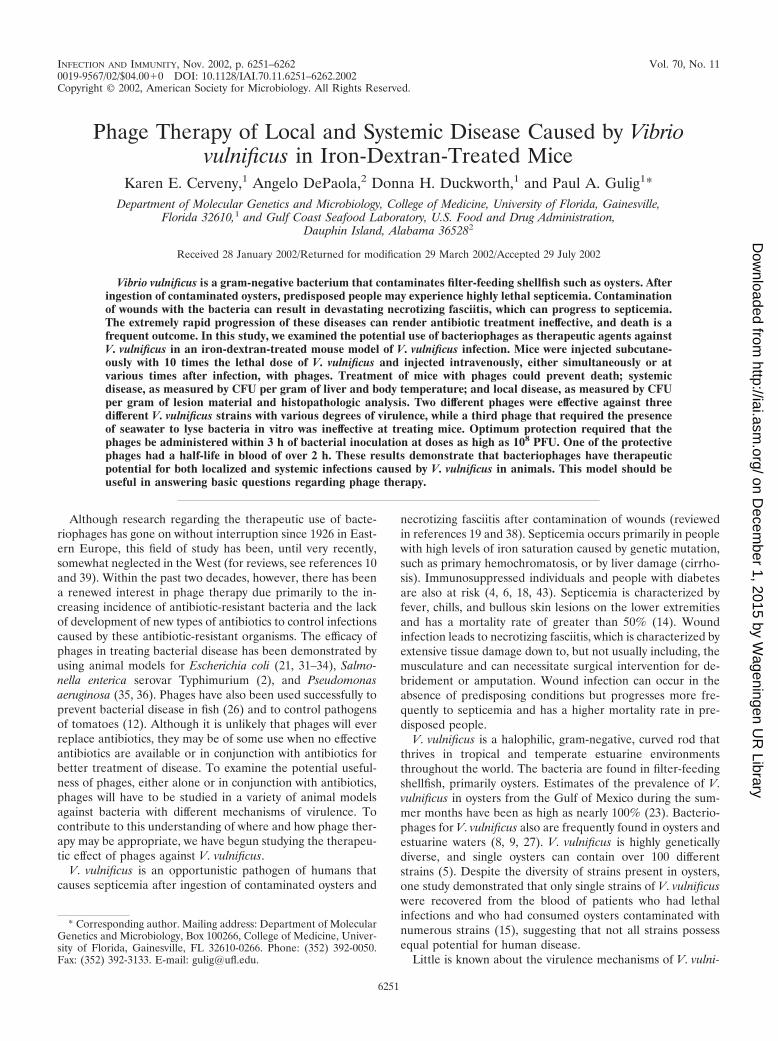
To determine whether phage CK-2 could protect against V.vulnificus infection, mice were injected i.p. with iron dextran,inoculated s.c. with 106 CFU (100 times the lethal dose) ofMLT403, and immediately injected i.v. with 108 PFU of phage.Control mice were treated with iron dextran and infected withMLT403 but received BSG instead of phage. After 14 h ofinfection, all four of the untreated (control) mice were visiblyill, lethargic, and scruffy, with rectal temperatures ranging from27.5 to 28.2°C (Fig. 1). All control mice had large s.c. lesionswith a score of 3 (out of a maximum of 3) and with character-istic hemorrhage and edema at the site of inoculation. Thecontrol mice had a mean of 108 CFU/g of lesion tissue, andtheir livers contained a mean of nearly 105 CFU/g of tissue. Incontrast, the four mice in the phage-treated group appeared to
be only slightly ill and had normal rectal temperatures of 36.5to 37.6°C. Phage-treated mice euthanized at 18 h postinfectionhad lesions with a mean score of 2.3 and with some discolor-ation but little edema. Two of these mice had undetectablebacteria in their lesion samples and were assigned a minimumdetectable level of log 3.2 and log 2.9 CFU/g (these numbersare different because different amounts of tissue were sam-pled), while the other two mice had log 7.6 and log 4.8 CFU/gof lesion tissue. For all three criteria used to measure disease,the phage-treated mice were significantly protected comparedwith the control mice. The survival, determined by countingeither actual deaths or a temperature of �33°C, of controlmice was none of four at 18 h; in comparison, four of fourphage-treated mice survived (�2 analysis; P � 0.005).
To determine whether the protection that we observed inthe first experiment was only a delay in the onset of disease orwhether the mice were destined to remain healthy, a long-termprotection experiment was conducted. As in the first experi-ment, after 1 day of infection, control mice had severe lesionswith high numbers of CFU in the skin and liver and depressedrectal temperatures, while phage CK-2-treated mice were sig-nificantly protected from CFU in the skin and liver (data notshown). A second set of six phage-treated mice was observedfor 8 days. One of these mice died on day 5 of an unknowncause, and the remaining mice continued to appear healthyand were euthanized on day 8; no detectable bacteria werefound in their skin or liver. Therefore, mice treated with CK-2appeared to be protected from disease for the long term.
In a final set of experiments with phage CK-2, we examinedhow long phage treatment could be delayed without affectingits efficacy by injecting the phage 6 and 12 h after bacterialinoculation. As described above, V. vulnificus-infected micewere either treated with buffer (control) or 108 PFU of phageCK-2 at various times postinfection. Of the 10 mice in the
FIG. 1. Short-term protection of V. vulnificus MLT403-infected mice by phage CK-2. Iron-dextran-treated mice were inoculated s.c. with 106
CFU of V. vulnificus MLT403. Mice were immediately injected i.v. with BSG (C) or phage CK-2 (CK-2), and samples were harvested 14 to 18 hpostinfection. Data are means and standard deviations for log CFU per gram of skin lesion (Lesion CFU), log CFU per gram of liver (Liver CFU),and rectal temperature (Temp). The temperature was divided by 10 and then plotted to fit the same scale as the other values. For lesion score(Lesion), the mean is shown. All groups contained five animals. An asterisk indicates that the difference between control and phage-treated groupswas statistically significant, as determined by the Student t test, as follows: lesion CFU, P � 0.016; liver CFU, P � 1.6 � 10�4; temperature, P �7.6 � 10�8.
VOL. 70, 2002 PHAGE THERAPY FOR V. VULNIFICUS DISEASE 6253
on Decem
ber 1, 2015 by Wageningen U
R Library
http://iai.asm.org/
Dow
nloaded from
control groups, only 1 survived, with a temperature of �33°C.Only mice treated with phage immediately after bacterial in-oculation had a 1-day survival rate significantly higher thanthat of their control group counterparts (four of four mice) (P� 0.0027). The survival rates for the 6- and 12-h treatmentdelay groups were three of five and one of five mice, respec-tively. Results for the 0-h treatment delay group were similarto those in the initial experiment; mice in the group weresignificantly protected in terms of all quantitative criteria (P �0.015), except for liver CFU (P � 0.07; although there was agreater than 1,000-fold difference in CFU, the high standarddeviation in the control group reduced significance). Further-more, the 0-h treatment delay group was significantly protectedin terms of all criteria compared with the 12-h treatment delaygroup (P � 0.03) and was significantly protected in terms oftemperature compared with the 6-h treatment delay group (P� 0.025). Therefore, delays of as little as 6 h in phage treat-ment rendered such treatment ineffective in this model withthis particular phage-host pair.
Analysis of V. vulnificus MO6/24-0 with two different V. vulni-ficus-specific phage. V. vulnificus MLT403, used in the initialexperiment, is a less virulent environmental strain which re-quires a relatively high inoculum to cause disease (37). Wetherefore tested phages 153A-5 and 153A-7, which had lytic
activity against V. vulnificus MO6/24-0, a widely studied, highlyvirulent clinical isolate (16, 45). As few as 100 CFU of MO6/24-0 can cause lethal infection in s.c. inoculated, iron-dextran-treated mice (16). Phage 153A-5 was lytic for MO6/24-0 inLB-N or LB-SS (20 ppt sea salts), while phage 153A-7 requiredsea salts (LB-SS). We examined phage 153A-5 for its ability toprotect mice from V. vulnificus infection and examinedwhether the requirement of phage 153A-7 for sea salts to lyseV. vulnificus would be an impediment to its effectiveness inpreventing V. vulnificus infection in mice.
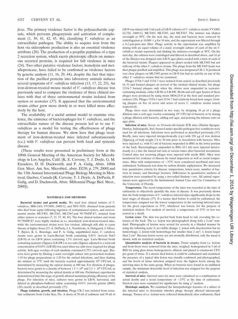
When mice were injected s.c. with 418 CFU of V. vulnificusMO6/24-0 and given a simultaneous i.v. injection with 108 PFUof phage 153A-5, significant protection was observed over a1-day period in terms of CFU per gram of lesion and CFU pergram of liver (Fig. 2). Images of lesions from control (infectedbut not phage treated), phage 153A-5-treated, and phage153A-7-treated mice are shown in Fig. 3. Phage 153A-5-treatedmice had minor s.c. skin lesions at 19 h postinfection, with amean lesion score of less than 1.5. Although body tempera-tures were more normal for phage 153A-5-treated mice, thedifferences were not statistically significant in comparison withthe values for control mice. The survival rate for the phage153A-5 group examined at 18 h (five of five mice) was signif-icantly higher than that for the control group (two of five mice)
FIG. 2. Protection of V. vulnificus MO6-24/0-infected mice by phage 153A-5 but not phage 153A-7. Iron-dextran-treated mice were inoculateds.c. with 417 CFU of V. vulnificus MO6-24/0 and immediately injected i.v. with BSG (control) (C), phage 153A-5 (A-5S and A-5L), or phage 153A-7(A-7S). Samples were harvested at 14 h (C, A-5S, and A-7S) or 6 days (A-5L) after infection. Data are means and standard deviations for log CFUper gram of skin lesion (Lesion CFU), log CFU per gram of liver lesion (Liver CFU), and rectal temperature (Temp). For lesion score (Lesion),the mean is shown. For the A-5L liver data, the bar represents the minimum detectable level, since no CFU were recovered. All groups containedfive animals. Protection was demonstrated for A-5S Lesion CFU and Temp (P � 0.01) and for A-5L Lesion CFU (P � 0.01); marginal protectionwas observed for Liver CFU (P � 0.05). Asterisks indicate significant differences from control. Mice treated with phage 153A-7 did not show anysignificant protection.
6254 CERVENY ET AL. INFECT. IMMUN.
on Decem
ber 1, 2015 by Wageningen U
R Library
http://iai.asm.org/
Dow
nloaded from
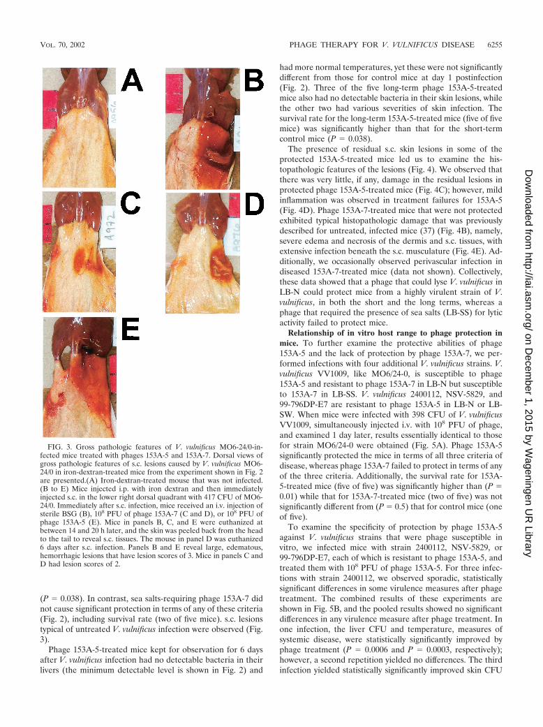
(P � 0.038). In contrast, sea salts-requiring phage 153A-7 didnot cause significant protection in terms of any of these criteria(Fig. 2), including survival rate (two of five mice). s.c. lesionstypical of untreated V. vulnificus infection were observed (Fig.3).
Phage 153A-5-treated mice kept for observation for 6 daysafter V. vulnificus infection had no detectable bacteria in theirlivers (the minimum detectable level is shown in Fig. 2) and
had more normal temperatures, yet these were not significantlydifferent from those for control mice at day 1 postinfection(Fig. 2). Three of the five long-term phage 153A-5-treatedmice also had no detectable bacteria in their skin lesions, whilethe other two had various severities of skin infection. Thesurvival rate for the long-term 153A-5-treated mice (five of fivemice) was significantly higher than that for the short-termcontrol mice (P � 0.038).
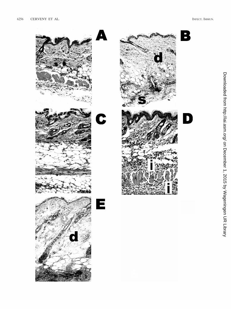
The presence of residual s.c. skin lesions in some of theprotected 153A-5-treated mice led us to examine the his-topathologic features of the lesions (Fig. 4). We observed thatthere was very little, if any, damage in the residual lesions inprotected phage 153A-5-treated mice (Fig. 4C); however, mildinflammation was observed in treatment failures for 153A-5(Fig. 4D). Phage 153A-7-treated mice that were not protectedexhibited typical histopathologic damage that was previouslydescribed for untreated, infected mice (37) (Fig. 4B), namely,severe edema and necrosis of the dermis and s.c. tissues, withextensive infection beneath the s.c. musculature (Fig. 4E). Ad-ditionally, we occasionally observed perivascular infection indiseased 153A-7-treated mice (data not shown). Collectively,these data showed that a phage that could lyse V. vulnificus inLB-N could protect mice from a highly virulent strain of V.vulnificus, in both the short and the long terms, whereas aphage that required the presence of sea salts (LB-SS) for lyticactivity failed to protect mice.
Relationship of in vitro host range to phage protection inmice. To further examine the protective abilities of phage153A-5 and the lack of protection by phage 153A-7, we per-formed infections with four additional V. vulnificus strains. V.vulnificus VV1009, like MO6/24-0, is susceptible to phage153A-5 and resistant to phage 153A-7 in LB-N but susceptibleto 153A-7 in LB-SS. V. vulnificus 2400112, NSV-5829, and99-796DP-E7 are resistant to phage 153A-5 in LB-N or LB-SW. When mice were infected with 398 CFU of V. vulnificusVV1009, simultaneously injected i.v. with 108 PFU of phage,and examined 1 day later, results essentially identical to thosefor strain MO6/24-0 were obtained (Fig. 5A). Phage 153A-5significantly protected the mice in terms of all three criteria ofdisease, whereas phage 153A-7 failed to protect in terms of anyof the three criteria. Additionally, the survival rate for 153A-5-treated mice (five of five) was significantly higher than (P �0.01) while that for 153A-7-treated mice (two of five) was notsignificantly different from (P � 0.5) that for control mice (oneof five).
To examine the specificity of protection by phage 153A-5against V. vulnificus strains that were phage susceptible invitro, we infected mice with strain 2400112, NSV-5829, or99-796DP-E7, each of which is resistant to phage 153A-5, andtreated them with 108 PFU of phage 153A-5. For three infec-tions with strain 2400112, we observed sporadic, statisticallysignificant differences in some virulence measures after phagetreatment. The combined results of these experiments areshown in Fig. 5B, and the pooled results showed no significantdifferences in any virulence measure after phage treatment. Inone infection, the liver CFU and temperature, measures ofsystemic disease, were statistically significantly improved byphage treatment (P � 0.0006 and P � 0.0003, respectively);however, a second repetition yielded no differences. The thirdinfection yielded statistically significantly improved skin CFU
FIG. 3. Gross pathologic features of V. vulnificus MO6-24/0-in-fected mice treated with phages 153A-5 and 153A-7. Dorsal views ofgross pathologic features of s.c. lesions caused by V. vulnificus MO6-24/0 in iron-dextran-treated mice from the experiment shown in Fig. 2are presented.(A) Iron-dextran-treated mouse that was not infected.(B to E) Mice injected i.p. with iron dextran and then immediatelyinjected s.c. in the lower right dorsal quadrant with 417 CFU of MO6-24/0. Immediately after s.c. infection, mice received an i.v. injection ofsterile BSG (B), 108 PFU of phage 153A-7 (C and D), or 108 PFU ofphage 153A-5 (E). Mice in panels B, C, and E were euthanized atbetween 14 and 20 h later, and the skin was peeled back from the headto the tail to reveal s.c. tissues. The mouse in panel D was euthanized6 days after s.c. infection. Panels B and E reveal large, edematous,hemorrhagic lesions that have lesion scores of 3. Mice in panels C andD had lesion scores of 2.
VOL. 70, 2002 PHAGE THERAPY FOR V. VULNIFICUS DISEASE 6255
on Decem
ber 1, 2015 by Wageningen U
R Library
http://iai.asm.org/
Dow
nloaded from
6256 CERVENY ET AL. INFECT. IMMUN.
on Decem
ber 1, 2015 by Wageningen U
R Library
http://iai.asm.org/
Dow
nloaded from
and liver CFU (P � 0.00014 and P � 0.03, respectively);however the temperatures were not statistically different be-tween the groups in the last two infections (32 and 29°C,respectively). These low temperatures indicate that the micewere moribund. Because we observed occasional improvementin terms of various measures of virulence with phage 153-A5treatment for strain 2400112, which is not lysed by this phagein vitro, we examined whether the bacteria might becomephage susceptible during growth in the mouse host. As a sur-rogate, we examined the susceptibility of the bacteria to phage153-A5 during growth in rat serum. As a positive control forphage susceptibility, strain MO6/24-0 was also examined. Inboth LB-N and rat serum, strain MO6/24-0 was killed by phage153-A5, while strain 2400112 was not affected by the phage atall (data not shown).
Two infection and protection experiments each were per-formed with strains NSV-5829 and 99-796DP-E7 and phage153-A5, and the combined results are shown in Fig. 5B. Nostatistically significant differences were observed for lesionCFU, liver CFU, or temperature with phage treatment. How-ever, in one experiment, strain NSV-5829 produced marginallylower skin lesion CFU with phage treatment (P � 0.03).
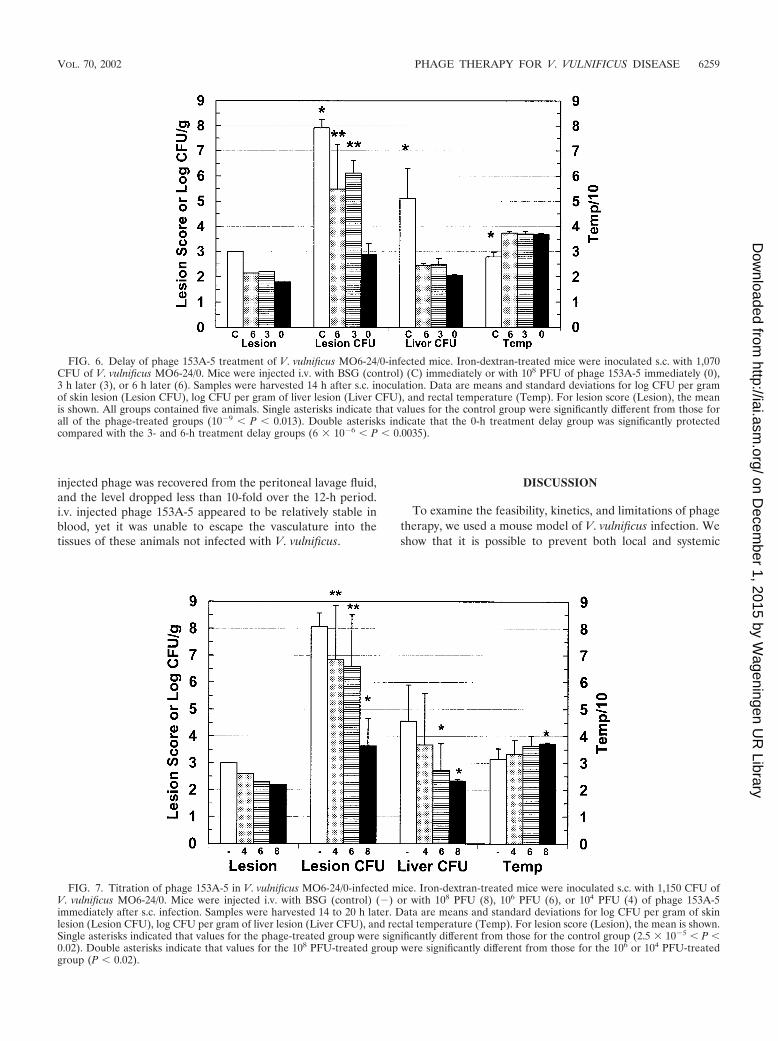
Effects of delaying phage 153A-5 treatment on infection withMO6/24-0. As was attempted for phage CK-2 and V. vulnificusMLT403, we examined how long phage treatment could bedelayed after s.c. administration of a lethal dose of V. vulnificusto iron-dextran-treated mice. Our failure to observe significantprotection with delays of 6 and 12 h led us to examine delaysof 6 h or less. Mice infected with 1,070 CFU of MO6/24-0 wereinjected with 108 PFU of phage 153A-5 at 0, 3, and 6 h postin-fection and then examined for disease at day 1 postinfection.As shown in Fig. 6, all three times of administering the phagetreatment resulted in significant protection in terms of all fourcriteria in comparison with the results for the control group.However, the numbers of CFU per gram of lesion were signif-icantly higher for the 3- and 6-h treatment delay groups thanfor the 0-h treatment delay group. The numbers of CFU pergram of liver and temperature were not significantly differentamong the phage-treated groups. The survival rates for allphage-treated groups (five of five mice) were significantlyhigher than that for the control group (zero of five mice) (P �0.0016). Delaying phage treatment for as long as 6 h seemed toprovide protection over a short infection period of 1 day forthis phage-host pair.
In a follow-up experiment, we examined whether the miceprotected over a 1-day period with 6-h delayed phage treat-ment were destined to recover or whether the delayed phagetreatment had only caused a temporary delay in lethal V. vulni-ficus infection. Seven mice were infected with V. vulnificusMO6/24-0 and treated with 108 PFU of phage 153A-5 6 h
postinfection. The experiment had to be terminated at 2 dayspostinfection because three mice died. One of the remainingmice had 106.2 CFU/g of s.c. lesion, and the other three hadundetectable bacteria in skin lesions. Therefore, althoughsome longer-term protection was observed with a 6-h delayedphage treatment, the efficiency of protection was compromisedcompared with that obtained with simultaneous V. vulnificusinfection and phage treatment.
Dose response in mice for phage 153A-5 protection againstV. vulnificus MO6/24-0. Since we had been treating mice withphage 153A-5 doses of up to 106-fold higher than the V. vulni-ficus inoculum, we titrated the minimum protective dose of thephage for a 1-day V. vulnificus infection (Fig. 7). Iron-dextran-treated mice were injected s.c. with 1,150 CFU of V. vulnificusMO6/24-0 and immediately injected i.v. with 108, 106, or 104
PFU of phage 153A-5. Only the highest dose of phage, 108
PFU, afforded significant protection to the mice in terms ofevery criterion. The lower doses of phage did not significantlyprotect the mice over the course of a 1-day infection, except forthe CFU per gram of liver in the group treated with 106 PFU.The trends in terms of all four criteria progressed from thecontrol group to the highest-dose phage-treated group, givingthe appearance of a dose response; however, the values for thegroups treated with 0, 104, and 106 PFU were generally notsignificantly different from each other. The survival rate foronly the group treated with 108 PFU (five of five mice) wassignificantly higher than that for the control group (one of fivemice) (P � 0.01). The survival rate for the group treated with106 PFU (four of five mice) was nearly significantly higher (P� 0.06). However, when similarly infected mice treated with106 PFU of phage 153A-5 were monitored for 2 days, three offive mice died and a fourth had nearly 107 CFU/g of skin lesion(data not shown). Therefore, it appeared that very high mul-tiplicities of phage treatment were required for significant pro-tection against s.c. infection by V. vulnificus in iron-dextran-treated mice.
Half-life of phage in mouse tissues. Since delayed treatmentwith phages resulted in decreased efficacy of protection, wemeasured the stability of phage 153A-5 in mice after i.v. injec-tion. Mice that were not infected with V. vulnificus were in-jected i.v. with 108 PFU of phage, and the numbers of phagewere measured in the blood, liver, and peritoneal fluid. From1 h postinjection to 12 h postinjection, the PFU per milliliter ofblood dropped over 43-fold from 106.4 to 104.9 PFU/ml, yield-ing a half-life of 2.2 h (Fig. 8). The PFU per gram of liver wasapproximately 10% the PFU per milliliter of blood at 1 hpostinjection, and liver PFU dropped only 10-fold over thecourse of the experiment. The ability of the i.v. injected phageto translocate out of the vasculature into the peritoneal cavitywas very low. At 1 h postinjection, only about 0.1% the dose of
FIG. 4. Histopathologic features of mice infected with MO6-24/0 and treated with phages 153A-5 and 153A-7. The micrographs depict thehistologic features of the mice from the experiment shown in Fig. 3. (A) Healthy mouse that received only an i.p. injection of iron. (B to E) Miceinjected s.c. with 417 CFU of MO6-24/0. Tissues were collected from each mouse at death, fixed in buffered formalin, embedded in paraffin, andcut into 5-�m sections. Sections were stained with hematoxylin and eosin and observed at a magnification of �200. (B) Control mouse (V. vulnificusinfected, not phage treated). Necrosis and edema in the dermis (d) and subcutis (s) are indicated. (C) Phage 153A-5-treated mouse with nohistologic damage. (D) Failure of phage 153A-5 treatment, with a lesion score of 2. The dermis and subcutis are intact, but inflammation (i) isindicated. (E) Failure of phage 153A-7 treatment. Necrosis in the dermis (d) and subcutis and edema were observed.
VOL. 70, 2002 PHAGE THERAPY FOR V. VULNIFICUS DISEASE 6257
on Decem
ber 1, 2015 by Wageningen U
R Library
http://iai.asm.org/
Dow
nloaded from
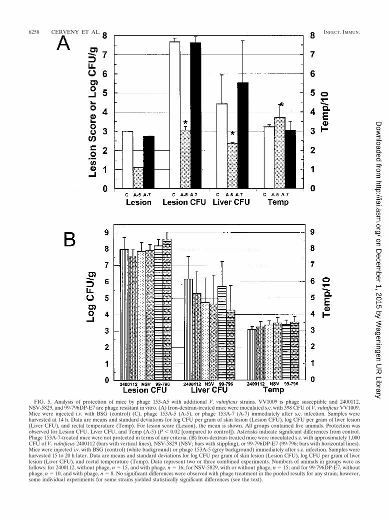
FIG. 5. Analysis of protection of mice by phage 153-A5 with additional V. vulnificus strains. VV1009 is phage susceptible and 2400112,NSV-5829, and 99-796DP-E7 are phage resistant in vitro. (A) Iron-dextran-treated mice were inoculated s.c. with 398 CFU of V. vulnificus VV1009.Mice were injected i.v. with BSG (control) (C), phage 153A-5 (A-5), or phage 153A-7 (A-7) immediately after s.c. infection. Samples wereharvested at 14 h. Data are means and standard deviations for log CFU per gram of skin lesion (Lesion CFU), log CFU per gram of liver lesion(Liver CFU), and rectal temperature (Temp). For lesion score (Lesion), the mean is shown. All groups contained five animals. Protection wasobserved for Lesion CFU, Liver CFU, and Temp (A-5) (P � 0.02 [compared to control]). Asterisks indicate significant differences from control.Phage 153A-7-treated mice were not protected in terms of any criteria. (B) Iron-dextran-treated mice were inoculated s.c. with approximately 1,000CFU of V. vulnificus 2400112 (bars with vertical lines), NSV-5829 (NSV; bars with stippling), or 99-796DP-E7 (99-796; bars with horizontal lines).Mice were injected i.v. with BSG (control) (white background) or phage 153A-5 (grey background) immediately after s.c. infection. Samples wereharvested 15 to 20 h later. Data are means and standard deviations for log CFU per gram of skin lesion (Lesion CFU), log CFU per gram of liverlesion (Liver CFU), and rectal temperature (Temp). Data represent two or three combined experiments. Numbers of animals in groups were asfollows: for 2400112, without phage, n � 15, and with phage, n � 16; for NSV-5829, with or without phage, n � 15; and for 99-796DP-E7, withoutphage, n � 10, and with phage, n � 8. No significant differences were observed with phage treatment in the pooled results for any strain; however,some individual experiments for some strains yielded statistically significant differences (see the text).
6258 CERVENY ET AL. INFECT. IMMUN.
on Decem
ber 1, 2015 by Wageningen U
R Library
http://iai.asm.org/
Dow
nloaded from
injected phage was recovered from the peritoneal lavage fluid,and the level dropped less than 10-fold over the 12-h period.i.v. injected phage 153A-5 appeared to be relatively stable inblood, yet it was unable to escape the vasculature into thetissues of these animals not infected with V. vulnificus.
DISCUSSION
To examine the feasibility, kinetics, and limitations of phagetherapy, we used a mouse model of V. vulnificus infection. Weshow that it is possible to prevent both local and systemic
FIG. 6. Delay of phage 153A-5 treatment of V. vulnificus MO6-24/0-infected mice. Iron-dextran-treated mice were inoculated s.c. with 1,070CFU of V. vulnificus MO6-24/0. Mice were injected i.v. with BSG (control) (C) immediately or with 108 PFU of phage 153A-5 immediately (0),3 h later (3), or 6 h later (6). Samples were harvested 14 h after s.c. inoculation. Data are means and standard deviations for log CFU per gramof skin lesion (Lesion CFU), log CFU per gram of liver lesion (Liver CFU), and rectal temperature (Temp). For lesion score (Lesion), the meanis shown. All groups contained five animals. Single asterisks indicate that values for the control group were significantly different from those forall of the phage-treated groups (10�9 � P � 0.013). Double asterisks indicate that the 0-h treatment delay group was significantly protectedcompared with the 3- and 6-h treatment delay groups (6 � 10�6 � P � 0.0035).
FIG. 7. Titration of phage 153A-5 in V. vulnificus MO6-24/0-infected mice. Iron-dextran-treated mice were inoculated s.c. with 1,150 CFU ofV. vulnificus MO6-24/0. Mice were injected i.v. with BSG (control) (�) or with 108 PFU (8), 106 PFU (6), or 104 PFU (4) of phage 153A-5immediately after s.c. infection. Samples were harvested 14 to 20 h later. Data are means and standard deviations for log CFU per gram of skinlesion (Lesion CFU), log CFU per gram of liver lesion (Liver CFU), and rectal temperature (Temp). For lesion score (Lesion), the mean is shown.Single asterisks indicated that values for the phage-treated group were significantly different from those for the control group (2.5 � 10�5 � P �0.02). Double asterisks indicate that values for the 108 PFU-treated group were significantly different from those for the 106 or 104 PFU-treatedgroup (P � 0.02).
VOL. 70, 2002 PHAGE THERAPY FOR V. VULNIFICUS DISEASE 6259
on Decem
ber 1, 2015 by Wageningen U
R Library
http://iai.asm.org/
Dow
nloaded from

disease caused by V. vulnificus in an iron-dextran-treatedmouse model by administering bacteriophages that are lytic forthe infecting bacterial strain. Phage CK-2 could prevent infec-tion by V. vulnificus MLT403, and phage 153A-5 could preventinfection by V. vulnificus MO6/24-0 and VV1009. We quanti-tatively measured disease and protection by using four criteria:s.c. lesion score and CFU per gram of s.c. lesion as measures oflocal disease and body temperature and CFU per gram of liveras measures of systemic disease. We also measured the survivalof mice. In most, but not all, treated mice, these phages couldsignificantly reduce these measures of disease and in manyinstances resulted in apparently sterile lesions and livers.Achievement of this level of protection required the adminis-tration of high doses of phage (108 PFU/mouse) relatively soonafter inoculation.
We hypothesize the phage-bacterium interaction in thismouse model for V. vulnificus disease to be follows. After s.c.inoculation into iron-dextran-treated mice, the vibrios begin toreplicate at an extremely rapid rate. In the initial characteriza-tion of this model, the slowest doubling time of the bacteria inthe mice was calculated to be 45 min (37). Although the vibriosand phages are often administered to mice simultaneously,they may not come into contact until some time after injection.Early on, the vibrios may be sequestered in the intercellularfluid in the s.c. tissues, while the phages are present in thebloodstream. Particles as large as phages may not freely passthrough the vascular endothelium. The very small amounts ofphages obtained from the peritoneal lavage fluid over a 12-htime course are in agreement with this hypothesis. However, asthe vibrios reach sufficient numbers in the tissues, they inducevasodilation and vascular permeability (3), thereby enablingthe phages access to the bacteria as edema floods the infectedtissues. Therefore, the bacteria may actually induce their owndemise in this particular model system. The fact that phages
administered i.v. can be effective at clearing local infection ofthe skin tissues suggests that such treatment may be useful ina clinical setting for V. vulnificus disease, where i.v. adminis-tration would be most efficient at delivering the phagesthroughout the body.
With regard to the mechanism whereby phages elicit theirprotective effect, we have shown that it is, as expected, killingof the bacteria. We generally did not observe protection whenthe chosen phage was unable to lyse the inoculating V. vulni-ficus strain in vitro. This relationship was shown in two ways.First, we noted that phage 153A-7 formed plaques on the hostbacteria only when bacterial medium containing sea salts orseawater was used. Since these conditions are not present inanimals, such phages should not be able to lyse the bacteria inthe infected animal host. We demonstrated that phage 153A-7failed to protect mice from infection with V. vulnificus MO6/24-0 (Fig. 2) and VV1009 (Fig. 5). There are at least twopossible reasons for the failure of phage 153A-7 to formplaques on V. vulnificus in the absence of sea salts. Either thereceptor on V. vulnificus cells is not expressed in the absence ofsea salts or the salts are required for the binding of the phageto the receptor. We have yet to examine these possibilities.Clearly, phages such as phage 153A-7 would not be clinicallyuseful.
A second reason that phages failed to form plaques was thelimited host range for the V. vulnificus strains in any mediatested. We would not expect these phage-bacterium mixturesto yield protection in our mouse model. As shown in Fig. 5B,phage 153A-5 failed to consistently protect mice from s.c. in-fection with V. vulnificus 2400112, NSV-5829, and 99-796DP-E7, which were resistant to 153A-5. Figure 5B depicts thecombined results of three experiments for strain 2400112 andtwo experiments for NSV-5829 and 99-796DP-E7 because itwas noted that for strains 2400112 and NSV-5829, variousmeasures of virulence were sporadically significantly amelio-rated by phage treatment. The occasional statistically signifi-cant differences were not nearly on the same order as thoseseen with the productive phage-host strain combinations, inwhich the mice were completely protected. These statisticallysignificant differences for the apparently resistant V. vulnificusstrains were of questionable biological significance. The reasonfor these sporadic differences in the absence of in vitro plaque-forming activity of the phage is unknown. One possibility isthat the phage is able to infect and kill or debilitate the bacteriawithout releasing enough progeny phage to form a plaque invitro. In this scenario, the phage in the bloodstream of the micewould be able to kill vibrios that leak into the vasculature asthe disease progresses in the skin. However, we found thatstrain 2400112 was not killed by phage 153-A5 in either LB-Nor rat serum. It is also possible that residual bacterial compo-nents in the phage lysates, e.g., lipopolysaccharide, induced aninflammatory response upon i.v. injection and that this inflam-matory response slightly boosted the resistance of the mice toinfection. However, such a phenomenon was not always ap-parent, because phage 153-A7, which could lyse its host bac-teria only in the presence of seawater, offered no such protec-tion.
To examine the limitations of our phage therapy model, weeither delayed treatment with phages after bacterial infectionor titrated the minimum numbers of phages required for pro-
FIG. 8. Half-life of phages in mouse tissues. Mice were treated withiron dextran but not s.c. infected with V. vulnificus. Phage 153A-5 wasinjected i.v. at 108 PFU per mouse. Samples of the blood, liver, andperitoneal lavage fluid were sampled at 1, 6, and 12 h after injection ofthe phage, and numbers of PFU per milliliter or PFU per gram oftissue were enumerated. The half-life of the phage in the blood wascalculated to be 2.2 h. Data are means and standard deviations.
6260 CERVENY ET AL. INFECT. IMMUN.
on Decem
ber 1, 2015 by Wageningen U
R Library
http://iai.asm.org/
Dow
nloaded from

tection. As shown in Fig. 6, delaying phage treatment for morethan 3 h resulted in failure. Even though mice were signifi-cantly protected at 1 day postinfection, many became ill within2 days. The reason for the inability to confer protection afterdelayed treatment in our model is unknown. The progressionof disease in our model is extremely rapid, with death occur-ring as soon as 12 h after infection with a minimum lethal dose.Since the histopathologic features of the s.c. lesions consist ofthrombosis of capillaries (37), it is possible that with delayedphage treatment, the localized vasculature is clotted off,thereby preventing the entry of subsequently administeredphages into the infected tissues. In contrast, when the phagesare present early during the infection, they may leak into theinfected tissues before thrombosis occurs and begin to kill andreplicate in the infecting vibrios. Another possibility is that thenumbers of bacteria have increased to such a high level that thephages cannot kill them all. Figure 7 shows that a high level ofphage, i.e., 108 PFU, is required to protect against a minimumlethal dose of bacteria, and the half-life of phage 153A-5 inmice is approximately 2.2 h. It therefore appears that thephages are relatively stable in mouse fluids, particularly thevasculature. The reason for the ineffectiveness of lower dosesof the phages is unknown. However, very high levels of phagesdo not appear to have harmful side effects.
Phage therapy has been widely used in Eastern Europe formany decades, and animal model studies have been investi-gated in the West since the early 1980s (10, 39). Almost all ofthese studies have shown that phage therapy has great poten-tial, but more research needs to be done to understand theparameters that limit the effectiveness of phage therapy indifferent diseases. Phages have some unique advantageous fea-tures that could make them highly efficacious under certainconditions. (i) Phages are capable of replicating in a treatedpatient by infecting the bacteria, with the subsequent release of100-fold more phage. This effect is in marked contrast to thatof antibiotics, which only decrease in concentration from thetime when they are administered. (ii) Phages are specific tobacterial species; hence, the normal microbial flora will bepreserved. In contrast, the use of many antibiotics disrupts thenormal flora, a process which may lead to secondary symptoms.(iii) Phages can be isolated that recognize virulence factors asreceptors so that phage-resistant mutants that have lost theirreceptors will be attenuated. (iv) Because of their specificity tobacteria, phages have very few, if any, side effects in the treatedpatient. Many antibiotics exert toxic side effects. (v) Phages canbe used topically as well as in the environment to controlpopulations of bacterial pathogens. In spite of these advan-tages, phage therapy also has its drawbacks. The specificity ofphages for host bacteria necessitates the use of broad-host-range phages or pools of phages with broad collective activityor at least the identification of phages with activity against theinfecting bacterial strains. Bacteria can become resistant tophages by spontaneous mutation. In theory, the production ofantiphage antibodies by phage-treated hosts could interferewith subsequent treatments with the same phage. Finally, inour experimental system, high numbers of phages needed to beadministered soon after bacterial infection for treatment to beeffective. Optimizing the beneficial attributes of phage therapywhile decreasing the potential problems will require continuinganalysis of animal models of infection.
In summary, our results demonstrate that treatment of V.vulnificus-infected mice can prevent local and systemic diseaseas well as death. These results contribute to the growing bodyof evidence that phage therapy is a viable alternative treatmentmodality for bacterial infectious disease. Our V. vulnificusphage therapy model will now be used to define the majorbenefits and limitations of phage therapy.
ACKNOWLEDGMENTS
This work was supported by Department of Commerce sea grantR/LR-Q-20 to D.H.D. and P.A.G. and a focused giving award fromJohnson and Johnson, Inc., to P.A.G.
We thank Anita C. Wright for review of the manuscript and ThomasJ. Doyle, Julio Martin, and Eric Wilkening for expert technical assis-tance.
REFERENCES
1. Amako, K., K. Okada, and S. Miake. 1984. Evidence for the presence of acapsule in Vibrio vulnificus. J. Gen. Microbiol. 130:2741–2743.
2. Berchieri, A., M. A. Lovell, and P. A. Barrow. 1991. The activity in thechicken alimentary tract of bacteriophages lytic for Salmonella typhimurium.Res. Microbiol. 142:541–549.
3. Bowdre, J. H., M. D. Poole, and J. D. Oliver. 1981. Edema and hemocon-centration in mice experimentally infected with Vibrio vulnificus. Infect.Immun. 32:1193–1199.
4. Brennt, C. E., A. C. Wright, S. K. Dutta, and J. G. Morris, Jr. 1991. Growthof Vibrio vulnificus in serum from alcoholics: association with high transferriniron saturation. J. Infect. Dis. 164:1030–1032.
5. Buchrieser, C., V. V. Gangar, R. L. Murphree, M. L. Tamplin, and C. W.Kaspar. 1995. Multiple Vibrio vulnificus strains in oysters as demonstrated byclamped homogeneous electric field gel electrophoresis. Appl. Environ. Mi-crobiol. 61:1163–1168.
6. Bullen, J. J., P. B. Spalding, C. G. Ward, and J. M. Gutteridge. 1991.Hemochromatosis, iron and septicemia caused by Vibrio vulnificus. Arch.Intern. Med. 151:1606–1609.
7. Cook, D. W., P. Oleary, J. C. Hunsucker, E. M. Sloan, J. C. Bowers, R. J.Blodgett, and A. DePaola. 2002. Vibrio vulnificus and Vibrio parahaemolyticusin U.S. retail shell oysters: a national survey from June 1998 to July 1999. J.Food Prot. 65:79–87.
8. DePaola, A., S. McLeroy, and G. McManus. 1997. Distribution of Vibriovulnificus phage in oyster tissues and other estuarine habitats. Appl. Environ.Microbiol. 63:2464–2467.
9. DePaola, A., M. L. Motes, A. M. Chan, and C. A. Suttle. 1998. Phagesinfecting Vibrio vulnificus are abundant and diverse in oysters (Crassostreavirginica) collected from the Gulf of Mexico. Appl. Environ. Microbiol.64:346–351.
10. Duckworth, D. H., and P. A. Gulig. 2001. Bacteriophages: potential treat-ment for bacterial infections. BioDrugs 16:57–62.
11. Fan, J. J., C. P. Shao, Y. C. Ho, C. K. Yu, and L. I. Hor. 2001. Isolation andcharacterization of a Vibrio vulnificus mutant deficient in both extracellularmetalloprotease and cytotysin. Infect. Immun. 69:5943–5948.
12. Flaherty, J. E., J. B. Jones, B. K. Harbaugh, G. C. Somodi, and L. E.Jackson. 2000. Control of bacterial spot on tomato in the greenhouse andfield with H-mutant bacteriophages. Hortscience 35:882–884.
13. Gray, L. D., and A. S. Kreger. 1985. Purification and characterization of anextracellular cytolysin produced by Vibrio vulnificus. Infect. Immun. 48:62–72.
14. Hlady, W. G., and K. C. Klontz. 1996. The epidemiology of Vibrio infectionsin Florida, 1981–1993. J. Infect. Dis. 173:1176–1183.
15. Jackson, J. K., R. L. Murphree, and M. L. Tamplin. 1997. Evidence thatmortality from Vibrio vulnificus infection results from single strains amongheterogeneous populations in shellfish. J. Clin. Microbiol. 35:2098–2101.
16. Jeong, K. C., H. S. Jeong, J. H. Rhee, S. E. Lee, S. S. Chung, A. M. Starks,G. M. Escudero, P. A. Gulig, and S. H. Choi. 2000. Construction and phe-notypic evaluation of a Vibrio vulnificus vvpE mutant for elastolytic protease.Infect. Immun. 68:5096–5106.
17. Kook, H., S. E. Lee, Y. H. Baik, S. S. Chung, and J. H. Rhee. 1996. Vibriovulnificus hemolysin dilates rat thoracic aorta by activating guanylate cyclase.Life Sci. 59:PL41-PL47.
18. Kraffert, C. A., and D. J. Hogan. 1992. Vibrio vulnificus infection and ironoverload. J. Am. Acad. Dermatol. 26:140.
19. Linkous, D. A., and J. D. Oliver. 1999. Pathogenesis of Vibrio vulnificus.FEMS Microbiol. Lett. 174:207–214.
20. Litwin, C. M., T. W. Rayback, and J. Skinner. 1996. Role of catecholsiderophore synthesis in Vibrio vulnificus virulence. Infect. Immun. 64:2834–2838.
21. Merril, C. R., B. Biswas, R. Carlton, N. C. Jensen, G. J. Creed, S. Zullo, and
VOL. 70, 2002 PHAGE THERAPY FOR V. VULNIFICUS DISEASE 6261
on Decem
ber 1, 2015 by Wageningen U
R Library
http://iai.asm.org/
Dow
nloaded from
S. Adhya. 1996. Long-circulating bacteriophage as antibacterial agents. Proc.Natl. Acad. Sci. USA 93:3188–3192.
22. Miyoshi, S., and S. Shinoda. 1988. Role of the protease in the permeabilityenhancement by Vibrio vulnificus. Microbiol. Immunol. 32:1025–1032.
23. Motes, M. L., A. DePaola, D. W. Cook, J. E. Veazey, J. C. Hunsucker, W. E.Garthright, R. J. Blodgett, and S. J. Chirtel. 1998. Influence of water tem-perature and salinity on Vibrio vulnificus in Northern Gulf and Atlantic Coastoysters (Crassostrea virginica). Appl. Environ. Microbiol. 64:1459–1465.
24. Paranjpye, R. N., J. C. Lara, J. C. Pepe, C. M. Pepe, and M. S. Strom. 1998.The type IV leader peptidase/N-methyltransferase of Vibrio vulnificus con-trols factors required for adherence to HEp-2 cells and virulence in iron-overloaded mice. Infect. Immun. 66:5659–5668.
25. Park, J. W., S. N. Ma, E. S. Song, C. H. Song, M. R. Chae, B. H. Park, R. W.Rho, S. D. Park, and H. R. Kim. 1996. Pulmonary damage by Vibrio vulnificuscytolysin. Infect. Immun. 64:2873–2876.
26. Park, S. C., I. Shimamura, M. Fukunaga, K. I. Mori, and T. Nakai. 2000.Isolation of bacteriophages specific to a fish pathogen, Pseudomonas pleco-glossicida, as a candidate for disease control. Appl. Environ. Microbiol.66:1416–1422.
27. Pelon, W., R. J. Siebeling, J. Simonson, and R. B. Luftig. 1995. Isolation ofbacteriophage infectious for Vibrio vulnificus. Curr. Microbiol. 30:331–336.
28. Provence, D. L., and R. Curtiss III. 1994. Gene transfer in gram-negativebacteria, p. 317–347. In P. Gerhardt, R. G. E. Murray, W. A. Wood, andN. R. Krieg (ed.), Methods for general and molecular bacteriology. Amer-ican Society for Microbiology, Washington, D.C.
29. Shao, C. P., and L. I. Hor. 2000. Metalloprotease is not essential for Vibriovulnificus virulence in mice. Infect. Immun. 68:3569–3573.
30. Shinoda, S., M. Kobayashi, H. Yamada, S. Yoshida, M. Ogawa, and Y.Mizuguchi. 1987. Inhibitory effect of capsular antigen of Vibrio vulnificus onbactericidal activity of human serum. Microbiol. Immunol. 31:393–401.
31. Smith, H. W., and M. B. Huggins. 1982. Successful treatment of experimen-tal Escherichia coli infections in mice using phage: its general superiority overantibiotics. J. Gen. Microbiol. 128:307–318.
32. Smith, H. W., and M. B. Huggins. 1983. Effectiveness of phages in treatingexperimental Escherichia coli diarrhoea in calves, piglets and lambs. J. Gen.Microbiol. 129:2659–2675.
33. Smith, H. W., M. B. Huggins, and K. M. Shaw. 1987. Factors influencing thesurvival and multiplication of bacteriophages in calves and in their environ-ment. J. Gen. Microbiol. 133:1127–1135.
34. Smith, H. W., M. B. Huggins, and K. M. Shaw. 1987. The control of exper-imental Escherichia coli diarrhoea in calves by means of bacteriophages.J. Gen. Microbiol. 133:1111–1126.
35. Soothill, J. S. 1992. Treatment of experimental infections of mice withbacteriophages. J. Med. Microbiol. 37:258–261.
36. Soothill, J. S. 1994. Bacteriophage prevents destruction of skin grafts byPseudomonas aeruginosa. Burns 20:209–211.
37. Starks, A. M., T. R. Schoeb, M. L. Tamplin, S. Parveen, T. J. Doyle, P. E.Bomeisl, G. M. Escudero, and P. A. Gulig. 2000. Pathogenesis of infection byclinical and environmental strains of Vibrio vulnificus in iron-dextran-treatedmice. Infect. Immun. 68:5785–5793.
38. Strom, M. S., and R. N. Paranjpye. 2000. Epidemiology and pathogenesis ofVibrio vulnificus. Microbes Infect. 2:177–188.
39. Sulakvelidze, A., Z. Alavidze, and J. G. Morris. 2001. Bacteriophage therapy.Antimicrob. Agents Chemother. 45:649–659.
40. Tamplin, M. L., J. K. Jackson, C. Buchrieser, R. L. Murphree, K. M. Portier,V. Gangar, L. G. Miller, and C. W. Kaspar. 1996. Pulsed-field gel electro-phoresis and ribotype profiles of clinical and environmental Vibrio vulnificusisolates. Appl. Environ. Microbiol. 62:3572–3580.
41. Tamplin, M. L., S. Specter, G. E. Rodrick, and H. Friedman. 1983. Differ-ential complement activation and susceptibility to human serum bactericidalaction by Vibrio species. Infect. Immun. 42:1187–1190.
42. Tamplin, M. L., S. Specter, G. E. Rodrick, and H. Friedman. 1985. Vibriovulnificus resists phagocytosis in the absence of serum opsonins. Infect.Immun. 49:715–718.
43. Vollberg, C. M., and J. L. Herrera. 1997. Vibrio vulnificus infection: animportant cause of septicemia in patients with cirrhosis. South. Med. J.90:1040–1042.
44. Wright, A. C., and J. G. Morris, Jr. 1991. The extracellular cytolysin of Vibriovulnificus: inactivation and relationship to virulence in mice. Infect. Immun.59:192–197.
45. Wright, A. C., L. M. Simpson, J. D. Oliver, and J. G. Morris, Jr. 1990.Phenotypic evaluation of acapsular transposon mutants of Vibrio vulnificus.Infect. Immun. 58:1769–1773.
46. Yoshida, S., M. Ogawa, and Y. Mizuguchi. 1985. Relation of capsular ma-terials and colony opacity to virulence of Vibrio vulnificus. Infect. Immun.47:446–451.
Editor: J. T. Barbieri
6262 CERVENY ET AL. INFECT. IMMUN.
on Decem
ber 1, 2015 by Wageningen U
R Library
http://iai.asm.org/
Dow
nloaded from