pet scans - radmd | radmd-home guidelines/cms (medic… · · 2016-09-13pet scans original date:...
TRANSCRIPT
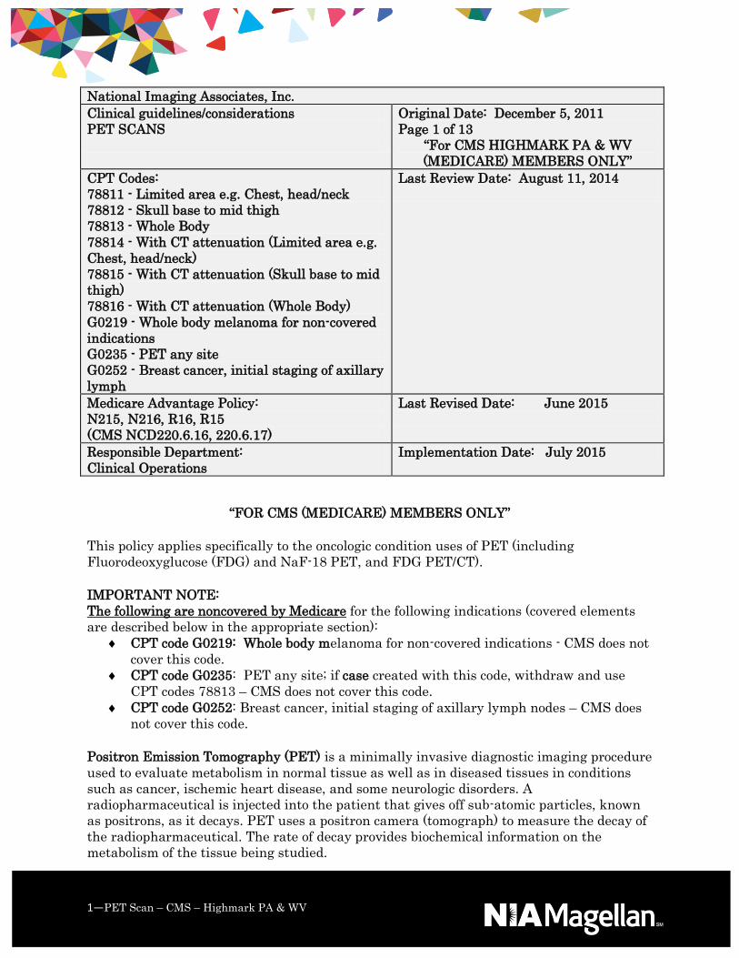
National Imaging Associates, Inc.
Clinical guidelines/considerations
PET SCANS
Original Date: December 5, 2011
Page 1 of 13
“For CMS HIGHMARK PA & WV
(MEDICARE) MEMBERS ONLY”
CPT Codes:
78811 - Limited area e.g. Chest, head/neck
78812 - Skull base to mid thigh
78813 - Whole Body
78814 - With CT attenuation (Limited area e.g.
Chest, head/neck)
78815 - With CT attenuation (Skull base to mid
thigh)
78816 - With CT attenuation (Whole Body)
G0219 - Whole body melanoma for non-covered
indications
G0235 - PET any site
G0252 - Breast cancer, initial staging of axillary
lymph
Last Review Date: August 11, 2014
Medicare Advantage Policy:
N215, N216, R16, R15
(CMS NCD220.6.16, 220.6.17)
Last Revised Date: June 2015
Responsible Department:
Clinical Operations
Implementation Date: July 2015
1—PET Scan – CMS – Highmark PA & WV
“FOR CMS (MEDICARE) MEMBERS ONLY”
This policy applies specifically to the oncologic condition uses of PET (including
Fluorodeoxyglucose (FDG) and NaF-18 PET, and FDG PET/CT).
IMPORTANT NOTE:
The following are noncovered by Medicare for the following indications (covered elements
are described below in the appropriate section):
CPT code G0219: Whole body melanoma for non-covered indications - CMS does not
cover this code.
CPT code G0235: PET any site; if case created with this code, withdraw and use
CPT codes 78813 – CMS does not cover this code.
CPT code G0252: Breast cancer, initial staging of axillary lymph nodes – CMS does
not cover this code.
Positron Emission Tomography (PET) is a minimally invasive diagnostic imaging procedure
used to evaluate metabolism in normal tissue as well as in diseased tissues in conditions
such as cancer, ischemic heart disease, and some neurologic disorders. A
radiopharmaceutical is injected into the patient that gives off sub-atomic particles, known
as positrons, as it decays. PET uses a positron camera (tomograph) to measure the decay of
the radiopharmaceutical. The rate of decay provides biochemical information on the
metabolism of the tissue being studied.

2— PET Scan – CMS – Highmark PA & WV
FDG (2-[F18] fluoro-2-deoxy-D-glucose) Positron Emission Tomography (PET) is a
minimally-invasive diagnostic imaging procedure used to evaluate glucose metabolism in
normal tissue as well as in diseased tissues in conditions such as cancer, ischemic heart
disease, and some neurologic disorders. FDG is an injected radionuclide (or
radiopharmaceutical) that emits sub-atomic particles, known as positrons, as it decays.
FDG PET uses a positron camera (tomograph) to measure the decay of FDG. The rate of
FDG decay provides biochemical information on glucose metabolism in the tissue being
studied. As malignancies can cause abnormalities of metabolism and blood flow, FDG PET
evaluation may indicate the probable presence or absence of a malignancy based upon
observed differences in biologic activity compared to adjacent tissues.
NA F-18 Positron Emission Tomography (PET). A positron camera (tomograph) is used to
produce cross-sectional tomographic images, which are obtained from positron-emitting
radioactive tracer substances (radiopharmaceuticals) such as F-18 sodium fluoride. NaF-18
PET has been recognized as an excellent technique for imaging areas of altered osteogenic
activity in bone. The clinical value of detecting and assessing the initial extent of metastatic
cancer in bone is attested by a number of professional guidelines for oncology. Imaging to
detect bone metastases is also recommended when a patient, following completion of initial
treatment, is symptomatic with bone pain suspicious for metastases from a known primary
tumor.
Refer to Medicare Advantage Medical Policy Bulletin R-15 for information on PET and
PET/CT scans for non-oncologic conditions.
Indications and Limitations of Coverage
PET (FDG) for Oncologic Conditions (NCD 220.6.17)
Framework for FDG Imaging
Effective for dates of service on or after June 11, 2013.
CMS is adopting a coverage framework that ends the prospective data collection
requirements under CED for all oncologic uses of FDG PET imaging.
Initial Anti-tumor Treatment Strategy for FDG Imaging
CMS continues to believe that the evidence is adequate to determine that the results of
FDG PET imaging are useful in determining the appropriate initial treatment strategy for
patients with suspected cancer and improve health outcomes and thus are medically
necessary.
Effective for dates of service on or after June 11, 2013:
Only one (1) FDG PET study is covered for patients who have cancers that are biopsy
proven or strongly suspected based on other diagnostic testing when the patient's treating
physician determines that the FDG PET study is needed to determine the location and/or
3— PET Scan – CMS – Highmark PA & WV
extent of the tumor for the following therapeutic purposes related to the initial treatment
anti-tumor strategy:
To determine whether or not the patient is an appropriate candidate for an invasive
diagnostic or therapeutic procedure; or
To determine the optimal anatomic location for an invasive procedure; or
To determine the anatomic extent of tumor when the recommended anti-tumor
treatment reasonably depends on the extent of the tumor.
Prostate
FDG PET imaging is not covered for treatment strategy in patients who have
adenocarcinoma of the prostate.
Breast
FDG PET imaging is covered for the initial anti-tumor treatment strategy for male
and female breast cancer only when used in staging distant metastasis.
FDG PET imaging is non-covered for diagnosis of breast cancer and initial staging of
axillary nodes.
Melanoma
FDG PET is covered to determine initial anti-tumor treatment strategy
for melanoma other than the evaluation of regional lymph nodes.
FDG PET imaging is non-covered for initial anti-tumor treatment strategy for the
evaluation of regional lymph nodes in melanoma.
Cervical Cancer
FDG PET imaging is covered for the detection of pre-treatment metastasis (i.e.,
staging) in newly diagnosed cervical cancers following conventional imaging.
FDG PET imaging is non-covered for the diagnosis of cervical cancer related to
initial anti-tumor treatment strategy.
Refer to the summary grid in the policy attachment section.
Subsequent Anti-tumor Treatment Strategy for FDG Imaging
Effective for dates of service on or after June 11, 2013:
Three (3) Fluorodeoxyglucose Positron Emission Tomography (FDG PET) scans are covered
without the Coverage with Evidence Development (CED) requirement when used to guide
subsequent management of anti-tumor treatment strategy after completion of initial anti-
cancer therapy for the same cancer diagnosis.

4— PET Scan – CMS – Highmark PA & WV
Coverage of any additional FDG PET scans that is, beyond three (3) used to guide
subsequent management of anti-tumor treatment strategy after completion of initial anti-
cancer therapy for the same diagnosis will be considered not medically necessary.
PET Sodium Fluoride-18 (NaF-18) to Identify Bone Metastases of Cancer (NCD 220.6.19)
Sodium Fluoride-18 (NaF-18) PET imaging is covered to inform initial treatment strategy
or subsequent treatment strategy for suspected or biopsy-proven bone metastases through
CED, only in the context of a clinical study.
A NaF-18 PET clinical study that is designed to collect additional information at the time of
the scan to assist in initial antitumor treatment planning or to guide subsequent treatment
strategy by the identification, location and quantification of bone metastases in patients in
whom bone metastases are strongly suspected based on clinical symptoms or the results of
other diagnostic studies. Qualifying clinical studies must ensure that:
specific hypotheses are addressed;
appropriate data elements are collected;
hospitals and providers are qualified to provide the PET scan and interpret the
results;
participating hospitals and providers accurately report data on all enrolled patients
not included in other qualifying trials through adequate auditing mechanisms; and
all patient confidentiality, privacy, and other Federal laws must be followed.
The clinical studies for which Medicare Advantage will provide coverage must answer one
or more of the following questions: prospectively, in patients whose treating physician
determines that the NaF-18 PET study results are needed to determine the initial
antitumor treatment strategy or guide subsequent antitumor treatment strategy after the
completion of initial treatment, does the addition of NaF-18 PET imaging lead to:
A change in patient management to more appropriate palliative care; or
A change in patient management to more appropriate curative care; or
Improved quality of life; or
Improved survival?
The study must adhere to the standards of scientific integrity and relevance to the
Medicare population.
The principal purpose of the research study is to test whether a particular
intervention potentially improves the participants’ health outcomes.
The research study is well-supported by available scientific and medical information
or it is intended to clarify or establish the health outcomes of interventions already
in common clinical use.
The research study does not unjustifiably duplicate existing studies.
The research study design is appropriate to answer the research question being
asked in the study.
The research study is sponsored by an organization or individual capable of
executing the proposed study successfully.

5— PET Scan – CMS – Highmark PA & WV
The research study is in compliance with all applicable Federal regulations
concerning the protection of human subjects found in the Code of Federal
Regulations (CFR) at 45 CFR Part 46. If a study is regulated by the Food and Drug
Administration (FDA), it also must be in compliance with 21 CFR Parts 50 and 56.
All aspects of the research study are conducted according to the appropriate
standards of scientific integrity.
The research study has a written protocol that clearly addresses, or incorporates by
reference, the Medicare standards.
The clinical research study is not designed to exclusively test toxicity or disease
pathophysiology in healthy individuals. Trials of all medical technologies measuring
therapeutic outcomes as one of the objectives meet this standard only if the disease
or condition being studied is life-threatening as defined in 21 CFR §312.81(a) and
the patient has no other viable treatment options.
The clinical research study is registered on the www.ClinicalTrials.gov Web site by
the principal sponsor/investigator prior to the enrollment of the first study subject.
The research study protocol specifies the method and timing of public release of all
pre-specified outcomes to be measured including release of outcomes if outcomes are
negative or study is terminated early. The results must be made public within 24
months of the end of data collection. If a report is planned to be published in a peer-
reviewed journal, then that initial release may be an abstract that meets the
requirements of the International Committee of Medical Journal Editors. However,
a full report of the outcomes must be made public no later than three (3) years after
the end of data collection.
The research study protocol must explicitly discuss subpopulations affected by the
treatment under investigation, particularly traditionally underrepresented groups
in clinical studies, how the inclusion and exclusion criteria affect enrollment of these
populations, and a plan for the retention and reporting of said populations on the
trial. If the inclusion and exclusion criteria are expected to have a negative effect on
the recruitment or retention of underrepresented populations, the protocol must
discuss why these criteria are necessary.
The research study protocol explicitly discusses how the results are or are not
expected to be generalizable to the Medicare population to infer whether Medicare
patients may benefit from the intervention. Separate discussions in the protocol may
be necessary for populations eligible for Medicare due to age, disability or Medicaid
eligibility.
CMS indicates that the evidence is not sufficient to determine whether the results of NaF-
18 PET imaging to identify bone metastases improve health outcomes of patients with
cancer and is not medically necessary unless it meets the above criteria.
All other uses and clinical indications of NaF-18 PET are considered not medically
necessary.
Appropriate Indications for Subsequent PET Scans
1. Assessment of therapeutic effect after completion of potentially curative therapy, if
additional or alternative therapy will be employed for residual or progressive disease.

6— PET Scan – CMS – Highmark PA & WV
2. Assessment of therapeutic effect after localized, non-surgical therapy (e.g.,
stereotactic radiotherapy, radiofrequency or thermal ablation, etc.) of apparently
limited or clinically significant lesions.
3. Suspected recurrence/progression:
o New symptoms suggestive of tumor recurrence or progression;
o New physical findings (e.g., palpable lesion) suspicious for
recurrence/progression (consider biopsy, if easily accessible, and especially if
first recurrence);
o Rising tumor markers or other signs or laboratory values suggestive of
paraneoplastic phenomena;
o Findings on other imaging that are equivocal or suspicious.
4. Restaging after documentation of recurrence/progression, if extent of disease will
alter therapy or if new baseline needed prior to change in therapy.
5. Evaluating response to treatment, when a change in therapy is contemplated and
routine imaging (e.g., CT) either does not or is not expected to provide optimal
information for such decision.
6. Follow-up of indeterminate findings on a clinically indicated PET scan performed for
restaging or suspected recurrence/progression, to assess for trends in FDG uptake
within suspected lesions over time. Such follow-up studies typically are performed
no sooner than three months after the “indeterminate” PET scan.
Caveats Regarding Use of Oncologic PET
“Watchful waiting” does not constitute a “therapy.” After an Initial Therapy
Strategy PET, further PET restaging is not justified unless treatment has been
given or unless a significant change in patient status suggests
progression/transformation of disease.
Routine modalities (e.g., diagnostic CT, bone scintigraphy, MRI) should be employed
before PET if the clinical decision to be made is likely to be answered by those
modalities, such as:
o If there is specific clinical suspicion of involvement of an organ or region,
documentation of a single site of involvement is necessary/sufficient for
clinical decision making, and documentation of additional disease would not
change patient management (imaging should be targeted to the area/organ of
concern);
o If the major site of clinical concern is one that is typically poorly imaged by
FDG-PET, such as:
o If the clinical concern is brain involvement (use MRI, or contrast-enhanced
CT, if patient cannot undergo MRI);
o If the clinical concern is within the urinary tract consider CT,
ultrasonography, or MRI. If the patient’s tumor is known to be poorly FDG
avid;
o If disease is reasonably assumed to be limited to an organ or system
generally well imaged by other modalities (e.g., bone scintigraphy for disease
limited to skeleton).
PET should be employed instead of or before conventional imaging if:
o Available data suggest that PET is likely to obviate additional advanced
imaging tests or invasive procedures or is likely to be more accurate than

7— PET Scan – CMS – Highmark PA & WV
other modalities in detection or characterization of lesions that would change
patient management;
o Patient has contrast allergy or other contraindication to contrast
administration that renders other modalities less effective;
o Patient is being monitored for known disease that either has been previously
shown to be better characterized on PET than on other modalities or has
been shown to be adequately FDG-avid for follow-up with PET and was not
previously imaged with other modalities (e.g., bone metastases well
visualized on previous PET and either not as well visualized on bone
scintigraphy or bone scintigraphy not done).
Special care should be taken to avoid conditions that may promote false-positive or
false-negative FDG PET studies. Although clinical circumstances sometimes
warrant modification of these guidelines, if clinically feasible, routine follow-up PET
imaging should be delayed:
o For at least three weeks after chemotherapy;
o For at least 8-12 weeks following radiation therapy, unless the clinical
question involves a site outside the field of radiation;
o Until resolution or near-resolution of acute infectious or inflammatory
processes that may mimic or mask active tumor;
o At least one week after short-acting marrow stimulants and three weeks
after long-acting marrow stimulants.
Incidental findings are common on PET/CT studies, and further follow-up of such
findings should be tempered by clinical circumstances and patient prognosis:
o Reasonable efforts should be made to obtain previous imaging studies to
evaluate for chronicity of indeterminate findings before ordering follow-up
imaging studies or interventions;
o Clinical impact should be assessed in the individual patient (e.g., a
potentially malignant thyroid nodule may be of little overall significance in a
patient with advanced metastatic disease)
Relative Advantages of Various Advanced Imaging Modalities in Oncology
The advantages of metabolic imaging have made PET the imaging modality of choice for
staging, restaging, and therapy monitoring for many oncologic indications, especially in
advanced stages. However, the more precise anatomic detail offered by diagnostic-quality
CT and MRI, especially with the added information supplied by contrast enhancement, is
often sufficient or even preferable for a number of oncologic indications. More precise
delineation of tumor extent is often needed for tumor staging and planning of interventions.
More accurate size measurements are often needed for routine follow-up of therapeutic
efficacy. In most cases, contrast-enhanced CT is sufficient for such determinations, when
needed. However, the unique tissue contrast offered by MRI provides additional benefits in
many cases. A few general considerations, but not those which are applied to the pre-
payment of claims, include:
CT is typically used as the initial modality to help detect or characterize abnormal
growths; to help diagnose tumors; to provide information about the extent, or stage,
or disease; to help in guiding biopsy procedures or in planning treatment; to

8— PET Scan – CMS – Highmark PA & WV
determine whether a cancer is responding to treatment; and to monitor for
recurrence.
CT is also commonly used to guide local treatments, such as cryotherapy,
radiofrequency ablation, and the implantation of radioactive seeds, and to help plan
external-beam radiation therapy or surgery.
MRI is typically more accurate in evaluation of brain lesions, including known or
suspected primary or metastatic tumors.
MRI is typically more accurate than CT in the evaluation of musculoskeletal
neoplasms.
MRI typically shows greater sensitivity, though lesser specificity, for detection of
liver metastases than CT or PET, while CT shows greater sensitivity for pulmonary
metastases.
MRI is often more helpful in delineating extent of tumor in anatomically complex
regions (e.g., the skull base), in evaluating perineural or paravertebral spread of
tumor, in evaluation of specific types of lesions in specific organs (e.g., pancreas,
salivary gland), and in locoregional staging of breast cancer in suspected cases of
multifocal or multicentric disease, contralateral lesions, or regional metastases
(especially in patients with dense breasts).
In the above situations, MRI may be used for planning of interventions, as well.
PET is typically (for most solid tumors) the most accurate overall staging modality
for patients with known or suspected advanced disease, as well as the most accurate
restaging modality for suspected recurrence after therapy.
PET is typically much more accurate in early treatment monitoring, if such is
necessary to determine possible changes in therapy.
The utility of PET may be limited in small lesions, in areas of high background
activity (e.g., brain, urinary tract), or lesions that often show poor FDG avidity (e.g.,
castrate-sensitive prostate cancer).
Additional types of imaging may be more appropriate for diseases likely confined to
specific organ systems (e.g., bone scintigraphy for skeletal disease) or with specific
molecular properties (e.g., OctreoScan for neuroendocrine tumors or ProstaScint for
prostate cancer).
General Frequency Guidance on the Use of Advanced Imaging Modalities (CT/MRI/PET)
1. It is recommended that, when possible, the ordering physician should select a single
imaging modality if that modality is most likely to provide the most accurate
information for providing the most optimal patient care.
2. If the initially selected advanced imaging test does not provide sufficient information
for clinical decision-making, then it is appropriate to select a second imaging
modality if, in the opinion of the physician, this will likely provide clinically useful
information in patient management. In general, multiple such studies should be
employed successively, when needed, and should only be performed together when
both are reasonably expected to provide information independently important to
patient management.
3. There are multiple reasons for the longitudinal (e.g., excluding baseline assessment,
staging/re-staging) use of advanced imaging modalities (e.g.,, CT, MRI, and PET),
such as:

9— PET Scan – CMS – Highmark PA & WV
o Surveillance, whereby the patient is assumed to have either no known
disease, or stable or clinically insignificant disease: It is not unreasonable to
expect such surveillance to occur every 6-12 months for an overall duration
(e.g., five years) which is consistent with the tumor biology of that neoplasm.
o Suspected recurrence/progression (as detailed above in Section A), whereby
frequency guidance is not applicable to more of a timeline approach to
management.
o Evaluating response to treatment, when a change in therapy is contemplated:
In general, imaging should be no more often than after 2 cycles of
chemotherapy and/or 6-8 weeks since the prior imaging evaluation.
4. Finally, there are other applications of advanced imaging, including, but not
restricted to, image-directed biopsy and radiation therapy treatment planning,
where frequency guidance is not applicable.
PET/CT Scans
PET with concurrently acquired CT is reported with procedure codes 78814-78816 as
appropriate. These codes should not be reported for PET scans performed on a non-hybrid
scanner.
If a PET scan is obtained and, on the same date of service, diagnostic CT scan(s) are
obtained at a separate session, then both the PET scan and the CT scan(s) may be coded
individually. If a PET/CT study is performed concurrently on a hybrid PET/CT scanner and
an additional diagnostic CT scan is also obtained non-concurrently, it is appropriate to code
the PET/CT scan and the diagnostic CT scan(s) separately (whether the diagnostic CT
scans are performed on a hybrid PET/CT scanner or on a dedicated CT scanner). To further
clarify this, the CT component of a PET/CT scan is for concurrently obtained CT scans for
attenuation correction and localization and does not include any additional diagnostic CT
studies that may be requested.
When a diagnostic CT scan is performed concurrently with a PET scan, the appropriate
PET scan and the appropriate diagnostic CT code may be reported. If a medically necessary
diagnostic CT is performed non-concurrently with a PET/CT scan, either on the PET/CT
scanner or on an independent CT scanner, the appropriate PET/CT procedure code and the
diagnostic CT study(s) code may be reported.
Radiopharmaceutical Diagnostic Imaging Agents
The only radiopharmaceutical diagnostic imaging agents covered for oncologic PET imaging
are:
2-[F-18] Fluoro-D-Glucose (FDG) (A9552) and
NaF-18 (sodium fluoride-18) used for bone metastases of cancer (A9580).
All other uses and clinical indications of NaF-18 PET are not covered for oncologic
imaging. They are denied as not medically necessary.
Procedure code A4641 is not an applicable tracer for PET scans.

10— PET Scan – CMS – Highmark PA & WV
Reasons for Noncoverage
The following procedure codes are not covered. They will be denied as not medically
necessary.
78609 - Brain imaging, positron emission tomography (PET); perfusion evaluation
G0219 - PET imaging whole body; melanoma for non-covered indications
G0235 - PET imaging, any site, not otherwise specified
G0252 - PET imaging, full and partial-ring pet scanners only, for initial diagnosis of breast
cancer and/or surgical planning for breast cancer (e.g., initial staging of axillary lymph
nodes)
UTILIZATION GUIDELINES
In accordance with CMS Ruling 95-1 (V), utilization of these services should be consistent
with locally acceptable standards of practice. The preceding (see above) guidance can
hopefully influence such locally acceptable standards of practice in a positive manner.
Notice: This policy imposes utilization guideline limitations. Despite allowing up to these
maximums, each patient’s condition and response to treatment must medically warrant the
number of services reported for payment. Medicare Advantage requires the medical
necessity for each service reported to be clearly demonstrated in the patient’s medical
record. It is expected that patients will not routinely require the maximum allowable
number of services.
Documentation Requirements
PET scans are covered only when performed at a PET imaging center with a PET scanner
that has been approved or cleared by the FDA. When a claim is submitted, the provider is
certifying this and must be able to produce a copy of this approval upon request. An official
approval letter need not be submitted with the claim.
1. All documentation must be maintained in the patient’s medical record and available
to the contractor upon request.
2. Every page of the record must be legible and include appropriate patient
identification information (e.g., complete name, dates of service(s)). The
documentation must include the legible signature of the physician or non-physician
practitioner responsible for and providing the care to the patient.
3. The submitted medical record must support the use of the selected ICD-9-CM
code(s). The submitted CPT/HCPCS code must describe the service performed.
4. There is a special emphasis on the medical record as that opportunity for any
provider to clearly articulate why an advanced imaging examination, or its
particular context within a cluster and/or sequence of identical (or different) imaging
modalities, is medically necessary.
Attachments
The chart below summarizes FDG PET coverage (Effective 06/11/2013).

11— PET Scan – CMS – Highmark PA & WV
FDG PET for
Cancers
Tumor Type
Initial Treatment
Strategy
(formerly "diagnosis"
& "staging")
Subsequent Treatment
Strategy
(formerly "restaging" &
"monitoring response to
treatment")
Colorectal Cover Cover
Esophagus Cover Cover
Head & Neck (not
Thyroid, CNS)
Cover Cover
Lymphoma Cover Cover
Non-Small Cell
Lung
Cover Cover
Ovary Cover Cover
Brain Cover Cover
Cervix Cover with
exception1
Cover
Small Cell Lung Cover Cover
Soft Tissue Sarcoma Cover Cover
Pancreas Cover Cover
Testes Cover Cover
Prostate Non-cover Cover
Thyroid Cover Cover
Breast (female and
male)
Cover with
exception2
Cover
Melanoma Cover with
exception3
Cover
All Other Solid
Tumors
Cover Cover
Myeloma Cover Cover
All other cancers not
listed
Cover Cover

12— PET Scan – CMS – Highmark PA & WV
¹ Cervix: Nationally non-covered initial diagnosis of cervical cancer related to initial
treatment anti-tumor strategy. All other indications for initial treatment strategy for
cervical cancer are nationally covered.
² Breast: Nationally non-covered for initial diagnosis and/or staging of axillary lymph nodes;
nationally covered for initial staging of metastatic disease. All other indications for initial
anti-tumor treatment strategy for breast cancer are nationally covered.
³ Melanoma: Nationally non-covered for initial staging of regional lymph nodes. All other
uses for initial treatment anti-tumor strategy for melanoma are nationally covered.
Procedure Code Attachments
Diagnosis Codes
ICD-9 Diagnosis Codes
Refer to Medicare Advantage Medical Policy Bulletin R-15 for PET and PET/CT scans for
non-oncologic conditions.
Covered Diagnosis Codes for FDG PET
Covered Diagnosis Codes for procedure codes 78608, 78811, 78812, 78813, 78814, 78815,
78816 FDG PET Initial Treatment Strategy (PI modifier)
140.0-184.9 186.0-209.36 209.70 209.73
230.0-230.6 231.0-231.8 235.0-239.9 518.89
733.90 793.11* V10.00-V10.45 V10.47-V10.59
V10.60-V10.69 V10.71-V10.89 V10.90-V10.91 V71.1
*Use diagnosis code 793.11 for suspicion of solitary pulmonary nodule.
Covered Diagnosis Codes for procedure codes 78608, 78811, 78812, 78813, 78814, 78815,
78816 FDG PET Subsequent Treatment Strategy (PS modifier)
140.0-209.36 209.70 209.73 230.0-230.1
230.3-230.6 231.0-231.8 235.0-239.9 518.89
733.90 793.11 V10.00-V10.03 V10.04
V10.05-V10.06 V10.07-V10.09 V10.11-V10.3 V10.40
V10.41 V10.42 V10.43 V10.44-V10.81
V10.82 V10.83-V10.86 V10.87 V10.88-V10.91
V71.1
Covered Diagnosis Codes for NaF-18 PET
13— PET Scan – CMS – Highmark PA & WV
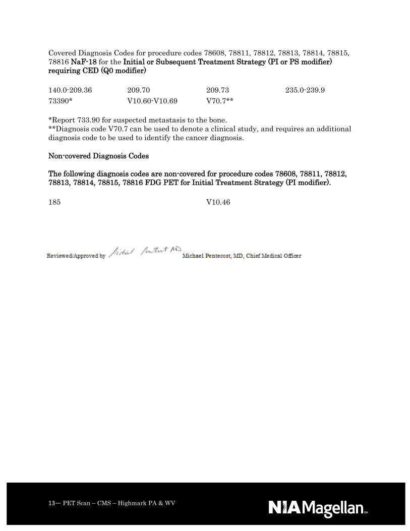
Covered Diagnosis Codes for procedure codes 78608, 78811, 78812, 78813, 78814, 78815,
78816 NaF-18 for the Initial or Subsequent Treatment Strategy (PI or PS modifier)
requiring CED (Q0 modifier)
140.0-209.36 209.70 209.73 235.0-239.9
73390* V10.60-V10.69 V70.7**
*Report 733.90 for suspected metastasis to the bone.
**Diagnosis code V70.7 can be used to denote a clinical study, and requires an additional
diagnosis code to be used to identify the cancer diagnosis.
Non-covered Diagnosis Codes
The following diagnosis codes are non-covered for procedure codes 78608, 78811, 78812,
78813, 78814, 78815, 78816 FDG PET for Initial Treatment Strategy (PI modifier).
185 V10.46