periodontal therapy in siblings with papillon–lefèvre syndrome and tinea capitis: a report of two...
TRANSCRIPT

Periodontal therapy in siblingswith Papillon–Lefevre syndromeand tinea capitis: a report of twocasesSchacher B, Baron F, Ludwig B, Valesky E, Noack B, Eickholz P. Periodontal therapyin siblings with Papillon–Lefevre syndrome and tinea capitis: a report of two cases.J Clin Periodontol 2006; 33: 829–836. doi 10.1111/j.1600-051X.2006.00992.x
AbstractObjective: Report of clinical and microbiological periodontal findings before and6 months after treatment of two siblings with Papillon–Lefevre syndrome (PLS) andtinea capitis.
Methods: Two brothers, RG 3 years and NG 5 years of age, were referred fortreatment due to premature mobility of their deciduous teeth. Probing depths (PPD),attachment levels (PAL-V), and furcation involvements were examined clinically.Panoramic radiographs were taken. Subgingival plaque samples within the deepestpocket of each tooth were taken and analysed by real-time polymerase chain reaction(PCR) for Actinobacillus actinomycetemcomitans (AA), Porphyromonas gingivalis,Tannerella forsythensis, Treponema denticola, Fusobacterium nucleatum, andPrevotella intermedia. One-stage full-mouth scaling and extraction of hopeless teethwere performed under general anaesthesia, followed by systemic amoxicillin andmetronidazole for 7 days. Clinical and microbiological analyses were performed6 months after treatment.
Results: Before treatment, both siblings had exhibited PPD of up to 13 mm, Class IIIfurcation defects at four teeth, and marginal suppuration. AA was detected in bothpatients and at all teeth at levels ranging from 3.0 � 102 to 5.1 � 106. Both patientsexhibited palmar and plantar hyperkeratosis. Seven teeth were extracted from RG, andnine from NG. Six months after treatment, PPD had been reduced to � 5 mm. AA wasnot detected in any of the remaining teeth.
Conclusion: Even periodontally affected deciduous teeth of PLS patients can betreated successfully. Suppression of AA to below detection level seems to be of highsignificance.
Key words: Actinobacillus actinomycetem-comitans-associated periodontitis; Papillon-Lefevre syndrome; tinea capitis
Accepted for publication 19 July 2006
Papillon–Lefevre syndrome (PLS) is char-acterized by palmar and plantar hyperker-atosis, combined with severe periodontaldestruction affecting both the primary andpermanent dentitions (Papillon & Lefevre1924). The disease is rare (one to fourcases per 1 million individuals) and isinherited in an autosomal recessive pat-tern (Gorlin et al. 1964). PLS may occurin siblings (Preus & Gjermo 1987, Firatliet al. 1996a, Rudiger et al. 1999, De Vreeet al. 2000, Cagli et al. 2005).
Microbiological studies of plaquesamples from patients with PLS haveshown the presence of Gram-negativeanaerobic pathogens, often but notalways including Actinobacillus actino-mycetemcomitans (AA) (Kressin et al.1995, Rudiger et al. 1999, Eickholzet al. 2001, Pacheco et al. 2002).
After localization of a gene for PLSto chromosome11q14–q21 (Fischeret al. 1997, Laass et al. 1997, Hartet al. 1998), it was elucidated that this
syndrome is caused by mutations in theCathepsin C (CTSC) gene (Hart et al.1999, Toomes et al. 1999). It was shownthat certain mutations result in a nearlycomplete loss of function of the cathe-psin C (Toomes et al. 1999). To date,more than 40 mutations of this genehave been identified (Selvaraju et al.2003, Noack et al. 2004).
Conventional periodontal therapy(oral hygiene instructions, scaling androot planing, periodontal surgery, sys-
B. Schacher1, F. Baron1, B. Ludwig2,E. Valesky3, B. Noack4 andP. Eickholz1
Departments of 1Periodontology;2Orthodontics, Center for Dental, Oral, and
Maxillofacial Medicine; 3Dermatology,
Hospital of the Johann Wolfgang Goethe-
University at Frankfurt, 60590 Frankfurt,
Germany; 4Periodontology, Department of
Conservative Dentistry, Center for Dental,
Oral, and Maxillofacial Medicine, University
Hospital, Dresden, Germany
J Clin Periodontol 2006; 33: 829–836 doi: 10.1111/j.1600-051X.2006.00992.x
829r 2006 The Authors. Journal compilation r 2006 Blackwell Munksgaard

temic antibiotics) often failed in PLSpatients (Bullon et al. 1993, Hattab et al.1995, De Vree et al. 2000). Some casereports described the early extractionof all erupted teeth, followed by anedentulous period, to prevent subse-quent infection of the non-erupted teeth(Baer & McDonald 1981, Preus 1988,Tinanoff et al. 1995, Wiebe et al. 2001,Ullbro et al. 2005). This conceptresulted in successful outcomes, butsuccessful cases have also beendescribed that did not include large-scale extractions (Ishikawa et al. 1994,Eickholz et al. 2001, Pacheco et al.2002, Lux et al. 2005). In these cases,AA had either not been detected beforeor had been eliminated by treatment.
Other findings beyond skin and perio-dontal lesions have also been reportedfor PLS, e.g. immunological changes(Van Dyke et al. 1984, Firatli et al.1996b, Ghaffar et al. 1999, Lundgrenet al. 2005) and increased suscepti-bility to infection (Haim & Munk1965, Haneke et al. 1975). Patientswith PLS seem to be predisposed todevelop pyogenic liver abscesses(Khandpur & Reddy 2001, Almuneefet al. 2003).
Especially in children, tinea capitisis a frequent fungal infection of the skinand hair with involvement of the hairshaft and the pilosebaceous unit. Thecausative organisms for tinea capitisin Germany are Microsporum (M.)canis (54.8%) and Tircophyton (T.)mentagrophytes (14.7%), followed byT. verrucosum (8.1%), T. violaceum(6.1%), and T. tonsurans (3.8%) (Tietzet al. 1999). The distribution of patho-gens shows geographical differences,e.g. in the United States T. tonsuranshas been replaced by M. adouinii, andM. canis (Gupta et al. 1999) comple-tely. The clinical pattern of tinea capitisvaries: The non-inflammatory typemanifests as a subtile clinical infec-tion with one or multiple patches ofalopecia, whereas the tinea capitis pro-funda shows an inflammatory, boggytender scalp with pustules and withpurulent discharge. In addition, thepatients may have occipital lymphade-nopathy, malaise, and fever (Guptaet al. 1999).
The present report describes theperiodontal, dermatological, and geneticfindings in two brothers having PLS andtinea capitis. The periodontal treatmentof the deciduous dentition with 6-monthresults and the dermatological treatmentare reported.
Case report
Patients
Two brothers, RG 3 years and NG5 years of age, were referred to theDepartment of Periodontology of theCenter for Dental, Oral, and Maxillofa-cial Medicine at the University Hospitalof Frankfurt. The family’s dentist hadobserved premature mobility of thedeciduous teeth and asked for additionaldiagnostics and therapy.
Both the children had palmoplantarhyperkeratosis since the first months oflife. Initially, the symptoms started withan erythema of the palms and the soles.Later on, the lesions spread to the backof the hands and feet. Additionally, RGshowed psoriasiform plaques on theknees and elbows.
The boys’ parents (father, age 42;mother, age 38) are of Eritrean origin,but had been living in Germany for 13years. Both parents were in generallygood health and had not previously had
any major or unusual dental or perio-dontal problems. No unusual dentalpathologies were reported in theirextended families. The parents are notblood related. Before scientific exami-nation and treatment of the patients, therisks, benefits, and procedures wereexplained to their parents and informedconsent was obtained. The investigationwas approved by the Institutional Re-view Boards for Human Studies of theUniversities of Frankfurt and Dresden.
Clinical and radiographic examination
At the time of the baseline examination,NG and RG presented with completedeciduous dentitions. Generalizedsupragingival plaque, bleeding as wellas suppuration on probing, and class I toIII tooth mobility were found in bothpatients (Fig. 1a and b). Owing to theyoung age of the patients and the highlyinflamed gingival tissues at the initialpresentation, a reduced periodontal
Fig. 1. Clinical situation before therapy: (a) NG, (b) RG.
830 Schacher et al.
r 2006 The Authors. Journal compilation r 2006 Blackwell Munksgaard

examination was performed revealingprobing depths (PPD) of 2–9 mm inboth and vertical attachment levels(PAL-V) of 2–9 mm in NG.
The panoramic radiographs for NGand RG showing generalized mainly
horizontal bone loss are presented inFig. 2a and b.
The clinical and radiological findingsyielded a diagnosis of periodontitis as amanifestation of PLS for both patients.The children’s teeth were professionally
cleaned, and they and their motherreceived comprehensive oral hygieneinstructions.
A comprehensive periodontal status,including assessment of PPD and PAL-V at six sites per tooth to the nearest1 mm (PCPUNC 15, Hu-Friedy, Chica-go, IL, USA), was obtained immediatelybefore subgingival debridement undergeneral anaesthesia. Furcation diagnosis(clinical horizontal attachment levels)was performed using a calibratedNabers probe (PQ2 N, Hu Friedy).
Microbiological examination
Immediately before subgingival instru-mentation, under general anaesthesia,subgingival plaque samples wereobtained from the deepest pocket ofeach tooth using sterile paper pointsand analysed for AA, Porphyromonasgingivalis (PG), Tannerella forsythensis(TF), Treponema denticola (TD), Fuso-bacterium nucleatum (FN), and Prevo-tella intermedia (PI) using acommercially available real-time poly-merase chain reaction (PCR) test (Mer-idol Paro Diagnostik, Gaba GmbH,Lorrach, Germany).
Genetic examination
Venous blood samples were taken. AfterDNA purification (DNA purification kit,Qiagen, Hilden, Germany), all sevenexons of the CTSC gene includingexon/intron boundary regions wereamplified by PCR and directlysequenced as described previously(Noack et al. 2004). Generatedsequences (ALFwin Sequence Analyser2.00 software, Amersham PharmaciaBiotech, Freiburg, Germany) werealigned with published CTSC sequences(GenBank accession no. NT_08984).DNA sequence variants were confirmedby sequencing at least two independentPCR products.
Periodontal treatment
NG’s teeth 51 and 85 had been extractedbefore systematic treatment due to acuteinflammation and pain. Systematic treat-ment commenced for both NG and RGwith subgingival scaling using ultraso-nic applicators and hand instrumentsunder general anaesthesia. The perio-dontally hopeless teeth (NG: teeth 55,54, 64, 74, 72, 71, 81, 82, and 83; RG:teeth 54, 51, 64, 74, 71, 81, and 84) weresubsequently removed.
Fig. 2. Panoramic radiographs before therapy: (a) NG, (b) RG.
Fig. 3. Tinea capitis in RG: (a) before and (b) 3 months after combined mechanic andantibiotic periodontal therapy: inflammatory erythematosquamous plaques with pustules,purulent discharge, and yellow crusts on the scalp.
Periodontal therapy in siblings with PLS and tinea capitis 831
r 2006 The Authors. Journal compilation r 2006 Blackwell Munksgaard

All remaining teeth received a subgin-gival instillation if a 1% chlorhexidinegel (Chlorhexamed Gel, GlaxoSmith-Kline Consumer Healthcare, Buhl, Ger-many). The parents were instructed tohave the children rinse with 0.12%chlorhexidine solution (GUM Paroex,John O. Butler, Kriftel, Germany) twicedaily and to brush their children’s teethwith 1% chlorhexidine gel. Both pa-tients received amoxicillin 250 mg andmetronidazole 250 mg three times dailyfor 1 week. Post-operative follow-upexaminations with professional toothcleaning and oral hygiene re-motivationsessions were performed after 1, 2, 3, 6,10, 14, 18, and 22 weeks. Space main-tainers were inserted for NG, 3 weekspost-operatively.
Dermatological treatment
Before periodontal therapy NG and RGwere admitted to the department ofdermatology for treatment of the hyper-keratotic skin lesions. After combinedmechanic and antibiotic periodontaltherapy in both siblings, the develop-ment of inflammatory erythematosqua-mous plaques with pustules, purulentdischarge, and yellow crusts on thescalp, accompanied by worsening ofpalmoplantar hyperkeratosis, wasobserved (Fig. 3).
RG’s disease was more pronounced.The clinical diagnosis of tinea capitiswas confirmed in RG with the Wood’slamp (filtered ultraviolet light peak of365 nm). The direct light microscopicexamination of RG’s hair showed afungal ectotrix infection, and the subse-quent culture (takes 14 days to grow)identified M. canis as the pathogenicorganism. In NG, the light microscopicexamination and the fungal culture werenegative. It is known in the literaturethat in a substantial number of cases(approximately 50%) the fungal culturecan be negative (Silverman & Elewski1998). Because of the mild manifesta-tion and the negative light micro-scopic and culture analysis, we decidedto treat NG only with an intensive localtreatment. The whole scalp was treatedwith ciclopiroxolamin cream underocclusion twice daily. Furthermore, aselenium sulphide shampoo was usedonce daily.
RG additionally received itracona-zole systemically (5 mg/kg weight) for4 weeks. No adverse events have beenobserved. After 1 month of treatment,
the light microscopy and consecutiveculture were negative.
The follow-up assessment showedminimal clinical signs of tinea capitisin RG and none in NG. The therapy withlocal ciclopiroxolamin cream was con-ducted in both siblings for a further 4weeks. The palmoplantar keratodermawas treated by intensive topical therapyusing 10–20% urea in an ointment andcream base twice daily as the basistreatment. The hyperkeratosis decreasedalmost completely.
Examination of the parents
Both parents were also clinically exam-ined, panoramic radiographs wereobtained, subgingival plaque sampleswere taken at the deepest site of eachquadrant, and analysed using the RNAprobe test (IAI Pado-Test 4.5s, Institutfur angewandte Immunologie, Zuchwil,
Switzerland). Finally, samples ofvenous blood were obtained.
Results
Clinical and microbiological parameters
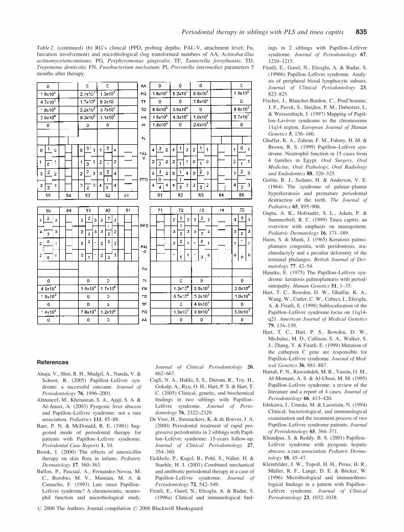
The detailed clinical and microbiologi-cal results for NG and RG are presentedin Table 1 and Table 2: before treat-ment, markedly increased PPD (up to13 mm) were found for nearly all teeth,with the findings more pronounced forNG than for RG (Table 1). Six monthsafter treatment, PPDs were generallyreduced to 2–4 mm with one site ineither sibling exhibiting PPD of 5 mm(Table 2).
Before treatment, AA at levels of3.0 � 102 –5.1 � 106 had been detectedin NG and RG (Table 1). Six monthsafter treatment, AA was not detectable ineither patient (Table 2).
Table 1. (a) NG’s clinical (PPD, probing depths; PAL-V, attachment level; Fu, furcationinvolvement) and microbiological (log transformed numbers of AA, Actinobacillus actinomy-cetemcomitans; PG, Porphyromonas gingivalis; TF, Tannerella forsythensis; TD, Treponemadenticola; FN, Fusobacterium nucleatum; PI, Prevotella intermedia) parameters before therapy
832 Schacher et al.
r 2006 The Authors. Journal compilation r 2006 Blackwell Munksgaard

The parents’ clinical and radiologicfindings were each consistent with ageneralized moderate chronic perio-dontitis. AA was not detected microbio-logically in sub-gingival plaque samplesof either parent.
Genetic parameters
Sequence analysis of the CTSC geneidentified a functionally relevant homo-zygote missense mutation in NG andRG in exon 5 (c.755A4T, p.Q252L)first described by Toomes et al. (1999).Both parents were heterozygous carriersof the c.755A4T mutation in the CTSCgene.
Discussion
The present report documents the diag-nostic measures and partial preservation
of teeth in two cases of generalizedperiodontitis in the deciduous dentitionof two brothers with PLS. To the best ofour knowledge, this is the first report ofsubgingival instrumentation and partialpreservation of deciduous teeth in treat-ment of PLS periodontitis. Both patientswere referred with severe periodontalinflammation including bleeding andsuppuration on probing, prematuremobility of deciduous teeth, attachmentloss, and furcation involvement (classIII). NG had acute symptoms with painand periodontal abscesses at two teeth.Both siblings also exhibited palmoplan-tar hyperkeratoses. The clinical diagno-sis of PLS was confirmed by geneticanalysis showing a corresponding muta-tion of the CTSC gene that was found tobe homozygotic in the children andheterozygotic in their parents.
Detailed microbiological examina-tion revealed subgingival presence of
AA at all teeth before treatment, con-firming the observation that this perio-dontal pathogen plays a key role inthe aetiology of periodontitis in PLS(Kleinfelder et al. 1996, De Vree et al.2000, Eickholz et al. 2001, Wara-aswa-pati et al. 2001, Lux et al. 2005). Owingto the fact that AA could not be detectedin either of the parents, the source of theAA-infection remains in question. Onthe one hand, it cannot be ruled outtotally that the parents harbour AA sub-gingivally because microbiologicalsampling was performed only at thefour teeth with most pronounced PPD.On the other hand, the source of infec-tion may be located in other relativeswho were not available for examination.However, on this issue we can onlyspeculate.
Combined mechanical and antibioticperiodontal therapy resulted in drasticclinical improvements in both patientsand suppressed AA to below the quitelow detection limit of real-time PCR(102). This primary result is in agree-ment with the results of other research-ers (Rudiger et al. 1999, Eickholz et al.2001, Pacheco et al. 2002). Presumably,the positive treatment outcome maypartially be attributed to the fact thatmechanical therapy, including thenecessary extractions, followed theprinciple of full-mouth disinfection(Quirynen et al. 1995). Amoxicillinand metronidazole, administered con-currently, are reliable and effective inthe treatment of AA-associated perio-dontitis (Van Winkelhoff et al. 1989),which was confirmed in the present case.
Thus, an overall favourable develop-ment may be expected. This presup-poses, however, that the young patientsare closely followed, with their parentsintricately involved, as the children arenot yet able to ensure adequate oralhygiene on their own.
Wiebe et al. (2001) reported a suc-cessful long-term therapeutic outcomein the case of a young boy with PLSwho had undergone periodontal treat-ment since the age of 4, despite med-iocre oral hygiene. In that case, theperiodontal infection could only beeliminated by extracting all deciduousteeth and a subsequent edentulous phaseafter metronidazole monotherapy hadfailed. Subsequent microbiological ana-lysis of the permanent dentition showedno indication of AA. Extraction of allperiodontally involved teeth causing anedentulous phase seems to be a reliablestrategy to eliminate AA. However, this
Table 1. (continued) (b) RG’s clinical (PPD, probing depths; PAL-V, attachment level; Fu, furcationinvolvement) and microbiological (log transformed numbers of AA, Actinobacillus actinomycetem-comitans; PG, Porphyromonas gingivalis; TF, Tannerella forsythensis; TD, Treponema denticola;FN, Fusobacterium nucleatum; PI, Prevotella intermedia) parameters before therapy.
Periodontal therapy in siblings with PLS and tinea capitis 833
r 2006 The Authors. Journal compilation r 2006 Blackwell Munksgaard

is a quite drastic measure possibly hav-ing psychological consequences for thechildren and likely to cause orthodonticproblems due to the lack of space-main-taining function of the deciduous teeth(Lux et al. 2005).
As AA was no longer found on thedeciduous teeth of either of the two boystreated, it may be assumed that theeruption of the first permanent teeth,which is currently in progress in NG,may occur in a healthy periodontalenvironment. The risk of AA reinfectionof the children is considered to be low,as the pathogen was not found in eitherof the parents. The orthodontic conse-quences due to the unavoidable loss ofthe most severely involved teeth weretaken care of by insertion of spacemaintainers.
Ahuja et al. (2005) described the caseof a 10-year-old PLS patient: after scal-ing and root planing, combined withsystemic administration of amoxicillinand metronidazole, failed to improve theperiodontal situation, flap surgeries witha second antibiotic treatment was suc-cessful. Despite systemic amoxicillinand metronidazole, it may be hypothe-sized that in this case, AA could not beeliminated by non-surgical therapy. Per-sistence of AA despite repeated mechan-ical therapy combined with amoxicillinand metronidazole had been describedbefore by Rudiger et al. (1999). Finally,renewed antibiotics combined with sur-gical therapy with its additional poten-tial to remove AA from the tissue weresuccessful. However, Ahuja et al. (2005)performed microbiological sampling
neither before nor after therapy. Thus,we cannot be sure of the reason behindthe initial failure and the final success oftherapy.
On the one hand, the systemic use ofretinoids has been reported to be effec-tive in patients with PLS (Lee et al.2005, Sethuraman et al. 2005). On theother, retinoid treatment may cause ske-letal abnormalities in children, slenderlong bones and premature epiphysealclosure as well as mucocutaneous dry-ness, abnormal liver function tests, andelevated triglycerides (Lee et al. 2005).Some authors reported that systemicretinoids not only improve skin lesionsbut also gingival inflammation by anunknown mechanism (Lee et al. 2005,Sethuraman et al. 2005). Others demon-strated that the level of periodontalinfection and the severity of skin affec-tion do not correlate at all (Ullbro et al.2003). Because of the mild to moderatemanifestation of the palmoplantar kera-toderma and the young age of thepatients, we initially decided to useonly local dermatological treatment inboth of them.
Tinea capitis, which had aggravatedin both children after combined mechan-ical and antibiotic treatment, is to beconsidered a separate disease. It canonly be speculated that children withPLS associated with abnormal func-tional or quantitative neutrophils in50% may be more susceptible to derma-tomycoses. Furthermore, the medicationwith systemic antibiotics (amoxicillinand metronidazole) may have supportedthe growth of the dermatophytes bychanging bacterial and fungal skin floraquantitatively as well as qualitatively(Brook 2000).
The dental treatment provider ofyoung PLS patients should make sureto keep an eye on the children’s generalhealth and to ensure that they see apaediatrician or appropriate specialistas required.
To the best of our knowledge, this isthe first report of (i) PLS with a mani-festation of tinea capitis and (ii) ofsubgingival instrumentation and at leastshort-term partial preservation of decid-uous teeth in the treatment of PLSperiodontitis.
Acknowledgement
This study was kindly supported byGABA GmbH, Lorrach, Germany.
Table 2. (a) NG’s clinical (PPD, probing depths; PAL-V, attachment level; Fu, furcationinvolvement) and microbiological (log transformed numbers of AA, Actinobacillus actinomy-cetemcomitans; PG, Porphyromonas gingivalis; TF, Tannerella forsythensis; TD, Treponemadenticola; FN, Fusobacterium nucleatum; PI, Prevotella intermedia) parameters 5 months aftertherapy
834 Schacher et al.
r 2006 The Authors. Journal compilation r 2006 Blackwell Munksgaard
References
Ahuja, V., Shin, R. H., Mudgil, A., Nanda, V. &
Schoor, R. (2005) Papillon–Lefevre syn-
drome: a successful outcome. Journal of
Periodontology 76, 1996–2001.
Almuneef, M., Khenaizan, S. A., Ajaji, S. A. &
Al-Anazi, A. (2003) Pyogenic liver abscess
and Papillon–Lefevre syndrome: not a rare
association. Pediatrics 111, 85–88.
Baer, P. N. & McDonald, R. E. (1981) Sug-
gested mode of periodontal therapy for
patients with Papillon–Lefevre syndrome.
Periodontal Case Reports 1, 10.
Brook, I. (2000) The effects of amoxicillin
therapy on skin flora in infants. Pediatric
Dermatology 17, 360–363.
Bullon, P., Pascual, A., Fernandez-Novoa, M.
C., Borobio, M. V., Muniain, M. A. &
Camacho, F. (1993) Late onset Papillon–
Lefevre syndrome? A chromosomic, neutro-
phil function and microbiological study.
Journal of Clinical Periodontology 20,
662–667.
Cagli, N. A., Hakki, S. S., Dursun, R., Toy, H.,
Gokalp, A., Ryu, O. H., Hart, P. S. & Hart, T.
C. (2005) Clinical, genetic, and biochemical
findings in two siblings with Papillon–
Lefevre syndrome. Journal of Perio-
dontology 76, 2322–2329.
De Vree, H., Steenackers, K. & de Boever, J. A.
(2000) Periodontal treatment of rapid pro-
gressive periodontitis in 2 siblings with Papil-
lon–Lefevre syndrome: 15-years follow-up.
Journal of Clinical Periodontology 27,
354–360.
Eickholz, P., Kugel, B., Pohl, S., Naher, H. &
Staehle, H. J. (2001) Combined mechanical
and antibiotic periodontal therapy in a case of
Papillon–Lefevre syndrome. Journal of
Periodontology 72, 542–549.
Firatli, E., Gurel, N., Efeoglu, A. & Badur, S.
(1996a) Clinical and immunological find-
ings in 2 siblings with Papillon–Lefevre
syndrome. Journal of Periodontology 67,
1210–1215.
Firatli, E., Gurel, N., Efeoglu, A. & Badur, S.
(1996b) Papillon–Lefevre syndrome. Analy-
sis of peripheral blood lymphocyte subsets.
Journal of Clinical Periodontology 23,
823–825.
Fischer, J., Blanchet-Bardon, C., Prud’homme,
J. F., Pavek, S., Steijlen, P. M., Dubertret, L.
& Weissenbach, J. (1997) Mapping of Papil-
lon–Levevre syndrome to the chromosome
11q14 region. European Journal of Human
Genetics 5, 156–160.
Ghaffar, K. A., Zahran, F. M., Fahmy, H. M. &
Brown, R. S. (1999) Papillon–Lefevre syn-
drome. Neutrophil function in 15 cases from
4 families in Egypt. Oral Surgery, Oral
Medicine, Oral Pathology, Oral Radiology
and Endodontics 88, 320–325.
Gorlin, R. J., Sedano, H. & Anderson, V. E.
(1964) The syndrome of palmar–plantar
hyperkeratosis and premature periodontal
destruction of the teeth. The Journal of
Pediatrics 65, 895–906.
Gupta, A. K., Hofstader, S. L., Adam, P. &
Summerbell, R. C. (1999) Tinea capitis: an
overview with emphasis on management.
Pediatric Dermatology 16, 171–189.
Haim, S. & Munk, J. (1965) Keratosis palmo-
plantaris congenita, with peridontosis, ara-
chnodactyly and a peculiar deformity of the
terminal phalanges. British Journal of Der-
matology 77, 42–54.
Haneke, E. (1975) The Papillon–Lefevre syn-
drome: keratosis palmoplantaris with period-
ontopathy. Human Genetics 51, 1–35.
Hart, T. C., Bowden, D. W., Ghaffar, K. A.,
Wang, W., Cutler, C. W., Cebeci, I., Efeoglu,
A. & Firatli, E. (1998) Sublocalization of the
Papillon–Lefevre syndrome locus on 11q14-
q21. American Journal of Medical Genetics
79, 134–139.
Hart, T. C., Hart, P. S., Bowden, D. W.,
Michalec, M. D., Callison, S. A., Walker, S.
J., Zhang, Y. & Firatli, E. (1999) Mutation of
the cathepsin C gene are responsible for
Papillon–Lefevre syndrome. Journal of Med-
ical Genetics 36, 881–887.
Hattab, F. N., Rawashdeh, M. R., Yassin, O. M.,
Al-Momani, A. S. & Al-Ubosi, M. M. (1995)
Papillon–Lefevre syndrome: a review of the
literature and a report of 4 cases. Journal of
Periodontology 66, 413–420.
Ishikawa, I., Umeda, M. & Laosrisin, N. (1994)
Clinical, bacteriological, and immunological
examination and the treatment process of two
Papillon–Lefevre syndrome patients. Journal
of Periodontology 65, 364–371.
Khandpur, S. & Reddy, B. S. (2001) Papillon–
Lefevre syndrome with pyogenic hepatic
abscess: a rare association. Pediatric Derma-
tology 18, 45–47.
Kleinfelder, J. W., Topoll, H. H., Preus, H. R.,
Muller, R. F., Lange, D. E. & Bocker, W.
(1996) Microbiological and immunohisto-
logical findings in a patient with Papillon–
Lefevre syndrome. Journal of Clinical
Periodontology 23, 1032–1038.
Table 2. (continued) (b) RG’s clinical (PPD, probing depths; PAL-V, attachment level; Fu,furcation involvement) and microbiological (log transformed numbers of AA, Actinobacillusactinomycetemcomitans; PG, Porphyromonas gingivalis; TF, Tannerella forsythensis; TD,Treponema denticola; FN, Fusobacterium nucleatum; PI, Prevotella intermedia) parameters 5months after therapy.
Periodontal therapy in siblings with PLS and tinea capitis 835
r 2006 The Authors. Journal compilation r 2006 Blackwell Munksgaard

Kressin, S., Herforth, A., Preis, S., Wahn, V. &
Lenard, H. G. (1995) Papillon–Lefevre syn-
drome–successful treatment with a combina-
tion of retinoid and concurrent systemic
periodontal therapy: case report. Quintes-
sence International 26, 795–803.
Laass, M. W., Hennies, H. C., Preis, S., Stevens,
H. P., Jung, M., Leigh, I. M., Wienker, T. F.
& Reis, A. (1997) Localization of a gene for
Papillon–Lefevre syndrome to chromosome
11q14-q21 by homozygosity mapping.
Human Genetics 101, 376–382.
Lee, M. R., Wong, L. C. & Fischer, G. O.
(2005) Papillon–Lefevre syndrome treated
with acitretin. The Australasian Journal of
Dermatology 46, 199–201.
Lundgren, T., Parhar, R. S., Renvert, S. &
Tatakis, D. N. (2005) Impaired cytotoxicity
in Papillon–Lefevre syndrome. Journal of
Dental Research 84, 414–417.
Lux, C. J., Kugel, B., Komposch, G., Pohl, S. &
Eickholz, P. (2005) Orthodontic treatment in
a patient with Papillon–Lefevre syndrome.
Journal of Periodontology 76, 149–157.
Noack, B., Gorgens, H., Hoffmann, T., Fangha-
nel, J., Kocher, T., Eickholz, P. & Schackert,
H. K. (2004) Novel mutations in the Cathe-
psin C Gene in patients with prepubertal
aggressive periodontitis and Papillon–
Lefevre syndrome. Journal of Dental
Research 83, 368–370.
Pacheco, J. J., Coelho, C., Salazar, F., Con-
treras, A., Slots, J. & Velazco, C. H. (2002)
Treatment of Papillon–Lefevre syndrome
periodontitis. Journal of Clinical Perio-
dontology 29, 370–374.
Papillon, M. M. & Lefevre, P. (1924) Deux cas
de keratodermie palmaire et plantaire syme-
trique familiale (maladie de Meleda) chez le
frere et la sœur. Coexistence dans les deus cas
d’alterations dentaires graves. Bulletin de la
Societe Francaise de Dermatologie et de
Syphiligraphie 31, 82–87.
Preus, H. R. (1988) Treatment of rapidly
destructive periodontitis in Papillon–Lefevre
syndrome. Journal of Clinical Perio-
dontology 15, 639–643.
Preus, H. R. & Gjermo, P. (1987) Clinical man-
agement of prepubertal periodontitis in 2 sib-
lings with Papillon–Lefevre syndrome. Journal
of Clinical Periodontology 14, 156–160.
Quirynen, M., Bollen, C. M. L., Vandekerc-
khove, B. N. A., Dekeyser, C., Papaioannou,
W. & Eyssen, H. (1995) Full- versus partial-
mouth disinfection in the treatment of perio-
dontal infections: short-term clinical and
microbiological observations. Journal of
Dental Research 74, 1459–1467.
Rudiger, S., Petersilka, G. & Flemming, T. F.
(1999) Combined systemic and local antimi-
crobiol therapy of periodontal disease in
Papillon–Lefevre syndrome. Journal of Clin-
ical Periodontology 26, 847–854.
Selvaraju, V., Markandaya, M., Prasad, P.,
Sathyan, P., Sethuraman, G., Srivastava, S.
C., Thakker, N. & Kumar, A. (2003) Muta-
tion analysis of the cathepsin C gene in
Indian families with Papillon–Lefevre syn-
drome. BMC Medical Genetics 4, 5.
Sethuraman, G., Malhotra, A. K., Khaitan, B. K.
& Sharma, V. K. (2005) Effectiveness of
isotretinoin in Papillon–Lefevre syndrome.
Pediatric Dermatology 22, 378–379.
Silverman, R. A. & Elewski, B. E. (1998)
Pediatric mycoses. In: Cutaneous Fungal
Infections, 2nd edition, pp. 261–285. Malden:
Blackwell Science.
Tinanoff, N., Tempro, P. & Maderazo, E. G.
(1995) Dental treatment of Papillon–Lefevre
syndrome: 15 year follow-up. Journal of
Clinical Periodontology 22, 609–612.
Tietz, H. J., Czaika, V., Ulbricht, H. M. &
Sterry, W. (1999) Tinea capitis in Germany.
A survey in 1998. Mycoses 42, 73–76.
Toomes, C., James, J., Wood, A. J., Wu, C. L.,
McCormick, D., Lench, N., Hewitt, C., Moy-
nihan, L., Roberts, E., Woods, C. G., Mark-
ham, A., Wong, M., Widmer, R., Ghaffar, K.
A., Pemberton, M., Hussein, I. R., Temtamy,
S. A., Davies, R., Read, A. P., Sloan, P.,
Dixon, M. J. & Thakker, N. S. (1999)
Loss-of-function mutations in the cathepsin
C gene result in periodontal disease and
palmoplantar keratosis. Nature Genetics 23,
421–424.
Ullbro, C., Brown, A. & Twetman, S. (2005)
Preventive periodontal regimen in Papillon–
Lefevre syndrome (in process citation).
Pediatric Dental Journal 27, 226–232.
Ullbro, C., Crossner, C. G., Nederfors, T.,
Alfadley, A. & Thestrup-Pedersen, K.
(2003) Dermatologic and oral findings in a
cohort of 47 patients with Papillon–Lefevre
syndrome. Journal of the American Academy
of Dermatology 48, 345–351.
Van Dyke, T. E., Taubman, M. A., Ebersole, J.
L., Haffajee, A. D., Socransky, S. S., Smith,
D. J. & Genco, R. J. (1984) The Papillon–
Lefevre syndrome: neutrophil dysfunction
with severe periodontal disease. Clinical
Immunology and Immunopathology 31,
419–429.
Van Winkelhoff, A. J., Rodenburg, J. P., Goene,
R. J., Abbas, E. G., Winkel, E. G. & de
Graaff, J. (1989) Metronidazole plus amox-
icillin in the treatment of Actinobacillus
actinomycetemcomitans-associated perio-
dontitis. Journal of Clinical Periodontology
16, 128–131.
Wara-aswapati, N., Lertsirivorakul, J., Naga-
sawa, T., Kawashima, Y. & Ishikawa, I.
(2001) Papillon–Lefevre syndrome: serum
immunoglobulin G (IgG) subclass antibody
response to periodontapathic bacteria. A -
case report. Journal of Periodontology 72,
1747–1753.
Wiebe, C. B., Hakkinen, L., Putkins, E. E.,
Walsh, P. & Larjava, H. S. (2001) Successful
periodontal maintenance of a case with Papil-
lon–Lefevre syndrome: 12-year follow-up
and review of the literature. Journal of Perio-
dontology 72, 824–830.
Address:
B. Schacher
Department of Periodontology
Johann Wolfgang Hospital
Goethe-University at Frankfurt
Theodor-Stern-Kai 7
60590 Frankfurt
Germany
E-mail: [email protected]
Clinical Relevance
Scientific rationale for study: PLS isa condition that, if untreated, willlead unevitably to total tooth lossbefore the age of 30. For therapy inaffected deciduous dentitions,extraction of all teeth with an eden-
tulous period is still recommended.This invasive concept has severalnegative orthodontic and psychologi-cal consequences.
Principal findings: By extractionof hopeless teeth as well as combinedmechanical and systemic antibiotic
therapy, clinical parameters wereimproved and AA was suppressedbelow detection limit.
Practical implication: PLS perio-dontitis in deciduous dentitions maybe treated successfully withoutextracting all teeth.
836 Schacher et al.
r 2006 The Authors. Journal compilation r 2006 Blackwell Munksgaard