percutaneous treatment of large pyogenic liver abscess
TRANSCRIPT
The Egyptian Journal of Radiology and Nuclear Medicine (2014) 45, 109–115
Egyptian Society of Radiology and Nuclear Medicine
The Egyptian Journal of Radiology andNuclearMedicine
www.elsevier.com/locate/ejrnmwww.sciencedirect.com
ORIGINAL ARTICLE
Percutaneous treatment of large pyogenic
liver abscess
* Corresponding author. Tel.: +20 1094274500; fax: +20 934602963.
E-mail addresses: [email protected] (M.A. Abusedera), elbadryam@
yahoo.com (A.M. El-Badry).
Peer review under responsibility of Egyptian Society of Radiology and
Nuclear Medicine.
Production and hosting by Elsevier
0378-603X � 2014 Production and hosting by Elsevier B.V. on behalf of Egyptian Society of Radiology and Nuclear Medicine.
http://dx.doi.org/10.1016/j.ejrnm.2013.11.005
Mohammad Alaa Abusedera a,*, Ashraf Mohammad El-Badry b
a Sohag University, Nasser City, Sohag 82524, Egyptb Sohag University, Sohag University Street, Nasser City, Sohag 82524, Egypt
Received 6 June 2013; accepted 10 November 2013Available online 15 December 2013
KEYWORDS
Liver;
Abscess;
Drainage;
Aspiration
Abstract Surgical drainage has been the traditional mode of treatment of pyogenic liver abscess
but this was replaced by IV broad-spectrum antibiotics and imaging-guided percutaneous drainage
either via needle aspiration or percutaneous catheter drainage (CD). There is a debate about which
is better intermittent needle aspiration or CD.
Our objective is to compare the outcome of CD versus intermittent needle aspiration of pyogenic
liver abscess and to compare the single step Trocar technique versus the modified Seldinger tech-
nique.
Patients and methods: 88 patients, 65 men and 23 women, mean age 44.6 (18–73) years had pyo-
genic liver abscess. Patients were divided in two groups randomly; aspiration group with maximum
of three attempts and the CD group. Ultrasound or CT was used.
Results: Aspiration was successful in 60% of cases (26/43). CD was successful in 98% (44/45).
Three patients were treated by surgical drainage (two patients of the aspiration group and one of
the CD group) with favorable outcome. Both Seldinger and single step Trocar techniques were com-
parable as regards outcome and procedure-related pain but the procedure time of Trocar was sig-
nificantly shorter. No major complications were encountered.
Conclusion: CD is more efficient than needle aspiration. Aspiration can be used for simple small
abscesses. Trocar technique is less time-consuming than the Seldinger technique.� 2014 Production and hosting by Elsevier B.V. on behalf of Egyptian Society of Radiology and Nuclear
Medicine.
1. Introduction
Pyogenic liver abscess is a rare, life-threatening disease thathas an increasing incidence rate in the United States and
Europe. The high morbidity and mortality rates associatedwith the treatment of pyogenic liver abscess were improved sig-nificantly with the introduction of ultrasound (US) and com-
puted tomography (CT) guided percutaneous drainage (1–6).
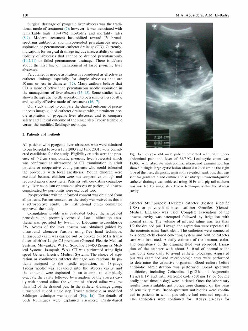
Fig. 1a 65 year old male patient presented with right upper
abdominal pain and fever of 38.7 �C. Leukocytic count was
18,000, with absolute neutrophilia, ultrasound examination has
shown a single large cystic lesion about 8 · 7 · 6 cm at the right
lobe of the liver, diagnostic aspiration revealed frank pus, that was
sent for gram stain and culture and sensitivity, ultrasound-guided
catheter drainage was achieved using 10 Fr and pig tail catheter
was inserted by single step Trocar technique within the abscess
cavity.
110 M.A. Abusedera, A.M. El-Badry
Surgical drainage of pyogenic liver abscess was the tradi-tional mode of treatment (7); however, it was associated withremarkably high (10–47%) morbidity and mortality rates
(8,9). Modern treatment has shifted toward IV broad-spectrum antibiotics and image-guided percutaneous needleaspiration or percutaneous catheter drainage (CD). Currently,
indications for surgical drainage include inaccessibility or mul-tiplicity of abscesses that cannot be drained percutaneously(10,2,11) or failed percutaneous drainage. There is debate
about the first line of management of large pyogenic liverabscesses.
Percutaneous needle aspiration is considered as effective ascatheter drainage especially for simple abscesses that are
50 mm or less in diameter (12). Many authors believe thatCD is more effective than percutaneous needle aspiration inthe management of liver abscess (13–15). Some studies have
shown therapeutic needle aspiration to be a simpler, less costly,and equally effective mode of treatment (16,17).
Our study aimed to compare the clinical outcome of percu-
taneous image-guided catheter drainage with intermittent nee-dle aspiration of pyogenic liver abscesses and to comparesafety and clinical outcome of the single step Trocar technique
versus the modified Seldinger technique.
2. Patients and methods
All patients with pyogenic liver abscesses who were admittedto our hospital between July 2003 and June 20013 were consid-ered candidates for the study. Eligibility criteria were the pres-ence of >2 cm symptomatic pyogenic liver abscess(s) which
was confirmed at ultrasound or CT examination in adultpatients or cooperative young patients who could toleratedthe procedure with local anesthesia. Young children were
excluded because children were not cooperative enough andrequired general anesthesia. Patients with coexisting coagulop-athy, liver neoplasm or amoebic abscess or perforated abscess
complicated by peritonitis were excluded too.Pre-procedure written informed consent was obtained from
all patients. Patient consent for the study was waived as this is
a retrospective study. The institutional ethics committeeapproved the study.
Coagulation profile was evaluated before the scheduledprocedure and promptly corrected. Local infiltration anes-
thesia was provided by 6–8 ml of Lidocaine hydrochloride2%. Access of the liver abscess was obtained guided byultrasound whenever feasible using free hand technique.
Ultrasound exam was carried out by convex 3–5 MHz trans-ducer of either Logic C5 premium (General Electric MedicalSystems, Milwaukee, WI) or Sonoline 51–450 (Siemens Med-
ical Systems, Issaquah, WA). CT was performed using lightspeed General Electric Medical Systems. The choice of aspi-ration or continuous catheter drainage was random. In pa-tients assigned to the needle aspiration, an 18-gauge
Trocar needle was advanced into the abscess cavity andthe contents were aspirated in an attempt to completelyevacuate the cavity followed by irrigation of the abscess cav-
ity with normal saline; the volume of infused saline was lessthan 1/2 of the drained pus. In the catheter drainage group,ultrasound guided single step Trocar technique or modified
Seldinger technique was applied (Fig. 1a). The details ofboth techniques were explained elsewhere. Plastic-based
catheter Multipurpose Flexiema catheter (Boston scientificUSA) or polyurethane-based catheter Genoflex (GenesisMedical England) was used. Complete evacuation of the
abscess cavity was attempted followed by irrigation withnormal saline. The volume of infused saline was less than1/2 the drained pus. Lavage and aspiration were repeated till
the contents came back clear. The catheters were connectedto a completely closed collecting system and routine cathetercare was instituted. A daily estimate of the amount, color,
and consistency of the drainage fluid was recorded. Irriga-tion of the catheter with about 5–10 ml of normal salinewas done once daily to avoid catheter blockage. Aspirated
pus was examined and microbiologic tests were performedto determine the causative organism. Blood culture beforeantibiotic administration was performed. Broad spectrumantibiotics, including Cefazoline 1 g/12 h and Augmentin
1.2 g/8 h IV and with Metronidazole (500 mg IV or 500 mgorally three times a day) were initiated. Once the laboratoryresults were available, antibiotics were changed on the basis
of sensitivity tests. Broad-spectrum antibiotics were contin-ued in patients in whom pus culture had returned negative.The antibiotics were continued for 10 days (14 days for
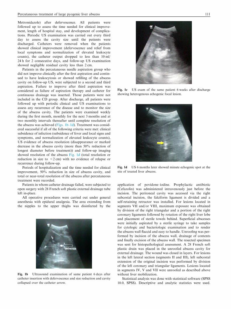
Fig. 1c US exam of the same patient 6 weeks after discharge
showing heterogenous echogenic focal lesion.
Fig. 1d US 6 months later showed minute echogenic spot at the
site of treated liver abscess.
Percutaneous treatment of large pyogenic liver abscess 111
Metronidazole) after defervescence. All patients werefollowed up to assess the time needed for clinical improve-ment, length of hospital stay, and development of complica-
tions. Periodic US examination was carried out every thirdday to assess the cavity size until the patients weredischarged. Catheters were removed when the patients
showed clinical improvement (defervescence and relief fromlocal symptoms and normalization of elevated leukocytecounts), the catheter output dropped to less than 10 ml/
24 h for 2 consecutive days, and follow-up US examinationshowed negligible residual cavity less than 2 cm.
Patients in the percutaneous needle aspiration group whodid not improve clinically after the first aspiration and contin-
ued to have leukocytosis or showed refilling of the abscesscavity on follow-up US, were subjected to a second and thirdaspiration. Failure to improve after third aspiration was
considered as failure of aspiration therapy and catheter forcontinuous drainage was inserted. Those patients were notincluded in the CD group. After discharge, all patients were
followed up with periodic clinical and US examinations toassess any recurrence of the disease and to monitor the sizeof the abscess cavity. The patients were examined weekly
during the first month, monthly for the next 3 months and attwo monthly intervals thereafter until complete resolution ofthe abscess was achieved (Figs. 1b–1d). Treatment was consid-ered successful if all of the following criteria were met: clinical
subsidence of infection (subsidence of fever and local signs andsymptoms, and normalization of elevated leukocyte counts),US evidence of abscess resolution (disappearance or marked
decrease in the abscess cavity (more than 50% reduction oflongest diameter before treatment)) and follow-up imagingshowed resolution of the abscess Fig. 1d (total resolution or
reduction in size to <2 cm) with no evidence of relapse orrecurrence during follow-up.
Periods of hospitalization and the time needed for clinical
improvement, 50% reduction in size of abscess cavity, andtotal or near-total resolution of the abscess after percutaneoustreatment were recorded.
Patients in whom catheter drainage failed, were subjected to
open surgery with 28 French soft plastic external drainage tubeleft in-place.
All operative procedures were carried out under general
anesthesia with epidural analgesia. The area extending fromthe nipples to the upper thighs was disinfected by the
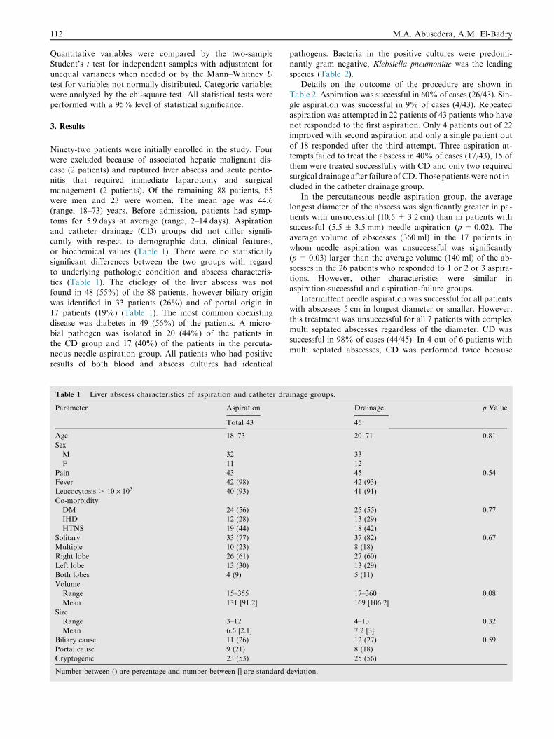
Fig. 1b Ultrasound examination of same patient 6 days after
catheter insertion with defervescence and size reduction and cavity
collapsed over the catheter arrow.
application of povidone–iodine. Prophylactic antibiotic
(Cefazolin) was administered intravenously just before theincision. The peritoneal cavity was accessed via the rightsubcostal incision, the falciform ligament is divided and a
self-retaining retractor was installed. For lesions located insegments VII and/or VIII, maximum exposure was obtainedby division of the right triangular and a portion of the right
coronary ligaments followed by rotation of the right liver lobeand placement of sterile towels behind. Superficial abscesseswere initially aspirated by a sterile syringe to take samplesfor cytologic and bacteriologic examination and to render
the abscess wall flaccid and easy to handle. Unroofing was per-formed by incision of the abscess wall, drainage of contentsand finally excision of the abscess wall. The resected specimen
was sent for histopathological assessment. A 28 French softplastic drain was placed in the unroofed abscess cavity forexternal drainage. The wound was closed in layers. For lesions
in the left lateral section (segments II and III), left subcostalextension of the original incision was performed by divisionof the left coronary and triangular ligaments. Lesions located
in segments IV, V and VII were unroofed as described abovewithout liver mobilization.
Statistical analysis was done with statistical software (SPSS10.0, SPSS). Descriptive and analytic statistics were used.
112 M.A. Abusedera, A.M. El-Badry
Quantitative variables were compared by the two-sampleStudent’s t test for independent samples with adjustment forunequal variances when needed or by the Mann–Whitney U
test for variables not normally distributed. Categoric variableswere analyzed by the chi-square test. All statistical tests wereperformed with a 95% level of statistical significance.
3. Results
Ninety-two patients were initially enrolled in the study. Four
were excluded because of associated hepatic malignant dis-ease (2 patients) and ruptured liver abscess and acute perito-nitis that required immediate laparotomy and surgical
management (2 patients). Of the remaining 88 patients, 65were men and 23 were women. The mean age was 44.6(range, 18–73) years. Before admission, patients had symp-
toms for 5.9 days at average (range, 2–14 days). Aspirationand catheter drainage (CD) groups did not differ signifi-cantly with respect to demographic data, clinical features,or biochemical values (Table 1). There were no statistically
significant differences between the two groups with regardto underlying pathologic condition and abscess characteris-tics (Table 1). The etiology of the liver abscess was not
found in 48 (55%) of the 88 patients, however biliary originwas identified in 33 patients (26%) and of portal origin in17 patients (19%) (Table 1). The most common coexisting
disease was diabetes in 49 (56%) of the patients. A micro-bial pathogen was isolated in 20 (44%) of the patients inthe CD group and 17 (40%) of the patients in the percuta-neous needle aspiration group. All patients who had positive
results of both blood and abscess cultures had identical
Table 1 Liver abscess characteristics of aspiration and catheter dra
Parameter Aspiration
Total 43
Age 18–73
Sex
M 32
F 11
Pain 43
Fever 42 (98)
Leucocytosis > 10 · 103 40 (93)
Co-morbidity
DM 24 (56)
IHD 12 (28)
HTNS 19 (44)
Solitary 33 (77)
Multiple 10 (23)
Right lobe 26 (61)
Left lobe 13 (30)
Both lobes 4 (9)
Volume
Range 15–355
Mean 131 [91.2]
Size
Range 3–12
Mean 6.6 [2.1]
Biliary cause 11 (26)
Portal cause 9 (21)
Cryptogenic 23 (53)
Number between () are percentage and number between [] are standard d
pathogens. Bacteria in the positive cultures were predomi-nantly gram negative, Klebsiella pneumoniae was the leadingspecies (Table 2).
Details on the outcome of the procedure are shown inTable 2. Aspiration was successful in 60% of cases (26/43). Sin-gle aspiration was successful in 9% of cases (4/43). Repeated
aspiration was attempted in 22 patients of 43 patients who havenot responded to the first aspiration. Only 4 patients out of 22improved with second aspiration and only a single patient out
of 18 responded after the third attempt. Three aspiration at-tempts failed to treat the abscess in 40% of cases (17/43), 15 ofthem were treated successfully with CD and only two requiredsurgical drainage after failure of CD.Those patients were not in-
cluded in the catheter drainage group.In the percutaneous needle aspiration group, the average
longest diameter of the abscess was significantly greater in pa-
tients with unsuccessful (10.5 ± 3.2 cm) than in patients withsuccessful (5.5 ± 3.5 mm) needle aspiration (p = 0.02). Theaverage volume of abscesses (360 ml) in the 17 patients in
whom needle aspiration was unsuccessful was significantly(p = 0.03) larger than the average volume (140 ml) of the ab-scesses in the 26 patients who responded to 1 or 2 or 3 aspira-
tions. However, other characteristics were similar inaspiration-successful and aspiration-failure groups.
Intermittent needle aspiration was successful for all patientswith abscesses 5 cm in longest diameter or smaller. However,
this treatment was unsuccessful for all 7 patients with complexmulti septated abscesses regardless of the diameter. CD wassuccessful in 98% of cases (44/45). In 4 out of 6 patients with
multi septated abscesses, CD was performed twice because
inage groups.
Drainage p Value
45
20–71 0.81
33
12
45 0.54
42 (93)
41 (91)
25 (55) 0.77
13 (29)
18 (42)
37 (82) 0.67
8 (18)
27 (60)
13 (29)
5 (11)
17–360 0.08
169 [106.2]
4–13 0.32
7.2 [3]
12 (27) 0.59
8 (18)
25 (56)
eviation.
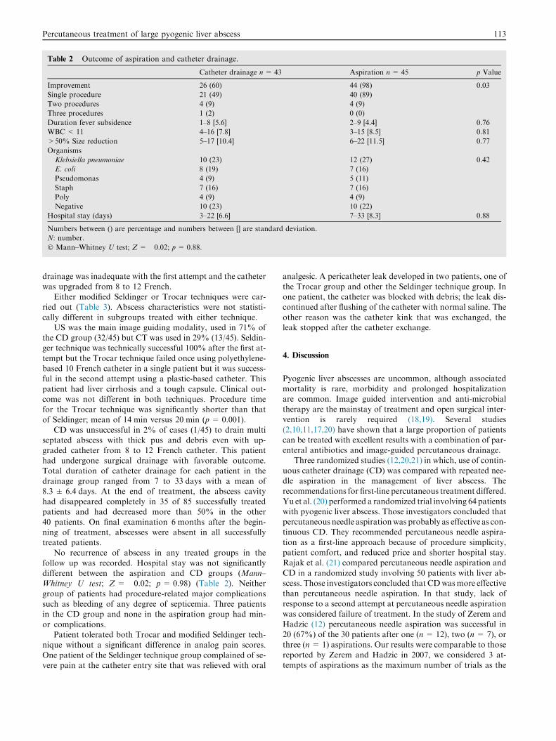
Table 2 Outcome of aspiration and catheter drainage.
Catheter drainage n= 43 Aspiration n= 45 p Value
Improvement 26 (60) 44 (98) 0.03
Single procedure 21 (49) 40 (89)
Two procedures 4 (9) 4 (9)
Three procedures 1 (2) 0 (0)
Duration fever subsidence 1–8 [5.6] 2–9 [4.4] 0.76
WBC< 11 4–16 [7.8] 3–15 [8.5] 0.81
>50% Size reduction 5–17 [10.4] 6–22 [11.5] 0.77
Organisms
Klebsiella pneumoniae 10 (23) 12 (27) 0.42
E. coli 8 (19) 7 (16)
Pseudomonas 4 (9) 5 (11)
Staph 7 (16) 7 (16)
Poly 4 (9) 4 (9)
Negative 10 (23) 10 (22)
Hospital stay (days) 3–22 [6.6] 7–33 [8.3] 0.88
Numbers between () are percentage and numbers between [] are standard deviation.
N: number.
� Mann–Whitney U test; Z= �0.02; p= 0.88.
Percutaneous treatment of large pyogenic liver abscess 113
drainage was inadequate with the first attempt and the catheter
was upgraded from 8 to 12 French.Either modified Seldinger or Trocar techniques were car-
ried out (Table 3). Abscess characteristics were not statisti-
cally different in subgroups treated with either technique.US was the main image guiding modality, used in 71% of
the CD group (32/45) but CT was used in 29% (13/45). Seldin-ger technique was technically successful 100% after the first at-
tempt but the Trocar technique failed once using polyethylene-based 10 French catheter in a single patient but it was success-ful in the second attempt using a plastic-based catheter. This
patient had liver cirrhosis and a tough capsule. Clinical out-come was not different in both techniques. Procedure timefor the Trocar technique was significantly shorter than that
of Seldinger; mean of 14 min versus 20 min (p = 0.001).CD was unsuccessful in 2% of cases (1/45) to drain multi
septated abscess with thick pus and debris even with up-
graded catheter from 8 to 12 French catheter. This patienthad undergone surgical drainage with favorable outcome.Total duration of catheter drainage for each patient in thedrainage group ranged from 7 to 33 days with a mean of
8.3 ± 6.4 days. At the end of treatment, the abscess cavityhad disappeared completely in 35 of 85 successfully treatedpatients and had decreased more than 50% in the other
40 patients. On final examination 6 months after the begin-ning of treatment, abscesses were absent in all successfullytreated patients.
No recurrence of abscess in any treated groups in thefollow up was recorded. Hospital stay was not significantlydifferent between the aspiration and CD groups (Mann–Whitney U test; Z= �0.02; p = 0.98) (Table 2). Neither
group of patients had procedure-related major complicationssuch as bleeding of any degree of septicemia. Three patientsin the CD group and none in the aspiration group had min-
or complications.Patient tolerated both Trocar and modified Seldinger tech-
nique without a significant difference in analog pain scores.
One patient of the Seldinger technique group complained of se-vere pain at the catheter entry site that was relieved with oral
analgesic. A pericatheter leak developed in two patients, one of
the Trocar group and other the Seldinger technique group. Inone patient, the catheter was blocked with debris; the leak dis-continued after flushing of the catheter with normal saline. The
other reason was the catheter kink that was exchanged, theleak stopped after the catheter exchange.
4. Discussion
Pyogenic liver abscesses are uncommon, although associatedmortality is rare, morbidity and prolonged hospitalizationare common. Image guided intervention and anti-microbial
therapy are the mainstay of treatment and open surgical inter-vention is rarely required (18,19). Several studies(2,10,11,17,20) have shown that a large proportion of patients
can be treated with excellent results with a combination of par-enteral antibiotics and image-guided percutaneous drainage.
Three randomized studies (12,20,21) in which, use of contin-
uous catheter drainage (CD) was compared with repeated nee-dle aspiration in the management of liver abscess. Therecommendations for first-line percutaneous treatment differed.
Yu et al. (20) performed a randomized trial involving 64 patientswith pyogenic liver abscess. Those investigators concluded thatpercutaneous needle aspirationwas probably as effective as con-tinuous CD. They recommended percutaneous needle aspira-
tion as a first-line approach because of procedure simplicity,patient comfort, and reduced price and shorter hospital stay.Rajak et al. (21) compared percutaneous needle aspiration and
CD in a randomized study involving 50 patients with liver ab-scess. Those investigators concluded that CDwasmore effectivethan percutaneous needle aspiration. In that study, lack of
response to a second attempt at percutaneous needle aspirationwas considered failure of treatment. In the study of Zerem andHadzic (12) percutaneous needle aspiration was successful in
20 (67%) of the 30 patients after one (n = 12), two (n = 7), orthree (n= 1) aspirations. Our results were comparable to thosereported by Zerem and Hadzic in 2007, we considered 3 at-tempts of aspirations as the maximum number of trials as the
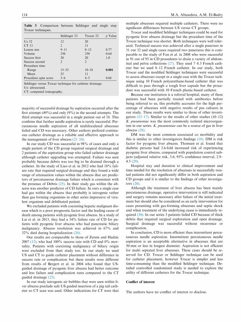
Table 3 Comparison between Seldinger and single step
Trocar techniques.
Seldinger 23 Trocar 22 p Value
Us 32 12 20
CT 13 2 11
Lesion size 9–11 8–12 0.77
Volume 250 230 0.64
Success first 20 20 1.0
Success second 2 2
Procedure time
Range 15–35 10–18 0.001
Mean 25 11
Procedure apin score 3–6 4–5 0.66
Seldinger versus Trocar technique for catheter drainage.
Us: ultrasound.
CT: computed tomography.
114 M.A. Abusedera, A.M. El-Badry
majority of successful drainage by aspiration occurred after the
first attempt (49%) and only (9%) in the second attempts. Thethird attempt was successful in a single patient out of 18. Thisconfirms that further needle aspiration is rarely successful. Per-cutaneous needle aspiration of all multiloculated abscesses
failed and CD was necessary. Other authors prefered continu-ous catheter drainage as a reliable and effective approach tothe management of liver abscess (22–24).
In our study CD was successful in 98% of cases and only asingle patient of the CD group required surgical drainage and2 patients of the aspiration group who failed catheter drainage
although catheter upgrading was attempted. Failure was seenprobably because debris was too big to be drained through acatheter. In the study of Liao et al. in 2012 who had 18% fail-ure rate that required surgical drainage and they found a wide
range of attenuation values within the abscess that are predic-tors of percutaneous drainage failure which is consistent withthe presence of Debris (25). In their study gas within the ab-
scess was another predictor of CD failure. In ours a single casehad gas within the abscess that probably is iatrogenic ratherthan gas forming organisms in other series impressive of viru-
lent organism and debilitated patient.We excluded patients with coexisting hepatic malignant dis-
ease which is a poor prognostic factor and the leading cause of
death among patients with pyogenic liver abscess. In a study ofLai et al. in 2013, they had a 34% failure rate of CD for pa-tients with pyogenic liver abscess who had pancreatic biliarymalignancy. Abscess resolution was achieved in 67% and
32% died during hospitalization (26).Our results are comparable to those of Zerem and Hadzic
2007 (12), who had 100% success rate with CD and 0% mor-
tality. Patients with coexisting malignancy of biliary originwere excluded from their study too. In our study we usedUS and CT to guide catheter placement without difference in
success rate or complication but these results were differentfrom results of Bergert et al. in 2004 who found that US-guided drainage of pyogenic liver abscess had better outcomeand less failure and complication rates compared to the CT
guided drainage (22).In our study iatrogenic air bubbles that were seen within li-
ver abscess preclude safe US guided insertion of a pig tail cath-
eter so CT scan was used. Other indications for CT were that
multiple abscesses required multiple catheters. There were nosignificant differences between US versus CT groups.
Trocar and modified Seldinger techniques could be used for
pyogenic liver abscess drainage but the procedure time of theTrocar technique was shorter. Both techniques were well toler-ated. Technical success was achieved after a single puncture in
31 out 32 and single cases required two punctures this is com-parable to the study of Fan et al. in 2008 who were successfulin 91 out of 93 in CD procedures to drain a variety of abdom-
inal and pelvic collections (27). They used 7–8.3 French cath-eter but we used 8–12 French catheter. In our study, bothTrocar and the modified Seldinger techniques were successfulto access abscesses except in a single case with the Trocar tech-
nique using 10 French polyurethane-based catheter that wasdifficult to pass through a tough liver capsule but the proce-dure was successful with 10 French plastic-based catheter.
Because our institution is a referral hospital, many of thesepatients had been partially treated with antibiotics beforebeing referred to us; this probably accounts for the high per-
centage of abscesses with negative results of pus cultures inour study. These results were similar to those of other investi-gators (15–17). Similar to the results of other studies (10–12)
K. pneumoniae was the most commonly isolated microorgan-ism in our series. K. pneumoniae can be a source of gas in liverabscess (28).
DM was the most common associated co morbidity and
this is similar to other investigators findings (18). DM is riskfactor for pyogenic liver abscess. Thomsen et al. found thatdiabetic persons had 3.6-fold increased risk of experiencing
pyogenic liver abscess, compared with population control sub-jects (adjusted relative risk, 3.6; 95% confidence interval, 2.9–4.5 (29).
Hospital stay and duration to clinical improvement andtime needed for the resolution of abscesses in successfully trea-ted patients did not significantly differ in both aspiration and
CD groups and it is similar to the findings of other investiga-tors (20).
Although the treatment of liver abscess has been mainlypercutaneous drainage, operative intervention is still indicated
and surgery remains necessary after failure of the initial treat-ment but should also be considered as an early intervention forcases presenting with gas-forming abscesses and septic shock
and when treatment of the underlying cause is immediately re-quired (30). In our series 3 patients failed CD because of thickdebris that required surgical exploration and open drainage.
Surgical drainage was successful without recurrence orcomplication.
In conclusion, CD is more efficient than intermittent percu-taneous needle aspiration. Intermittent percutaneous needle
aspiration is an acceptable alternative in abscesses that are50 mm or less in longest diameter. Aspiration is not efficientfor multi septated liver abscesses. These cases should be re-
served for CD. Trocar or Seldinger technique can be usedfor catheter placement, however Trocar is simpler and lesstime-consuming than the modified Seldinger technique. De-
tailed controlled randomized study is needed to explore theutility of different catheters for the Trocar technique.
Conflict of interest
The authors have no conflict of interest to disclose.
Percutaneous treatment of large pyogenic liver abscess 115
References
(1) Branum GD, Tyson GS, Branum MA, Meyers WC. Hepatic
abscess: changes in etiology, diagnosis, and management. Ann
Surg 1990;212:655–62.
(2) Huang CJ, Pitt HA, Lipsett PA, Osterman FA Jr, Lillemoe KD,
Cameron JL, et al. Pyogenic hepatic abscess: changing trends
over 42 years. Ann Surg 1996;223:600–7.
(3) Neoptolemos JP, Macpherson DS, Holm J, Fossard DP.
Pyogenic liver abscess: a study of forty-four cases in two centres.
Acta Chir Scand 1982;148:415–21.
(4) Northover JM, Jones BJ, Dawson JL, Williams R. Difficulties in
the diagnosis and management of pyogenic liver abscess. Br J
Surg 1982;69:48–51.
(5) Miedema BW, Dineen P. The diagnosis and treatment of
pyogenic liver abscesses. Ann Surg 1984;200:328–35.
(6) Silver S, Weinstein AJ, Cooperman A. Changes in the pathogen-
esis and detection of intrahepatic abscess. Am J Surg
1979;137:608–10.
(7) Satani B, Davidson ED. Hepatic abscesses: improvement in
mortality with early diagnosis and treatment. Am J Surg
1978;135:647–50.
(8) Gerzof SG, Johnson WC, Robbins AH, Nabseth DC. Intrahe-
patic pyogenic abscesses: treatment by percutaneous drainage.
Am J Surg 1985;149:487–94.
(9) Lee JF, Block GE. The changing clinical pattern of hepatic
abscesses. Arch Surg 1972;104:465–70.
(10) Barakate MS, Stephen MS, Waugh RC, Gallagher PJ, Solomon
MJ, Storey DW, et al. Pyogenic liver abscess: a review of 10 years
experience in management. Aust NZ J Surg 1999;69:205–9.
(11) Mohan S, Talwar N, Chaudhary A, Andley M, Ravi B, Kumar A.
Liver abscess: a clinicopathological analysis of 82 cases. Int Surg
2006;91:228–33.
(12) Zerem Enver, Hadzic Amir. Sonographically guided percutaneous
catheter drainage versus needle aspiration in the management of
pyogenic liver abscess. AJR 2007;189:W138–42.
(13) Seeto RK, Rockey DC. Pyogenic liver abscess: change in etiology,
management and outcome. Medicine 1996;75:I 99–I 128.
(14) Robert JH, Miresew D, Ambroseui P, Khoury G, Greenstein Al,
Rohner A. Critical review of the treatment of pyogenic liver
abscess. Surg Gynecol Obstet 1992;l74:97–102.
(15) Hashimoto L, Hermann R, Broniatowski 5G. Pyogenic hepatic
abscess: results of current management. Am Surg 1995;61:407–11.
(16) Back SY, Lee MG, Cho KS, Lee SC, Sung KB, Auh YH.
Therapeutic percutaneous aspiration of hepatic abscesses: effec-
tiveness in 25 patients. AJR 1993;160:799–802.
(17) Giorgio A, Tarantino L, Mariniello N, Francica G, Scala E,
Amoroso P, et al. Pyogenic liver abscesses: 13 years of experience
in percutaneous needle aspiration with US guidance. Radiology
1995;l95:122–4.
(18) O’Farrell N, Collins CG, McEntee GP. Pyogenic liver abscesses:
diminished role for operative treatment. Surgeon 2010;8(4):192–6.
(19) Mezhir JJ, Fong Y, Jacks LM, Getrajdman GI, Brody LA, Covey
AM, et al. Current management of pyogenic liver abscess:
surgery is now second-line treatment. J Am Coll Surg
2010;210(6):975–83.
(20) Yu SC, Ho SS, Lau WY, Yeung DT, Yuen EH, Lee PS, et al.
Treatment of pyogenic liver abscess: prospective randomized
comparison of catheter drainage and needle aspiration. Hepatol-
ogy 2004;39:932–8.
(21) Rajak CL, Gupta S, Jain S, Chawla Y, Gulati M, Suri S.
Percutaneous treatment of liver abscesses: needle aspiration
versus catheter drainage. AJR 1998;170:1035–9.
(22) Bergert H, Kersting S, Pyrc J, Saeger HD, Bunk A. Therapeutic
options in the treatment of pyogenic liver abscess. Ultraschall
Med 2004;25:356–62.
(23) Lee KT, Wong SR, Sheen PC. Pyogenic liver abscess: an audit of
10 years’ experience and analysis of risk factors. Dig Surg
2001;18:459–66.
(24) Alvarez Perez JA, Gonzalez JJ, Baldonedo RF, Sanz L, Carreno
G, Junco A, et al. Clinical course, treatment, and multivariate
analysis of risk factors for pyogenic liver abscess. Am J Surg
2001;181:177–86.
(25) Liao W-I, Tsai S-H, Yu C-Y, Huang G-S, Lin Y-Y, Hsu C-W,
et al. Pyogenic liver abscess treated by percutaneous catheter
drainage: MDCT measurement for treatment outcome. Eur J
Radiol 2012;81(4):609–15.
(26) Lai KC, Cheng KS, Jeng LB, Huang CC, Lee YT, Chang HR,
et al. Factors associated with treatment failure of percutaneous
catheter drainage for pyogenic liver abscess in patients with
hepatobiliary–pancreatic cancer. Am J Surg 2013;205(1):52–7.
(27) Fan WC, Chan CC, Chan JCS. Image-guided drainage using the
Trocar technique. J HK Coll Radiol 2008;11:69–71.
(28) Yu C, Lee C. Pyogenic liver abscess. N Engl J Med
2011;364:1154.
(29) Thomsen Reimar W, Jepsen Peter, Sørensen Henrik T. Diabetes
mellitus and pyogenic liver abscess: risk and prognosis. Clin Infect
Dis 2007;44(9):1194–201.
(30) Alkofer B, Dufay C, Parienti JJ, Lepennec V, Dargere S, Chiche
L. Are pyogenic liver abscesses still a surgical concern? A western
experience. HPB Surg 2012;316013(10):19.