pediatric tb radiographs - home | curry international ...nid... · pediatric tb radiographs ... the...
TRANSCRIPT
1
Pediatric TB radiographs
Ann M. Loeffler, MDCurry International Tuberculosis Center
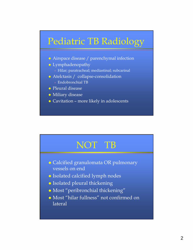
Radiology
Best quality frontal and lateral views of the chest
Reading by experienced pediatric radiologist
Avoid overreading –» If questionable – consider other infection, reactive
airways disease –» Treat other causes if feasible and then repeat» Avoid CT scans (diagnose subradiographic nodes)
2
Pediatric TB Radiology
Airspace disease / parenchymal infection Lymphadenopathy
» Hilar; paratracheal; mediastinal; subcarinal
Atelctasis / collapse-consolidation» Endobronchial TB
Pleural disease Miliary disease Cavitation – more likely in adolescents
NOT TB
Calcified granulomata OR pulmonary vessels on end
Isolated calcified lymph nodes Isolated pleural thickening Most “peribronchial thickening” Most “hilar fullness” not confirmed on
lateral
3
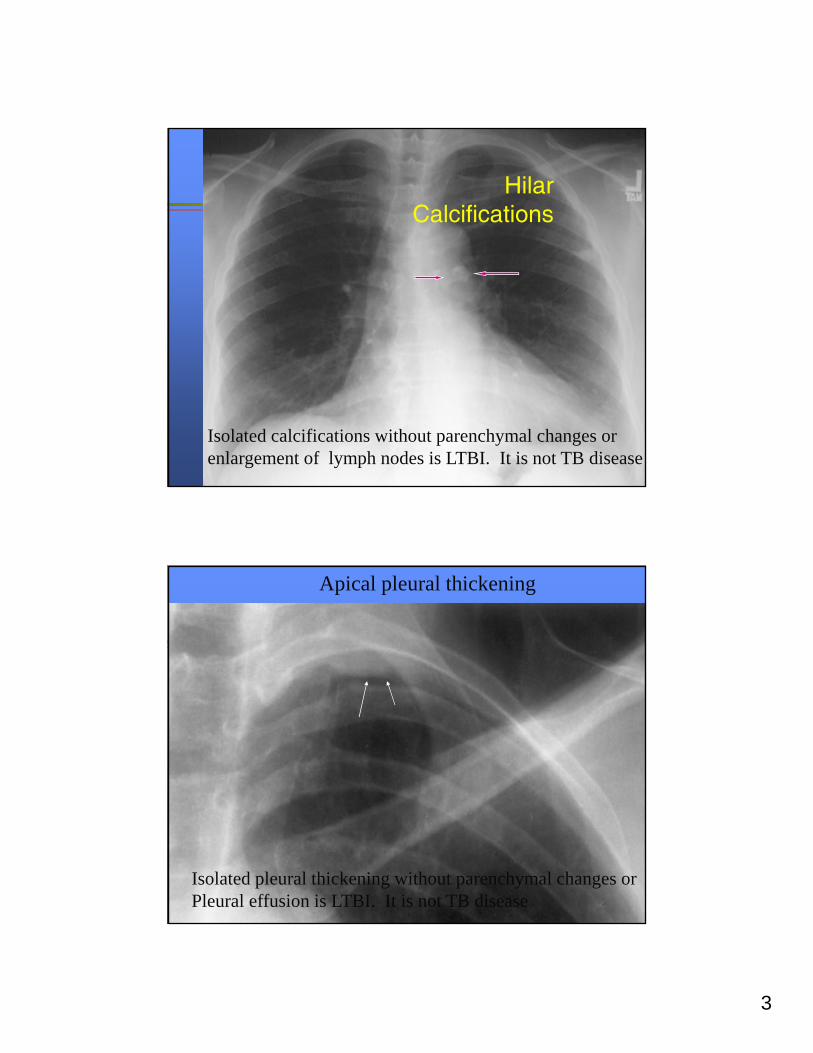
Hilar Calcifications
Isolated calcifications without parenchymal changes or enlargement of lymph nodes is LTBI. It is not TB disease
Apical pleural thickening
Isolated pleural thickening without parenchymal changes or Pleural effusion is LTBI. It is not TB disease
4
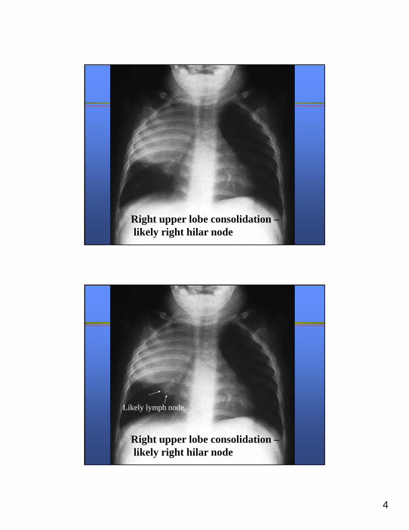
Right upper lobe consolidation –likely right hilar node
Right upper lobe consolidation –likely right hilar node
Likely lymph node
5
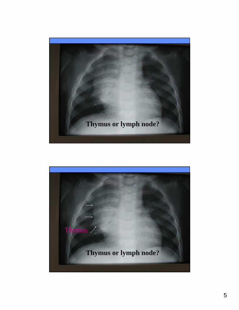
Thymus or lymph node?
Thymus or lymph node?
Thymus
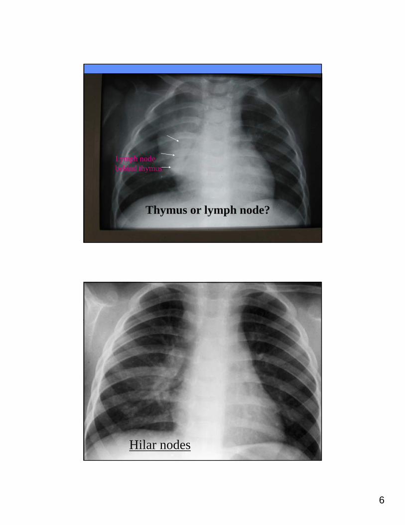
6
Thymus or lymph node?
Lymph nodebehind thymus
Hilar nodes
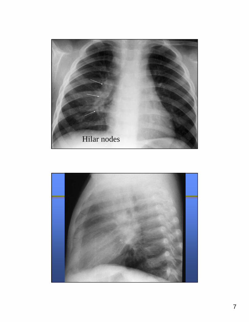
7
Hilar nodes
8
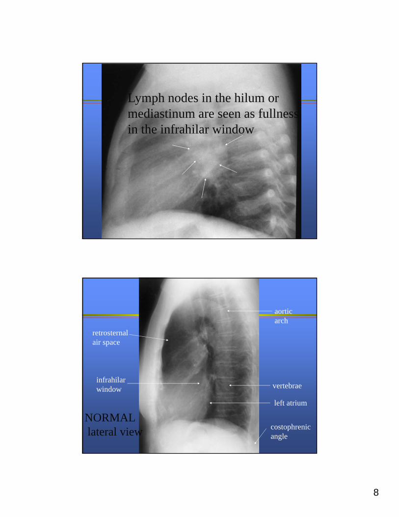
Lymph nodes in the hilum or mediastinum are seen as fullness in the infrahilar window
vertebrae
aorticarch
retrosternalair space
infrahilarwindow
left atrium
costophrenicangle
NORMALlateral view
9
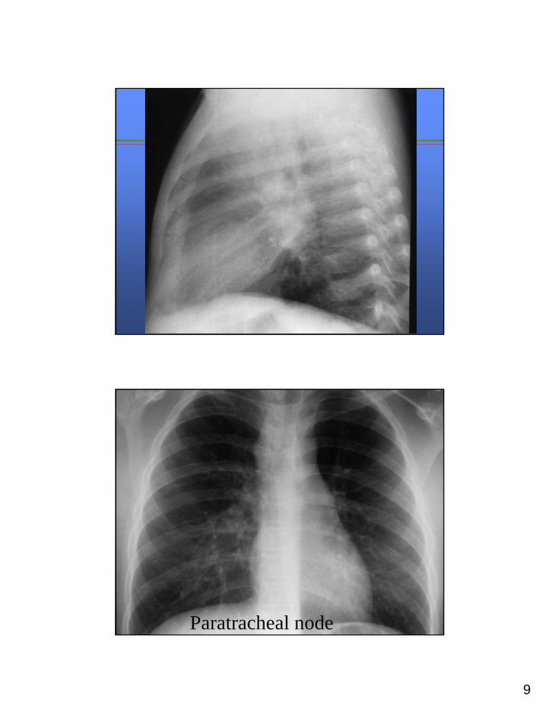
Paratracheal node
10
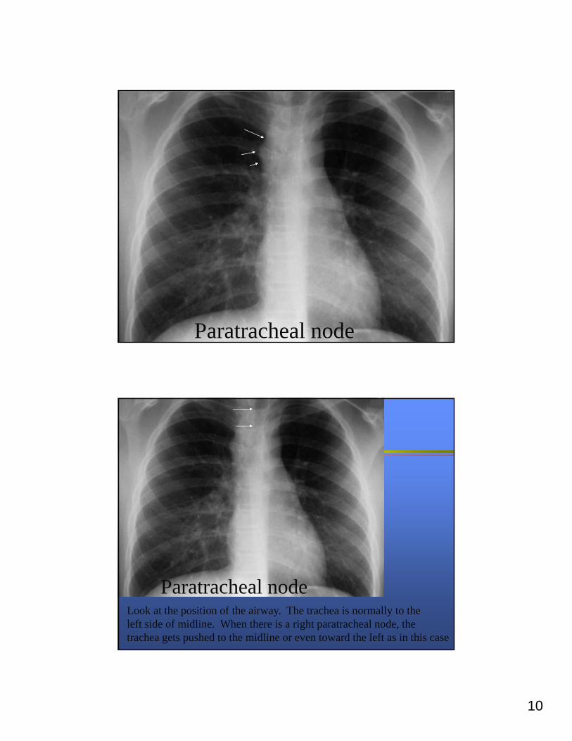
Paratracheal node
Paratracheal nodeLook at the position of the airway. The trachea is normally to the left side of midline. When there is a right paratracheal node, the trachea gets pushed to the midline or even toward the left as in this case
11
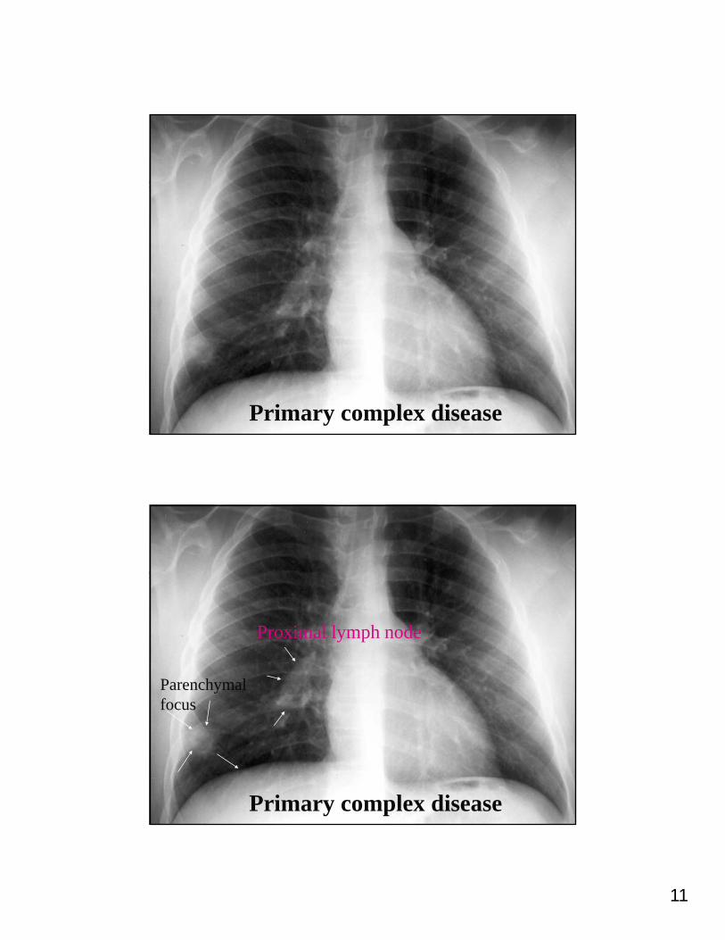
Primary complex disease
Primary complex disease
Parenchymal focus
Proximal lymph node
12
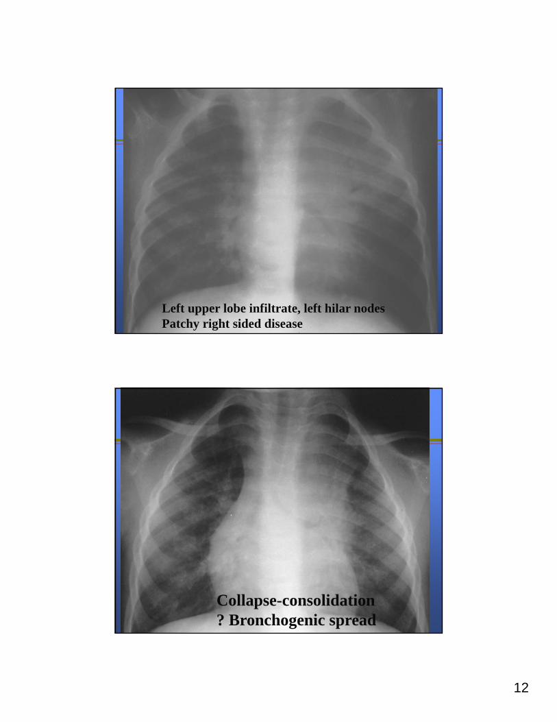
Left upper lobe infiltrate, left hilar nodesPatchy right sided disease
Collapse-consolidation? Bronchogenic spread

13
Collapse-consolidation
Diffuse patchy changes suggest bronchogenic spread
Miliary TB
14
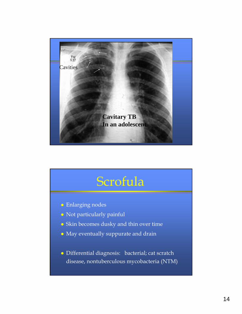
Cavitary TBIn an adolescent
Cavities
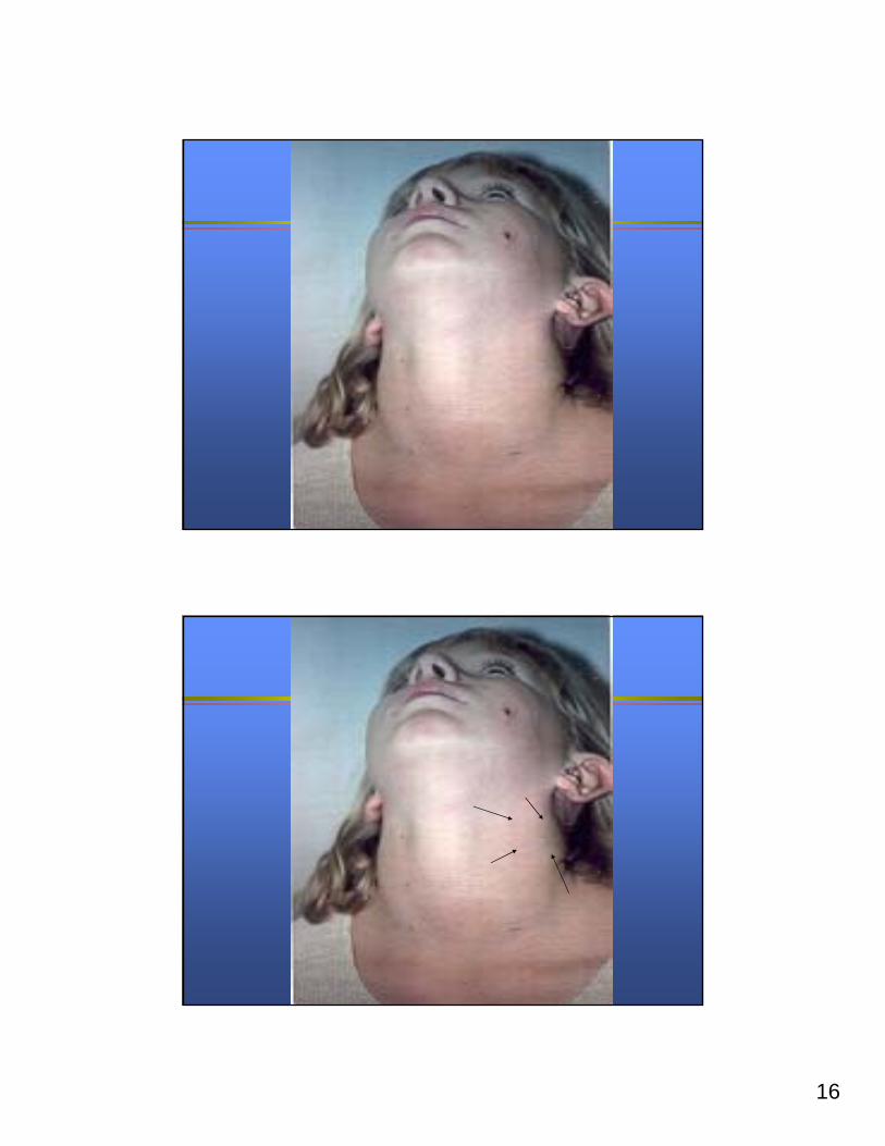
Scrofula
Enlarging nodes
Not particularly painful
Skin becomes dusky and thin over time
May eventually suppurate and drain
Differential diagnosis: bacterial; cat scratch disease, nontuberculous mycobacteria (NTM)
15
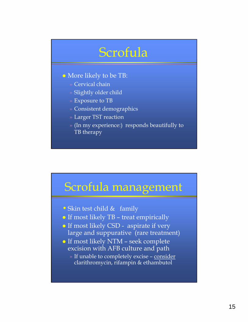
Scrofula
More likely to be TB:» Cervical chain» Slightly older child» Exposure to TB» Consistent demographics» Larger TST reaction» (In my experience:) responds beautifully to
TB therapy
Scrofula management
• Skin test child & family If most likely TB – treat empirically If most likely CSD - aspirate if very
large and suppurative (rare treatment) If most likely NTM – seek complete
excision with AFB culture and path» If unable to completely excise – consider
clarithromycin, rifampin & ethambutol
16
17
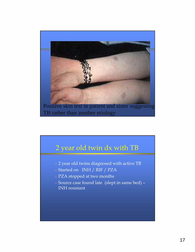
Positive skin test in patient and sister suggesting TB rather than another etiology
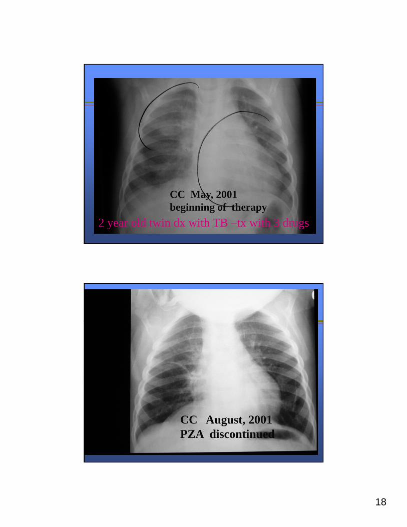
2 year old twin dx with TB
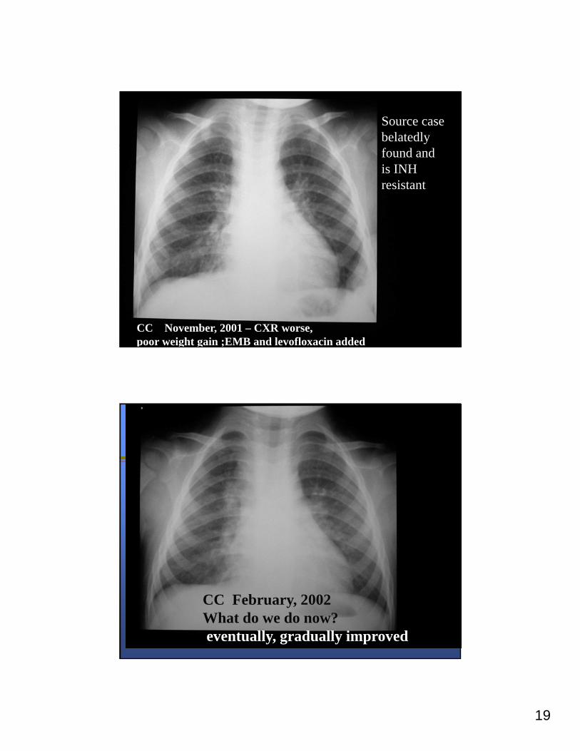
» 2 year old twins diagnosed with active TB» Started on INH / RIF / PZA » PZA stopped at two months» Source case found late (slept in same bed) –
INH resistant
18
CC May, 2001 beginning of therapy
2 year old twin dx with TB –tx with 3 drugs
CC August, 2001PZA discontinued
19
CC November, 2001 – CXR worse, poor weight gain ;EMB and levofloxacin added
Source casebelatedly found andis INHresistant
CC February, 2002What do we do now? eventually, gradually improved
20
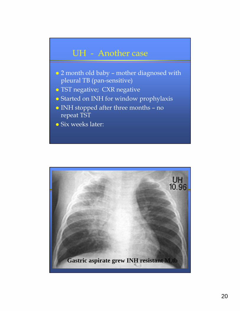
UH - Another case
2 month old baby – mother diagnosed with pleural TB (pan-sensitive)
TST negative; CXR negative Started on INH for window prophylaxis INH stopped after three months – no
repeat TST Six weeks later:
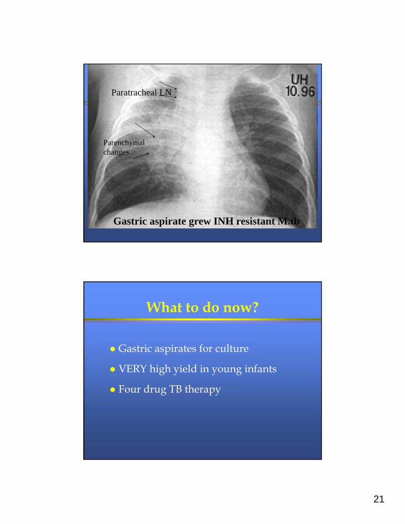
Gastric aspirate grew INH resistant M.tb
21
Gastric aspirate grew INH resistant M.tb
Paratracheal LN
Parenchymalchanges
What to do now?
Gastric aspirates for culture
VERY high yield in young infants
Four drug TB therapy
22
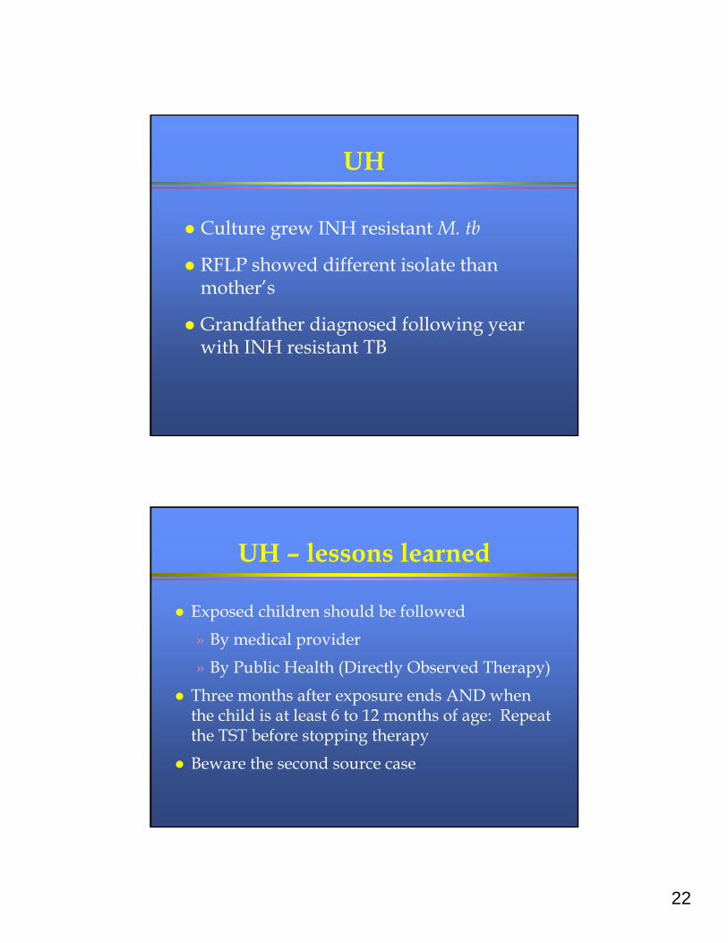
UH
Culture grew INH resistant M. tb
RFLP showed different isolate than mother’s
Grandfather diagnosed following year with INH resistant TB
UH – lessons learned
Exposed children should be followed
» By medical provider
» By Public Health (Directly Observed Therapy)
Three months after exposure ends AND when the child is at least 6 to 12 months of age: Repeat the TST before stopping therapy
Beware the second source case
23
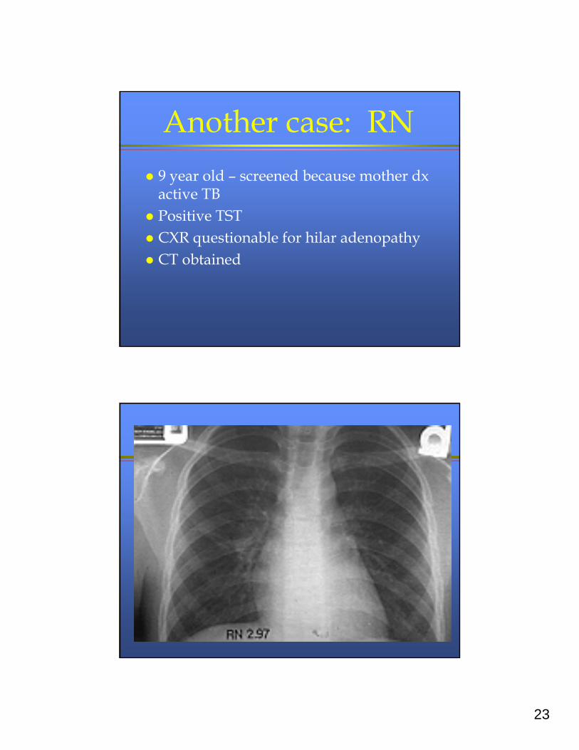
Another case: RN
9 year old – screened because mother dx active TB
Positive TST CXR questionable for hilar adenopathy CT obtained
24
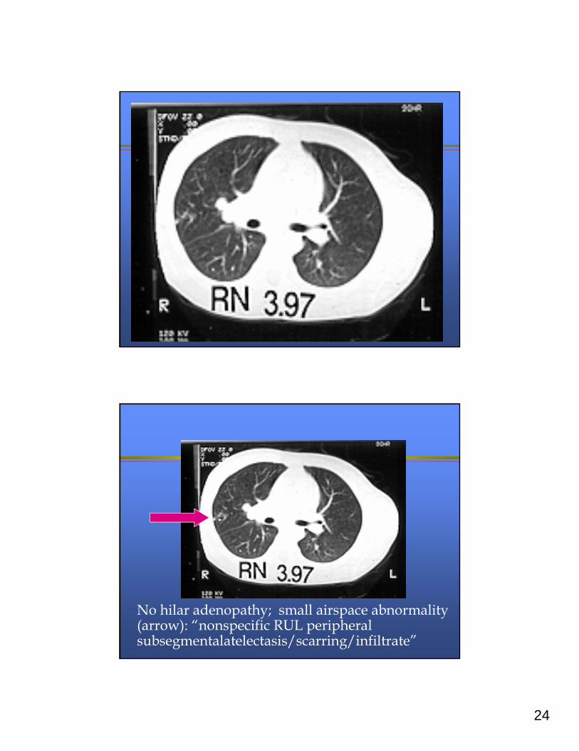
No hilar adenopathy; small airspace abnormality (arrow): “nonspecific RUL peripheral subsegmentalatelectasis/scarring/infiltrate”
25
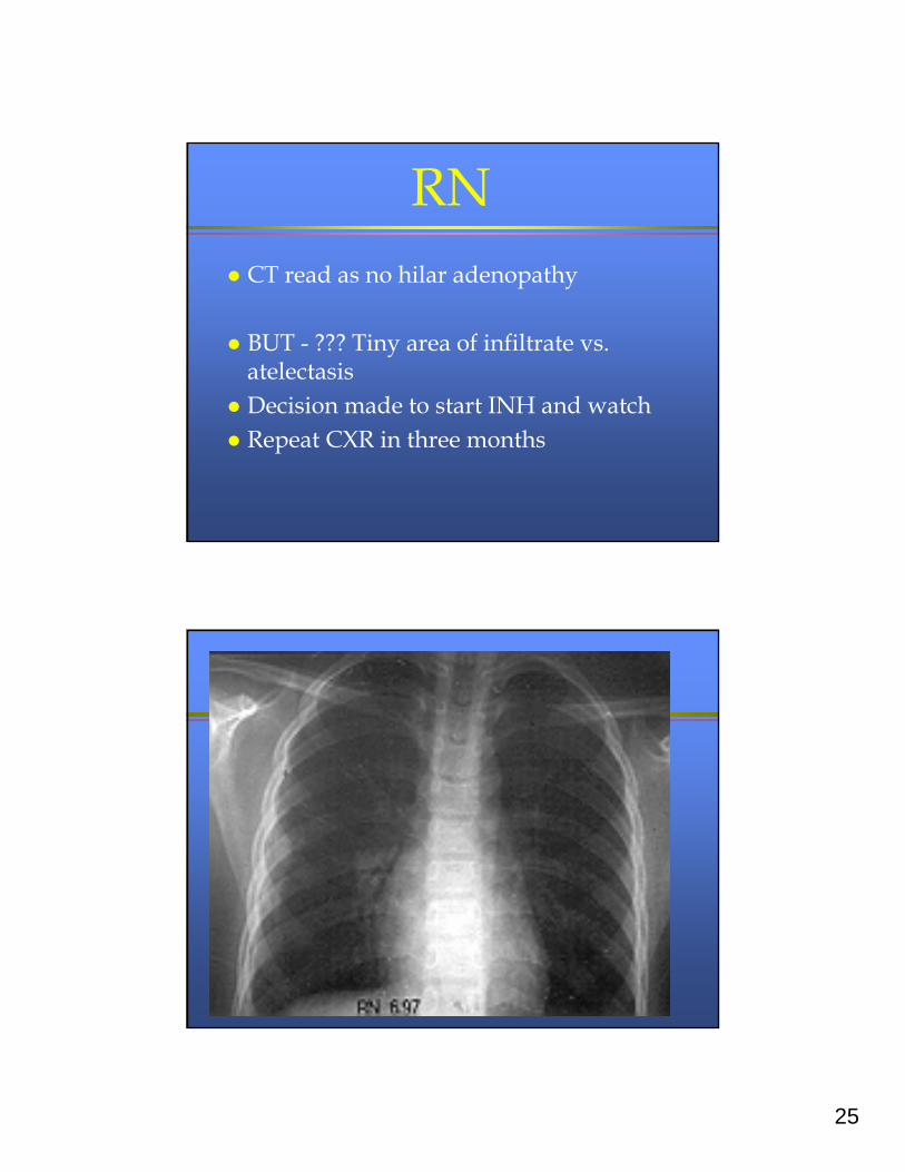
RN CT read as no hilar adenopathy
BUT - ??? Tiny area of infiltrate vs. atelectasis
Decision made to start INH and watch Repeat CXR in three months
26
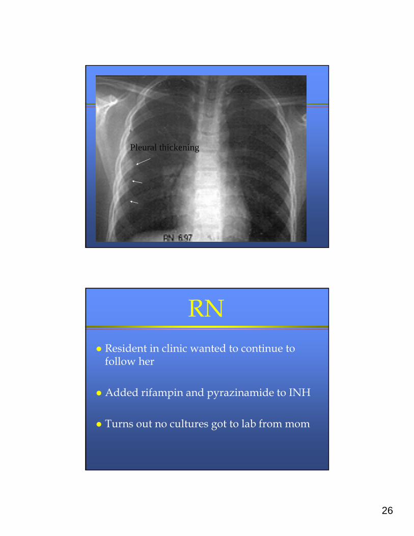
Pleural thickening
RN Resident in clinic wanted to continue to
follow her
Added rifampin and pyrazinamide to INH
Turns out no cultures got to lab from mom
27
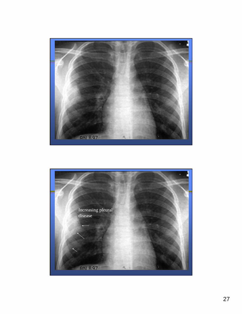
Increasing pleural disease
28
RN Surgical biopsy revealed INH resistant
M. tb
She did beautifully on RIF, PZA, EMB
Siblings also treated for INH resistant LTBI
Lessons learned
Completely evaluate adult source cases
Do not start INH alone if active TB is considered
Review all films together
» (CT and plain film both show small airspace abnormality)

29
Lessons learned (2)
If possible:
» Treat patient in dedicated TB clinic
If lesion increased on INH alone, likely resistant
» INH kills first 95% of organisms
» Children rarely have enough organisms to induce resistance
Another patient: RG 17 month old
» Fever without source for two weeks
» Some sweats, no respiratory symptoms
» Evaluated with blood work, TST by MD
» TB skin test not read
» Fever eventually subsided
30
RG
During routine follow-up, PMD notes that TST not read and repeats it
TST now 25 mm
Mother swears that this is different
What to do?
Chest radiograph, PA, and lateral
Physical exam
History
Source case investigation
31
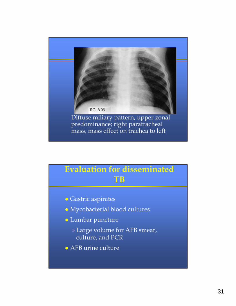
Diffuse miliary pattern, upper zonal predominance; right paratracheal mass, mass effect on trachea to left
Evaluation for disseminated TB
Gastric aspirates
Mycobacterial blood cultures
Lumbar puncture
» Large volume for AFB smear, culture, and PCR
AFB urine culture
32
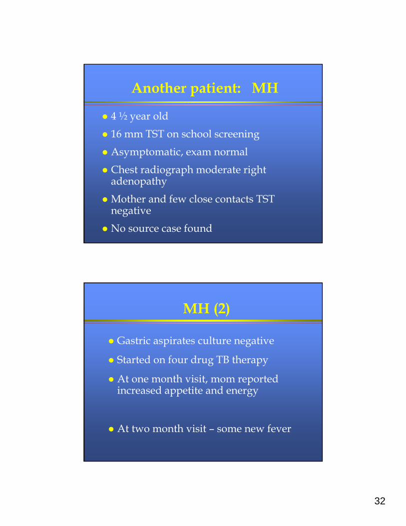
Another patient: MH
4 ½ year old
16 mm TST on school screening
Asymptomatic, exam normal
Chest radiograph moderate right adenopathy
Mother and few close contacts TST negative
No source case found
MH (2)
Gastric aspirates culture negative
Started on four drug TB therapy
At one month visit, mom reported increased appetite and energy
At two month visit – some new fever
33
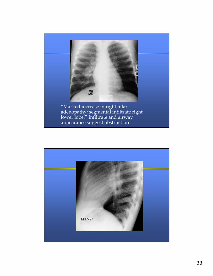
“Marked increase in right hilar adenopathy; segmental infiltrate right lower lobe.” Infiltrate and airway appearance suggest obstruction
34
MH – what to do?
Review:
» Adherence – nurse had begged to d/c DOT
» Ingestion – swallows whole dose
» Absorption – no reason for concern
– (HIV negative, no diarrhea, weight good)
» Resistance – still no source case found
MH – what to do? (2)
Bronchoscopy done – caseous material, cultures negative
Other causes of endobronchial granulomatous disease considered
Steroids, quinolone, and streptomycin added
35
MH – what to do? (3)
One month later, increased infiltrate –presumed to be post-obstructive;
Treated with steroids and azithromycin
I usually treat post-obstructive pneumonia with steroids and augmentin to cover mouth flora
MH - what to do? (4)
Marked clinical improvement!!!
Two and ½ weeks off azithromycin
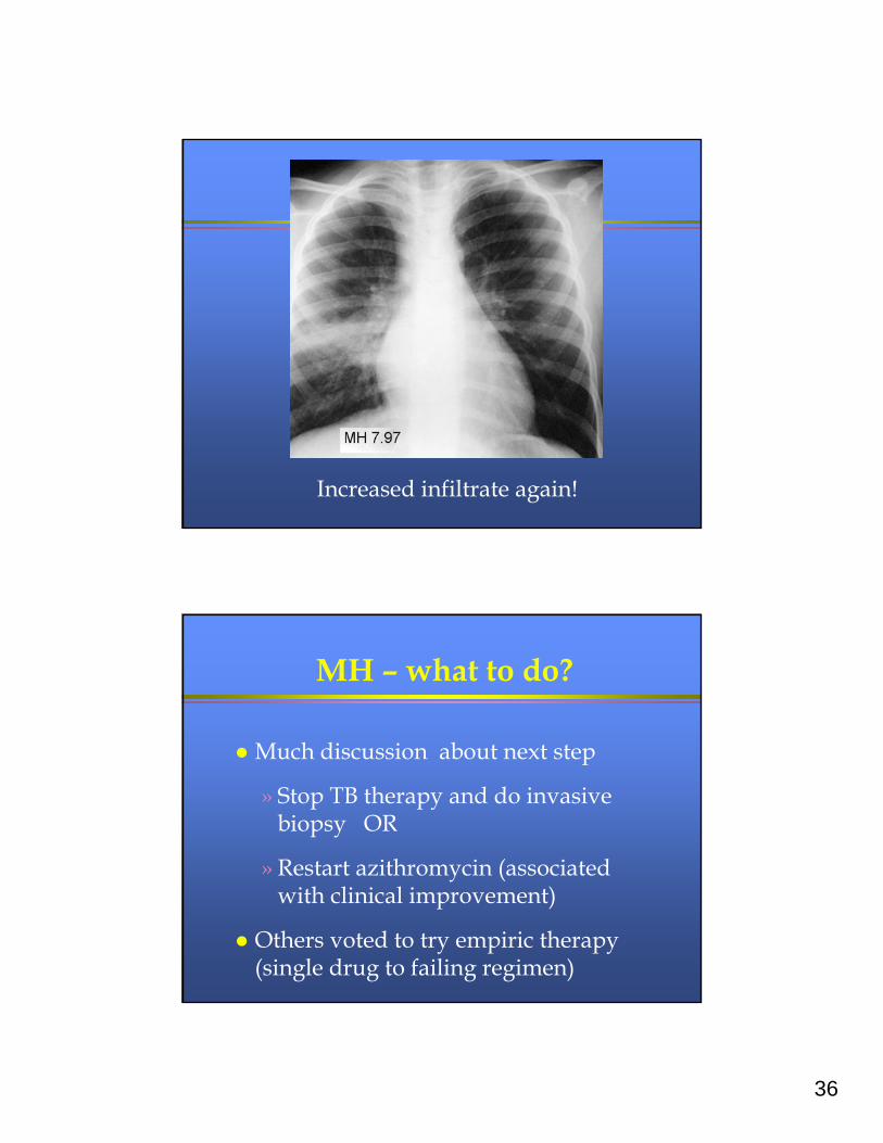
36
Increased infiltrate again!
MH – what to do?
Much discussion about next step
» Stop TB therapy and do invasive biopsy OR
» Restart azithromycin (associated with clinical improvement)
Others voted to try empiric therapy (single drug to failing regimen)
37
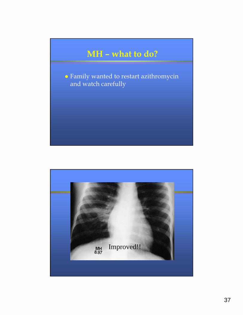
MH – what to do?
Family wanted to restart azithromycin and watch carefully
Improved!!
38
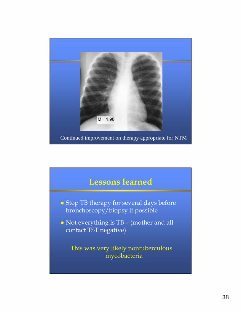
Continued improvement on therapy appropriate for NTM
Lessons learned
Stop TB therapy for several days before bronchoscopy/biopsy if possible
Not everything is TB – (mother and all contact TST negative)
This was very likely nontuberculous mycobacteria
39
Lessons learned
Stop TB therapy for several days before bronchoscopy/biopsy if possible
Not everything is TB – (mother and all contact TST negative)
This was very likely non-tuberculous mycobacteria
Another patient: BC
22 month old previously healthy boy
5 months of intermittent wheezing
Wax and wane cough, wheeze,
congestion
40
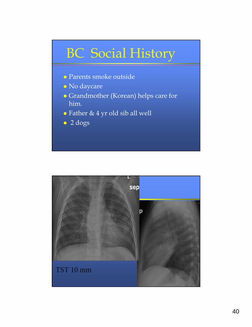
BC Social History
Parents smoke outside No daycare Grandmother (Korean) helps care for
him. Father & 4 yr old sib all well 2 dogs
TST 10 mm
41
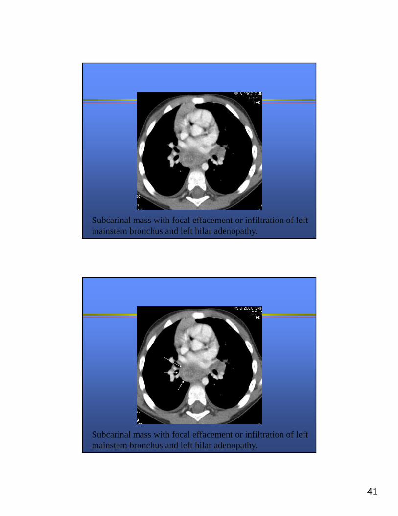
Subcarinal mass with focal effacement or infiltration of left mainstem bronchus and left hilar adenopathy.
Subcarinal mass with focal effacement or infiltration of left mainstem bronchus and left hilar adenopathy.
42
COURSE
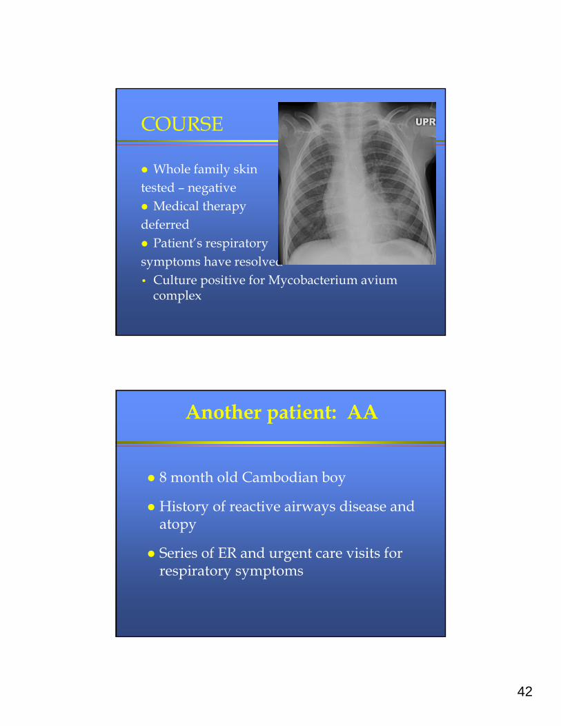
Whole family skin tested – negative Medical therapy deferred Patient’s respiratory symptoms have resolved• Culture positive for Mycobacterium avium
complex
Another patient: AA
8 month old Cambodian boy
History of reactive airways disease and atopy
Series of ER and urgent care visits for respiratory symptoms
43
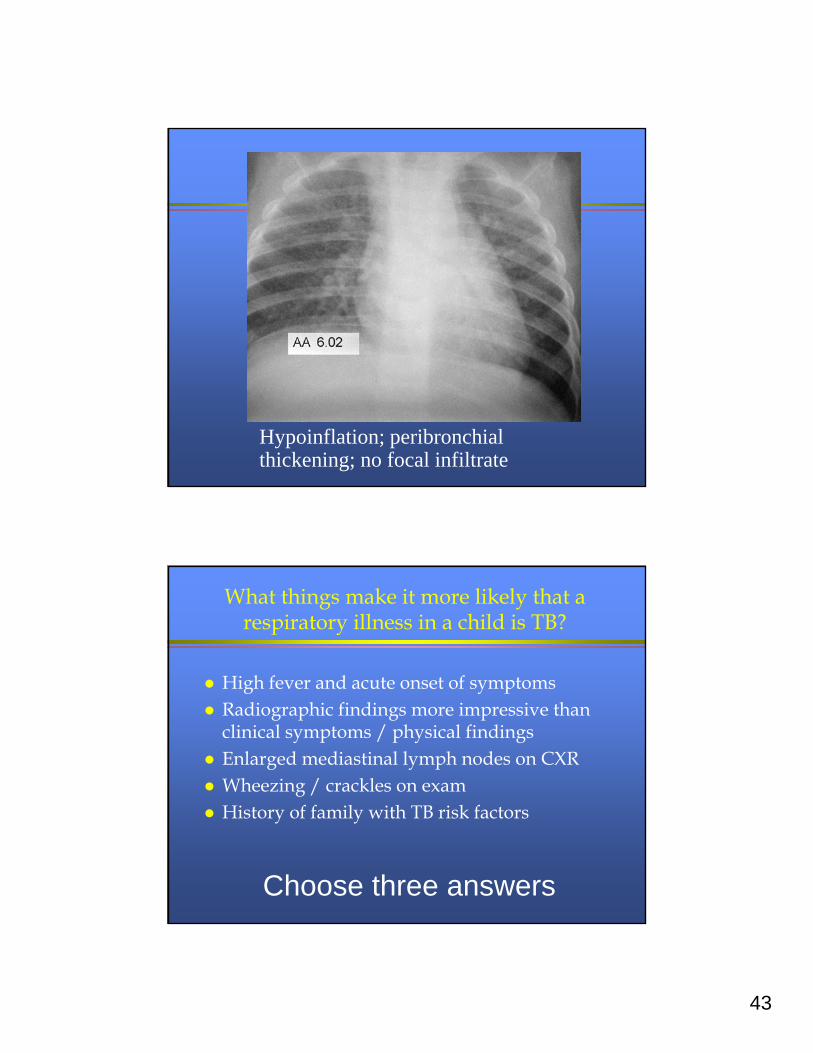
Hypoinflation; peribronchial thickening; no focal infiltrate
What things make it more likely that a respiratory illness in a child is TB?
High fever and acute onset of symptoms Radiographic findings more impressive than
clinical symptoms / physical findings Enlarged mediastinal lymph nodes on CXR Wheezing / crackles on exam History of family with TB risk factors
Choose three answers
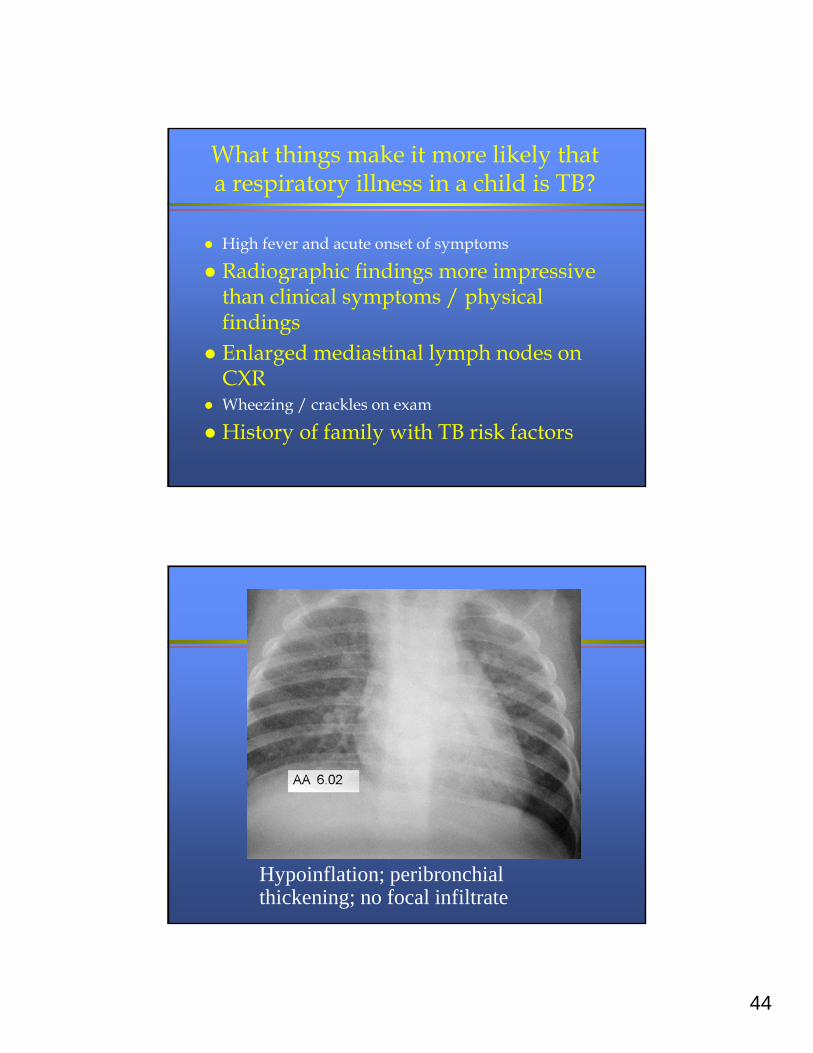
44
What things make it more likely that a respiratory illness in a child is TB?
High fever and acute onset of symptoms
Radiographic findings more impressive than clinical symptoms / physical findings
Enlarged mediastinal lymph nodes on CXR
Wheezing / crackles on exam
History of family with TB risk factors
Hypoinflation; peribronchial thickening; no focal infiltrate
45
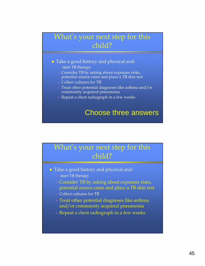
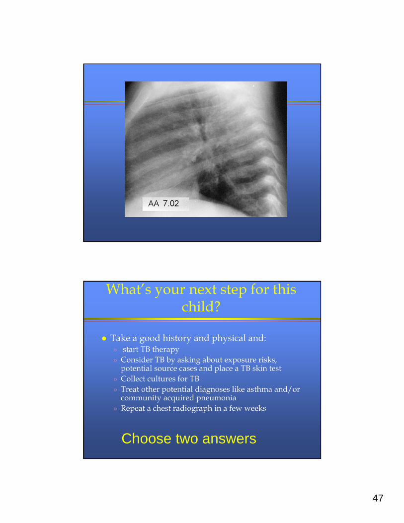
What’s your next step for this child?
Take a good history and physical and:» start TB therapy» Consider TB by asking about exposure risks,
potential source cases and place a TB skin test» Collect cultures for TB» Treat other potential diagnoses like asthma and/or
community acquired pneumonia» Repeat a chest radiograph in a few weeks
Choose three answers
What’s your next step for this child?
Take a good history and physical and:» start TB therapy
» Consider TB by asking about exposure risks, potential source cases and place a TB skin test
» Collect cultures for TB
» Treat other potential diagnoses like asthma and/or community acquired pneumonia
» Repeat a chest radiograph in a few weeks
46
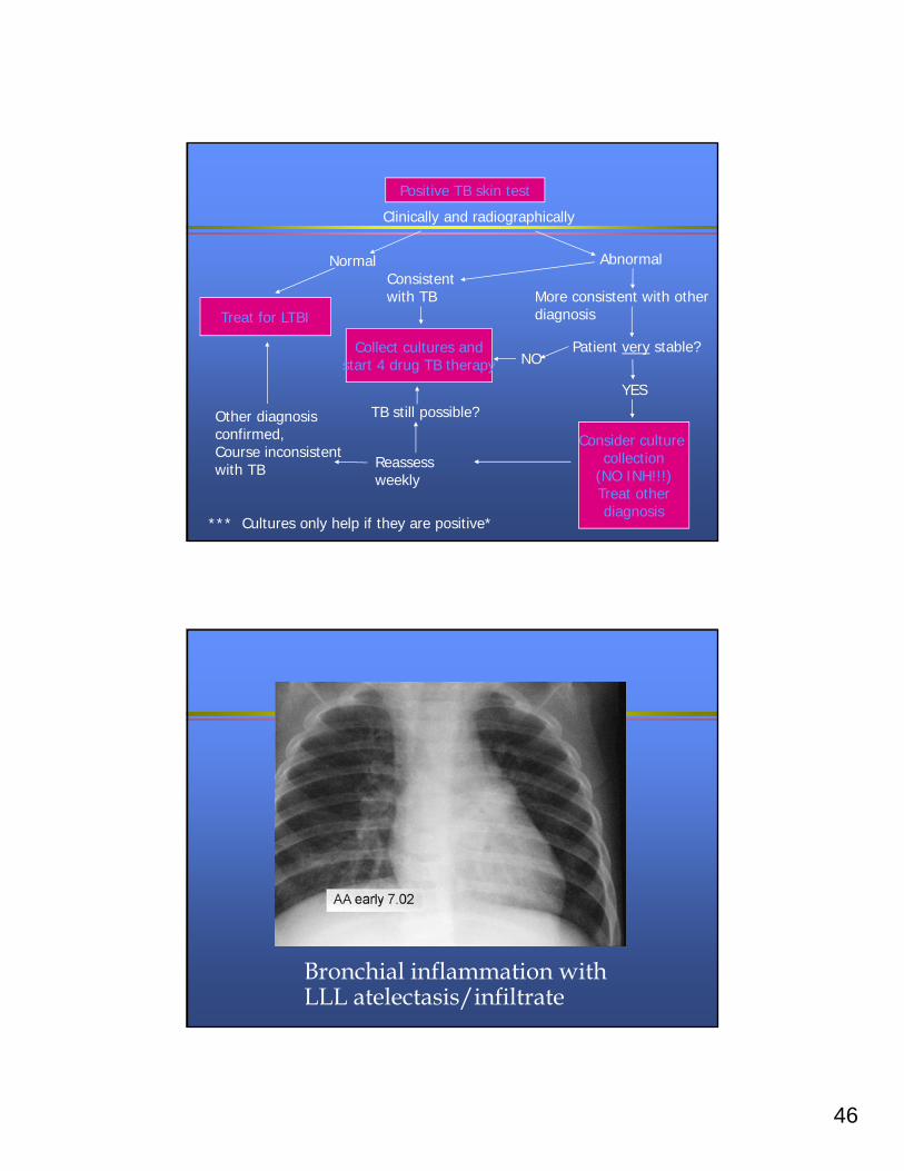
Clinically and radiographically
Normal AbnormalConsistent with TB More consistent with other
diagnosis
Patient very stable?
Positive TB skin test
Treat for LTBI
Collect cultures andstart 4 drug TB therapy NO
YES
Consider culture collection
(NO INH!!!)Treat otherdiagnosis
Reassess weekly
Other diagnosis confirmed,Course inconsistent with TB
TB still possible?
*** Cultures only help if they are positive*
Bronchial inflammation with LLL atelectasis/infiltrate
47
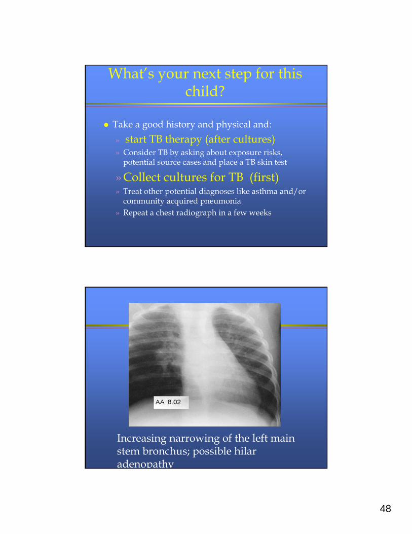
What’s your next step for this child?
Take a good history and physical and:» start TB therapy» Consider TB by asking about exposure risks,
potential source cases and place a TB skin test» Collect cultures for TB» Treat other potential diagnoses like asthma and/or
community acquired pneumonia» Repeat a chest radiograph in a few weeks
Choose two answers
48
What’s your next step for this child?
Take a good history and physical and:
» start TB therapy (after cultures)» Consider TB by asking about exposure risks,
potential source cases and place a TB skin test
» Collect cultures for TB (first)» Treat other potential diagnoses like asthma and/or
community acquired pneumonia» Repeat a chest radiograph in a few weeks
Increasing narrowing of the left main stem bronchus; possible hilar adenopathy
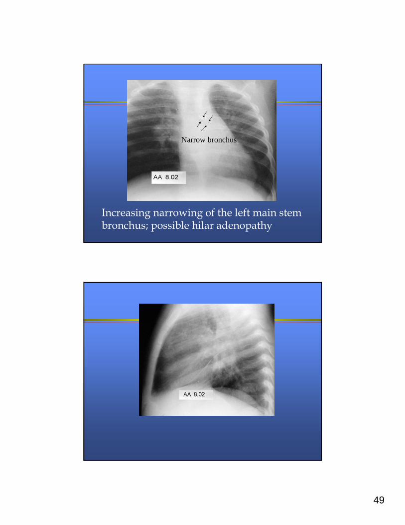
49
Increasing narrowing of the left main stem bronchus; possible hilar adenopathy
Narrow bronchus
50
Infectious disease consulted
Diagnosis of TB made
Source case investigation revealed:
» Mom had pleural TB
» Family friend had 4+ smear positive TB
» Three children had active TB
» Everyone else had LTBI
» Subsequent secondary case in adult
How are you going to treat this child?
Isoniazid suspension 5 mg/kg/day Isoniazid tablets 10 – 15 mg/kg/day Rifampin capsules 10 – 15
mg/kg/day Rifampin suspension 10 – 15
mg/kg/day Pyrazinamide 20-40 mg/kg/day Pyrazinamide 25 mg/kg/day Ethambutol 15 – 25 mg/kg/dayPick some
51
How are you going to treat this child?By Directly observed therapy!!
Isoniazid suspension 5 mg/kg/day Isoniazid tablets 10 – 15 mg/kg/day Rifampin capsules 10 – 15 mg/kg/day
Rifampin suspension 10 – 15 mg/kg/day
Pyrazinamide 20-40 mg/kg/day Pyrazinamide 25 mg/kg/day
Ethambutol 15 – 25 mg/kg/day
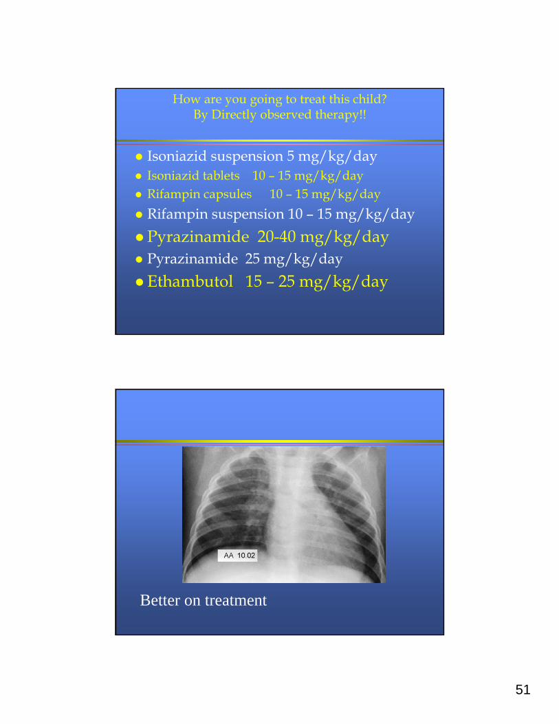
Better on treatment
52
Lessons learned
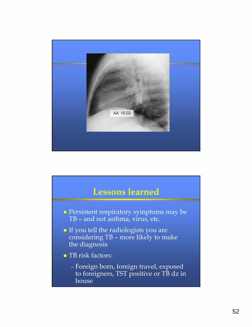
Persistent respiratory symptoms may be TB – and not asthma, virus, etc.
If you tell the radiologists you are considering TB – more likely to make the diagnosis
TB risk factors:
» Foreign born, foreign travel, exposed to foreigners, TST positive or TB dz in house
53
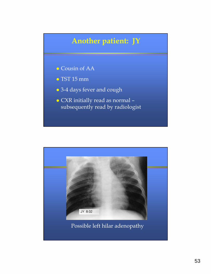
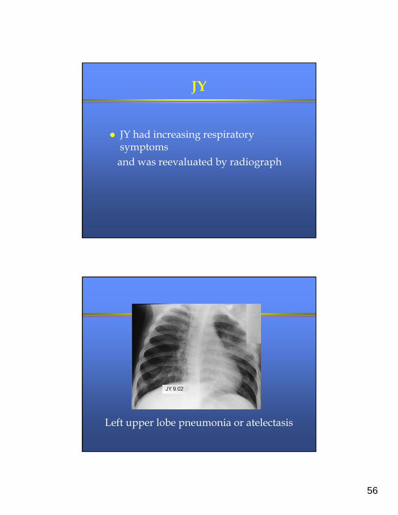
Another patient: JY
Cousin of AA
TST 15 mm
3-4 days fever and cough
CXR initially read as normal –subsequently read by radiologist
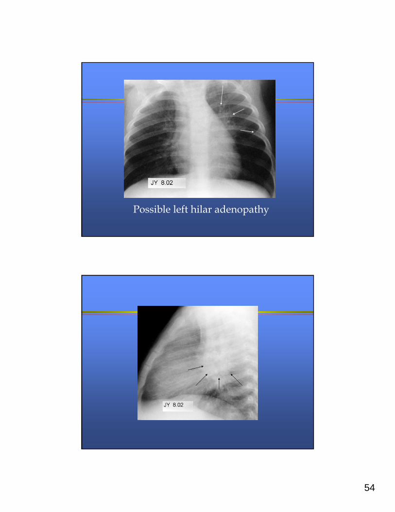
Possible left hilar adenopathy
54
Possible left hilar adenopathy
55
What things make it more likely that a respiratory illness in a child is TB?
High fever and acute onset of symptoms Radiographic findings more impressive than
clinical symptoms / physical findings
Enlarged mediastinal lymph nodes on CXR
Wheezing / crackles on exam History of family with TB risk factors
What’s your next step for this child?
Take a good history and physical and:
» start TB therapy (after cultures)» Consider TB by asking about exposure risks,
potential source cases and place a TB skin test
» Collect cultures for TB (first)» Treat other potential diagnoses like asthma and/or
community acquired pneumonia» Repeat a chest radiograph in a few weeks
Because this child is a TB contact – you start Tx NOW
56
JY
JY had increasing respiratory symptomsand was reevaluated by radiograph
Left upper lobe pneumonia or atelectasis
57
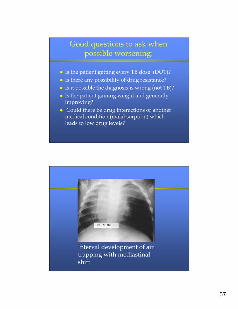
Good questions to ask when possible worsening:
Is the patient getting every TB dose (DOT)? Is there any possibility of drug resistance? Is it possible the diagnosis is wrong (not TB)? Is the patient gaining weight and generally
improving? Could there be drug interactions or another
medical condition (malabsorption) which leads to low drug levels?
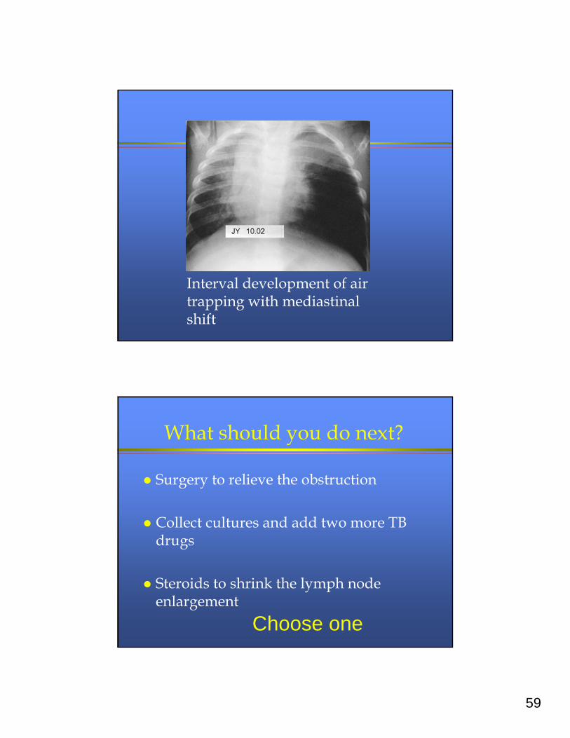
Interval development of air trapping with mediastinal shift
58
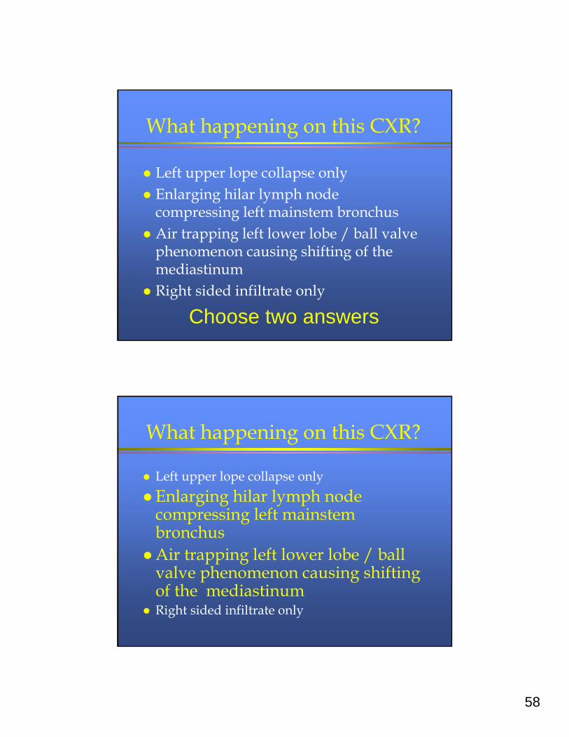
What happening on this CXR?
Left upper lope collapse only Enlarging hilar lymph node
compressing left mainstem bronchus Air trapping left lower lobe / ball valve
phenomenon causing shifting of the mediastinum
Right sided infiltrate only
Choose two answers
What happening on this CXR?
Left upper lope collapse only
Enlarging hilar lymph node compressing left mainstem bronchus
Air trapping left lower lobe / ball valve phenomenon causing shifting of the mediastinum
Right sided infiltrate only
59
Interval development of air trapping with mediastinal shift
What should you do next?
Surgery to relieve the obstruction
Collect cultures and add two more TB drugs
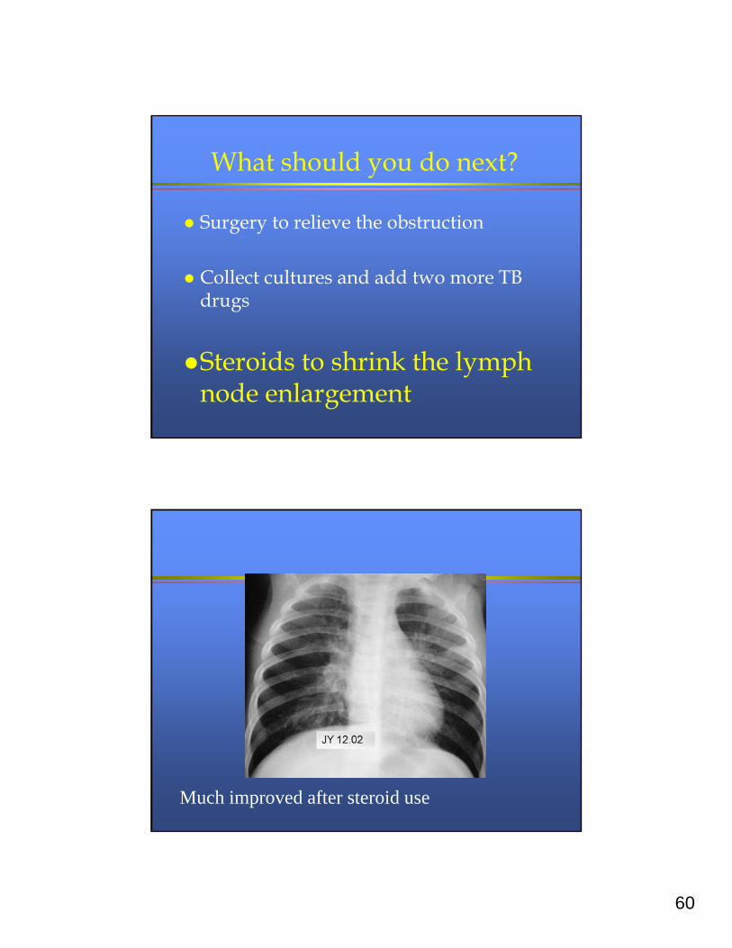
Steroids to shrink the lymph node enlargement
Choose one
60
What should you do next?
Surgery to relieve the obstruction
Collect cultures and add two more TB drugs
Steroids to shrink the lymph node enlargement
Much improved after steroid use
61
Lessons learned
Read radiograph more aggressively for:
» Exposure to TB
» Symptoms of TB
Endobronchial TB:
» Post-obstructive pneumonia
» Ball-valve air trapping
» Bronchogenic spread
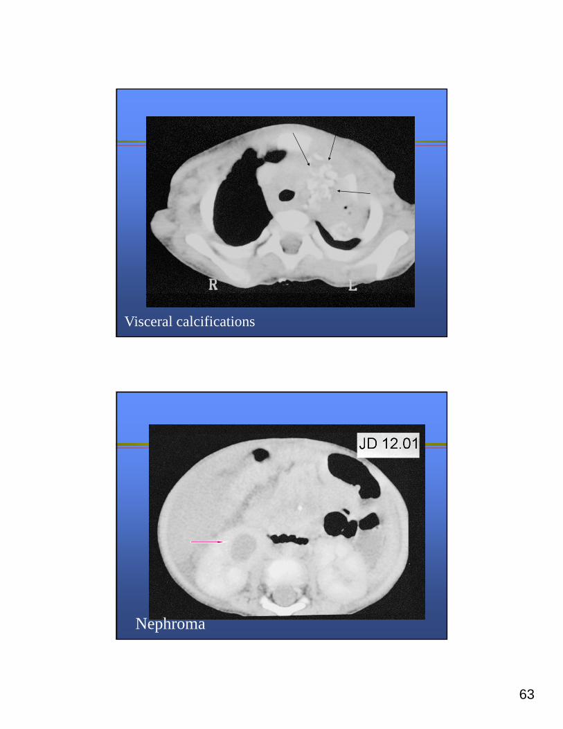
Another patient: JD
15 month old girl
Bullous pemphigus skin disease
Treated with Cellcept and prednisone
One month of cough and anorexia
Six pound weight loss
62
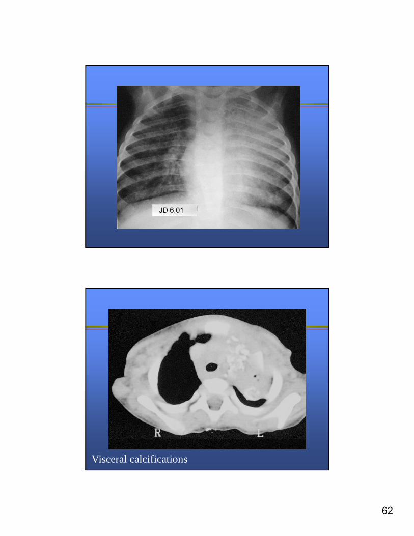
Visceral calcifications
63
Visceral calcifications
Nephroma
64
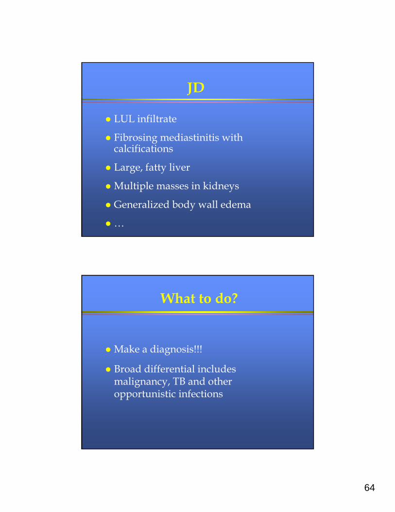
JD
LUL infiltrate
Fibrosing mediastinitis with calcifications
Large, fatty liver
Multiple masses in kidneys
Generalized body wall edema
…
What to do?
Make a diagnosis!!!
Broad differential includes malignancy, TB and other opportunistic infections
65
JD
History:
» Dad had been treated for active TB before JD was born
» Rest of family was TST positive, but treated for 6-9 months with INH
JD (2)
More history
Dad had a friend who developed TB after JD was born
Their “friendship” was not initially known to public health
Two different PHNs
“Oh, that household is taken care of”
66
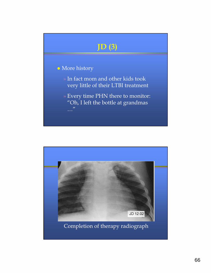
JD (3)
More history
» In fact mom and other kids took very little of their LTBI treatment
» Every time PHN there to monitor: “Oh, I left the bottle at grandmas …”
Completion of therapy radiograph
67
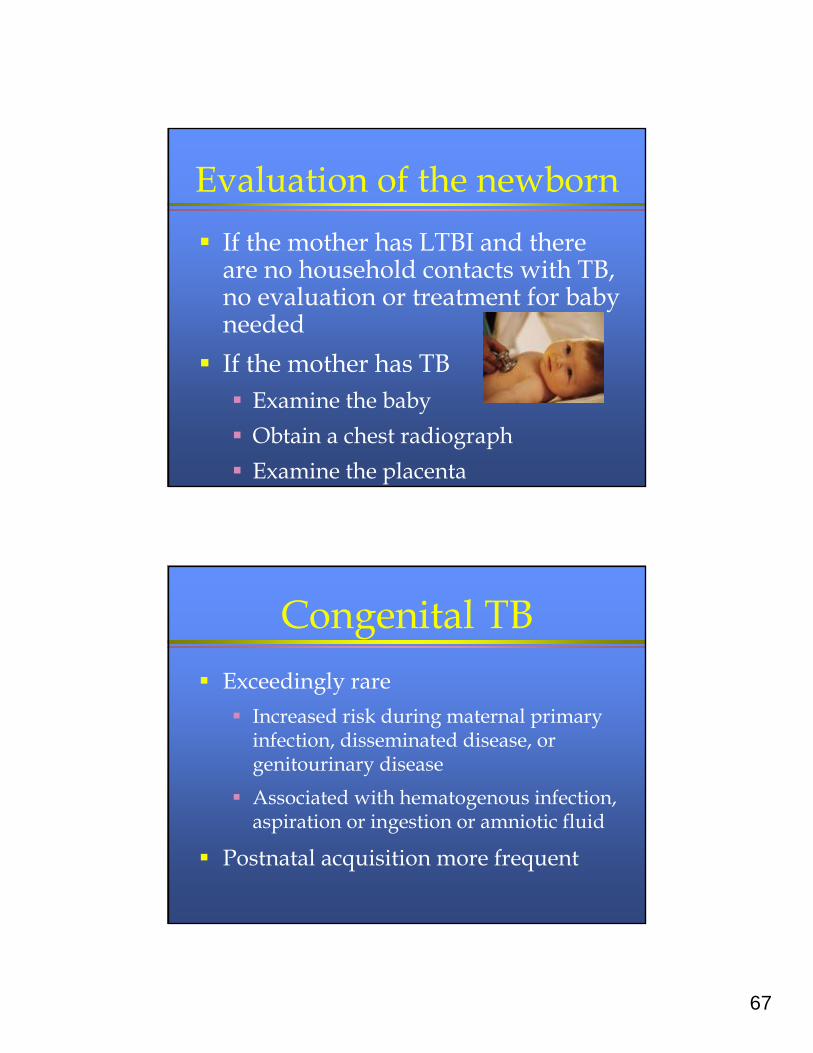
Evaluation of the newborn
If the mother has LTBI and there are no household contacts with TB, no evaluation or treatment for baby needed
If the mother has TB Examine the baby
Obtain a chest radiograph
Examine the placenta
Congenital TB
Exceedingly rare
Increased risk during maternal primary infection, disseminated disease, or genitourinary disease
Associated with hematogenous infection, aspiration or ingestion or amniotic fluid
Postnatal acquisition more frequent
68
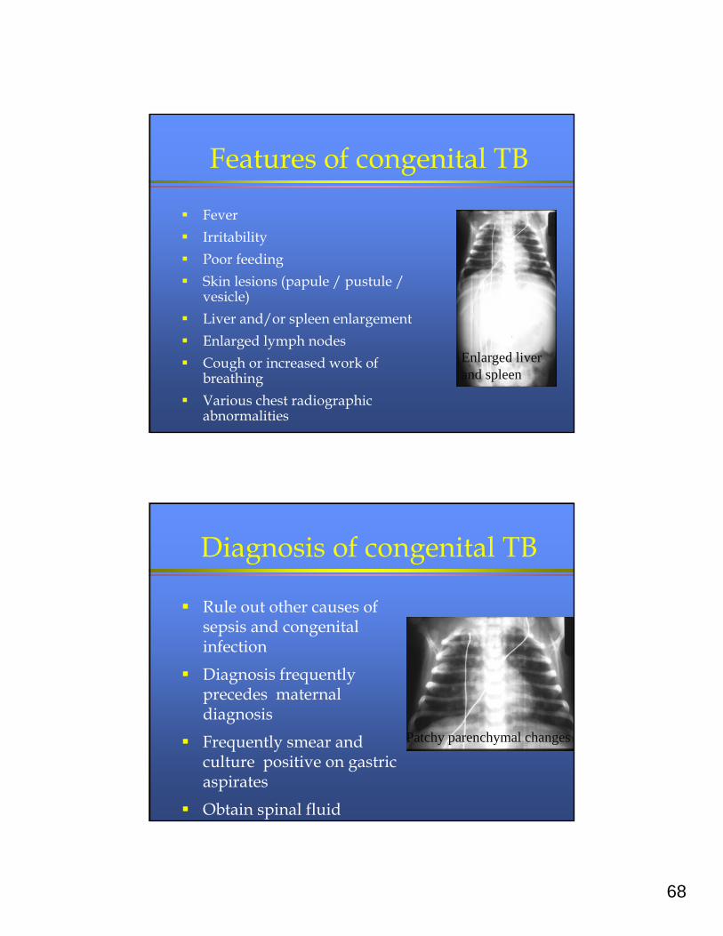
Features of congenital TB
Fever
Irritability
Poor feeding
Skin lesions (papule / pustule / vesicle)
Liver and/or spleen enlargement
Enlarged lymph nodes
Cough or increased work of breathing
Various chest radiographic abnormalities
Enlarged liver and spleen
Diagnosis of congenital TB
Rule out other causes of sepsis and congenital infection
Diagnosis frequently precedes maternal diagnosis
Frequently smear and culture positive on gastric aspirates
Obtain spinal fluid
Patchy parenchymal changes
69
Treatment of newborn Congenital TB - treat based on maternal
drug susceptibility pattern or empiric 3 – 4 drug regimen
Normal exam and radiograph INH unless mother is very clearly no longer
contagious and no second source case
Treat 3 – 9 months and then place a TST
Reunite mother and baby as soon as INH tolerated
Another patient: KH
10 year old Ethiopian adoptee Treated for smear positive TB in
Ethiopia (no cultures) Initially, lack of clinical improvement Seizure in Ethiopia
70
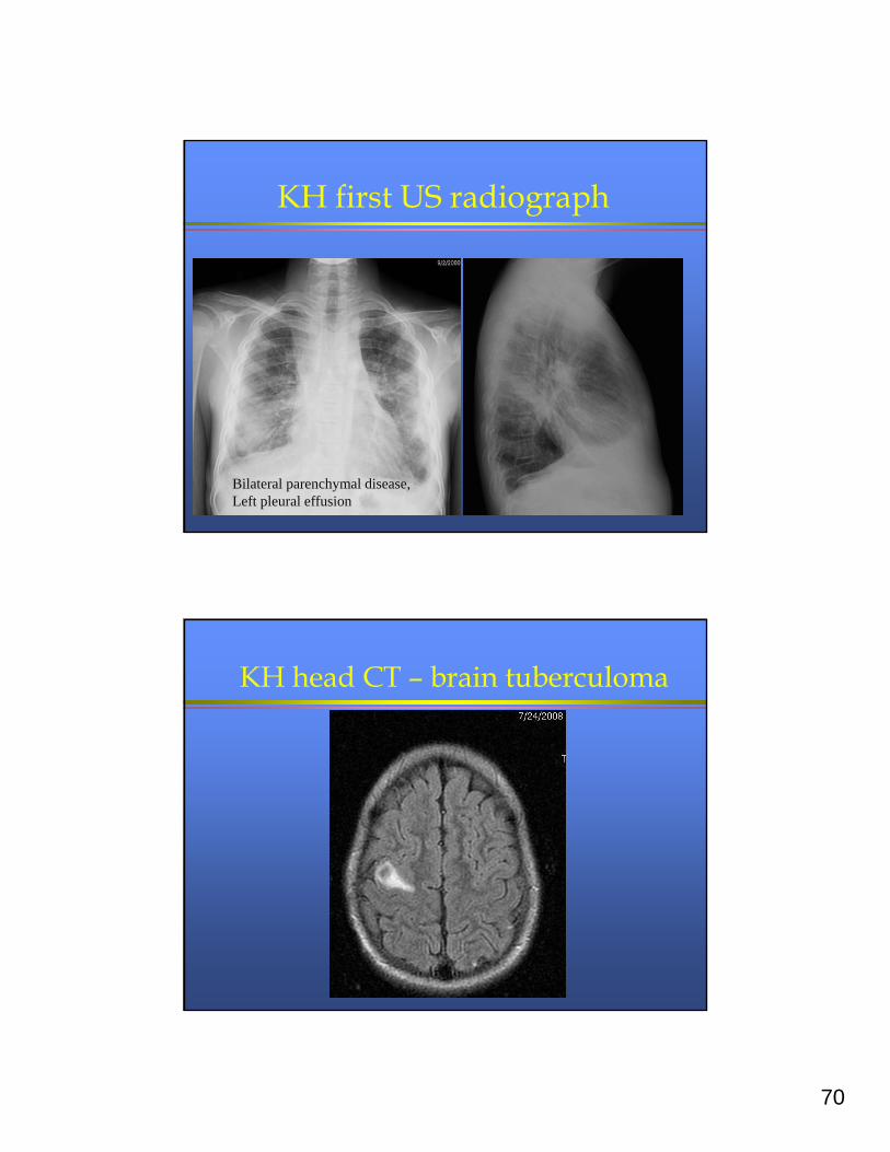
KH first US radiograph
Bilateral parenchymal disease,Left pleural effusion
KH head CT – brain tuberculoma
71
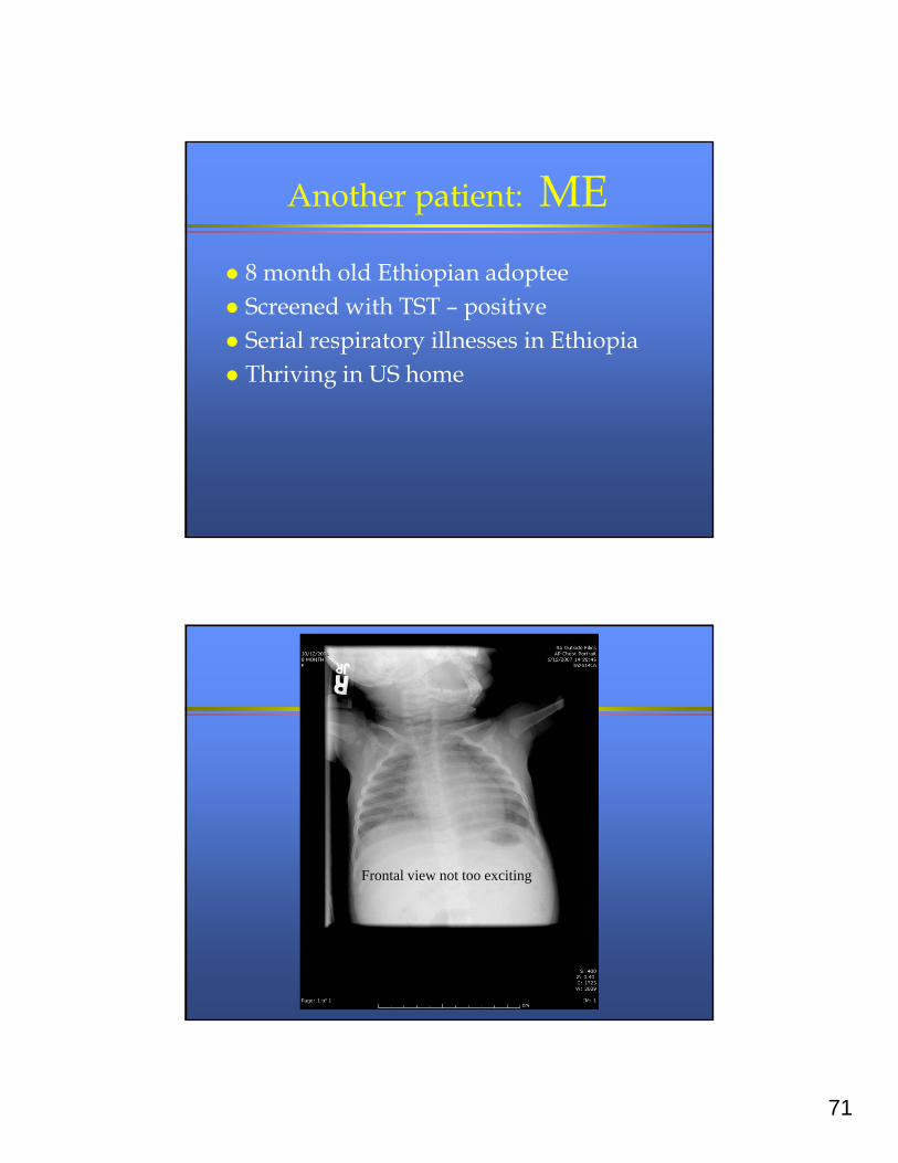
Another patient: ME
8 month old Ethiopian adoptee Screened with TST – positive Serial respiratory illnesses in Ethiopia Thriving in US home
Frontal view not too exciting
72
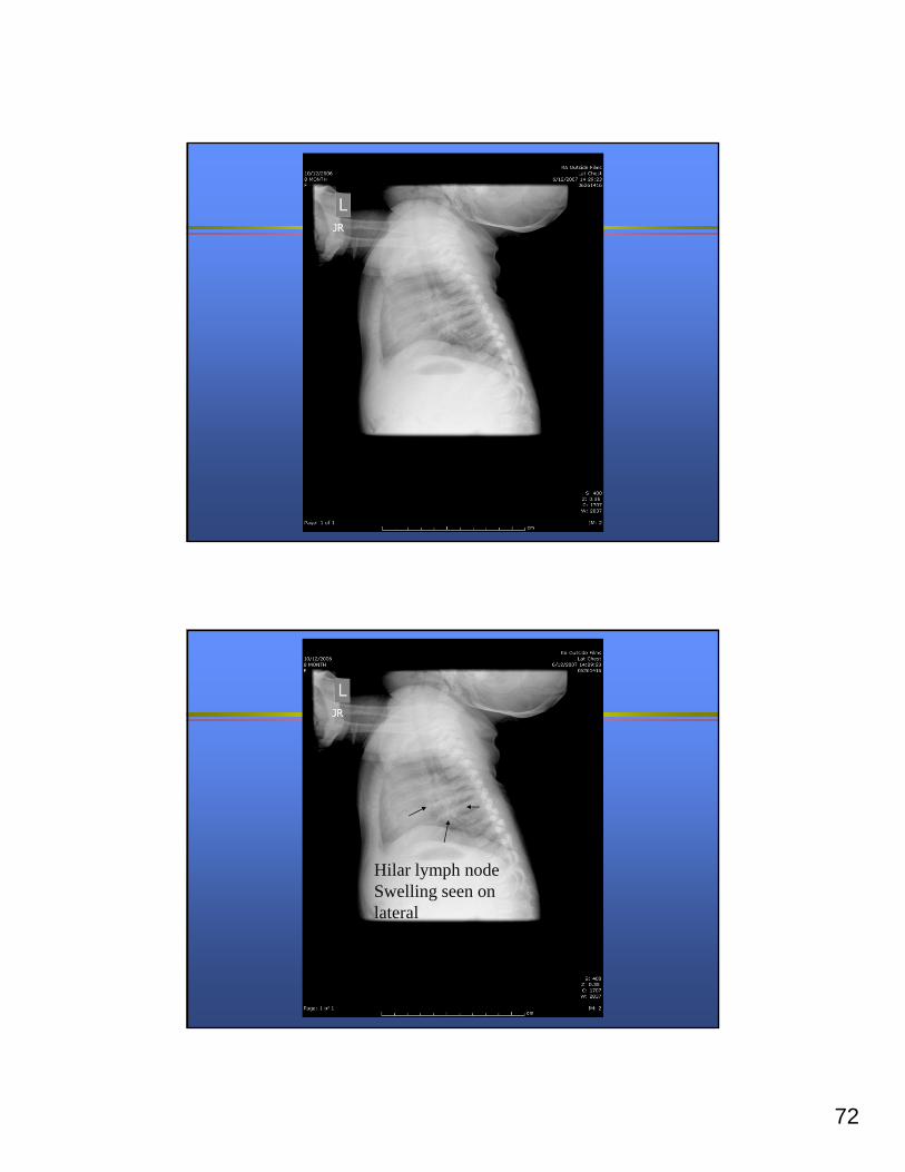
Hilar lymph node Swelling seen on lateral
73
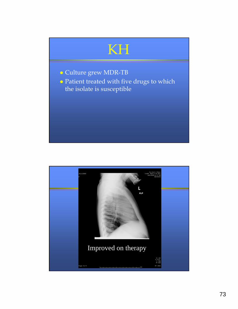
KH Culture grew MDR-TB Patient treated with five drugs to which
the isolate is susceptible
Improved on therapy

74
Another patient VC
5 month old asymptomatic VC (sister of index case), TST 8 mm, has an abnormal chest radiograph
Upon further questioning: » 3 days of minimal cough» 2 days of decreased appetite » 1 day of fever» no URI symptoms
Right sided disease
75
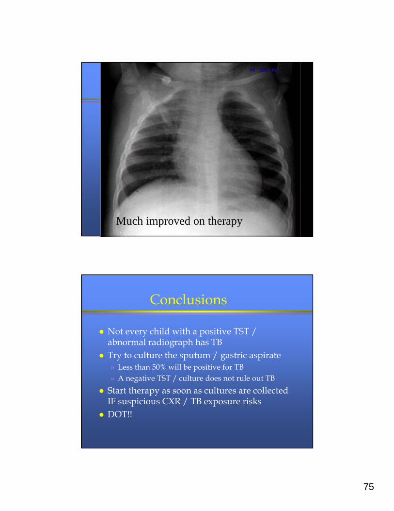
Much improved on therapy
Conclusions
Not every child with a positive TST / abnormal radiograph has TB
Try to culture the sputum / gastric aspirate» Less than 50% will be positive for TB» A negative TST / culture does not rule out TB
Start therapy as soon as cultures are collected IF suspicious CXR / TB exposure risks
DOT!!
76
Conclusions
If the patient has clinical or radiographic worsening:» Evaluate adherence to therapy» Consider possibility of drug resistance /
other diagnosis / low drug levels (check doses)
Consider steroids / antibiotics for obstructive phenomenon if confident of dx and TB susceptibility