pediatric hematology/oncology grand rounds: neuroblastoma ... · • - focal involvement by...
TRANSCRIPT
Disclosure Information: a) Moderators/panelists/presenters: Dr. Sophia Bornstein has nothing to disclose. b) Planning Committee: Drs. Linda Stork, Suman Malempati and Michael Recht have nothing to disclose.
Learning Objectives: 1. Pathophysiology and workup 2. Studies 3. Radiation oncology techniques
Pediatric Hematology/Oncology Grand Rounds: Neuroblastoma 9/4/13
Sophia Bornstein, PGY4 Radiation Medicine
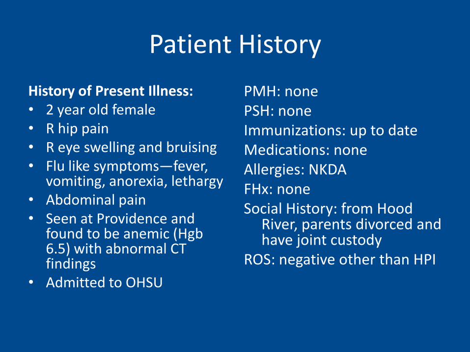
Patient History
History of Present Illness: • 2 year old female • R hip pain • R eye swelling and bruising • Flu like symptoms—fever,
vomiting, anorexia, lethargy • Abdominal pain • Seen at Providence and
found to be anemic (Hgb 6.5) with abnormal CT findings
• Admitted to OHSU
PMH: none PSH: none Immunizations: up to date Medications: none Allergies: NKDA FHx: none Social History: from Hood
River, parents divorced and have joint custody
ROS: negative other than HPI

Patient LS Physical Exam
Ht86cm(41%), Wt10.5kg(7%), BP89/65, HR147, T 37.3 °C, RR 40, SpO2 100% • General: Sleeping, NAD • HEENT: Eyes PERRL, significant bruising around right eye. Unable to palpate
head due to positioning and irritability. Nose: normal. Mouth: Normal pharynx, mucosa and teeth.
• Neck: Supple • Lungs: chest clear to auscultation bilaterally, respirations even and
unlabored. • Heart: regular rate and rhythm, no extra sounds. • Abdomen: L abdominal fullness. BS normal. Pt guards abdomen and is fussy
during exam, unable to palpate mass, very limited exam. • Musculoskeletal: well developed, good perfusion. • Skin: + bruising to face, as above. No petechiae.
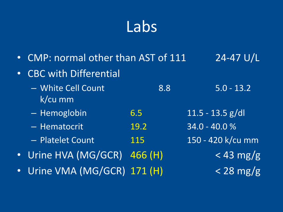
Labs
• CMP: normal other than AST of 111 24-47 U/L
• CBC with Differential
– White Cell Count 8.8 5.0 - 13.2 k/cu mm
– Hemoglobin 6.5 11.5 - 13.5 g/dl
– Hematocrit 19.2 34.0 - 40.0 %
– Platelet Count 115 150 - 420 k/cu mm
• Urine HVA (MG/GCR) 466 (H) < 43 mg/g
• Urine VMA (MG/GCR) 171 (H) < 28 mg/g
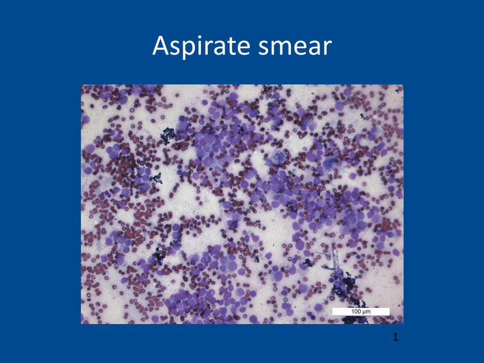
Aspirate smear
1
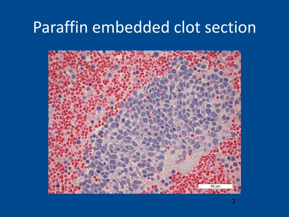
Paraffin embedded clot section
2
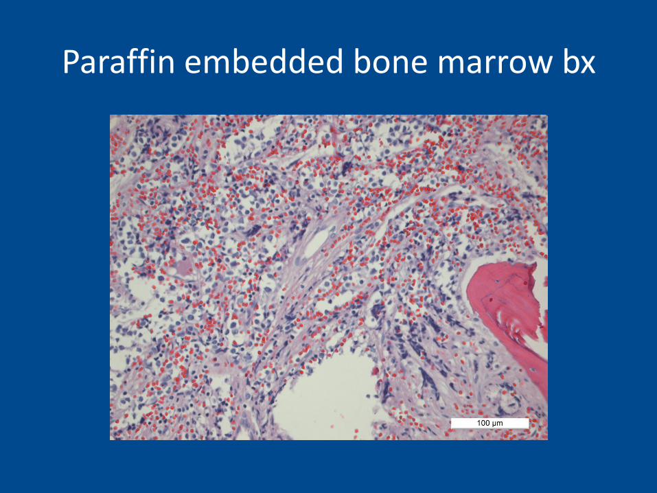
Paraffin embedded bone marrow bx
Work-up
• Neuroblastoma, r/o other SRBCT of childhood (Ewings)
• Synaptophysin, chromogranin (CD56, NSE): positive in neuroblastoma, can also be positive in Ewings/PNET and some rhabdomyosarcomas may pick up synapto
• Neuroblastoma is CD99 negative
• Consider: CK, desmin, myogenin, WT-1, CD45, ect
Cytogenetics
• Patient: 11/20 metaphase cells examined comprised a single abnormal clone, most with an isochromosome 1q, and additional material of unknown origin on chromosomes 2p and 13q; all cells had tens of double minute chromosomes, which stained positive for N-MYC by FISH (below). Nine cells appeared normal female.
• FISH was performed with the N-MYC probe set, as listed below. 89% of cells had >10 signals for N-MYC and two for the CEP2, consistent with N-MYC amplification, which was observed on available metaphases as hybridizing to double minute chromosomes.
• Poor prognosis: N-myc amplification, -1p, +17q • Good prognosis: age <1 year, hyperdiploidy
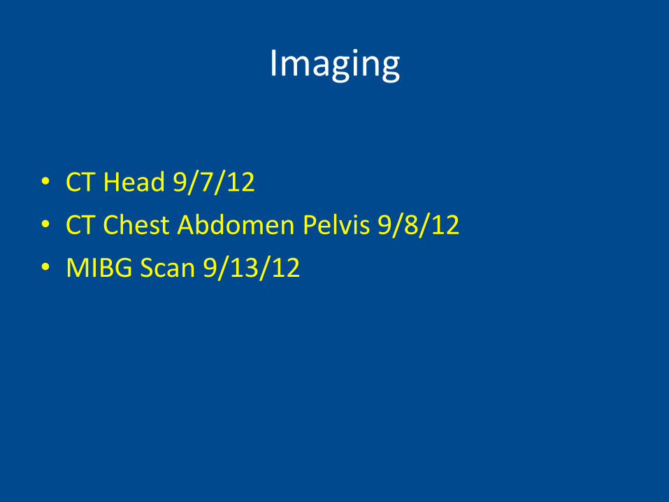
Imaging
• CT Head 9/7/12
• CT Chest Abdomen Pelvis 9/8/12
• MIBG Scan 9/13/12
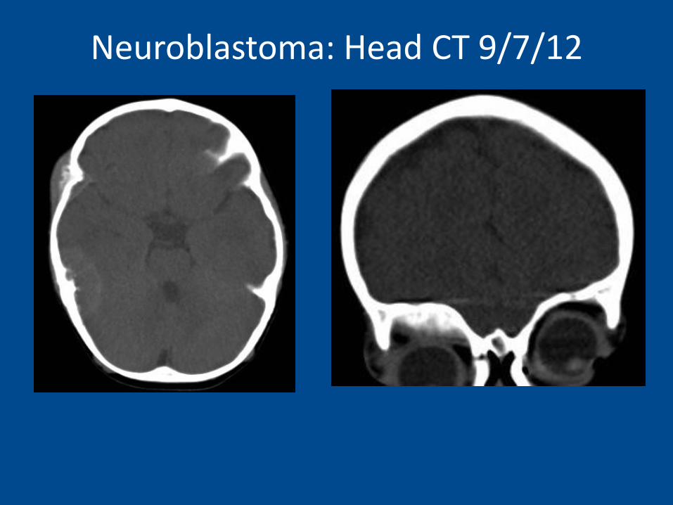
Neuroblastoma: Head CT 9/7/12
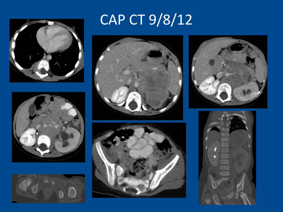
CAP CT 9/8/12
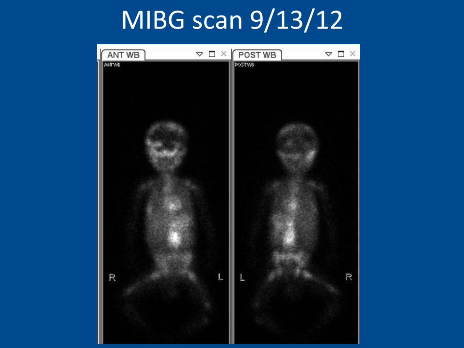
MIBG scan 9/13/12
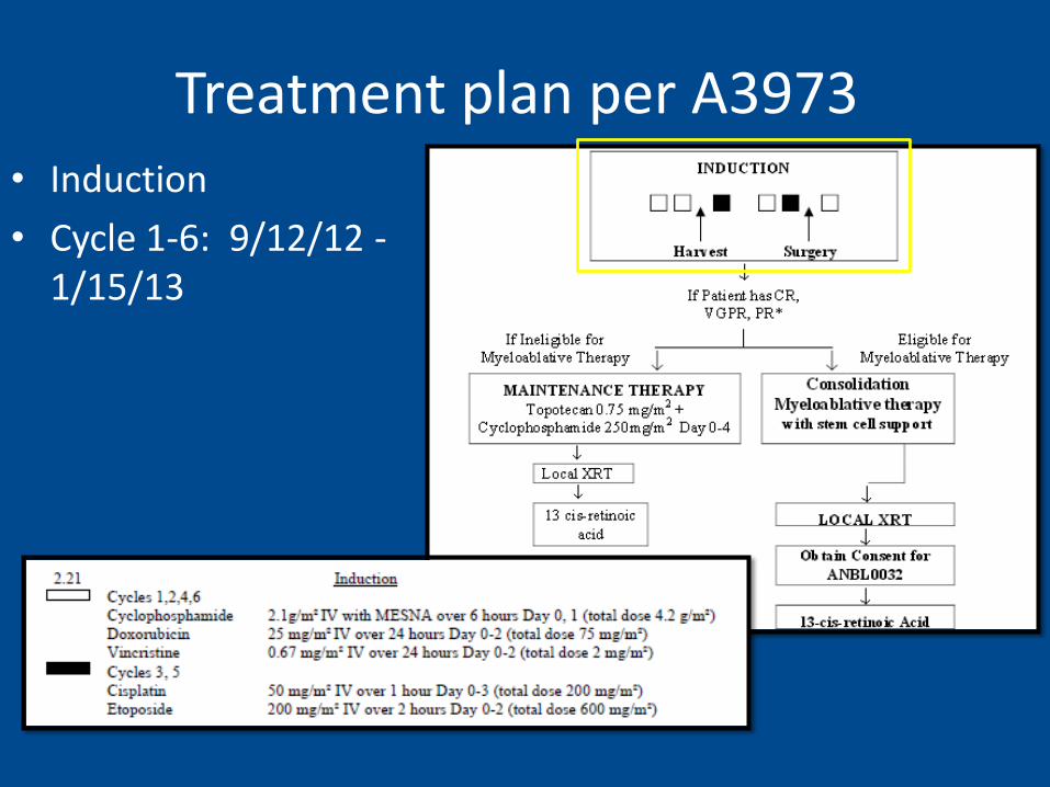
Treatment plan per A3973 • Induction
• Cycle 1-6: 9/12/12 - 1/15/13
Treatment plan per A3973
• Restaging: CT Chest Abdomen Pelvis 1/31/13 (pre-op)
• MIBG scan 2/21/13 (pre-BMT)
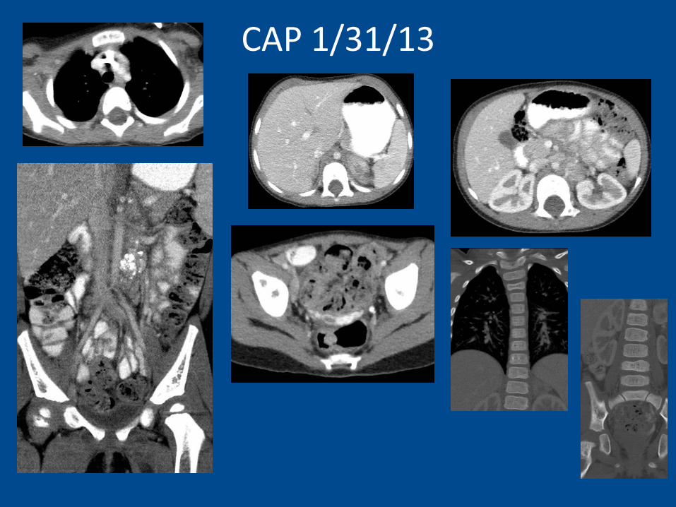
CAP 1/31/13
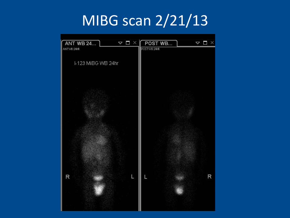
MIBG scan 2/21/13
Treatment plan per A3973
• Surgery on 2/11/13—resection of retroperitoneal tumor with L adrenal gland, debulking of peri-aortic and peri-SMA tumor.
– Per the op note, estimated 60% resected
– Surgical Pathology 2/11/13
Marrow • Final Pathologic Diagnosis: • Peripheral blood: • - Normocytic anemia
• Right and left bone marrow aspirate, clots, and core biopsies: • - Mildly hypocellular bone marrow (80 - 85%) with trilineage
hematopoiesis • - Focal involvement by neuroblastoma (see note)
• Note: There are rare single cells and small groups of 2 or 3 cells that stain
with synaptophysin and chromogranin consistent with focal residual involvement by neuroblastoma. Overall, these cells comprise much less than 1% of the overall cellularity.
• All twenty metaphase cells examined appeared normal female.
• FISH was performed with the N-MYC probe set, as listed below. All results were within the normal limits established by our laboratory.

Treatment plan per A3973
• Transplant: Day 0 3/7/13, discharged 3/26/13.
• Seen in Radiation Oncology Day 26 after transplant.
• Was doing will since DC: eating, talking. Still requiring RBC and platelet transfusions.
• Radiation Treatment: 4/8-4/24/13 (to be discussed later)
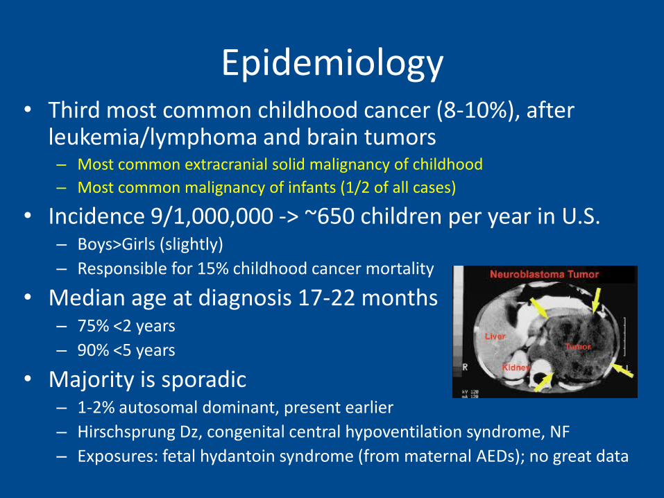
Epidemiology • Third most common childhood cancer (8-10%), after
leukemia/lymphoma and brain tumors – Most common extracranial solid malignancy of childhood
– Most common malignancy of infants (1/2 of all cases)
• Incidence 9/1,000,000 -> ~650 children per year in U.S. – Boys>Girls (slightly)
– Responsible for 15% childhood cancer mortality
• Median age at diagnosis 17-22 months – 75% <2 years
– 90% <5 years
• Majority is sporadic – 1-2% autosomal dominant, present earlier
– Hirschsprung Dz, congenital central hypoventilation syndrome, NF
– Exposures: fetal hydantoin syndrome (from maternal AEDs); no great data
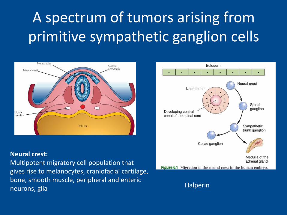
A spectrum of tumors arising from primitive sympathetic ganglion cells
Halperin
Neural crest: Multipotent migratory cell population that gives rise to melanocytes, craniofacial cartilage, bone, smooth muscle, peripheral and enteric neurons, glia

Neuroblastoma Sites
Sympathetic nervous system Adrenal gland 40% Abdominal ganglia 25% Thoracic ganglia 15% (infants) Cervical ganglia 5% Pelvic ganglia 5%
Abdomen primary in 50-80%
Halperin
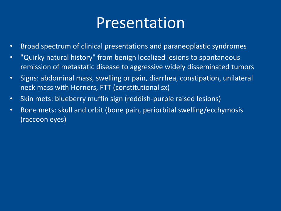
Presentation • Broad spectrum of clinical presentations and paraneoplastic syndromes
• "Quirky natural history" from benign localized lesions to spontaneous remission of metastatic disease to aggressive widely disseminated tumors
• Signs: abdominal mass, swelling or pain, diarrhea, constipation, unilateral neck mass with Horners, FTT (constitutional sx)
• Skin mets: blueberry muffin sign (reddish-purple raised lesions)
• Bone mets: skull and orbit (bone pain, periorbital swelling/ecchymosis (raccoon eyes)
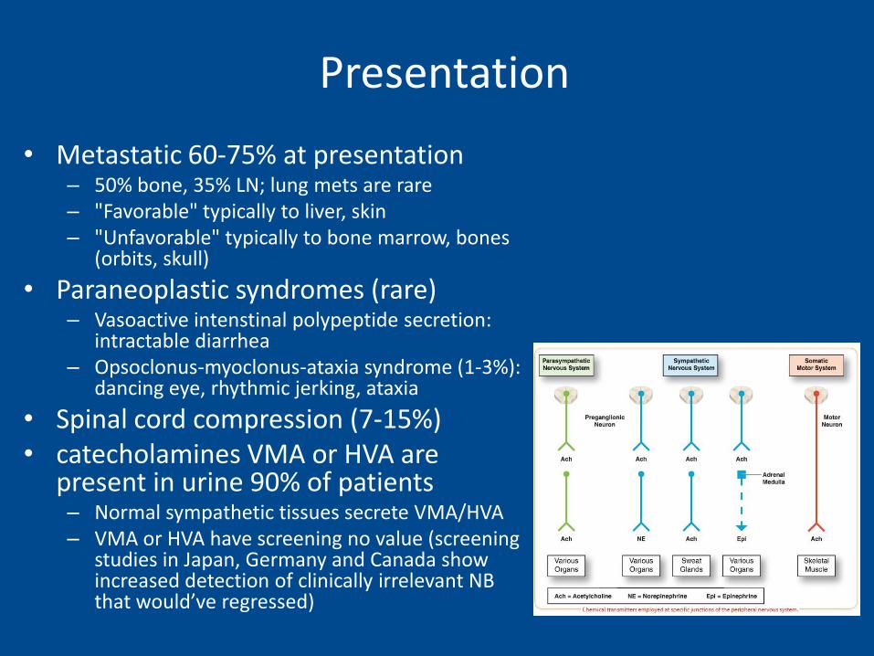
• Metastatic 60-75% at presentation – 50% bone, 35% LN; lung mets are rare – "Favorable" typically to liver, skin – "Unfavorable" typically to bone marrow, bones
(orbits, skull)
• Paraneoplastic syndromes (rare) – Vasoactive intenstinal polypeptide secretion:
intractable diarrhea – Opsoclonus-myoclonus-ataxia syndrome (1-3%):
dancing eye, rhythmic jerking, ataxia
• Spinal cord compression (7-15%) • catecholamines VMA or HVA are
present in urine 90% of patients – Normal sympathetic tissues secrete VMA/HVA – VMA or HVA have screening no value (screening
studies in Japan, Germany and Canada show increased detection of clinically irrelevant NB that would’ve regressed)
Presentation
Workup
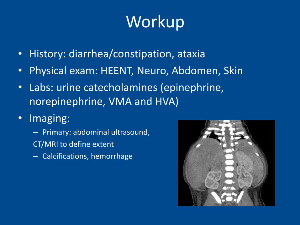
• History: diarrhea/constipation, ataxia
• Physical exam: HEENT, Neuro, Abdomen, Skin
• Labs: urine catecholamines (epinephrine, norepinephrine, VMA and HVA)
• Imaging: – Primary: abdominal ultrasound,
CT/MRI to define extent
– Calcifications, hemorrhage
Workup
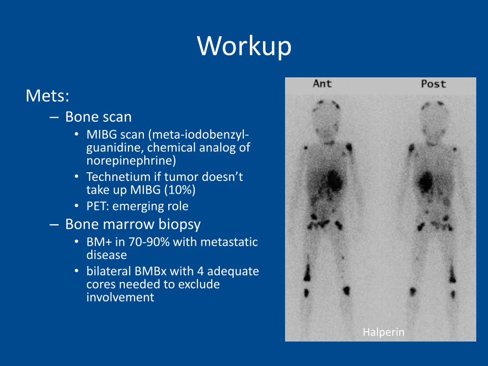
Mets: – Bone scan
• MIBG scan (meta-iodobenzyl-guanidine, chemical analog of norepinephrine)
• Technetium if tumor doesn’t take up MIBG (10%)
• PET: emerging role
– Bone marrow biopsy • BM+ in 70-90% with metastatic
disease • bilateral BMBx with 4 adequate
cores needed to exclude involvement
Halperin
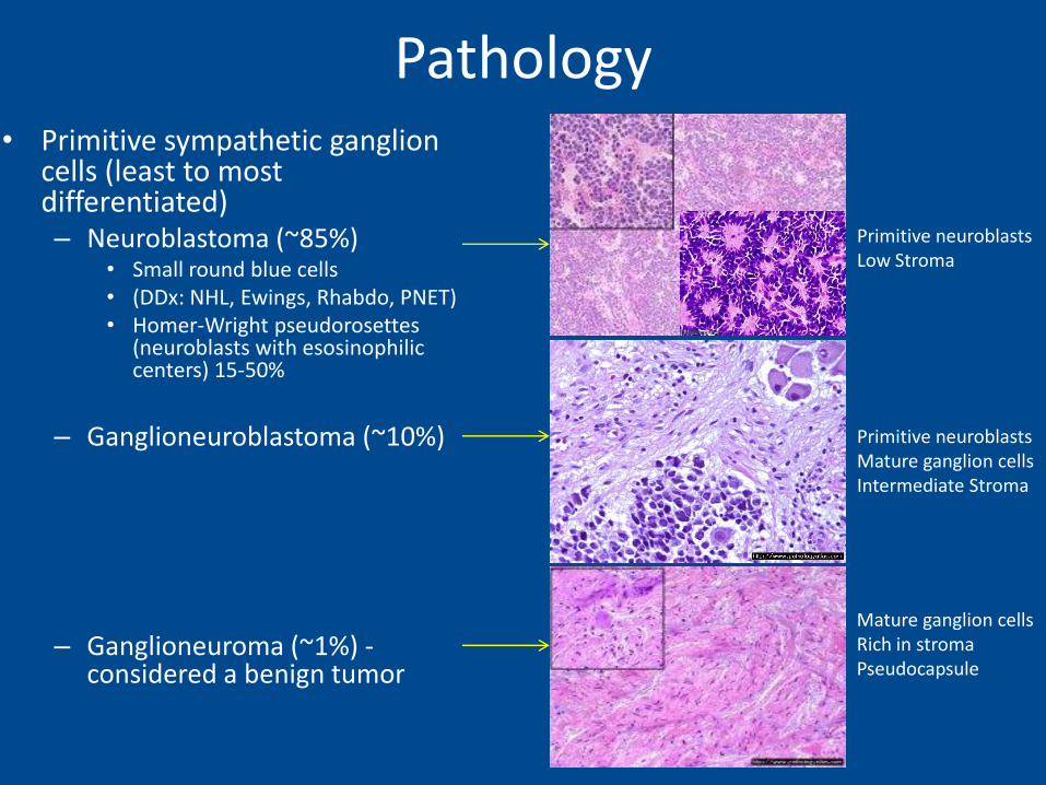
Pathology • Primitive sympathetic ganglion
cells (least to most differentiated) – Neuroblastoma (~85%)
• Small round blue cells • (DDx: NHL, Ewings, Rhabdo, PNET) • Homer-Wright pseudorosettes
(neuroblasts with esosinophilic centers) 15-50%
– Ganglioneuroblastoma (~10%)
– Ganglioneuroma (~1%) - considered a benign tumor
Primitive neuroblasts Low Stroma
Primitive neuroblasts Mature ganglion cells Intermediate Stroma
Mature ganglion cells Rich in stroma Pseudocapsule
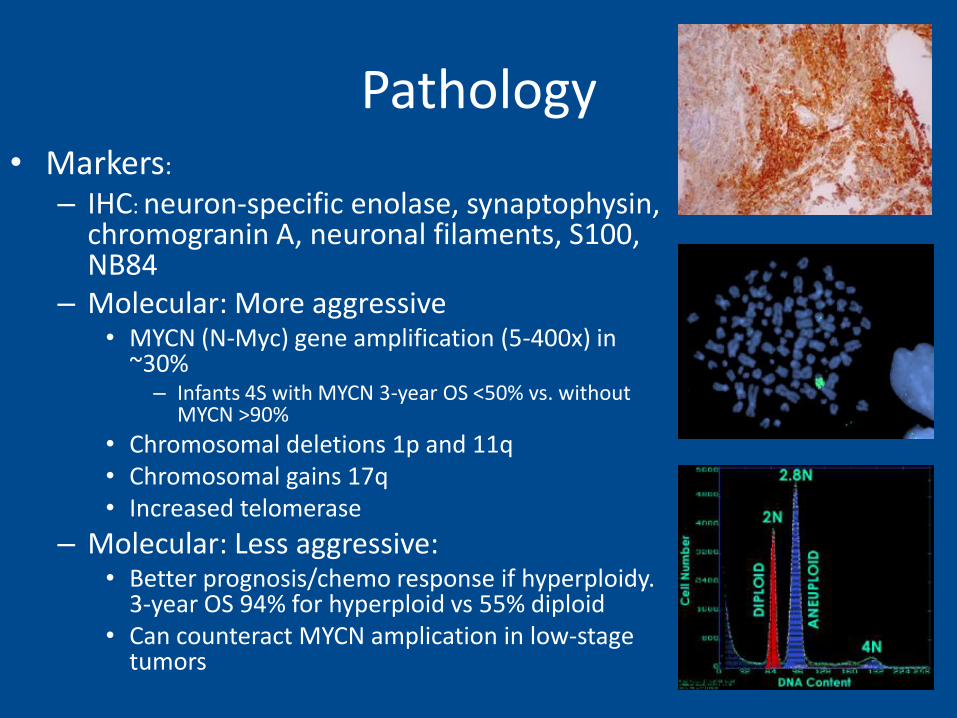
Pathology • Markers:
– IHC: neuron-specific enolase, synaptophysin, chromogranin A, neuronal filaments, S100, NB84
– Molecular: More aggressive • MYCN (N-Myc) gene amplification (5-400x) in
~30% – Infants 4S with MYCN 3-year OS <50% vs. without
MYCN >90%
• Chromosomal deletions 1p and 11q • Chromosomal gains 17q • Increased telomerase
– Molecular: Less aggressive: • Better prognosis/chemo response if hyperploidy.
3-year OS 94% for hyperploid vs 55% diploid • Can counteract MYCN amplication in low-stage
tumors
Staging (first consensus system, relies on surgical staging)
International Neuroblastoma Staging System (INSS) • Stage 1 - Localized tumor with GTR (can have microscopic+ margins)
– Representative ipsilateral lymph nodes negative microscopically (nodes attached and removed with the primary tumor may be positive)
• Stage 2A - Localized tumor with STR – Representative ipsilateral nonadherent lymph nodes negative microscopically
• Stage 2B - Localized tumor GTR or STR – Positive nonadherent ipsilateral lymph nodes. – Enlarged contralateral lymph nodes negative microscopically
• Stage 3 - Unresectable tumor – Infiltrating across the midline with or without regional lymph node involvement. – Unilateral tumor with contralateral regional lymph node involvement. Midline tumor with
bilateral extension by infiltration (unresectable) or by regional lymph node involvement
• Stage 4S - Localized primary tumor (Primary fits Stage 1, 2A or 2B) – Dissemination limited to liver, skin and/or bone marrow (in infants <1 year of age)
• Stage 4 - Dissemination of tumor to distant lymph nodes, bone, bone marrow, liver, and/or other organs (but not stage 4S)
Infant by definition
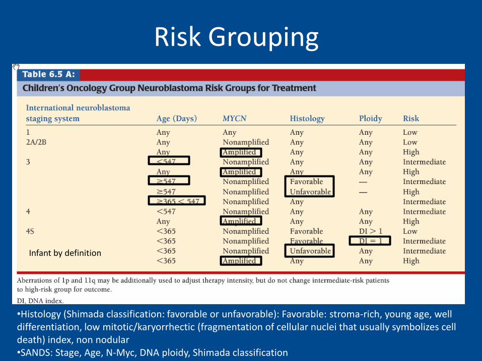
Risk Grouping
•Histology (Shimada classification: favorable or unfavorable): Favorable: stroma-rich, young age, well differentiation, low mitotic/karyorrhectic (fragmentation of cellular nuclei that usually symbolizes cell death) index, non nodular •SANDS: Stage, Age, N-Myc, DNA ploidy, Shimada classification

55% of pts are high risk at diagnosis vs 30% low risk
3 yr OS Low: 95-100%
Int: 75-98%
High: <30%
Risk Grouping
Treatment Overview
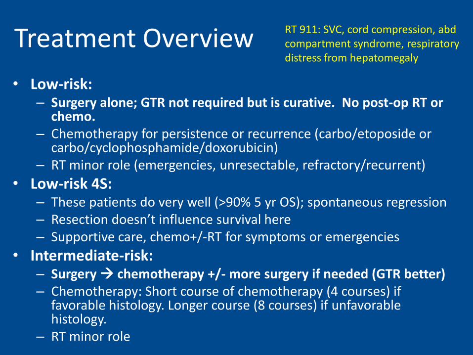
• Low-risk: – Surgery alone; GTR not required but is curative. No post-op RT or
chemo. – Chemotherapy for persistence or recurrence (carbo/etoposide or
carbo/cyclophosphamide/doxorubicin) – RT minor role (emergencies, unresectable, refractory/recurrent)
• Low-risk 4S: – These patients do very well (>90% 5 yr OS); spontaneous regression – Resection doesn’t influence survival here – Supportive care, chemo+/-RT for symptoms or emergencies
• Intermediate-risk: – Surgery chemotherapy +/- more surgery if needed (GTR better) – Chemotherapy: Short course of chemotherapy (4 courses) if
favorable histology. Longer course (8 courses) if unfavorable histology.
– RT minor role
RT 911: SVC, cord compression, abd compartment syndrome, respiratory distress from hepatomegaly
Treatment Overview
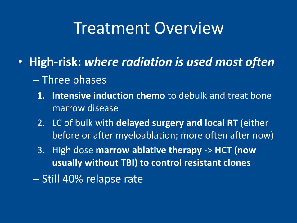
• High-risk: where radiation is used most often
– Three phases
1. Intensive induction chemo to debulk and treat bone marrow disease
2. LC of bulk with delayed surgery and local RT (either before or after myeloablation; more often after now)
3. High dose marrow ablative therapy -> HCT (now usually without TBI) to control resistant clones
– Still 40% relapse rate
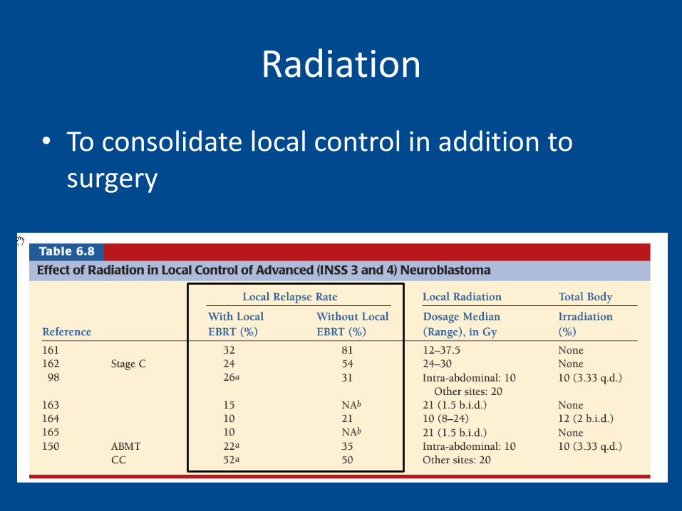
Radiation
• To consolidate local control in addition to surgery
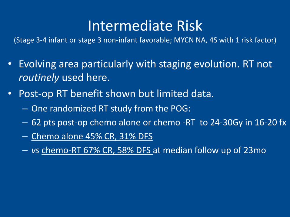
Intermediate Risk (Stage 3-4 infant or stage 3 non-infant favorable; MYCN NA, 4S with 1 risk factor)
• Evolving area particularly with staging evolution. RT not routinely used here.
• Post-op RT benefit shown but limited data.
– One randomized RT study from the POG:
– 62 pts post-op chemo alone or chemo -RT to 24-30Gy in 16-20 fx
– Chemo alone 45% CR, 31% DFS
– vs chemo-RT 67% CR, 58% DFS at median follow up of 23mo
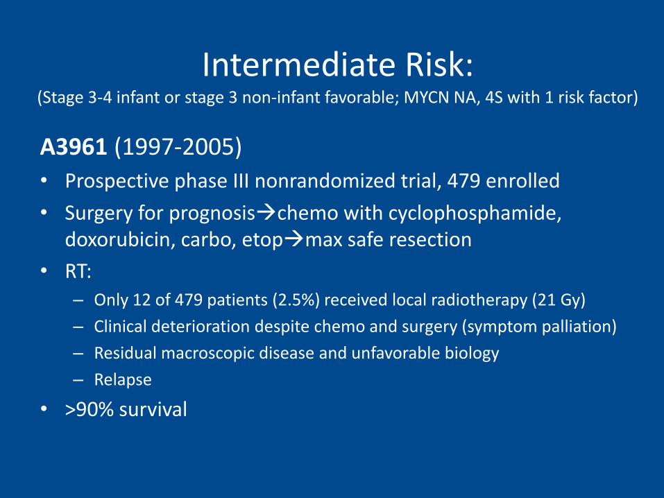
Intermediate Risk: (Stage 3-4 infant or stage 3 non-infant favorable; MYCN NA, 4S with 1 risk factor)
A3961 (1997-2005)
• Prospective phase III nonrandomized trial, 479 enrolled
• Surgery for prognosischemo with cyclophosphamide, doxorubicin, carbo, etopmax safe resection
• RT: – Only 12 of 479 patients (2.5%) received local radiotherapy (21 Gy)
– Clinical deterioration despite chemo and surgery (symptom palliation)
– Residual macroscopic disease and unfavorable biology
– Relapse
• >90% survival
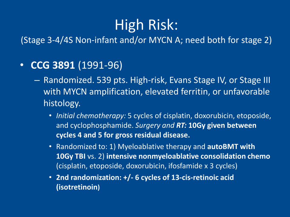
High Risk: (Stage 3-4/4S Non-infant and/or MYCN A; need both for stage 2)
• CCG 3891 (1991-96)
– Randomized. 539 pts. High-risk, Evans Stage IV, or Stage III with MYCN amplification, elevated ferritin, or unfavorable histology. • Initial chemotherapy: 5 cycles of cisplatin, doxorubicin, etoposide,
and cyclophosphamide. Surgery and RT: 10Gy given between cycles 4 and 5 for gross residual disease.
• Randomized to: 1) Myeloablative therapy and autoBMT with 10Gy TBI vs. 2) intensive nonmyeloablative consolidation chemo (cisplatin, etoposide, doxorubicin, ifosfamide x 3 cycles)
• 2nd randomization: +/- 6 cycles of 13-cis-retinoic acid (isotretinoin)
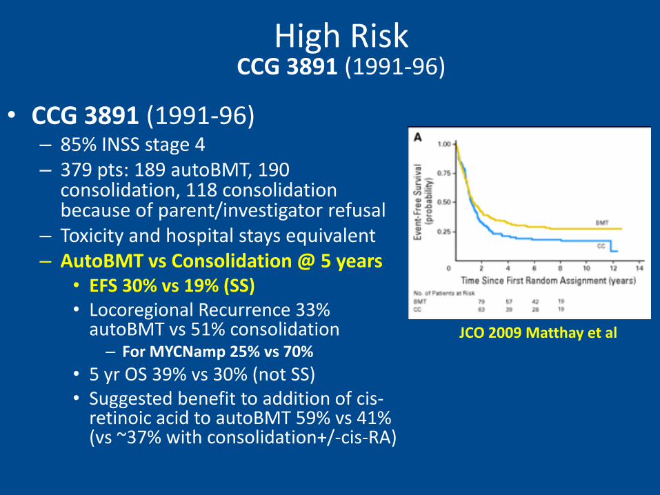
• CCG 3891 (1991-96) – 85% INSS stage 4 – 379 pts: 189 autoBMT, 190
consolidation, 118 consolidation because of parent/investigator refusal
– Toxicity and hospital stays equivalent – AutoBMT vs Consolidation @ 5 years
• EFS 30% vs 19% (SS) • Locoregional Recurrence 33%
autoBMT vs 51% consolidation – For MYCNamp 25% vs 70%
• 5 yr OS 39% vs 30% (not SS) • Suggested benefit to addition of cis-
retinoic acid to autoBMT 59% vs 41% (vs ~37% with consolidation+/-cis-RA)
High Risk CCG 3891 (1991-96)
JCO 2009 Matthay et al
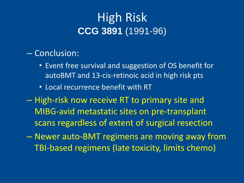
High Risk CCG 3891 (1991-96)
– Conclusion:
• Event free survival and suggestion of OS benefit for autoBMT and 13-cis-retinoic acid in high risk pts
• Local recurrence benefit with RT
– High-risk now receive RT to primary site and MIBG-avid metastatic sites on pre-transplant scans regardless of extent of surgical resection
– Newer auto-BMT regimens are moving away from TBI-based regimens (late toxicity, limits chemo)
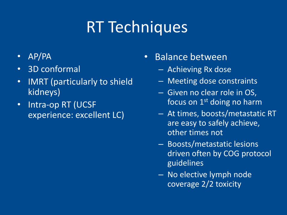
RT Techniques
• AP/PA
• 3D conformal
• IMRT (particularly to shield kidneys)
• Intra-op RT (UCSF experience: excellent LC)
• Balance between – Achieving Rx dose
– Meeting dose constraints
– Given no clear role in OS, focus on 1st doing no harm
– At times, boosts/metastatic RT are easy to safely achieve, other times not
– Boosts/metastatic lesions driven often by COG protocol guidelines
– No elective lymph node coverage 2/2 toxicity
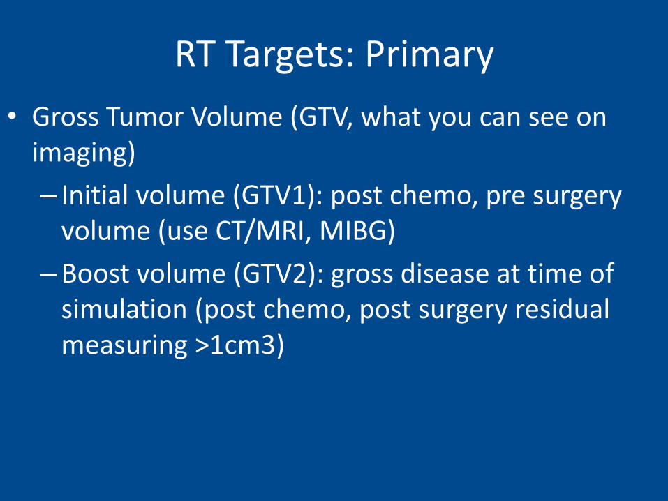
RT Targets: Primary
• Gross Tumor Volume (GTV, what you can see on imaging)
– Initial volume (GTV1): post chemo, pre surgery volume (use CT/MRI, MIBG)
–Boost volume (GTV2): gross disease at time of simulation (post chemo, post surgery residual measuring >1cm3)
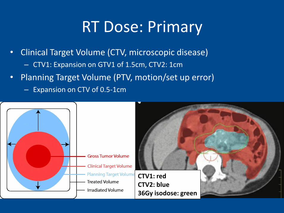
RT Dose: Primary
• Clinical Target Volume (CTV, microscopic disease) – CTV1: Expansion on GTV1 of 1.5cm, CTV2: 1cm
• Planning Target Volume (PTV, motion/set up error) – Expansion on CTV of 0.5-1cm
CTV1: red CTV2: blue 36Gy isodose: green
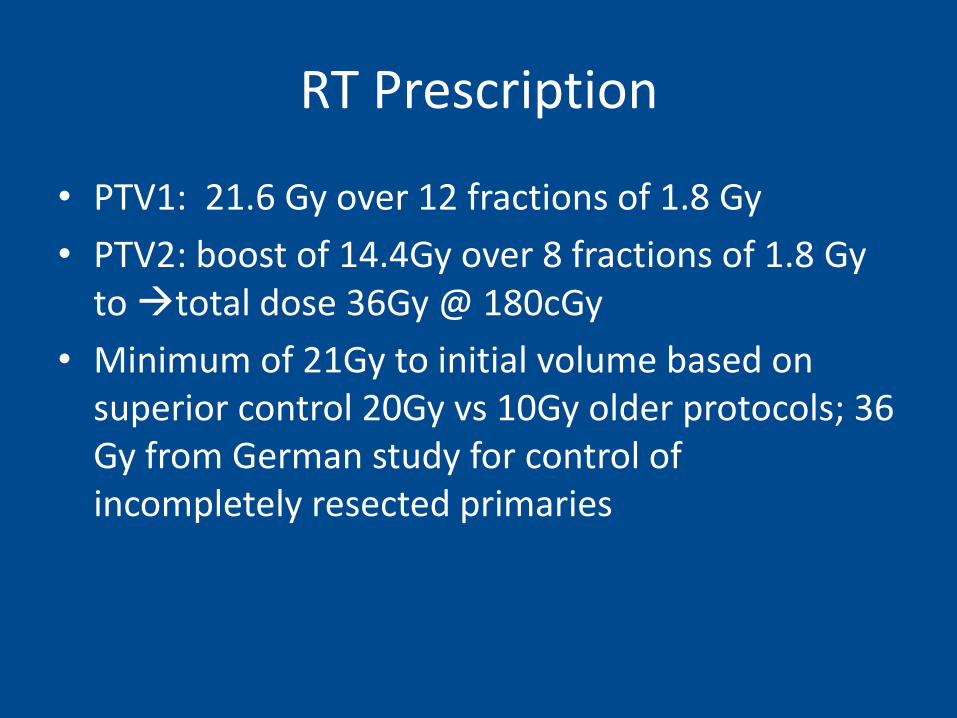
RT Prescription
• PTV1: 21.6 Gy over 12 fractions of 1.8 Gy
• PTV2: boost of 14.4Gy over 8 fractions of 1.8 Gy to total dose 36Gy @ 180cGy
• Minimum of 21Gy to initial volume based on superior control 20Gy vs 10Gy older protocols; 36 Gy from German study for control of incompletely resected primaries
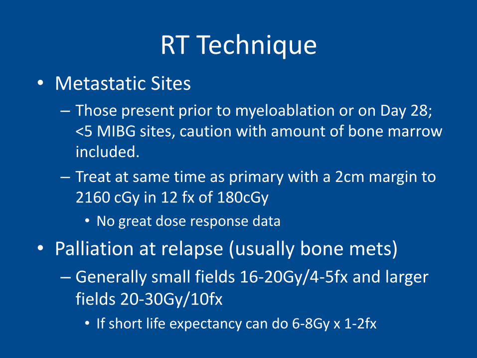
RT Technique • Metastatic Sites
– Those present prior to myeloablation or on Day 28; <5 MIBG sites, caution with amount of bone marrow included.
– Treat at same time as primary with a 2cm margin to 2160 cGy in 12 fx of 180cGy
• No great dose response data
• Palliation at relapse (usually bone mets)
– Generally small fields 16-20Gy/4-5fx and larger fields 20-30Gy/10fx
• If short life expectancy can do 6-8Gy x 1-2fx
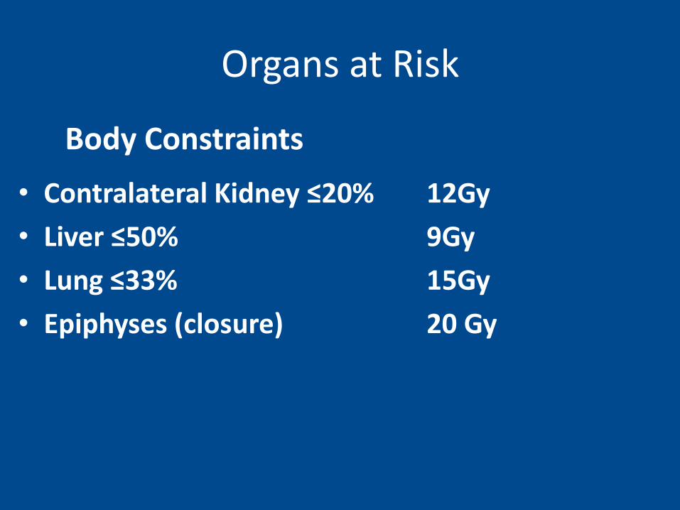
Organs at Risk
Body Constraints
• Contralateral Kidney ≤20% 12Gy
• Liver ≤50% 9Gy
• Lung ≤33% 15Gy
• Epiphyses (closure) 20 Gy
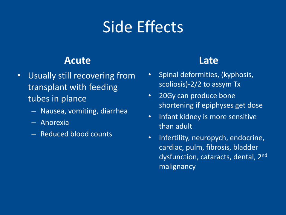
Side Effects
Acute
• Usually still recovering from transplant with feeding tubes in plance – Nausea, vomiting, diarrhea
– Anorexia
– Reduced blood counts
Late • Spinal deformities, (kyphosis,
scoliosis)-2/2 to assym Tx
• 20Gy can produce bone shortening if epiphyses get dose
• Infant kidney is more sensitive than adult
• Infertility, neuropych, endocrine, cardiac, pulm, fibrosis, bladder dysfunction, cataracts, dental, 2nd malignancy
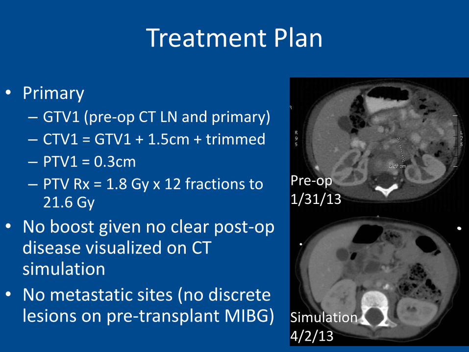
Treatment Plan
• Primary – GTV1 (pre-op CT LN and primary)
– CTV1 = GTV1 + 1.5cm + trimmed
– PTV1 = 0.3cm
– PTV Rx = 1.8 Gy x 12 fractions to 21.6 Gy
• No boost given no clear post-op disease visualized on CT simulation
• No metastatic sites (no discrete lesions on pre-transplant MIBG)
Simulation 4/2/13
Pre-op 1/31/13
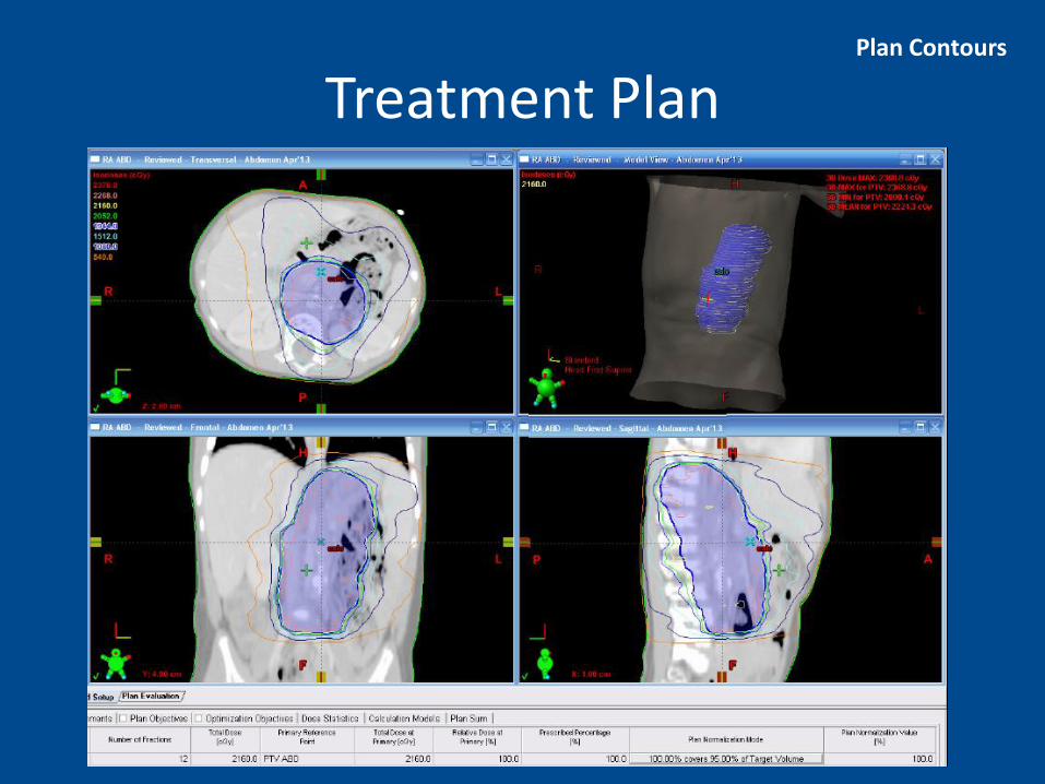
Treatment Plan Plan Contours
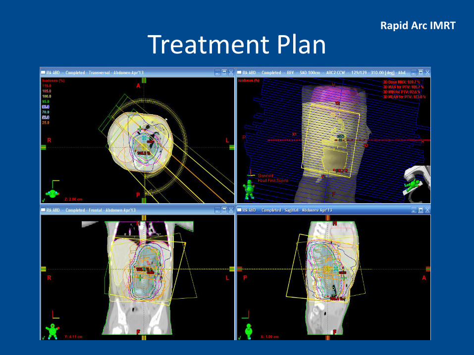
Rapid Arc IMRT
Treatment Plan
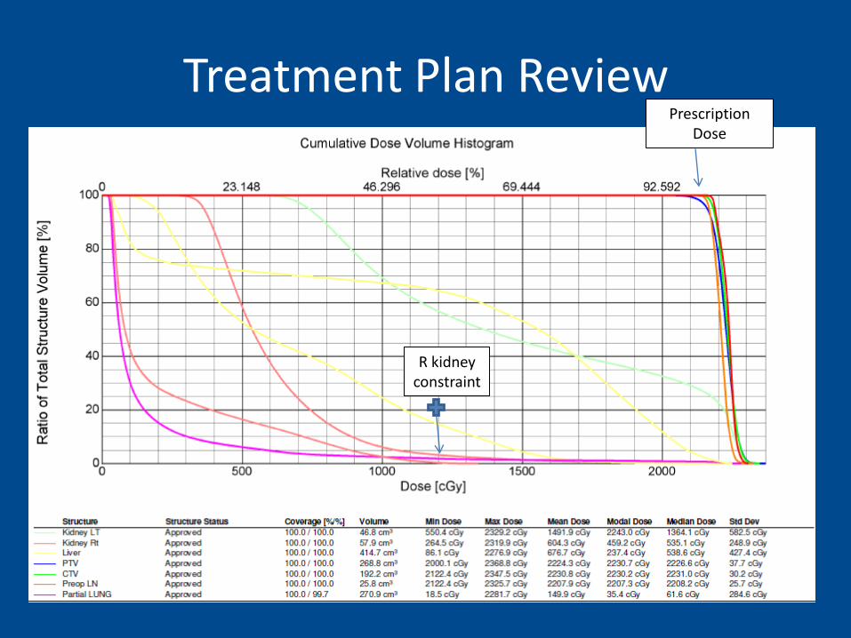
Treatment Plan Review
R kidney constraint
Prescription Dose
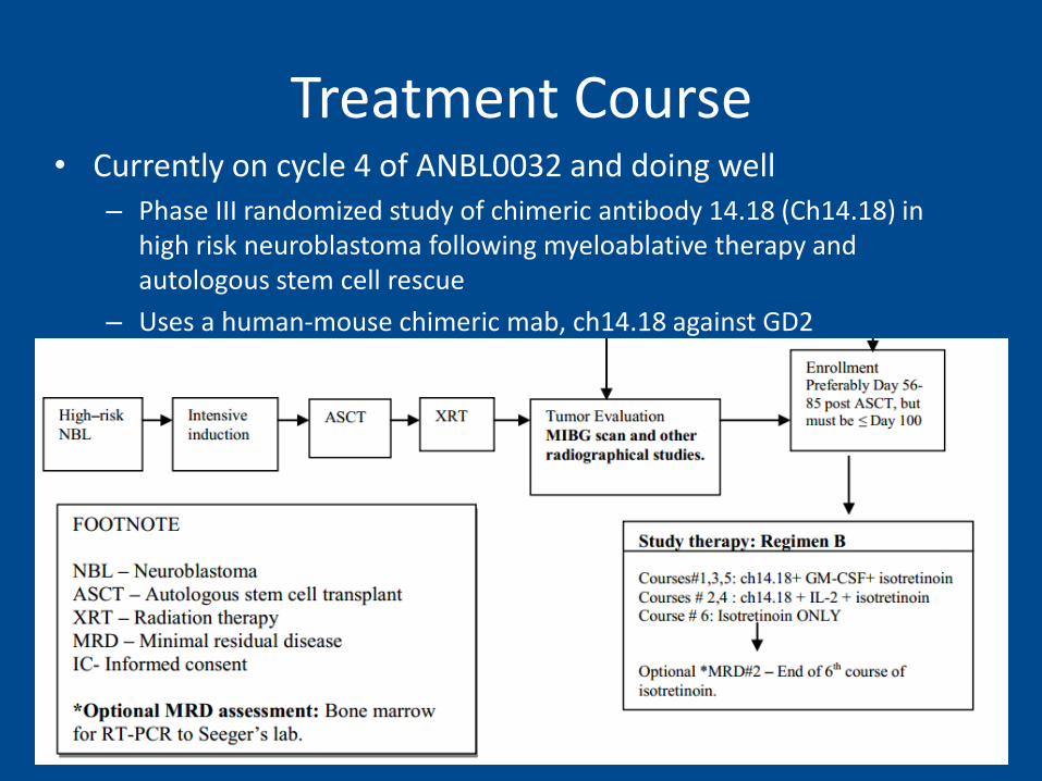
Treatment Course • Currently on cycle 4 of ANBL0032 and doing well
– Phase III randomized study of chimeric antibody 14.18 (Ch14.18) in high risk neuroblastoma following myeloablative therapy and autologous stem cell rescue
– Uses a human-mouse chimeric mab, ch14.18 against GD2
Acknowledgements
• PHO Grand Rounds Organizers – Linda Stork, Suman Malempati and Michael Recht – Beth Wamala
• Peds Heme/Onc – Lara Riegler
• Pathology – Ellen Flatley
• Radiology – Petra Vajtai
• Radiation Medicine – Carol Marquez