pe rspective - wyss institute | wyss institute at harvard organs-on... · these systems can...
TRANSCRIPT
760 volume 32 number 8 august 2014 nature biotechnology
1Department of Electrical Engineering & Computer Science, Koch Institute and Institute for Medical Engineering and Science, Massachusetts Institute of Technology and Broad Institute, Cambridge, Massachusetts, USA. 2Department of Medicine, Brigham and Women’s Hospital, Boston, Massachusetts, USA. 3Wyss Institute for Biologically Inspired Engineering at Harvard University, Boston, Massachusetts, USA. 4Vascular Biology Program, Departments of Pathology & Surgery, Boston Children’s Hospital and Harvard Medical School, Boston, Massachusetts, USA. 5School of Engineering and Applied Sciences, Harvard University, Cambridge, Massachusetts, USA. Correspondence should be addressed to D.E.I. ([email protected]).
Received 13 January; accepted 10 July; published online 5 August 2014; doi:10.1038/nbt.2989
Microfluidic organs-on-chipsSangeeta N Bhatia1,2 & Donald E Ingber3–5
An organ-on-a-chip is a microfluidic cell culture device created with microchip manufacturing methods that contains continuously perfused chambers inhabited by living cells arranged to simulate tissue- and organ-level physiology. By recapitulating the multicellular architectures, tissue-tissue interfaces, physicochemical microenvironments and vascular perfusion of the body, these devices produce levels of tissue and organ functionality not possible with conventional 2D or 3D culture systems. They also enable high-resolution, real-time imaging and in vitro analysis of biochemical, genetic and metabolic activities of living cells in a functional tissue and organ context. This technology has great potential to advance the study of tissue development, organ physiology and disease etiology. In the context of drug discovery and development, it should be especially valuable for the study of molecular mechanisms of action, prioritization of lead candidates, toxicity testing and biomarker identification.
Conventional two-dimensional (2D) cell cultures were developed almost a century ago1. Despite their demonstrated value in biomedical research, they cannot support the tissue-specific, differentiated func-tions of many cell types or accurately predict in vivo tissue functions and drug activities2. These limitations have led to increased interest in more complex 2D models, such as those that incorporate multiple cell types or involve cell patterning, and in three-dimensional (3D) mod-els, which better represent the spatial and chemical complexity of liv-ing tissues. 3D cell cultures, developed over 50 years ago3, usually rely on hydrogels, composed of either natural extracellular matrix (ECM) molecules or synthetic polymers, which induce cells to polarize and to interact with neighboring cells. They can take many forms, including cells randomly interspersed in ECM or clustered in self-assembling cellular microstructures known as organoids. 3D models have been very useful for studying the molecular basis of tissue function and better capture signaling pathways and drug responsiveness in some disease states compared with 2D models4–7. Nonetheless, they also
have limitations. For example, organoids are highly variable in size and shape, and it is difficult to maintain cells in consistent positions in these structures for extended analysis. Another drawback of 3D models is that functional analysis of entrapped cells—for example, to quantify transcellular transport, absorption or secretion—is often hampered by the difficulty of sampling luminal contents, and it is difficult to harvest cellular components for biochemical and genetic analysis. In addition, many systems lack multiscale architecture and tissue-tissue interfaces, such as the interface between vascular endo-thelium and surrounding connective tissue and parenchymal cells, which are crucial to the function of nearly all organs. Furthermore, cells are usually not exposed to normal mechanical cues, including fluid shear stress, tension and compression, which influence organ development and function in health and disease8,9. The absence of fluid flow also precludes the study of how cultured cells interact with circulating blood and immune cells.
Microfluidic organs-on-chips offer the possibility of overcoming all of these limitations. In this Perspective, we discuss the value of this new approach to scientists in basic and applied research. We also describe the technical challenges that must be overcome to develop organs-on-chips into robust, predictive models of human physiol-ogy and disease, and into tools for drug discovery and development.
What are organs-on-chips?Microfluidic culture devices. Organs-on-chips are microfluidic devices for culturing living cells in continuously perfused, micrometer- sized chambers in order to model physiological functions of tissues and organs. The goal is not to build a whole living organ but rather to syn-thesize minimal functional units that recapitulate tissue- and organ-level functions. The simplest system is a single, perfused microfluidic chamber containing one kind of cultured cell (e.g., hepatocytes or kidney tubu-lar epithelial cells) that exhibits functions of one tissue type. In more complex designs, two or more microchannels are connected by porous membranes, lined on opposite sides by different cell types, to recreate interfaces between different tissues (e.g., lung alveolar-capillary interface or blood-brain barrier). These systems can incorporate physical forces, including physiologically relevant levels of fluid shear stress, cyclic strain and mechanical compression, and permit analysis of organ-specific responses, including recruitment of circulating immune cells, in reaction to drugs, toxins or other environmental perturbations. Similar analyses can be conducted with chips lined by cells from different organs that are linked fluidically, either directly from one interstitial tissue compartment to another, or potentially through a second channel lined with vascu-lar endothelium, to mimic physiological interactions between different organs or to study drug distribution in vitro.
The word ‘chip’ in organ-on-a-chip stems from the original fabrica-tion method, a modified form of photolithographic etching used to
p e R s p e c t i v enp
g©
2014
Nat
ure
Am
eric
a, In
c. A
ll rig
hts
rese
rved
.
nature biotechnology volume 32 number 8 august 2014 761
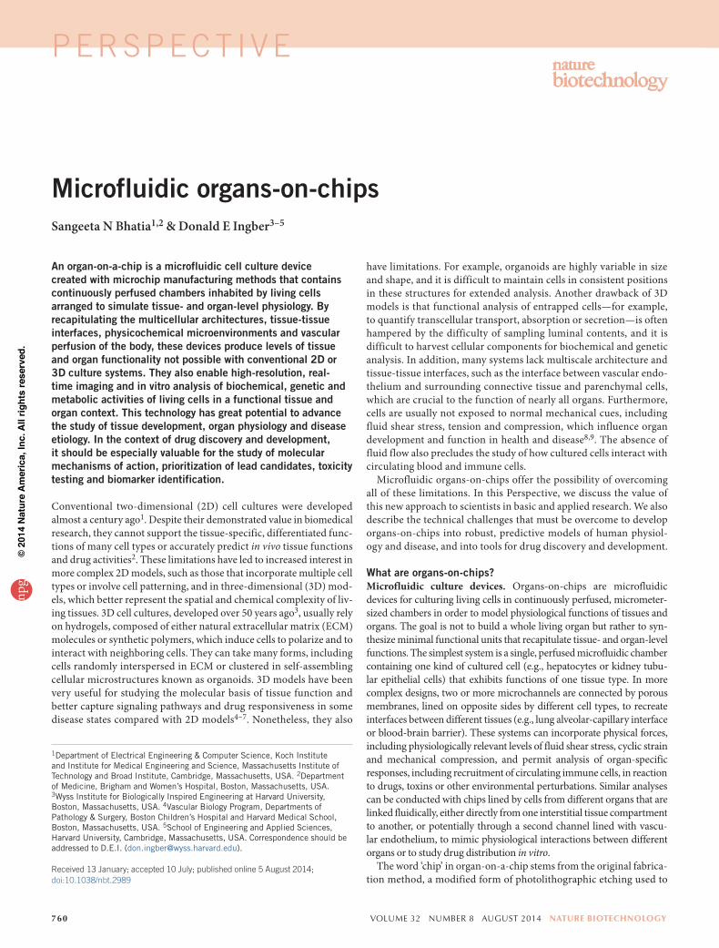
manufacture computer microchips, which allows control of surface feature shapes and sizes on the same scale (nm to µm) that living cells sense and respond to in their natural tissue milieu. Microfluidic culture systems are often made by ‘soft lithography’, a means of rep-licating patterns etched into silicon chips in more biocompatible and flexible materials10. This is done by pouring a liquid polymer, such as poly-dimethylsiloxane (PDMS), on an etched silicon substrate and allowing it to polymerize into an optically clear, rubber-like material, essentially creating a rubber stamp (Fig. 1a). Soft lithography was first used to pattern microscale adhesive islands made of ECM molecules as a way to specify the shape, position and function of cells cultured on silicon chips11, and later on conventional culture substrates12,13 (work by D.E.I. and colleagues). Subsequently, this approach was modified by inverting the PDMS mold and conformally sealing it to a flat smooth substrate, such as glass, to create open cavities in the form of small (cross section < 1 mm × 1 mm), linear, hollow chambers, or ‘microfluidic channels,’ with openings at both ends of the polymer block for perfusion of fluids (Fig. 1b). Miniaturized perfusion bio-reactors for culturing cells were made by coating the surface of the central channel with ECM molecules, flowing cells into the channel so that they adhere to the ECM substrate, and then perfusing the chan-nel continuously with culture medium14,15 (Fig. 2a). A key feature of PDMS culture systems is that they are optically clear (Fig. 1b), which allows real-time, high-resolution optical imaging of cellular responses
to environmental cues. Similar devices were later fabricated out of various materials (e.g., silicon, plastic, glass, silk) using micromold-ing, microetching, laser etching, injection molding, photopolymerization, solid object printing and other microscale manufacturing approaches.
Control of system parameters. Microfluidic chips provide control over many system parameters that are not easily controlled in 3D static cultures or bioreactors, facilitat-ing study of a broad array of physiological phenomena. Because these devices are fully microengineered, they can be integrated with microsensors that report on the cultured cells or microenvironmental conditions, which is usually not feasible in self-organized 3D cul-tures. Microsensors incorporated in chips have been used for analysis of tissue barrier integ-rity16, cell migration17 and fluid pressure18. In the future, it may be possible to detect a range of other chemical and culture conditions (glu-cose, lactate, oxygen, pH)19.
Control of fluid flow in chips has proved enormously useful. For example, because viscous forces dominate over inertial ones at small length scales, the flow is laminar if the diameter of the ‘microfluidic’ channel is less than about one millimeter. This allows the generation of physical and chemical gra-dients, which have been exploited for nonin-vasive study of directional cell migration20–22, cardiac tissue formation23, nerve axon out-growth24, and graded metabolic25, differentia-tion26 and neurotoxin27 responses, as well as analysis of subcellular structure20 and cell-cell
junctional integrity28. Fluid shear stresses can be controlled indepen-dently of physical and chemical gradients by altering flow rates or chan-nel dimensions29,30, and by separating cells from the flow path using a nanoporous membrane29 or microengineered posts that restrict cell passage31. Fluid-mechanical computational models can be applied to optimize microchannel geometry and enhance oxygen and nutrient delivery, thereby increasing cell survival and function29.
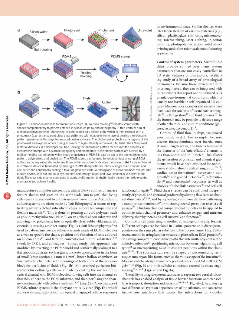
Control of cell patterning is another advantage of the chip format. Different cell types can be plated in distinct patterns or in direct juxta-position on the same planar substrate in the microchannel (Fig. 2b) by several methods: using laminar streams to plate cells or ECM proteins20, designing complex microchannel paths that intermittently contact the adhesive substrate32, positioning microposts between neighboring cell types31 or microprinting ECM in distinct positions within the chan-nels33–35. The substrate can even be shaped by micromolding tech-niques into organ-like forms, such as the villus shape of the intestine36. More recent chip designs have incorporated cells embedded in 3D ECM gels37–41 (Fig. 3) and multicellular constructs created by tissue engi-neering35,41–43 (Figs. 2c and Fig. 4a).
The ability to integrate porous substrates to separate two parallel micro-channels has enabled analysis of tissue barrier functions and transcel-lular transport, absorption and secretion16,44–49 (Fig. 4b,c). By culturing two different cell types on opposite sides of the substrate, one can create tissue-tissue interfaces that mimic the interactions of vascular
Figure 1 Fabrication methods for microfluidic chips. (a) Replica molding10 creates stamps with shapes complementary to patterns etched in silicon chips by photolithography. A thin uniform film of a photosensitive material (photoresist) is spin-coated on a silicon chip, which is then overlaid with a photomask (e.g., a transparent glass plate patterned with opaque chrome layers) bearing a microscale pattern generated with computer-assisted design software. The photomask protects some regions of the photoresist and exposes others during exposure to high-intensity ultraviolet (UV) light. The UV-exposed material dissolves in a developer solution, leaving the microscale pattern etched into the photoresist. Elastomeric stamps with a surface topography complementary to the etched surface are created by a replica-molding technique in which liquid prepolymer of PDMS is cast on top of the etched photoresist pattern, polymerized and peeled off. The PDMS stamp can be used for microcontact printing of ECM molecules on any substrate, including those within microfluidic devices (not shown). (b) A single-channel microfluidic device is fabricated by making a PDMS stamp with two inlets, a single main channel and one outlet and conformally sealing it to a flat glass substrate. A photograph of a two-chamber microfluidic culture device, with red and blue dye are perfused through upper and lower channels, is shown at the right. The clear side channels are used to apply cyclic suction to rhythmically distort the flexible central membrane and adherent cells.
Silicon wafera
b
Spin-coating
Replicamolding
InletsOutlet
Photoresist
PDMS PDMS stamp
Photomask Ultraviolet
Kim
cae
sar/
Nat
ure
pub
lishi
ng G
roup
PERSPECT IVEnp
g©
2014
Nat
ure
Am
eric
a, In
c. A
ll rig
hts
rese
rved
.
762 volume 32 number 8 august 2014 nature biotechnology
endothelium and parenchymal tissues that define nearly all organs44,45,47 (Fig. 4d).
Chips also allow the complex mechanical microenvironment of liv-ing tissues to be recapitulated in vitro. Cyclic mechanical strain can be produced using flexible side chambers and applying cyclic suction that rhythmically stretches and relaxes the lateral wall and attached central membrane44,45,48,49 (Fig. 4c,d). Cells in tissues adhered to a flexible mem-brane can be exposed simultaneously to cyclic mechanical deformation and fluid shear stresses, similar to what most cells experience in living organs during processes such as breathing, peristalsis and cardiovascular cycling35,44,45,48. Tissues that normally respond to compressive forces can be actively compressed by increasing pressure in an air chamber separated from the culture chamber by a membrane50. Electrical fields can also be applied; these have been used to pace contractile cells35 and to stimulate wound healing51 on chips.
Although most studies with organs-on-chips have been carried out on established cell lines or primary cells, in principle the technology is suited to any cell type amenable to culture, including plant and insect cells (e.g., to identify pesticides or defoliants that are not toxic to humans). Chips have been applied to investigate stem cells (embryonic stem cells52,53, induced pluripotent stem (iPS) cells54, mesenchymal stem cells54 and neural stem55,56 cells) differentiated to specific lineages, as well as the dif-ferentiation process itself57,58. The use of stem cells, and particularly iPS cells, is exciting because of the potential to model diseased organs with patient- and disease-specific cells. Current directed-differentiation proto-cols generally produce immature cells, such as immature cardiomyocytes, hepatocytes and endothelial cells53,59,60, but the problem of maturation is being addressed by multiple approaches61,62. Culture of stem cells in lineage-specific physical microenvironments on chips may also be ben-eficial in promoting differentiation. These lines of research may eventually lead to personalized ‘humans-on-chips’ in which all the organ chips are derived from a single patient.
Can organs-on-chips mimic organ-level functions and disease?Organs-on-chips have great potential for the investigation of basic mecha-nisms of organ physiology and disease. They are particularly well-suited to the study of biological phenomena that depend on tissue microarchi-tecture and perfusion, and that involve relatively acute (<1 month dura-tion) pathophysiological processes. Researchers have fabricated chips for the study of the liver29,31,33,43,63–70, kidney46,71–73, intestine36,48,49,74, lung44,45,63,75–77, heart35,40,78–80, smooth and striated muscle81, fat43,63, bone50,82,83, marrow84, cornea85, skin86, blood vessels18,44,45,47,87,88, nerve24,41,89–91 and blood-brain barrier30,47,92–94, among others, over the past decade. Many of these devices cannot be considered models of organs because only one cell type was cultured in one microchannel. However, they revealed that application of fluid flow and shear stress alone has potent effects on cell form and function. For example, a mixed popula-tion of primary hepatocytes in a microfluidic chamber formed two com-partments of periportal-like and perivenous-like cells after 24 h under dynamic flow67 (work by S.N.B. and colleagues). The depletion of soluble species caused by flow produced gradients of both dissolved oxygen and soluble growth factors, and oxygen tension was shown to be the major determinant of the ‘zonation’ response67.
Improved function through microengineering. Cell-cell interactions are crucial for maintaining tissue structure and function, and many cells respond to both homotypic and heterotypic interactions. A liver-on-a-chip was engineered to mimic heterotypic interactions by sepa-rating primary human hepatocytes, cultured at a high homotypic cell density in a low shear stress and diffusion-dominated in vivo-like envi-ronment, from the active flow channel by very small (1 × 2 × 30 µm)
microchannels designed to resemble the natural endothelial barrier of the liver sinusoid31. The device maintained the metabolic activity of the hepatocytes for over 7 d and permitted analysis of the hepatoxicity of the drug diclofenac (Voltaren), which depends on its chemical modification by functional liver cells. Heterotypic cell-cell interactions stabilize human hepatocyte function and improve the predictivity of drug metabolism and toxicity assays, even in the absence of flow95,96, but flow-based chips enable dynamic monitoring of metabolite production68–70.
Dynamic variation of oxygen tension has been used in organs-on-chips to invoke disease states, such as heart ischemia40 or vaso-occlusion in sickle-cell disease due to polymerization of hemoglobin S in deoxygenated erythrocytes (work by S.N.B. and colleagues)97. The latter model allows evaluation of drug candidates aimed at treating this life-threatening condi-tion because it provides a window onto pathophysiology that occurs deep inside living organs.
Application of low levels of fluid shear stress similar to that observed within the collecting ducts and proximal tubules of the living kidney enhanced differentiation (e.g., epithelial cell polarization, formation of primary cilia), increased molecular and drug transport functions, and produced more in vivo-like toxicity responses when primary rat46, dog72 and human73 cells derived from these tissues were cultured in chips. More recently, a kidney-on-a-chip lined by immortalized human proximal
Flow
a
b
c
Flow
Flow
Figure 2 Examples of increasingly complex single-channel, organ-on-chip designs. (a) Cells of a single type are cultured as a monolayer on a planar rigid (e.g., glass) or flexible (e.g., PDMS) substrate on one side of a microfluidic channel through which medium is perfused. (b) Cells of two types are cultured in direct juxtaposition by micropatterning ECM adhesive islands within the microfluidic chamber that preferentially support one cell population (e.g., hepatocyte). These cells are delivered first, and the empty spaces are then filled with the second cell population (e.g., fibroblast). (c) Cells in a tissue construct engineered with ECM are cultured in a microfluidic channel. In this example, microcontact printing of ECM in a linear pattern on a thin PDMS layer coated over the substrate is used to orient muscle cells to create an anisotropic muscle tissue layer. When parts of the PDMS film are released from the substrate, they bend up when the cells contract, allowing measurement of cell contraction forces under flow35.
Kat
ie v
icar
i/Nat
ure
pub
lishi
ng G
roup
PERSPECT IVEnp
g©
2014
Nat
ure
Am
eric
a, In
c. A
ll rig
hts
rese
rved
.
nature biotechnology volume 32 number 8 august 2014 763
tubule cells was used to recreate the epithelial-to-mesenchymal transition that causes renal interstitial fibrosis during the development of protein-uric nephropathy98. This study revealed that exposure of renal epithelial cells to flowing medium containing human serum proteins or comple-ment C3a induced apoptosis or a mesenchymal phenotype, providing new insight into this pathological process. The shape of a microchannel also can be altered to model disturbed flow. For example, a narrowed or stenotic region in an endothelial cell-lined microchannel provided insight into how the abnormal hemodynamics around an atherosclerotic plaque promotes thrombus formation99.
Combining fluid flow and mechanical forcing regimens similar to those found in vivo can improve tissue- and organ-specific functions. Application of dynamic hydraulic compression to human bone mar-row– and adipocyte-derived stem-cells-on-chips increased bone differ-entiation as measured by osteogenic gene expression and production of bone-inducing ECM components50. Human intestinal tumor–derived Caco-2 cells grow as poorly differentiated, flattened epithelial monolay-ers with low permeability barriers in static culture systems, but they are frequently used by pharmaceutical scientists to estimate intestinal bar-rier function because there are few alternatives. When Caco-2 cells are cultured on a flexible, porous ECM-coated membrane within a micro-fluidic device exposed both to trickling flow, analogous to that in the gut lumen, and to cyclic mechanical distortion, which mimics peristalsis-like motions of the living intestine, they reorganize into 3D undulating tissue structures lined by columnar epithelial cells that resemble the architecture of the villus of the small intestine48,49 (work by D.E.I. and colleagues). Their relevant specialized features include reestablishment of functional basal proliferative cell crypts, differentiation of all four cell lineages of the small intestine types (absorptive, mucus-secretory, enteroendocrine and Paneth), secretion of high levels of mucin and formation of a higher-resistance epithelial barrier. In addition, it was possible to culture the human intestinal cells with living commensal bacteria in the lumen of the gut-on-a-chip without compromising cell viability, which opens a new avenue for microbiome research.
Microengineering has also been applied to model and measure the mechanical activities of contractile cells and tissues on chips. For instance, lines of ECM molecules patterned on thin layers of flexible PDMS by microcontact printing support adhesion of muscle cells and promote formation of oriented and highly contractile muscular tissue thin films in vitro100,101 (work by S.N.B. and colleagues). When these thin films are partially released from the substrate, passive and active tension generated by cardiac, striated and smooth muscle cells can be quantified by measur-ing deformation of the flexible films78,81. This approach has been adapted to create a model of cardiac failure by mimicking mechanical overload102,
and to model the mitochondrial cardiomyopathy of Barth syndrome using patient-derived iPS cells and gene deletion in normal cardiomyocytes103. The recent demonstration that muscular thin films can be integrated into a microfluidic device35 (Fig. 2c) raises the possibility of exploring how fluid flow, tissue-tissue interactions, and other mechanical and electrical cues contribute to cardiac disease development. A simpler microfluidic model of heart ischemia/perfusion injury has been created by subjecting primary porcine cardiomyocytes cultured in a microchannel under flow to periods of hypoxia followed by normoxia40.
Integrating ECM and synthetic-polymer gels used in 3D culture systems into microfluidic channels provides a way to incorporate greater complex-ity of the tissue microenvironment on chips. For example, hepatocytes and fibroblasts were co-cultured on a microtextured surface to produce uni-form cellular aggregates that were encapsulated in a synthetic 3D hydrogel using a microfluidic droplet generator to form hepatic microtissues104 (work by S.N.B. and colleagues). These microtissues were introduced into a chip (Fig. 4a) under flow and exposed to drugs. Such microtissues have the advantage that cell states can be nondestructively sampled within the intact device by harvesting individual microtissues over time104,105.
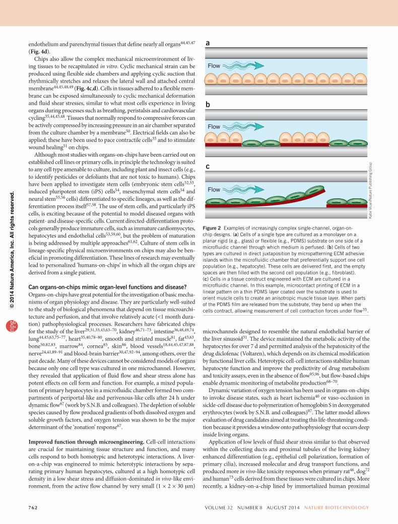
Multiple on-chip models of angiogenesis and microvascular function have been created as well. One includes a 3D stroma permeated by func-tional capillary networks that can sprout freely106–108, whereas another model used biodegradable chips composed entirely of ECM that con-tained internal networks of microchannels filled with sacrificial mate-rial, which was dissolved before plating cells either inside the channels (endothelial cells) or in the surrounding perfused ECM (e.g., tumor cells, fibroblasts)37,38 (Fig. 3a). This latter model is interesting in that newly formed microvessels become perfused once they functionally integrate into the existing vascular network. Such angiogenesis models enable high-resolution analysis of how spatial diffusive gradients influence angiogenic sprouting, and they have shed light on the mechanism of action of angio-genesis inhibitors37,38,106–108. A chip with a central, endothelium-lined channel separated by an ECM gel from a side channel containing chemoat-tractants was used to study transendothelial migration of neutrophils in response to chemical gradients39 (Fig. 3b). A more recent development is the fabrication of multiplexed arrays of nearly identical ECM gels contain-ing well-formed human microvascular networks on a single chip, which may be useful to study the effects of multiple perturbations on angiogenic responses109.
Modeling organ-level physiology and disease. Although culture of a single cell type can mimic some facets of the tissue microenvironment on chips, it is not usually sufficient to generate organ-like functionality. An organ is a hierarchical structure composed of two or more different
Figure 3 Microfluidic chip models of angiogenesis and immune cell invasion that incorporate ECM gels. (a) Sacrificial materials are deposited in linear patterns in an ECM gel and later removed to create two channels (left). One is populated with vascular endothelial (EC) cells38, and the other is used to deliver angiogenic factors. Angiogenic stimuli induce the endothelial cells to undergo sprouting angiogenesis and then functionally link to the source channel, forming new microvessels that support fluid flow (right). Red indicates microparticles flowing in the medium. Fluorescence image reprinted from ref. 38 with permission. (b) A gradient-generating microfluidic culture device for analyzing immune cell migration through ECM gels when stimulated with a chemotactic gradient39.
Chemoattractantgradient
a b
Beadpath
Factor source channel
Parent channel
Cross-section
Collagen 1
Cover glass
Angiogenicfactors
ECchannel
400 μm
1 mm 3 m
m
Side channels
ECM
ECM
Growthmedium
Neutrophils
PERSPECT IVEnp
g©
2014
Nat
ure
Am
eric
a, In
c. A
ll rig
hts
rese
rved
.
764 volume 32 number 8 august 2014 nature biotechnology
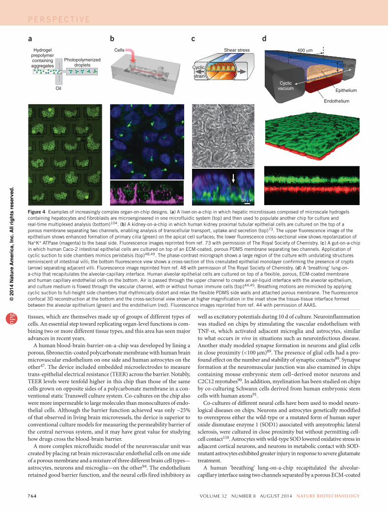
tissues, which are themselves made up of groups of different types of cells. An essential step toward replicating organ-level functions is com-bining two or more different tissue types, and this area has seen major advances in recent years.
A human blood-brain-barrier-on-a-chip was developed by lining a porous, fibronectin-coated polycarbonate membrane with human brain microvascular endothelium on one side and human astrocytes on the other47. The device included embedded microelectrodes to measure trans-epithelial electrical resistance (TEER) across the barrier. Notably, TEER levels were tenfold higher in this chip than those of the same cells grown on opposite sides of a polycarbonate membrane in a con-ventional static Transwell culture system. Co-cultures on the chip also were more impermeable to large molecules than monocultures of endo-thelial cells. Although the barrier function achieved was only ~25% of that observed in living brain microvessels, the device is superior to conventional culture models for measuring the permeability barrier of the central nervous system, and it may have great value for studying how drugs cross the blood-brain barrier.
A more complex microfluidic model of the neurovascular unit was created by placing rat brain microvascular endothelial cells on one side of a porous membrane and a mixture of three different brain cell types—astrocytes, neurons and microglia—on the other94. The endothelium retained good barrier function, and the neural cells fired inhibitory as
well as excitatory potentials during 10 d of culture. Neuroinflammation was studied on chips by stimulating the vascular endothelium with TNF-a, which activated adjacent microglia and astrocytes, similar to what occurs in vivo in situations such as neuroinfectious disease. Another study modeled synapse formation in neurons and glial cells in close proximity (<100 µm)89. The presence of glial cells had a pro-found effect on the number and stability of synaptic contacts89. Synapse formation at the neuromuscular junction was also examined in chips containing mouse embryonic stem cell–derived motor neurons and C2C12 myotubes90. In addition, myelination has been studied on chips by co-culturing Schwann cells derived from human embryonic stem cells with human axons91.
Co-cultures of different neural cells have been used to model neuro-logical diseases on chips. Neurons and astrocytes genetically modified to overexpress either the wild-type or a mutated form of human super oxide dismutase enzyme 1 (SOD1) associated with amyotrophic lateral sclerosis, were cultured in close proximity but without permitting cell-cell contact110. Astrocytes with wild-type SOD lowered oxidative stress in adjacent cortical neurons, and neurons in metabolic contact with SOD-mutant astrocytes exhibited greater injury in response to severe glutamate treatment.
A human ‘breathing’ lung-on-a-chip recapitulated the alveolar- capillary interface using two channels separated by a porous ECM-coated
a b c d
Oil
Hydrogelprepolymercontainingaggregates
Cells Shear stress
Photopolymerizeddroplets
strain
Cyclic
Epithelium
Endothelium
Cyclicvacuum
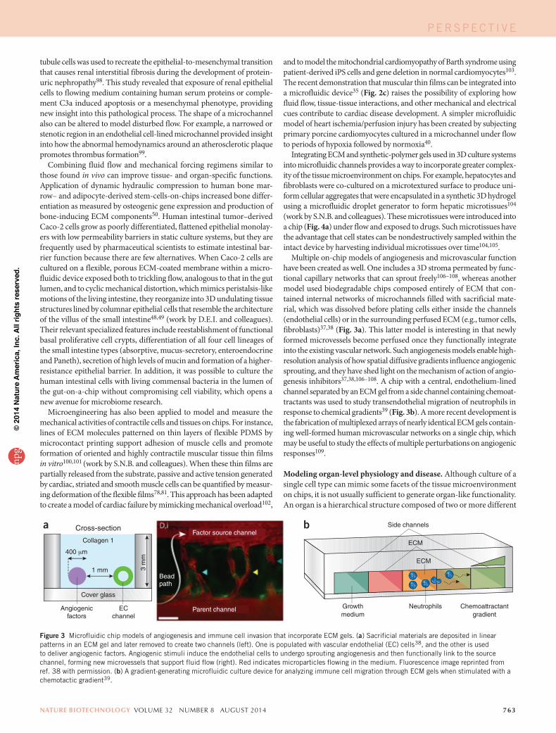
Figure 4 Examples of increasingly complex organ-on-chip designs. (a) A liver-on-a-chip in which hepatic microtissues composed of microscale hydrogels containing hepatocytes and fibroblasts are microengineered in one microfluidic system (top) and then used to populate another chip for culture and real-time multiplexed analysis (bottom)104. (b) A kidney-on-a-chip in which human kidney proximal tubular epithelial cells are cultured on the top of a porous membrane separating two channels, enabling analysis of transcellular transport, uptake and secretion (top)73. The upper fluorescence image of the epithelium shows enhanced formation of primary cilia (green) on the apical cell surfaces; the lower fluorescence cross-sectional view shows repolarization of Na+K+ ATPase (magenta) to the basal side. Fluorescence images reprinted from ref. 73 with permission of The Royal Society of Chemistry. (c) A gut-on-a-chip in which human Caco-2 intestinal epithelial cells are cultured on top of an ECM-coated, porous PDMS membrane separating two channels. Application of cyclic suction to side chambers mimics peristalsis (top)48,49. The phase-contrast micrograph shows a large region of the culture with undulating structures reminiscent of intestinal villi; the bottom fluorescence view shows a cross-section of this crenulated epithelial monolayer confirming the presence of crypts (arrow) separating adjacent villi. Fluorescence image reprinted from ref. 48 with permission of The Royal Society of Chemistry. (d) A ‘breathing’ lung-on-a-chip that recapitulates the alveolar-capillary interface. Human alveolar epithelial cells are cultured on top of a flexible, porous, ECM-coated membrane and human capillary endothelial cells on the bottom. Air is passed through the upper channel to create an air-liquid interface with the alveolar epithelium, and culture medium is flowed through the vascular channel, with or without human immune cells (top)44,45. Breathing motions are mimicked by applying cyclic suction to full-height side chambers that rhythmically distort and relax the flexible PDMS side walls and attached porous membrane. The fluorescence confocal 3D reconstruction at the bottom and the cross-sectional view shown at higher magnification in the inset show the tissue-tissue interface formed between the alveolar epithelium (green) and the endothelium (red). Fluorescence images reprinted from ref. 44 with permission of AAAS.
PERSPECT IVEnp
g©
2014
Nat
ure
Am
eric
a, In
c. A
ll rig
hts
rese
rved
.
nature biotechnology volume 32 number 8 august 2014 765
membrane44 (work by D.E.I. and colleagues). In the lower microvascular channel, the membrane was lined with lung microvascular endothelial cells; in the upper, air-filled channel, it was lined by human lung alveolar epithelial cells. The two channels were bordered by full-height hollow chambers through which cyclic suction was applied to rhythmically distort the flexible PDMS side walls and the linked central porous membrane with attached cells in order to mimic the cyclic mechanical strain (10%; 0.25 Hz) that cells experience in the alveolus from breathing motions (Fig. 4d). The combination of fluid flow in the vascular channel, genera-tion of an air-liquid interface in the alveolar channel and application of cyclic mechanical strain strongly promoted differentiation of the epithe-lial and endothelial cells lining the channels, as indicated by enhanced surfactant production and vascular barrier function (measured by both TEER and assays of macromolecular transport). When primary human neutrophils were flowed through the vascular channel under baseline flow conditions, the endothelium was quiescent. But addition of either the immune activator TNF-a or bacterial cells to the alveolar channel to mimic inflammation rapidly activated the endothelium, as measured by increased expression of surface ICAM-1 and recruitment of human neu-trophils perfused through the vascular channel. The recruited immune cells underwent diapedesis and migrated through both cell layers to the upper chamber, where they engulfed the living bacteria. Because the cells were precisely positioned in the optically clear chip, these analyses could be carried out in real-time using high-resolution optical, fluorescence and confocal microscopy as well as microfluorimetry.
Because the lung-on-a chip mimicked complex organ-level func-tions, it enabled new mechanistic insights into the role of mechani-cal breathing motions in lung disease. When silica nanoparticle simulants of environmental air-borne particulates were introduced into the air channel, production of reactive oxygen species, cellular uptake of nanoparticles, and transport of nanoparticles across both cell layers and into the vascular channel were many-fold higher if the cells were experiencing cyclic breathing motions. This dependence of nanoparticle transport on breathing movements was confirmed in a mouse ex vivo ventilation-perfusion model44. The lung-on-a-chip was also used to model pulmonary edema (‘fluid on the lungs’) by perfusing through the vascular channel the cancer drug interleukin-2 (IL-2), which induces pulmonary vascular permeability and lung edema as its major dose-limiting side effect (D.E.I. and colleagues, ref. 45). When IL-2 was added at the same dose used in patients, pathological shifts of fluid into the air channel (Fig. 4d), forma-tion of blood clots and associated compromise of oxygen transport occurred over the same 2- to 4-d time course as observed when the drug is administered in patients. The lung-on-a-chip also revealed that the mechanical forces of breathing motions contribute to the development of increased vascular leakage and pulmonary edema induced by IL-2, and that circulating immune cells are not required for the development of this life-threatening condition45. These results highlight the value of being able to vary cell types, chemical stimuli and mechanical perturbations independently in organs-on-chips.
Cancer-on-a-chip. Many groups have developed cancer-on-a-chip models. In a study of breast cancer progression, co-culture of human mammary epithelial cells with human mammary fibroblasts pro-moted a transition from ductal carcinoma in situ to invasive ductal carcinoma only when direct contact between the cells was permit-ted111. Effects of specific types of ECM on tumor-cell morphology and growth were detected when human breast carcinoma cells were co-cultured with stromal cells and various combinations of ECM com-ponents112. Taking advantage of the ability to install many different microenvironments on the same chip, another group discovered a correlation between the growth of breast cancer cells and ERa protein
downregulation113. A similar but higher-throughput approach was used to fabricate tunable cell microniches and to carry out flow-based analysis of large cell populations to measure differential responses of lung adenocarcinoma cells to various ECM molecules and soluble factors. This work led to the finding that tumor-cell growth is sensi-tive to TGF-b and TGFbR2 inhibitor drugs in 3D microenviron-ments but not in monolayer culture42. Use of a microfluidic vascular endothelium model that permitted site-specific stimulation with the chemokine CXCL12 from the basal side of the tissue layer under variable flow conditions revealed that CXCL12 acts through CXCR4 receptors on the endothelium, rather than on tumor cells, to promote adhesion of circulating breast cancer cells114. Tumor-cell invasion also has been studied by measuring invadopodia formation and ECM degradation by human non-small cell lung cancer cells growing in an ECM gel–filled microfluidic channel115. Both intravasation116 and extravasation107,117 of tumor cells have been analyzed in chip models of angiogenesis as well.
Taken together, the research described above provides strong evi-dence that organs-on-chips are capable of reproducing human organ physiology and organ-level features of disease at both the popula-tion and individual levels. Because they allow different features of a cell culture, such as relative cell and tissue position, fluid flow and mechanical cues, to be controlled independently, they also provide unprecedented flexibility in dissecting the cellular, molecular, chemi-cal and physical contributors to tissue and organ function, as well as disease development.
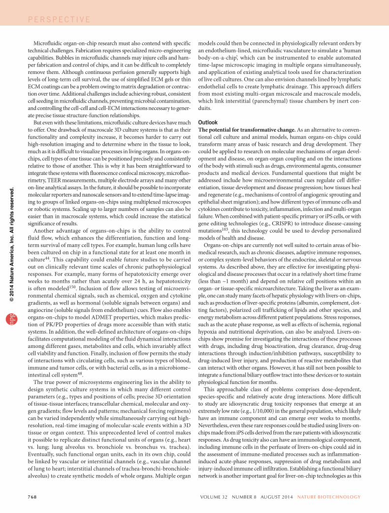
How might organs-on-chips be used for drug development?ADMET testing and PK/PD modeling. The first major step in adapting microphysiological systems for drug development and toxi-cology came in 2004, when the Shuler group created a microfluidic cell-culture analog of a mathematical pharmacokinetics (PK) model that represents the organs of the body as interconnected compart-ments43,63. The purpose of this microfluidic analog was to study the adsorption, distribution, metabolism, elimination and toxicity (ADMET) of chemicals entirely in vitro rather than in animals, as is usually done in the pharmaceutical industry43,63. The same group has since created many types of microscale devices43,63,74,118,119 that contain multiple culture chambers, each holding a single cell type representing a different organ (e.g., lung, liver, fat, marrow, tumor), and which are linked by microfluidic channels (Fig. 5a) in an order that mimics physiological coupling in vivo (Fig. 5b). When the toxi-cant naphthalene was introduced into one of these devices, it con-verted in the liver chamber into its reactive metabolites, which then circulated to the lung tissue chamber where they depleted cellular glutathione levels43,63. Adipocytes in the fat chamber moderated the glutathione depletion induced by naphthalene and preferentially accumulated hydrophobic compounds43,63. Thus, this simple chip mimicked basic organ-organ coupling and permitted analysis of drug ADMET characteristics that are usually determined in vivo. These early studies were limited by the use of cultured cell lines that were from different species and were not optimally differentiated. Another potential problem is that the interstitial fluids were flowed directly from one cell compartment to the other, which does not occur in distant living organs coupled by the vascular system. Nevertheless, these reports first raised the intriguing possibility of developing a ‘human-body-on-a-chip’—a microfluidic device containing several fluidically linked chambers representing different organs—that could be used to test drugs, chemicals or toxins.
Over the past ten years, many groups have explored the use of organs-on-chips to study drug ADMET properties, to support PK
PERSPECT IVEnp
g©
2014
Nat
ure
Am
eric
a, In
c. A
ll rig
hts
rese
rved
.
766 volume 32 number 8 august 2014 nature biotechnology
and pharmacodynamics (PD) modeling, and to measure drug efficacy. For exam-ple, livers-on-chips are useful for analyz-ing drug metabolism by liver tissue. In one study, HepG2/C3A cells exposed to the anticancer prodrug flutamide or to its active metabolite (hydroxyflutamide) were analyzed on a chip using nuclear magnetic resonance (NMR) spectroscopy to iden-tify response markers120. The prodrug and the active compound produced distinct metabolic signatures and hepatotoxicities, which correlated with effects on glutathi-one metabolism. NMR-based metabolomic footprinting was also used with chips as a high-throughput approach to determine the toxicities of several small molecules, includ-ing the environmental pollutant ammonia, the free radical-scavenging solvent dimethyl-sulfoxide and the hepatotoxic analgesic drug N-acetyl-para-aminophenol (acetaminophen or paracetamol)121. This effort identified sig-natures for ammonia and acetaminophen and dose-dependent metabolic responses to ammonia in chambers lined by liver (HepG2/C3A) cells, kidney cells and co-cultures of both cell types. However, another study on a liver-kidney chip found that HepRG cells derived from a human liver progenitor cell line metabolized ifosfamide into its toxic metabolite chloroacetaldehyde, whereas HepaG2/C3A cells did not122, which emphasizes the importance of cell selection123,124.
Some ADMET properties of acetaminophen were also demon-strated by coupling a microchamber lined by a monolayer of liver (HepG2/C3A) cells to a macroscale (5.6 mm high including top and bottom portions) chamber containing a mixture of intestinal cells74. Administration of acetaminophen resulted in glutathione depletion in the intestinal cells and further metabolic processing by the liver cells, causing dose-dependent hepatotoxicity. The experiment was designed so that the drug residence time in the liver chamber was similar to that measured in human liver (~1–2 min). These results were consistent with in vivo measurements.
Although tumor-derived hepatic cell lines were used to model liver metabolism and function in the multi-organ chips described above, more recent work has demonstrated that primary human hepatocytes are superior for predicting drug metabolism, clearance, drug-drug interactions and toxicity. Normal human hepatocytes cultured on chips maintained the functionality of multiple clinically relevant liver cytochromes P450 (CYP1A2, CYP3A4, CYP2C8, CYP2C19 and CYP2D6)125 and enabled measurement of hepatic clearance of six marketed drug compounds64. In the latter study, application of flow increased the duration of metabolic competency compared with static culture, consistent with other microfluidic studies using primary hepatocytes126, but hepatocytes under flow could not distin-guish high- and medium-clearance compounds, and neither model detected low-clearance compounds. Clearance of drug compounds with high, medium and low clearance values can be improved by cul-turing primary hepatocytes with nonparenchymal cells under flow70.
Unfortunately, despite these advances, livers-on-chips still cannot cap-ture certain key hepatic functions, such as directional biliary ductal clear-ance or sustained production of metabolic enzymes and blood proteins for the typical hepatocyte life span of approximately one year in vivo. In fact, in some cases, the presence of flow may actually be detrimental to
predictivity. For example, the US Food and Drug Administration (FDA) guidance on Metabolites in Safety Testing notes the importance of detec-tion of certain human metabolites that so far has relied on metabolite accumulation in small static environments owing to the sensitivity limita-tions of analytical methods127.
In certain chip designs, recirculating flow can effectively make them ‘well stirred’ models. This enables scaling of obtained clearance data to estimate human hepatic clearance without mathematical modeling, which is often required in conventional hepatocyte cultures or suspensions. For example, when hepatic clearance rates measured under flow on a chip or under static conditions were compared with in vivo data from the litera-ture, both formats generated predictions on the same scale as the reported values64. But the chip predictions did not require mathematical modeling, at least for this limited set of compounds. In this study, and in another one using human hepatocytes that found consistent results128, nonspecific adsorption of drugs into walls and tubing complicated the calculation of clearance rates. This remains a major problem for chips made of PDMS.
Microfluidic coupling has also been shown to be relevant for analysis of cancer drug metabolism and toxicity. For example, a study involving linked chambers containing liver (HepG2/C3A), bone marrow (MEG-01), uterine cancer (MES-SA) and a multidrug-resistant (MDR) variant of uterine cancer (MES-SA/DX-5) cells found that combining the chemo-therapeutic drug doxorubicin (Adriamycin, Doxil) with MDR modulators (cyclosporine and nicardipine) produced greater antiproliferative effects on the MDR cancer cell variant than any of these compounds alone; and, this result could not be obtained using a conventional static culture118. In another study, HCT-116 colon cancer cells (HCT-116) and hepatoma cells (HepG2/C3A) were encapsulated in an ECM gel (Matrigel) and cul-tured in linked tumor and liver chambers on-chips119. This microfluidic system was able to reproduce the metabolism of an oral prodrug of the anticancer drug, 5-fluorouracil (Carac, Efudex, Fluoroplex), to its active product in the liver chamber, and result in killing of the tumor cells when the metabolite flowed to the other chamber, whereas, again, cultures of similar cells in a 96-well microtiter plate did not.
Chips are also well suited to the study of drug transport and toxicity. Data from a blood-brain-barrier-on-a-chip analyzing the
LiverTumor Marrow
Liver
Tumor
MarrowReservoir
(other tissues)
Top frame
Channel layer
Base
Bottom frame
Cell culturechamber layer
Siliconegasket
Cell
Organ
kg
DeathKn
K1 KnK
ba
Figure 5 A multi-organ microfluidic framework used for PK/PD modeling. (a) Schematic diagram (top) and photograph (bottom) of a three-chamber chip used for PK modeling by flowing medium through liver, tumor and marrow cells cultured as monolayers in separate chambers and linked fluidically. (b) A flow diagram of the connections between the liver, tumor and marrow compartments in the chip shown in a (top), and a pharmacodynamics model for cell death in each compartment (bottom). Reprinted from ref. 156 with permission of The Royal Society of Chemistry.
PERSPECT IVEnp
g©
2014
Nat
ure
Am
eric
a, In
c. A
ll rig
hts
rese
rved
.

nature biotechnology volume 32 number 8 august 2014 767
permeability of a set of hydrophobic and hydrophilic drugs corre-lated well with corresponding in vivo values92. As discussed above, the human lung-on-a-chip permitted analysis of silica nanoparticle transport across the alveolar-capillary interface and detection of the increase of vascular permeability induced as a toxic side effect by the human anticancer drug IL-2, in addition to revealing the key role of physiological breathing motions in these responses44,45. Neither of these results had been reported previously, and it would be difficult to generate them using standard cell cultures, macroscale bioreactors or animal models.
A major problem with preclinical animal studies is that drug toxicities are difficult or impossible to identify if there are meaningful differences in drug uptake between the animal model and humans. In such cases, human cells or tissues are needed. For instance, the anticancer drug cis-platin (Platinol) has different toxicities in animals and humans owing to species differences in membrane transporters, such as OCT2, which medi-ate cellular accumulation of this drug and production of reactive oxygen species. A human kidney-proximal-tubule-on-a-chip measured OCT2-specific cisplatin toxicities that were undetectable in a static culture (work of D.E.I., ref. 73). The chip also measured the activity of P-glycoprotein ATP-binding cassette membrane (Pgp) transporters that mediate the efflux of certain cancer chemotherapeutic drugs in humans and thereby produce multidrug resistance. This system may provide a valuable in vitro screen for drug development given that new draft guidelines of the FDA require determination of whether a drug candidate is a substrate or inhibi-tor of Pgp transporters as drug interactions with Pgp can increase the toxicity of co-administered agents.
Efficacy testing and drug discovery. Organs-on-chips are beginning to be applied to assess drug efficacy as well. For example, a heart-on-a-chip, containing almost 20 rat cardiomyocyte thin films, stimulated electrically to contract, was used to test the inotropic effects of the beta-adrenergic agonist isoproterenol35. The results were similar to those previously deter-mined in rats. The chip also allowed multiplexed, high-resolution imaging of cell structure and function, as well as quantification of the effects of the drug on diastolic, systolic and twitch stress. In a related study, responses of muscular thin films constructed from cardiomyocytes or vascular smooth muscle cells to endothelin-1 and rho-associated kinase (ROCK) inhibitor were analyzed simultaneously on the same chip81. A high dose of endo-thelin-1 increased vascular smooth muscle contractility while suppressing cardiomyocyte contraction, and the ROCK inhibitor decreased tension in both types of muscular thin films. These findings show that the effects of drugs on different tissue types can be analyzed simultaneously on the same chip, which could improve assay throughput relative to animal studies or conventional cell cultures.
The effects of the clinical surfactant drug beractant (Survanta) were studied in a microfluidic pulmonary airway model that enables analysis of damage to the epithelium during reopening of airways occluded by liquid plugs (analogous to mucus plugs)77. These studies revealed that substantial cellular injury occurs owing to high mechanical stresses caused by plug propagation devoid of surfactant, whereas addition of a physiologic con-centration of Survanta protected the epithelium and significantly reduced cell death. Computational simulations also revealed a significant decrease in mechanical forces in the presence of surfactant, confirming the experi-mental observations.
Recent studies also raise the possibility of using organs-on-chips to discover new therapeutics or new uses for existing ones. When a cur-rent drug candidate that inhibits TRPV4 ion channels and suppresses mechanosensing in endothelial cells129 was tested in the lung-on-a-chip, it completely inhibited IL-2–induced human pulmonary edema45, and confirmatory results were obtained in a cardiogenic pulmonary edema
model in rodents and dogs130. The ability to culture a living microbiome in the human gut-on-a-chip revealed that co-culture of probiotic bacteria (lactobacillus GG) with human intestinal epithelial cells increases intestinal barrier integrity48, suggesting that this chip might be used to test the effi-cacy of other probiotic therapies. A recently developed bone-marrow-on-a-chip that permits culture of fully formed living marrow with a functional hematopoietic niche within a chip was used to model both organ-level marrow toxicity responses to radiation and the protective effects of radia-tion countermeasure drugs, whereas similar effects could not be measured with conventional bone marrow culture methods84 (work by D.E.I. and colleagues). Screening for drugs to protect against exposure to lethal radia-tion is an excellent example of how organs-on-chips can enable studies examining human responses that would otherwise be impossible to carry out in human patients due to ethical implications.
Cancer-on-a-chip devices also show promise for drug screening. Functional interrogation of hundreds of single chronic myeloid leu-kemia cells or normal hematopoietic stem cells allowed detection of tumor-specific cellular responses to the tyrosine kinase inhibitor dasat-inib (Sprycel), approved for the treatment of this cancer131. In another study, the responses of a lung cancer cell line, a mixture of lung cancer and stromal cell lines, and cells from fresh lung cancer tissues were com-pared when cultured in 3D gels and exposed to different concentrations of chemotherapeutic agents generated on-chip by a concentration-gradient generator132. The investigators assayed the sensitivities of different anti-cancer drugs in parallel and screened appropriate-dose, single-drug and combined-drug chemotherapy schemes for eight patients. Another exam-ple of work with potential relevance to personalized medicine involved use of a marrow-like environment containing human osteoblasts to culture mononuclear cells from clinical bone marrow aspirates as a way to expand patient-specific multiple myeloma cells that could be used to screen for individualized therapies83.
What are the advantages and disadvantages of organs-on-chips?Conventional 3D culture systems—including hydrogel-based methods, tissue-engineered constructs, static co-cultures, and bioreactors—have proved very useful for studying certain tissue- and organ-level behaviors and for developing disease models. Examples include mammary4, intes-tinal5 and brain6 organoids; co-cultures of hepatocytes with fibroblasts or endothelial cells in Petri dishes or bioreactors68,69,96,133–135; micro–cardiac muscles for measuring response to contractile modifiers78,136 and model-ing heart failure and cardiomyopathy102,103; atrophic skeletal muscle137 and mechanosensory circuits containing innervated muscle fibers138; and models of liver infection by hepatitis C virus139 and Plasmodium falciparum and Plasmodium vivax140. Macroscale culture chambers or bioreactors have been connected fluidically, either directly or through endothelium-lined channels to link multiple engineered mini-organs (e.g., liver, skin, hair, brain, bone marrow, lymphatic) for drug testing68,69,141–145, much like in microfluidic systems. These systems generate more tissue mass compared with microfluidic chips, which is advantageous when using analytical approaches (e.g., mass spectroscopy) that require larger experimental samples. It is also easier to retrieve larger numbers of cells for analysis from larger systems. Some organ functions—such as cognition in the brain and mechanical function in bone, ligaments and tendons—arise out of macroscale architecture and cannot be readily modeled on chips, although parts or sections of organs (e.g., artery146, pancreatic islets147, liver, intestine and brain148,149) have been cultured on chips to study cer-tain organ-level functions and responses to chemical or electrical signals. It is also difficult to reproduce on a microscale the spatial heterogeneity found in larger 3D organoids or tissue sections (e.g., in the lung, the spatial transition in epithelial structure from tracheal to bronchial to respiratory to alveolar).
PERSPECT IVEnp
g©
2014
Nat
ure
Am
eric
a, In
c. A
ll rig
hts
rese
rved
.
768 volume 32 number 8 august 2014 nature biotechnology
Microfluidic organ-on-chip research must also contend with specific technical challenges. Fabrication requires specialized micro-engineering capabilities. Bubbles in microfluidic channels may injure cells and ham-per fabrication and control of chips, and it can be difficult to completely remove them. Although continuous perfusion generally supports high levels of long-term cell survival, the use of simplified ECM gels or thin ECM coatings can be a problem owing to matrix degradation or contrac-tion over time. Additional challenges include achieving robust, consistent cell seeding in microfluidic channels, preventing microbial contamination, and controlling the cell-cell and cell-ECM interactions necessary to gener-ate precise tissue structure-function relationships.
But even with these limitations, microfluidic culture devices have much to offer. One drawback of macroscale 3D culture systems is that as their functionality and complexity increase, it becomes harder to carry out high-resolution imaging and to determine where in the tissue to look, much as it is difficult to visualize processes in living organs. In organs-on-chips, cell types of one tissue can be positioned precisely and consistently relative to those of another. This is why it has been straightforward to integrate these systems with fluorescence confocal microscopy, microfluo-rimetry, TEER measurements, multiple electrode arrays and many other on-line analytical assays. In the future, it should be possible to incorporate molecular reporters and nanoscale sensors and to extend time-lapse imag-ing to groups of linked organs-on-chips using multiplexed microscopes or robotic systems. Scaling up to larger numbers of samples can also be easier than in macroscale systems, which could increase the statistical significance of results.
Another advantage of organs-on-chips is the ability to control fluid flow, which enhances the differentiation, function and long-term survival of many cell types. For example, human lung cells have been cultured on chip in a functional state for at least one month in culture44. This capability could enable future studies to be carried out on clinically relevant time scales of chronic pathophysiological responses. For example, many forms of hepatotoxicity emerge over weeks to months rather than acutely over 24 h, as hepatotoxicity is often modeled150. Inclusion of flow allows testing of microenvi-ronmental chemical signals, such as chemical, oxygen and cytokine gradients, as well as hormonal (soluble signals between organs) and angiocrine (soluble signals from endothelium) cues. Flow also enables organs-on-chips to model ADMET properties, which makes predic-tion of PK/PD properties of drugs more accessible than with static systems. In addition, the well-defined architecture of organs-on-chips facilitates computational modeling of the fluid dynamical interactions among different gases, metabolites and cells, which invariably affect cell viability and function. Finally, inclusion of flow permits the study of interactions with circulating cells, such as various types of blood, immune and tumor cells, or with bacterial cells, as in a microbiome–intestinal cell system48.
The true power of microsystems engineering lies in the ability to design synthetic culture systems in which many different control parameters (e.g., types and positions of cells; precise 3D orientation of tissue-tissue interfaces; transcellular chemical, molecular and oxy-gen gradients; flow levels and patterns; mechanical forcing regimens) can be varied independently while simultaneously carrying out high- resolution, real-time imaging of molecular-scale events within a 3D tissue or organ context. This unprecedented level of control makes it possible to replicate distinct functional units of organs (e.g., heart vs. lung; lung alveolus vs. bronchiole vs. bronchus vs. trachea). Eventually, such functional organ units, each in its own chip, could be linked by vascular or interstitial channels (e.g., vascular channel of lung to heart; interstitial channels of trachea-bronchi-bronchiole-alveolus) to create synthetic models of whole organs. Multiple organ
models could then be connected in physiologically relevant orders by an endothelium-lined, microfluidic vasculature to simulate a ‘human body-on-a-chip’, which can be instrumented to enable automated time-lapse microscopic imaging in multiple organs simultaneously, and application of existing analytical tools used for characterization of live cell cultures. One can also envision channels lined by lymphatic endothelial cells to create lymphatic drainage. This approach differs from most existing multi-organ microscale and macroscale models, which link interstitial (parenchymal) tissue chambers by inert con-duits.
OutlookThe potential for transformative change. As an alternative to conven-tional cell culture and animal models, human organs-on-chips could transform many areas of basic research and drug development. They could be applied to research on molecular mechanisms of organ devel-opment and disease, on organ-organ coupling and on the interactions of the body with stimuli such as drugs, environmental agents, consumer products and medical devices. Fundamental questions that might be addressed include how microenvironmental cues regulate cell differ-entiation, tissue development and disease progression; how tissues heal and regenerate (e.g., mechanisms of control of angiogenic sprouting and epithelial sheet migration); and how different types of immune cells and cytokines contribute to toxicity, inflammation, infection and multi-organ failure. When combined with patient-specific primary or iPS cells, or with gene editing technologies (e.g., CRISPR) to introduce disease-causing mutations103, this technology could be used to develop personalized models of health and disease.
Organs-on-chips are currently not well suited to certain areas of bio-medical research, such as chronic diseases, adaptive immune responses, or complex system-level behaviors of the endocrine, skeletal or nervous systems. As described above, they are effective for investigating physi-ological and disease processes that occur in a relatively short time frame (less than ~1 month) and depend on relative cell positions within an organ- or tissue-specific microarchitecture. Taking the liver as an exam-ple, one can study many facets of hepatic physiology with livers-on-chips, such as production of liver-specific proteins (albumin, complement, clot-ting factors), polarized cell trafficking of lipids and other species, and energy metabolism across different patient populations. Stress responses, such as the acute phase response, as well as effects of ischemia, regional hypoxia and nutritional deprivation, can also be analyzed. Livers-on-chips show promise for investigating the interactions of these processes with drugs, including drug bioactivation, drug clearance, drug-drug interactions through induction/inhibition pathways, susceptibility to drug-induced liver injury, and production of reactive metabolites that can interact with other organs. However, it has still not been possible to integrate a functional biliary outflow tract into these devices or to sustain physiological function for months.
This approachable class of problems comprises dose-dependent, species-specific and relatively acute drug interactions. More difficult to study are idiosyncratic drug toxicity responses that emerge at an extremely low rate (e.g., 1/10,000) in the general population, which likely have an immune component and can emerge over weeks to months. Nevertheless, even these rare responses could be studied using livers-on-chips made from iPS cells derived from the rare patients with idiosyncratic responses. As drug toxicity also can have an immunological component, including immune cells in the perfusate of livers-on-chips could aid in the assessment of immune-mediated processes such as inflammation-induced acute-phase responses, suppression of drug metabolism and injury-induced immune cell infiltration. Establishing a functional biliary network is another important goal for liver-on-chip technologies as this
PERSPECT IVEnp
g©
2014
Nat
ure
Am
eric
a, In
c. A
ll rig
hts
rese
rved
.
nature biotechnology volume 32 number 8 august 2014 769
will afford the study of drug disposition across the basolateral cell surface to the apical domain—so-called phase III transport. Many diseases of the liver are chronic in nature, driven by stellate-cell activation and collagen remodeling that evolve over years. A subset of patients with fibrosis then progress to cirrhosis, a risk factor for hepatocellular carcinoma. Long-term processes such as these are less amenable to study in any in vitro format, although some of their features could be replicated on chips. For example, modeling the interactions of hepatocytes and peripheral adipose tissue in nonalcoholic fatty liver disease may offer valuable insights even if multiyear observation is not possible.
Directions for future research. There are many challenges that must be overcome before organs-on-chips will find widespread use in research laboratories. A basic problem that must be remedied is the material to use for fabrication. Most chips are made out of PDMS because it is easy to use and has high optical clarity, gas permeability and biocompatibility. But PDMS can absorb small organic compounds, including many drugs, and its high gas permeability can hinder some applications. Recently, other polymers, such as certain polyurethanes, were found that provide the benefits of PDMS but do not soak up small hydrophobic drugs151. However, more research is required to identify suitable materials that can be used to mass produce organs-on-chips at low cost. Another materials problem is that the ECM-coated PDMS membranes that serve as tissue-tissue interfaces may have transport, mechanical and structural proper-ties different from those of natural basement membrane. Given issues such as these, it will be many years before the community can consider developing generic production specifications for organs-on-chips.
Another major challenge is technical robustness. Various factors must align to achieve optimal function of organs-on-chips over a month or longer, including the cells, the ECM coatings, fluidic control, bubble removal and gradient maintenance. For systems consisting of multiple, linked organ chips, there is a need for a ‘universal blood substitute’—a single culture medium that supports all tissues, just as blood supplies all organs in the body. Existing culture media have been optimized for each cell type, so this remains a significant hurdle, especially if some tissues are grown in serum-containing medium and others in serum-free medium. In a study examining whether addition of growth factors, such as TGF-b, might enhance tissue functionality in a multiorgan chip containing chambers lined by liver, lung, kidney and adipose cells, the investigators discovered that this supplement enhanced the function of one cell type but inhibited the function of another152. They overcame this limitation by mixing controlled- release, gelatin microspheres containing TGF-b with the responsive cell type to create a local cell-specific microenviron-ment within the multiorgan system. Another possible solution is to flow either serum-containing medium, plasma or whole blood through micro-channels lined by a vascular endothelial barrier that regulates delivery of molecular factors to adjacent interstitial channels, as occurs in vivo.
The presence of flow in microfluidic systems has already made it pos-sible to develop PK models by combining data on clearance dynam-ics from single or coupled organs-on-chips with mathematical scaling approaches43,63,125,153. But there is a need for improved scaling approaches that more accurately match fluid flows, cultured tissue mass and volumes of distribution so as to ensure appropriate, relative, organ functional activity and to provide meaningful data for computational PK/PD models154,155. This goal would be more tractable with data from ‘next-generation’ chips that might include in-line sensors of critical control and functional param-eters (e.g., flow, pressure, temperature, pH, oxygen, glucose, lactate, TEER, electrical conduction) and integrated microscopic and microfluorimet-ric imaging capabilities to monitor overall system performance in real-time154,155. But broad acceptance of organs-on-chips in basic research and drug development will likely require automated instrumentation that
provides feedback control, continuous regulation, programmable moni-toring, and experimental sample collection and processing. Currently, laboratories that wish to work with organs-on-chips require significant in-house engineering capabilities and expertise. Automated control systems would allow nonspecialist academic researchers to replace existing 2D or 3D culture systems with microfluidic systems that provide similar levels of molecular analysis but in a functional-organ context. For example, once samples are collected from vascular or interstitial microfluidic channels, they can be analyzed using virtually any current method, such as histocy-tochemistry, western blots, PCR, gene microarrays and mass spectroscopy.
The greatest value of organs-on-chips for the pharmaceutical and bio-technology industries may lie in the validation and prioritization of lead drug candidates (rather than in high-throughput screening) and in the study of molecular mechanisms of action and toxicities. Sophisticated human organs-on-chips may also help to identify new biomarkers of drug efficacy, toxicity or disease response, which could be of value for clinical trials. If clinically relevant PK/PD modeling approaches can be developed, it may be possible to use this technology to determine drug dosing and safety margins for clinical trials as well. As drug companies will surely continue to rely on preclinical animal studies for many years to come, it will also be important to develop organs-on-chips lined with cells from the animal species now used for drug development or with human cells to study species differences and to refine in vitro–in vivo correlations and predictions.
Although there have been a few recent successes demonstrating that organs-on-chips can mimic specific organ-level functions, the field is still in its infancy. If the technology can be improved to the point that it effectively recapitulates a broad range of organ responses to chemicals, drugs and toxins, many new avenues for drug discovery, toxicology and personalized medicine will open up. These include testing of dangerous agents (e.g., highly infectious viruses, biothreat agents, chemical warfare agents and lethal doses of g-radiation) and ‘virtual’ pediatric clinical tri-als. In addition, organs-on-chips generated from iPS cells isolated from different genetic subpopulations, disease subgroups or individual patients might facilitate drug discovery targeted to specific subpopulations or clini-cal trial design.
Given the complexities of organ function and regulatory requirements, it is unlikely that organs-on-chips will replace animal testing anytime soon. However, as individual organs-on-chips are improved, it may be possible to progressively replace one animal-based assay at a time. Researchers in the field must recognize that there are major hurdles to overcome before this technology will have widespread acceptance and impact. But we can-not ignore its amazing potential, and we hope that others will share our excitement and jump into the fray.ACKNOWLEDGMENTSWe thank H. Fleming, S. Khetani, D. Levner, G. Hamilton and T. Bahinski for their helpful input. This work was supported by grants from the Defense Advanced Research Projects Agency (DARPA) (W911NF-12-2-0036), the FDA (HHSF223201310079C), the US National Institutes of Health (UH3 EB017103, R01 DK85713, R01 EB008396), Bill and Melinda Gates Foundation (OPP1023607, OPP1086223) and the Wyss Institute for Biologically Inspired Engineering at Harvard University. S.N.B. is an investigator of the Howard Hughes Medical Institute; D.E.I. is a recipient of a US Department of Defense (DoD) Breast Cancer Innovator Award (BC074986).
COMPETING FINANCIAL INTERESTSThe authors declare competing financial interests: details are available in the online version of the paper.
Reprints and permissions information is available online at http://www.nature.com/reprints/index.html.
1. Harrison, R.G. The outgrowth of the nerve fiber as a mode of protoplasmic movement. J. Exp. Zool. 9, 787–846 (1910).
2. Greek, R. & Menache, A. Systematic reviews of animal models: methodology versus epistemology. Int. J. Med. Sci. 10, 206–221 (2013).
PERSPECT IVEnp
g©
2014
Nat
ure
Am
eric
a, In
c. A
ll rig
hts
rese
rved
.
770 volume 32 number 8 august 2014 nature biotechnology
3. Ehrmann, R.L. & Gey, G.O. The growth of cells on a transparent gel of reconstituted rat-tail collagen. J. Natl. Cancer Inst. 16, 1375–1403 (1956).
4. Mroue, R. & Bissell, M.J. Three-dimensional cultures of mouse mammary epithelial cells. Methods Mol. Biol. 945, 221–250 (2013).
5. Sato, T. & Clevers, H. Growing self-organizing mini-guts from a single intestinal stem cell: mechanisms and applications. Science 340, 1190–1194 (2013).
6. Lancaster, M.A. et al. Cerebral organoids model human brain development and micro-cephaly. Nature 501, 373–379 (2013).
7. Muranen, T. et al. Inhibition of PI3K/mTOR leads to adaptive resistance in matrix-attached cancer cells. Cancer Cell 21, 227–239 (2012).
8. Mammoto, T., Mammoto, A. & Ingber, D.E. Mechanobiology and developmental control. Annu. Rev. Cell Dev. Biol. 29, 27–61 (2013).
9. Ingber, D.E. Mechanobiology and diseases of mechanotransduction. Ann. Med. 35, 564–577 (2003).
10. Duffy, D.C., McDonald, J.C., Schueller, O.J. & Whitesides, G.M. Rapid prototyping of microfluidic systems in poly(dimethylsiloxane). Anal. Chem. 70, 4974–4984 (1998).
11. Singhvi, R. et al. Engineering cell shape and function. Science 264, 696–698 (1994).
12. Chen, C.S., Mrksich, M., Huang, S., Whitesides, G. & Ingber, D.E. Geometric control of cell life and death. Science 276, 1425–1428 (1997).
13. Folch, A. & Toner, M. Cellular micropatterns on biocompatible materials. Biotechnol. Prog. 14, 388–392 (1998).
14. Kane, R.S., Takayama, S., Ostuni, E., Ingber, D.E. & Whitesides, G.M. Patterning proteins and cells using soft lithography. Biomaterials 20, 2363–2376 (1999).
15. Folch, A., Ayon, A., Hurtado, O., Schmidt, M.A. & Toner, M. Molding of deep polydimethylsiloxane microstructures for microfluidics and biological applications. J. Biomech. Eng. 121, 28–34 (1999).
16. Douville, N.J. et al. Fabrication of two-layered channel system with embedded elec-trodes to measure resistance across epithelial and endothelial barriers. Anal. Chem. 82, 2505–2511 (2010).
17. Nguyen, T.A., Yin, T.I., Reyes, D. & Urban, G.A. Microfluidic chip with integrated electrical cell-impedance sensing for monitoring single cancer cell migration in three-dimensional matrixes. Anal. Chem. 85, 11068–11076 (2013).
18. Liu, M.C. et al. Electrofluidic pressure sensor embedded microfluidic device: a study of endothelial cells under hydrostatic pressure and shear stress combinations. Lab Chip 13, 1743–1753 (2013).
19. Eklund, S.E. et al. Metabolic discrimination of select list agents by monitoring cel-lular responses in a multianalyte microphysiometer. Sensors (Basel) 9, 2117–2133 (2009).
20. Takayama, S. et al. Subcellular positioning of small molecules. Nature 411, 1016 (2001).
21. Li Jeon, N. et al. Neutrophil chemotaxis in linear and complex gradients of interleu-kin-8 formed in a microfabricated device. Nat. Biotechnol. 20, 826–830 (2002).
22. Prentice-Mott, H.V. et al. Biased migration of confined neutrophil-like cells in asym-metric hydraulic environments. Proc. Natl. Acad. Sci. USA 110, 21006–21011 (2013).
23. Radisic, M., Deen, W.M., Langer, R. & Vunjak-Novakovic, G. Mathematical model of oxygen distribution in engineered cardiac tissue with parallel channel array perfused with culture medium containing oxygen carriers. Am. J. Physiol.Heart Care Physiol. 288, H1278–H1289 (2005).
24. Xiao, R.R. et al. Simultaneous generation of gradients with gradually changed slope in a microfluidic device for quantifying axon response. Anal. Chem. 85, 7842–7850 (2013).
25. Peng, C.C., Liao, W.H., Chen, Y.H., Wu, C.Y. & Tung, Y.C. A microfluidic cell culture array with various oxygen tensions. Lab Chip 13, 3239–3245 (2013).
26. Cimetta, E. et al. Microfluidic device generating stable concentration gradients for long term cell culture: application to Wnt3a regulation of betacatenin signaling. Lab Chip 10, 3277–3283 (2010).
27. Seidi, A. et al. A microfluidic-based neurotoxin concentration gradient for the genera-tion of an in vitro model of Parkinson’s disease. Biomicrofluidics 5, 22214 (2011).
28. Chen, S. & Lee, L.P. Non-invasive microfluidic gap junction assay. Integr. Biol. (Camb) 2, 130–138 (2010).
29. Carraro, A. et al. In vitro analysis of a hepatic device with intrinsic microvascular-based channels. Biomed. Microdevices 10, 795–805 (2008).
30. Griep, L.M. et al. BBB on chip: microfluidic platform to mechanically and biochemi-cally modulate blood-brain barrier function. Biomed. Microdevices 15, 145–150 (2013).
31. Lee, P.J., Hung, P.J. & Lee, L.P. An artificial liver sinusoid with a microfluidic endothelial-like barrier for primary hepatocyte culture. Biotechnol. Bioeng. 97, 1340–1346 (2007).
32. Chiu, D.T. et al. Patterned deposition of cells and proteins onto surfaces by using three dimensional microfluidic systems. Proc. Natl. Acad. Sci. USA 97, 2408–2413 (2000).
33. Kane, B.J., Zinner, M.J., Yarmush, M.L. & Toner, M. Liver-specific functional stud-ies in a microfluidic array of primary mammalian hepatocytes. Anal. Chem. 78, 4291–4298 (2006).
34. Tumarkin, E. et al. High-throughput combinatorial cell co-culture using microfluidics. Integr. Biol. (Camb) 3, 653–662 (2011).
35. Agarwal, A., Goss, J.A., Cho, A., McCain, M.L. & Parker, K.K. Microfluidic heart on a chip for higher throughput pharmacological studies. Lab Chip 13, 3599–3608 (2013).
36. Esch, M.B. et al. On chip porous polymer membranes for integration of gastrointesti-nal tract epithelium with microfluidic ‘body-on-a-chip’ devices. Biomed. Microdevices
14, 895–906 (2012).37. Baker, B.M., Trappmann, B., Stapleton, S.C., Toro, E. & Chen, C.S. Microfluidics
embedded within extracellular matrix to define vascular architectures and pattern diffusive gradients. Lab Chip 13, 3246–3252 (2013).
38. Nguyen, D.H. et al. Biomimetic model to reconstitute angiogenic sprouting morpho-genesis in vitro. Proc. Natl. Acad. Sci. USA 110, 6712–6717 (2013).
39. Han, S. et al. A versatile assay for monitoring in vivo-like transendothelial migration of neutrophils. Lab Chip 12, 3861–3865 (2012).
40. Khanal, G., Chung, K., Solis-Wever, X., Johnson, B. & Pappas, D. Ischemia/reperfu-sion injury of primary porcine cardiomyocytes in a lowshear microfluidic culture and analysis device. Analyst (Lond.) 136, 3519–3526 (2011).
41. Tsantoulas, C. et al. Probing functional properties of nociceptive axons using a microfluidic culture system. PLoS ONE 8, e80722 (2013).
42. Li, C.Y., Wood, D.K., Huang, J.H. & Bhatia, S.N. Flow-based pipeline for systematic modulation and analysis of 3D tumor microenvironments. Lab Chip 13, 1969–1978 (2013).
43. Viravaidya, K. & Shuler, M.L. Incorporation of 3T3–L1 cells to mimic bioaccumulation in a microscale cell culture analog device for toxicity studies. Biotechnol. Prog. 20, 590–597 (2004).
44. Huh, D. et al. Reconstituting organ-level lung functions on a chip. Science 328, 1662–1668 (2010).
45. Huh, D. et al. A human disease model of drug toxicity-induced pulmonary edema in a lung-on-a-chip microdevice. Sci. Transl. Med. 4, 159ra147 (2012).
46. Jang, K.J. & Suh, K.Y. A multi-layer microfluidic device for efficient culture and analysis of renal tubular cells. Lab Chip 10, 36–42 (2010).
47. Booth, R. & Kim, H. Characterization of a microfluidic in vitro model of the blood-brain barrier (µBBB). Lab Chip 12, 1784–1792 (2012).
48. Kim, H.J., Huh, D., Hamilton, G. & Ingber, D.E. Human gut-on-a-chip inhabited by microbial flora that experiences intestinal peristalsis-like motions and flow. Lab Chip 12, 2165–2174 (2012).
49. Kim, H.J. & Ingber, D.E. Gut-on-a-Chip microenvironment induces human intestinal cells to undergo villus differentiation. Integr Biol (Camb) 5, 1130–1140 (2013).
50. Park, S.H. et al. Chip-based comparison of the osteogenesis of human bone marrow- and adipose tissue-derived mesenchymal stem cells under mechanical stimulation. PLoS ONE 7, e46689 (2012).
51. Sun, Y.S., Peng, S.W. & Cheng, J.Y. In vitro electrical-stimulated wound-healing chip for studying electric field-assisted wound healing process. Biomicrofluidics 6, 34117 (2012).
52. Wan, C.R., Chung, S. & Kamm, R.D. Differentiation of embryonic stem cells into car-diomyocytes in a compliant microfluidic system. Ann. Biomed. Eng. 39, 1840–1847 (2011).
53. Feinberg, A.W. et al. Functional differences in engineered myocardium from embry-onic stem cell-derived versus neonatal cardiomyocytes. Stem Cell Reports 1, 387–396 (2013).
54. Cimetta, E. et al. Microfluidic bioreactor for dynamic regulation of early mesodermal commitment in human pluripotent stem cells. Lab Chip 13, 355–364 (2013).
55. Chung, B.G. et al. Human neural stem cell growth and differentiation in a gradient generating microfluidic device. Lab Chip 5, 401–406 (2005).
56. Yang, K. et al. A microfluidic array for quantitative analysis of human neural stem cell self-renewal and differentiation in three-dimensional hypoxic microenvironment. Biomaterials 34, 6607–6614 (2013).
57. Trkov, S., Eng, G., Di Liddo, R., Parnigotto, P.P. & Vunjak-Novakovic, G. Micropatterned three-dimensional hydrogel system to study human endothelial-mesenchymal stem cell interactions. J. Tissue Eng. Regen. Med. 4, 205–215 (2010).
58. Han, S. et al. Three-dimensional extracellular matrix-mediated neural stem cell dif-ferentiation in a microfluidic device. Lab Chip 12, 2305–2308 (2012).
59. Kroon, E. et al. Pancreatic endoderm derived from human embryonic stem cells generates glucose-responsive insulin-secreting cells in vivo. Nat. Biotechnol. 26, 443–452 (2008).
60. Später, D. et al. A HCN4+ cardiomyogenic progenitor derived from the first heart field and human pluripotent stem cells. Nat. Cell Biol. 15, 1098–1106 (2013).
61. van Laake, L.W. et al. Reporter-based isolation of induced pluripotent stem cell- and embryonic stem cell-derived cardiac progenitors reveals limited gene expression variance. Circ. Res. 107, 340–347 (2010).
62. Shan, J. et al. High-throughput identification of small molecules for human hepato-cyte expansion and iPS differentiation. Nat. Chem. Biol. 9, 514–520 (2013).
63. Sin, A. et al. The design and fabrication of three-chamber microscale cell culture ana-log devices with integrated dissolved oxygen sensors. Biotechnol. Prog. 20, 338–345 (2004).
64. Chao, P., Maguire, T., Novik, E., Cheng, K.C. & Yarmush, M.L. Evaluation of a micro-fluidic based cell culture platform with primary human hepatocytes for the prediction of hepatic clearance in human. Biochem. Pharmacol. 78, 625–632 (2009).
65. Legendre, A. et al. Metabolic characterization of primary rat hepatocytes cultivated in parallel microfluidic biochips. J. Pharm. Sci. 102, 3264–3276 (2013).
66. Cheng, S., Prot, J.M., Leclerc, E. & Bois, F.Y. Zonation related function and ubiq-uitination regulation in human hepatocellular carcinoma cells in dynamic vs. static culture conditions. BMC Genomics 13, 54 (2012).
67. Allen, J.W. & Bhatia, S.N. Formation of steady-state oxygen gradients in vitro: applica-tion to liver zonation. Biotechnol. Bioeng. 82, 253–262 (2003).
68. Sivaraman, A. et al. A microscale in vitro physiological model of the liver: predictive screens for drug metabolism and enzyme induction. Curr. Drug Metab. 6, 569–591 (2005).
PERSPECT IVEnp
g©
2014
Nat
ure
Am
eric
a, In
c. A
ll rig
hts
rese
rved
.
nature biotechnology volume 32 number 8 august 2014 771
102. McCain, M.L., Sheehy, S.P., Grosberg, A., Goss, J.A. & Parker, K.K. Recapitulating maladaptive, multiscale remodeling of failing myocardium on a chip. Proc. Natl. Acad. Sci. USA 110, 9770–9775 (2013).
103. Wang, G. et al. Modeling the mitochondrial cardiomyopathy of Barth syndrome with induced pluripotent stem cell and heart-on-chip technologies. Nat. Med. 20, 616–623 (2014).
104. Li, C.Y. et al. Micropatterned cell-cell interactions enable functional encapsulation of primary hepatocytes in hydrogel microtissues. Tissue Eng. (in the press).
105. Chen, A.A., Underhill, G.H. & Bhatia, S.N. Multiplexed, high-throughput analysis of 3D microtissue suspensions. Integr. Biol. (Camb) 2, 517–527 (2010).
106. Chen, M.B., Whisler, J.A., Jeon, J.S. & Kamm, R.D. Mechanisms of tumor cell extravasation in an in vitro microvascular network platform. Integr. Biol. (Camb) 5, 1262–1271 (2013).
107. Moya, M.L., Hsu, Y.H., Lee, A.P., Hughes, C.C. & George, S.C. In vitro perfused human capillary networks. Tissue Eng. Part C Methods 19, 730–737 (2013).
108. Bischel, L.L., Young, E.W., Mader, B.R. & Beebe, D.J. Tubeless microfluidic angio-genesis assay with three-dimensional endothelial-lined microvessels. Biomaterials 34, 1471–1477 (2013).
109. Hsu, Y.H., Moya, M.L., Hughes, C.C., George, S.C. & Lee, A.P. A microfluidic platform for generating large-scale nearly identical human microphysiological vascularized tissue arrays. Lab Chip 13, 2990–2998 (2013).
110. Kunze, A. et al. Astrocyte-neuron co-culture on microchips based on the model of SOD mutation to mimic ALS. Integr. Biol. (Camb) 5, 964–975 (2013).
111. Sung, K.E. et al. Transition to invasion in breast cancer: a microfluidic in vitro model enables examination of spatial and temporal effects. Integr. Biol. (Camb) 3, 439–450 (2011).
112. Montanez-Sauri, S.I., Sung, K.E., Berthier, E. & Beebe, D.J. Enabling screening in 3D microenvironments: probing matrix and stromal effects on the morphology and proliferation of T47D breast carcinoma cells. Integr. Biol. (Camb) 5, 631–640 (2013).
113. Lang, J.D., Berry, S.M., Powers, G.L., Beebe, D.J. & Alarid, E.T. Hormonally responsive breast cancer cells in a microfluidic co-culture model as a sensor of microenviron-mental activity. Integr. Biol. (Camb) 5, 807–816 (2013).
114. Song, J.W. et al. Microfluidic endothelium for studying the intravascular adhesion of metastatic breast cancer cells. PLoS ONE 4, e5756 (2009).
115. Wang, S. et al. Study on invadopodia formation for lung carcinoma invasion with a microfluidic 3D culture device. PLoS ONE 8, e56448 (2013).
116. Zervantonakis, I.K. et al. Three-dimensional microfluidic model for tumor cell intrava-sation and endothelial barrier function. Proc. Natl. Acad. Sci. USA 109, 13515–13520 (2012).
117. Jeon, J.S., Zervantonakis, I.K., Chung, S., Kamm, R.D. & Charest, J.L. In vitro model of tumor cell extravasation. PLoS ONE 8, e56910 (2013).
118. Tatosian, D.A. & Shuler, M.L. A novel system for evaluation of drug mixtures for potential efficacy in treating multidrug resistant cancers. Biotechnol. Bioeng. 103, 187–198 (2009).
119. Sung, J.H. & Shuler, M.L. A micro cell culture analog (microCCA) with 3-D hydrogel culture of multiple cell lines to assess metabolism-dependent cytotoxicity of anti-cancer drugs. Lab Chip 9, 1385–1394 (2009).
120. Choucha Snouber, L. et al. Metabolomics-on-a-chip of hepatotoxicity induced by anticancer drug flutamide and Its active metabolite hydroxyflutamide using HepG2/C3a microfluidic biochips. Toxicol. Sci. 132, 8–20 (2013).
121. Shintu, L. et al. Metabolomics-on-a-chip and predictive systems toxicology in micro-fluidic bioartificial organs. Anal. Chem. 84, 1840–1848 (2012).
122. Choucha-Snouber, L. et al. Investigation of ifosfamide nephrotoxicity induced in a liver-kidney co-culture biochip. Biotechnol. Bioeng. 110, 597–608 (2013).
123. Gerets, H.H.J. et al. Characterization of primary human hepatocytes, HepG2 cells, and HepaRG cells at the mRNA level and CYP activity in response to inducers and their predictivity for the detection of human hepatotoxins. Cell Biol. Toxicol. 28, 69–87 (2012).
124. Wilkening, S., Stahl, F. & Bader, A. Comparison of primary human hepatocytes and hepatoma cell line Hepg2 with regard to their biotransformation properties. Drug Metab. Dispos. 31, 1035–1042 (2003).
125. Baudoin, R. et al. Evaluation of seven drug metabolisms and clearances by cryopre-served human primary hepatocytes cultivated in microfluidic biochips. Xenobiotica 43, 140–152 (2013).
126. Prot, J.M. et al. A cocktail of metabolic probes demonstrates the relevance of primary human hepatocyte cultures in a microfluidic biochip for pharmaceutical drug screen-ing. Int. J. Pharm. 408, 67–75 (2011).
127. Chan, T. et al. Meeting the challenge of predicting hepatic clearance of com-pounds slowly metabolized by cytochrome P450 using a novel hepatocyte model, HepatoPac™. Drug Metab. Dispos. 41, 2024–2032 (2013).
128. Baudoin, R. et al. Evaluation of a liver microfluidic biochip to predict in vivo clearances of seven drugs in rats. J. Pharm. Sci. 103, 706–718. (2014).
129. Thodeti, C.K. et al. TRPV4 channels mediate cyclic strain-induced endothelial cell reorientation through integrin-to-integrin signaling. Circ. Res. 104, 1123–1130 (2009).
130. Thorneloe, K.S. et al. An orally active TRPV4 channel blocker presents and resolves pulmonary edema induced by heart failure. Sci. Transl. Med. 4, 159ra148 (2012).
131. Faley, S.L. et al. Microfluidic single cell arrays to interrogate signalling dynamics of individual, patient-derived hematopoietic stem cells. Lab Chip 9, 2659–2664 (2009).
132. Xu, Z. et al. Application of a microfluidic chip-based 3D co-culture to test drug sensitivity for individualized treatment of lung cancer. Biomaterials 34, 4109–4117 (2013).
69. Toh, Y.C. et al. A microfluidic 3D hepatocyte chip for drug toxicity testing. Lab Chip 9, 2026–2035 (2009).
70. Novik, E., Maguire, T.J., Chao, P., Cheng, K.C. & Yarmush, M.L. A microfluidic hepatic coculture platform for cell-based drug metabolism studies. Biochem. Pharmacol. 79, 1036–1044 (2010).
71. Baudoin, R., Griscom, L., Monge, M., Legallais, C. & Leclerc, E. Development of a renal microchip for in vitro distal tubule models. Biotechnol. Prog. 23, 1245–1253 (2007).
72. Snouber, L.C. et al. Analysis of transcriptomic and proteomic profiles demonstrates improved Madin-Darby canine kidney cell function in a renal microfluidic biochip. Biotechnol. Prog. 28, 474–484 (2012).
73. Jang, K.J. et al. Human kidney proximal tubule-on-a-chip for drug transport and nephrotoxicity assessment. Integr. Biol. (Camb) 5, 1119–1129 (2013).
74. Mahler, G.J., Esch, M.B., Glahn, R.P. & Shuler, M.L. Characterization of a gastroin-testinal tract microscale cell culture analog used to predict drug toxicity. Biotechnol. Bioeng. 104, 193–205 (2009).
75. Huh, D. et al. Acoustically detectable cellular-level lung injury induced by fluid mechanical stresses in microfluidic airway systems. Proc. Natl. Acad. Sci. USA 104, 18886–18891 (2007).
76. Fritsche, C.S. et al. Pulmonary tissue engineering using dual-compartment polymer scaffolds with integrated vascular tree. Int. J. Artif. Organs. 32, 701–710 (2009).
77. Tavana, H. et al. Epithelium damage and protection during reopening of occluded airways in a physiologic microfluidic pulmonary airway model. Biomed. Microdevices 13, 731–742 (2011).
78. Grosberg, A., Alford, P.W., McCain, M.L. & Parker, K.K. Ensembles of engineered cardiac tissues for physiological and pharmacological study: heart on a chip. Lab Chip 11, 4165–4173 (2011).
79. Cheng, W., Klauke, N., Sedgwick, H., Smith, G.L. & Cooper, J.M. Metabolic monitoring of the electrically stimulated single heart cell within a microfluidic platform. Lab Chip 6, 1424–1431 (2006).
80. Giridharan, G.A. et al. Microfluidic cardiac cell culture model (µCCCM). Anal. Chem. 82, 7581–7587 (2010).
81. Grosberg, A. et al. Muscle on a chip: in vitro contractility assays for smooth and striated muscle. J. Pharmacol. Toxicol. Methods 65, 126–135 (2012).
82. Zhang, Y., Gazit, Z., Pelled, G., Gazit, D. & Vunjak-Novakovic, G. Patterning osteo-genesis by inducible gene expression in microfluidic culture systems. Integr. Biol. (Camb) 3, 39–47 (2011).
83. Zhang, W., Lee, W.Y., Siegel, D.S., Tolias, P. & Zilberberg, J. Patient-specific 3D micro-fluidic tissue model for multiple myeloma. Tissue Eng. Part C Methods doi:10.1089/ten.tec.2013.0490 (17 January 2014).
84. Torisawa, Y.S. et al. Bone marrow-on-a-chip replicates hematopoietic niche physiology in vitro. Nat. Methods 11, 663–669 (2014).
85. Puleo, C.M., McIntosh Ambrose, W., Takezawa, T., Elisseeff, J. & Wang, T.H. Integration and application of vitrified collagen in multilayered microfluidic devices for corneal microtissue culture. Lab Chip 9, 3221–3227 (2009).
86. O’Neill, A.T., Monteiro-Riviere, N.A. & Walker, G.M. Characterization of microfluidic human epidermal keratinocyte culture. Cytotechnology 56, 197–207 (2008).
87. Shin, M. et al. Endothelialized networks with a vascular geometry in microfabricated poly(dimethyl siloxane). Biomed. Microdevices 6, 269–278 (2004).
88. van der Meer, A.D., Orlova, V.V., ten Dijke, P., van den Berg, A. & Mummery, C.L. Three-dimensional co-cultures of human endothelial cells and embryonic stem cell-derived pericytes inside a microfluidic device. Lab Chip 13, 3562–3568 (2013).
89. Shi, M. et al. Glia co-culture with neurons in microfluidic platforms promotes the formation and stabilization of synaptic contacts. Lab Chip 13, 3008–3021 (2013).
90. Park, H.S., Liu, S., McDonald, J., Thakor, N. & Yang, I.H. Neuromuscular junc-tion in a microfluidic device. Conf. IEEE Eng. Med. Biol. Soc. 2013, 2833–2835 (2013).
91. Ziegler, L., Grigoryan, S., Yang, I.H., Thakor, N.V. & Goldstein, R.S. Efficient genera-tion of schwann cells from human embryonic stem cell-derived neurospheres. Stem Cell Rev. 7, 394–403 (2011).
92. Shayan, G., Choi, Y.S., Shusta, E.V., Shuler, M.L. & Lee, K.H. Murine in vitro model of the blood-brain barrier for evaluating drug transport. Eur. J. Pharm. Sci. 42, 148–155 (2011).
93. Shayan, G., Shuler, M.L. & Lee, K.H. The effect of astrocytes on the induction of bar-rier properties in aortic endothelial cells. Biotechnol. Prog. 27, 1137–1145 (2011).
94. Achyuta, A.K. et al. A modular approach to create a neurovascular unit-on-a-chip. Lab Chip 13, 542–553 (2013).
95. Khetani, S.R. et al. The use of micropatterned co-cultures to detect compounds that cause drug induced liver injury in humans. Toxicol. Sci. 132, 107–117 (2013).
96. Khetani, S.R. & Bhatia, S.N. Microscale culture of human liver cells for drug develop-ment. Nat. Biotechnol. 26, 120–126 (2008).
97. Wood, D.K., et al. A biophysical indicator of vaso-occlusive risk in sickle cell disease. Sci. Transl. Med. 4, 123ra26 (2012).
98. Zhou, M., Ma, H., Lin, H. & Qin, J. Induction of epithelial-to-mesenchymal transi-tion in proximal tubular epithelial cells on microfluidic devices. Biomaterials 35, 1390–1401 (2014).
99. Westein, E. et al. Atherosclerotic geometries exacerbate pathological thrombus forma-tion poststenosis in a von Willebrand factordependent manner. Proc. Natl. Acad. Sci. USA 110, 1357–1362 (2013).
100. Feinberg, A.W. et al. Muscular thin films for building actuators and powering devices. Science 317, 1366–1370 (2007).
101. Gopalan, S.M. et al. Anisotropic stretch-induced hypertrophy in neonatal ventricular myocytes micropatterned on deformable elastomers. Biotechnol. Bioeng. 81, 578–587 (2003).
PERSPECT IVEnp
g©
2014
Nat
ure
Am
eric
a, In
c. A
ll rig
hts
rese
rved
.
772 volume 32 number 8 august 2014 nature biotechnology
133. Allen, J.W., Khetani, S.R. & Bhatia, S.N. In vitro zonation and toxicity in a hepatocyte bioreactor. Toxicol. Sci. 84, 110–119 (2005).
134. Bhatia, S.N., Balis, U.J., Yarmush, M.L. & Toner, M. Effect of cell-cell interactions in preservation of cellular phenotype: cocultivation of hepatocytes and non parenchymal cells. FASEB J. 13, 1883–1900 (1999).
135. Kidambi, S. et al. Oxygen-mediated enhancement of primary hepatocyte metabolism, functional polarization, gene expression, and drug clearance. Proc. Natl. Acad. Sci. USA 106, 15714–15719 (2009).
136. Boudou, T. et al. A microfabricated platform to measure and manipulate the mechanics of engineered cardiac microtissues. Tissue Eng. Part A 18, 910–919 (2012).
137. Lee, P.H. & Vandenburgh, H.H. Skeletal muscle atrophy in bioengineered skeletal muscle: a new model system. Tissue Eng. Part A 19, 2147–2155 (2013).
138. Rumsey, J.W., Das, M., Bhalkikar, A., Stancescu, M. & Hickman, J.J. Tissue engineer-ing the mechanosensory circuit of the stretch reflex arc: sensory neuron innervation of intrafusal muscle fibers. Biomaterials 31, 8218–8227 (2010).
139. Ploss, A. et al. Persistent hepatitis C virus infection in microscale primary human hepatocyte cultures. Proc. Natl. Acad. Sci. USA 107, 3141–3145 (2010).
140. March, S. et al. A microscale human liver platform that supports the hepatic stages of Plasmodium falciparum and vivax. Cell Host Microbe 14, 104–115 (2013).
141. Wagner, I. et al. A dynamic multi-organ-chip for long-term cultivation and substance testing proven by 3D human liver and skin tissue co-culture. Lab Chip 13, 3538–3547 (2013).
142. Domansky, K. et al. Perfused multiwell plate for 3D liver tissue engineering. Lab Chip 10, 51–58 (2010).
143. Sonntag, F. et al. Design and prototyping of a chip-based multi-micro-organoid culture system for substance testing, predictive to human (substance) exposure. J. Biotechnol. 148, 70–75 (2010).
144. Ataç, B. et al. Skin and hair on-a-chip: in vitro skin models versus ex vivo tissue maintenance with dynamic perfusion. Lab Chip 13, 3555–3561 (2013).
145. Schimek, K. et al. Integrating biological vasculature into a multi-organ-chip micro-system. Lab Chip 13, 3588–3598 (2013).
146. Günther, A. et al. A microfluidic platform for probing small artery structure and function. Lab Chip 10, 2341–2349 (2010).
147. Silva, P.N., Green, B.J., Altamentova, S.M. & Rocheleau, J.V. A microfluidic device designed to induce media flow throughout pancreatic islets while limiting shear-induced damage. Lab Chip 13, 4374–4384 (2013).
148. van Midwoud, P.M., Merema, M.T., Verpoorte, E. & Groothuis, G.M. A microfluidic approach for in vitro assessment of interorgan interactions in drug metabolism using intestinal and liver slices. Lab Chip 10, 2778–2786 (2010).
149. Scott, A. et al. A microfluidic microelectrode array for simultaneous electrophysi-ology, chemical stimulation, and imaging of brain slices. Lab Chip 13, 527–535 (2013).
150. Kaplowitz, N. Idiosyncratic drug hepatotoxicity. Nat. Rev. Drug Discov. 4, 489 (2005).
151. Domansky, K. et al. Clear castable polyurethane elastomer for fabrication of micro-fluidic devices. Lab Chip 13, 3956–3964 (2013).
152. Zhang, C. et al. Towards a human-on-chip: culturing multiple cell types on a chip with compartmentalized microenvironments. Lab Chip 9, 3185–3192 (2009).
153. Sung, J.H., Esch, M.B. & Shuler, M.L. Integration of in silico and in vitro platforms for pharmacokinetic-pharmacodynamic modeling. Expert Opin. Drug Metab. Toxicol. 6, 1063–1081 (2010).
154. Wikswo, J.P. et al. Scaling and systems biology for integrating multiple organsona-Chip. Lab Chip 13, 3496–3511 (2013).
155. Wikswo, J.P. et al. Engineering challenges for instrumenting and controlling integrated organ-on-chip systems. IEEE Trans. Biomed. Eng. 60, 682–690 (2013).
156. Sung, J.H., Kam, C. & Shuler, M.L. A microfluidic device for a pharmacokinetic-pharmacodynamic (PK-PD) model on a chip. Lab Chip 10, 446–455 (2010).
PERSPECT IVEnp
g©
2014
Nat
ure
Am
eric
a, In
c. A
ll rig
hts
rese
rved
.