pds1 and esp1 control both anaphase and mitotic …physio.ucsf.edu/morgan/pdfs/genesdev13.pdf ·...
TRANSCRIPT
Pds1 and Esp1 control both anaphaseand mitotic exit in normal cellsand after DNA damageRachel L. Tinker-Kulberg and David O. Morgan1
Department of Physiology, University of California, San Francisco, California 94143-0444 USA
The separation of sister chromatids in anaphase is followed by spindle disassembly and cytokinesis. Theseevents are governed by the anaphase-promoting complex (APC), which triggers the ubiquitin-dependentproteolysis of key regulatory proteins: anaphase requires the destruction of the anaphase inhibitor Pds1,whereas mitotic exit requires the destruction of mitotic cyclins and the inactivation of Cdk1. We find thatPds1 is not only an inhibitor of anaphase, but also blocks cyclin destruction and mitotic exit by a mechanismindependent of its effects on sister chromatid separation. Pds1 is also required for the mitotic arrest andinhibition of cyclin destruction that occurs after DNA damage. Even in anaphase cells, where Pds1 levels arenormally low, DNA damage stabilizes Pds1 and prevents cyclin destruction and mitotic exit. Pds1 blockscyclin destruction by inhibiting its binding partner Esp1. Mutations in ESP1 delay cyclin destruction;overexpression of ESP1 causes premature cyclin destruction in cells arrested in metaphase by spindle defectsand in cells arrested in metaphase and anaphase by DNA damage. The effects of Esp1 are dependent on Cdc20(an activating subunit of the APC) and on several additional proteins (Cdc5, Cdc14, Cdc15, Tem1) that form aregulatory network governing mitotic exit. We speculate that the inhibition of cyclin destruction by Pds1 maycontribute to the ordering of late mitotic events by ensuring that mitotic exit is delayed until after anaphaseis initiated. In addition, the stabilization of Pds1 after DNA damage provides a mechanism to delay bothanaphase and mitotic exit while DNA repair occurs.
[Key Words: APC; cyclin; DNA damage; Esp1; mitotic exit; Pds1]
Received May 13, 1999; revised version accepted June 22, 1999.
Successful cell division requires that cell cycle eventsoccur in the correct order. In mitosis, for example, ana-phase is not initiated until the mitotic spindle is fullyassembled, and cytokinesis does not commence untilanaphase is complete. Although intrinsic timing mecha-nisms can initiate events in the correct sequence, theorder of events also depends on checkpoint mechanismsthat block the onset of events until preceding events arecompleted (Hartwell and Weinert 1989; Murray 1994;Elledge 1996). Checkpoints also help to enforce the cor-rect sequence of events after environmental insults suchas DNA damage or spindle damage. These mechanismsarrest cell cycle progression in response to damage, al-lowing the cell time to repair DNA damage or to com-plete spindle assembly before cell cycle progression re-sumes.
In late mitosis, the timing and coordination of eventsare controlled by the ubiquitin-dependent proteolytic de-struction of two major classes of regulatory molecules:anaphase inhibitors and mitotic cyclins (Murray 1995;Hershko 1997; Morgan 1999). In the budding yeast Sac-
charomyces cerevisiae, the destruction of the anaphaseinhibitor Pds1 is required for sister chromatid separation(Cohen-Fix et al. 1996; Yamamoto et al. 1996a). Pds1normally binds and inhibits Esp1, a protein that destroyssister chromatid cohesion; therefore, Pds1 destructionpromotes anaphase by liberating Esp1 (Funabiki et al.1996; Ciosk et al. 1998). After anaphase, destruction ofmitotic cyclins, leading to inactivation of mitotic cyclin-dependent kinase (Cdk1/Cdc28) activity, causes spindledisassembly and cytokinesis (Holloway et al. 1993;Surana et al. 1993). In budding yeast, up-regulation of theinhibitor Sic1 in late mitosis also contributes to Cdk1inactivation (Donovan et al. 1994; Schwab et al. 1997;Visintin et al. 1998).
Proteolysis of late mitotic regulators is controlled bya large, multisubunit ubiquitin–protein ligase knownas the anaphase-promoting complex (APC) or cyclo-some, which catalyzes the final step in the ubiquitina-tion of substrates containing a specific sequence termedthe destruction box (Glotzer et al. 1991; Hershko et al.1994; Irniger et al. 1995; King et al. 1995; Peters 1998;Morgan 1999). APC activity is regulated in the cell cycleand increases in late mitosis, primarily because ofchanges in its association with the activating subunits
1Corresponding author.E-MAIL [email protected]; FAX (415) 476-4929.
1936 GENES & DEVELOPMENT 13:1936–1949 © 1999 by Cold Spring Harbor Laboratory Press ISSN 0890-9369/99 $5.00; www.genesdev.org
Cdc20 and Hct1/Cdh1 (King et al. 1995; Lahav-Baratzet al. 1995; Sudakin et al. 1995; Zachariae and Nasmyth1996; Charles et al. 1998; Fang et al. 1998; Zachariaeet al. 1998). In yeast, these subunits may also confersubstrate specificity on the APC; genetic evidence hasled to the hypothesis that Cdc20 promotes degradationof Pds1, whereas Hct1 directs destruction of the majormitotic cyclin Clb2 (Schwab et al. 1997; Visintin et al.1997). This simple model is unlikely to be correct, how-ever, as some cyclins, such as Clb3 and Clb5, may betargeted by Cdc20 (Schwab et al. 1997; Alexandru et al.1999). In addition, Cdc20 is capable of inducing someClb2 destruction when overproduced in metaphase-ar-rested cells (Visintin et al. 1997). Nevertheless, althoughCdc20 possesses some cyclin-targeting activity, Hct1–APC activation appears essential for the complete de-struction of Clb2 (Schwab et al. 1997; Visintin et al.1997).
The activity of Hct1 is controlled by its phosphoryla-tion state. From S phase through early mitosis, phos-phorylation by Cdk1 restrains Hct1 activity; dephos-phorylation of Hct1 by the phosphatase Cdc14 then ini-tiates Hct1 activation in late mitosis (Zachariae et al.1998; Jaspersen et al. 1999). The inhibitor Sic1 is regu-lated by similar mechanisms: Cdk1-dependent phos-phorylation inhibits its synthesis and stability, andCdc14-dependent dephosphorylation reverses these ef-fects and leads to its accumulation (Moll et al. 1991;Toyn et al. 1996; Skowyra et al. 1997; Verma et al. 1997;Visintin et al. 1998). These regulatory relationships gen-erate a bistable switch in which a slight decrease in Cdkactivity (or an increase in Cdc14 activity) can rapidlytrigger complete Cdk inactivation. The signal that trig-gers this switch is not known, although there is evidencethat Hct1 activation and Sic1 accumulation involve acomplex ‘mitotic exit network’ comprising several sig-naling proteins, including the Ras-like protein Tem1, thePolo-like kinase Cdc5, and the protein kinase Cdc15(Schweitzer and Philippsen 1991; Wan et al. 1992; Kitadaet al. 1993; Shirayama et al. 1994, 1996; Taylor et al.1997; Jaspersen et al. 1998). These proteins are all re-quired for late mitotic destruction of Clb2 but not that ofPds1 (Jaspersen et al. 1998), but their mechanisms of ac-tion remain unclear.
Hct1 activation is also dependent on previous activa-tion of Cdc20, apparently because Cdc20 triggers the de-struction of proteins that inhibit Hct1 activation (Yama-moto et al. 1996b; Lim et al. 1998). One of these proteinsmay be Pds1; overexpression of a nondegradable mutantform of Pds1 (Pds1Ddb) blocks not only sister chromatidseparation but also delays cytokinesis, suggesting thatPds1 may be capable of inhibiting cyclin destruction (Co-hen-Fix et al. 1996). One explanation for the effects ofPds1Ddb is that cyclin destruction is dependent on thecompletion of sister chromatid separation. However,esp1-1 mutants are thought to undergo mitotic exit andcytokinesis without separating sister chromatids (Mc-Grew et al. 1992; Surana et al. 1993; Ciosk et al. 1998),favoring the possibility that Pds1 controls cyclin de-struction independently of sister chromatid separation.
As yet, however, there is no direct evidence for this pos-sibility.
Cells lacking both PDS1 and CDC20 arrest after ana-phase with stable Clb2 (Yamamoto et al. 1996b; Lim etal. 1998), suggesting that Cdc20 is required for cyclindestruction even in the absence of Pds1. Thus, activationof the Hct1–APC may require the Cdc20-dependent de-struction of at least two proteins: Pds1 and an unidenti-fied inhibitor of cyclin destruction.
Cdc20-dependent proteolysis appears to be inhibitedby checkpoint mechanisms that cause a metaphase ar-rest in cells with spindle defects or DNA damage(Elledge 1996; Rudner and Murray 1996; Weinert 1998).The spindle checkpoint component Mad2 binds and in-hibits directly the Cdc20–APC, presumably inhibitingthe destruction of cyclins as well as Pds1 (Li et al. 1997;Hwang et al. 1998; Kim et al. 1998; Alexandru et al.1999; Fesquet et al. 1999; Li 1999). There is also evidencesuggesting that Cdc20 is a target of the DNA damageresponse; overexpression of Cdc20 overrides the meta-phase arrest caused by DNA damage (Lim and Surana1996; Hwang et al. 1998). However, there is no evidencefor a direct regulation of Cdc20 activity by the DNAdamage response pathway.
Deletion of PDS1 allows cells to proceed through ana-phase and mitotic exit in the presence of DNA damage,but allows only anaphase to occur in the presence ofspindle defects (Yamamoto et al. 1996). In addition, Pds1is hyperphosphorylated in response to DNA damage butnot in response to spindle damage (Cohen-Fix and Kosh-land 1997). Therefore, it has been hypothesized that thephosphorylation of Pds1 after DNA damage protects itfrom Cdc20-dependent degradation. Stabilization of Pds1would lead not only to inhibition of anaphase, but mightalso block mitotic exit (as seen in studies of the Pds1Ddbmutant) (Cohen-Fix et al. 1996). Once again, however, itis not clear whether DNA damage (or Pds1Ddb) blockscytokinesis indirectly through the inhibition of sisterseparation or by a direct effect on the cyclin destructionmachinery.
We analyzed the role of Pds1 in the direct control ofcyclin destruction, both in normal cells and in cells ar-rested in mitosis by DNA damage. We found thatPds1Ddb and DNA damage both inhibit cyclin destruc-tion not only during metaphase but also in cells arrestedafter sister separation in anaphase, where DNA damageleads to Pds1 stabilization. Interestingly, our experi-ments also suggest that Pds1 acts by inhibiting its part-ner Esp1, which is capable of promoting cyclin destruc-tion.
Results
Pds1 is required for the DNA damage responsein metaphase and late anaphase
X-irradiation is known to delay exit from mitosis in cellsarrested transiently in metaphase by nocodazole treat-ment (Weinert and Hartwell 1988). We assessed the roleof Pds1 in this response by analyzing the effects of DNA
Control of cyclin destruction by Pds1 and Esp1
GENES & DEVELOPMENT 1937
damage in wild-type and pds1D cells. We performedthese experiments in cells lacking the RAD52 gene,which is required for recombinational double-strandedbreak repair (Kaytor and Livingston 1994; New et al.1998); because damage cannot be repaired in these cells,only mutants defective in the DNA damage checkpointare able to resume cell division after irradiation.
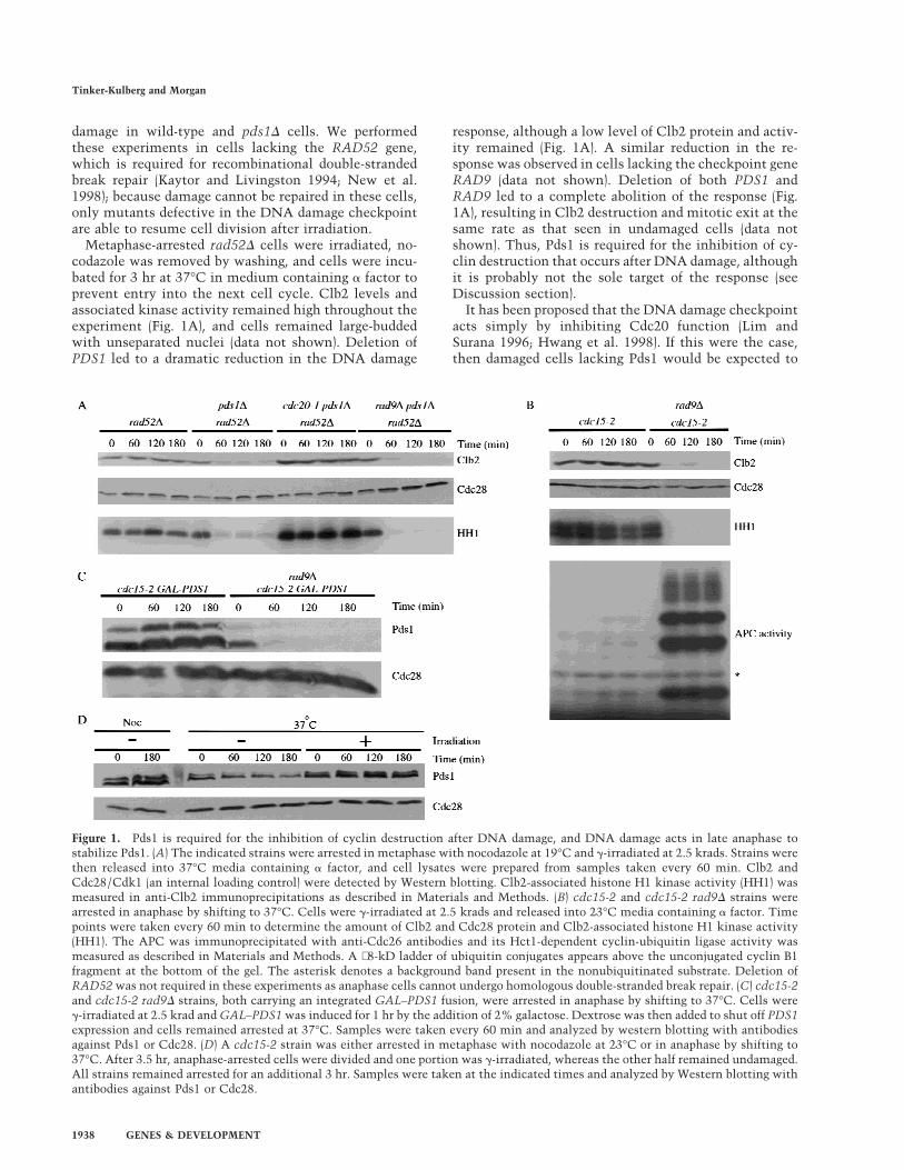
Metaphase-arrested rad52D cells were irradiated, no-codazole was removed by washing, and cells were incu-bated for 3 hr at 37°C in medium containing a factor toprevent entry into the next cell cycle. Clb2 levels andassociated kinase activity remained high throughout theexperiment (Fig. 1A), and cells remained large-buddedwith unseparated nuclei (data not shown). Deletion ofPDS1 led to a dramatic reduction in the DNA damage
response, although a low level of Clb2 protein and activ-ity remained (Fig. 1A). A similar reduction in the re-sponse was observed in cells lacking the checkpoint geneRAD9 (data not shown). Deletion of both PDS1 andRAD9 led to a complete abolition of the response (Fig.1A), resulting in Clb2 destruction and mitotic exit at thesame rate as that seen in undamaged cells (data notshown). Thus, Pds1 is required for the inhibition of cy-clin destruction that occurs after DNA damage, althoughit is probably not the sole target of the response (seeDiscussion section).
It has been proposed that the DNA damage checkpointacts simply by inhibiting Cdc20 function (Lim andSurana 1996; Hwang et al. 1998). If this were the case,then damaged cells lacking Pds1 would be expected to
Figure 1. Pds1 is required for the inhibition of cyclin destruction after DNA damage, and DNA damage acts in late anaphase tostabilize Pds1. (A) The indicated strains were arrested in metaphase with nocodazole at 19°C and g-irradiated at 2.5 krads. Strains werethen released into 37°C media containing a factor, and cell lysates were prepared from samples taken every 60 min. Clb2 andCdc28/Cdk1 (an internal loading control) were detected by Western blotting. Clb2-associated histone H1 kinase activity (HH1) wasmeasured in anti-Clb2 immunoprecipitations as described in Materials and Methods. (B) cdc15-2 and cdc15-2 rad9D strains werearrested in anaphase by shifting to 37°C. Cells were g-irradiated at 2.5 krads and released into 23°C media containing a factor. Timepoints were taken every 60 min to determine the amount of Clb2 and Cdc28 protein and Clb2-associated histone H1 kinase activity(HH1). The APC was immunoprecipitated with anti-Cdc26 antibodies and its Hct1-dependent cyclin-ubiquitin ligase activity wasmeasured as described in Materials and Methods. A ∼8-kD ladder of ubiquitin conjugates appears above the unconjugated cyclin B1fragment at the bottom of the gel. The asterisk denotes a background band present in the nonubiquitinated substrate. Deletion ofRAD52 was not required in these experiments as anaphase cells cannot undergo homologous double-stranded break repair. (C) cdc15-2and cdc15-2 rad9D strains, both carrying an integrated GAL–PDS1 fusion, were arrested in anaphase by shifting to 37°C. Cells wereg-irradiated at 2.5 krad and GAL–PDS1 was induced for 1 hr by the addition of 2% galactose. Dextrose was then added to shut off PDS1expression and cells remained arrested at 37°C. Samples were taken every 60 min and analyzed by western blotting with antibodiesagainst Pds1 or Cdc28. (D) A cdc15-2 strain was either arrested in metaphase with nocodazole at 23°C or in anaphase by shifting to37°C. After 3.5 hr, anaphase-arrested cells were divided and one portion was g-irradiated, whereas the other half remained undamaged.All strains remained arrested for an additional 3 hr. Samples were taken at the indicated times and analyzed by Western blotting withantibodies against Pds1 or Cdc28.
Tinker-Kulberg and Morgan
1938 GENES & DEVELOPMENT
arrest in late anaphase with high Clb2 levels, as Cdc20 isrequired for cyclin destruction as well as Pds1 destruc-tion (Lim et al. 1998). However, we found that CDC20 isrequired for mitotic exit in pds1D cells after DNA dam-age (Fig. 1A), implying that Cdc20 is active in these cells.These results suggest that DNA damage does not cause acomplete inhibition of Cdc20 activity (at least in pds1Dcells). A simpler interpretation is that the DNA damagecheckpoint acts through Pds1, not Cdc20, to inhibit bothanaphase and cytokinesis.
The majority of Pds1 is degraded at the metaphase-to-anaphase transition, and only a low level of the proteinpersists in mutants arrested in anaphase (Cohen-Fix etal. 1996; Jaspersen et al. 1998). Therefore, we reasonedthat if Pds1 is required for the DNA damage checkpoint,then anaphase-arrested cells might be only partially re-sponsive to DNA damage. To test this possibility,cdc15-2 cells were arrested in late anaphase, irradiated,and then returned to the permissive temperature in me-dium containing a factor. Interestingly, irradiated ana-phase cells exhibited a complete RAD9-dependent cellcycle arrest; Clb2 levels and activity remained high,whereas Hct1–APC activity was undetectable (Fig. 1B).We could not test directly the role of Pds1 in these ex-periments because pds1D cdc15-2 cells do not exhibit auniform, reversible anaphase arrest at the restrictivetemperature. However, our observation that pds1D cellsexit mitosis after DNA damage in metaphase (Fig. 1A)indicates that Pds1 is required not only for anaphase in-hibition after damage but also for the inhibition of mi-totic exit.
Pds1 is stabilized in response to DNA damage
If Pds1 is essential for the damage-induced cell cycle ar-rest, then it may be protected from degradation in re-sponse to irradiation. To test this possibility, we ana-lyzed Pds1 stability in cdc15-2 cells arrested in late ana-phase, where Pds1 is normally unstable (Jaspersen et al.1998). cdc15-2 and cdc15-2 rad9D strains carrying PDS1under the control of the regulatable GAL promoter werearrested in anaphase and irradiated. A transient pulse ofPDS1 expression was then induced by addition of galac-tose, after which glucose was added to repress PDS1 ex-pression. Irradiation resulted in the complete stabiliza-tion of Pds1 for >3 hr, whereas Pds1 was degraded within1 hr in rad9D cells (Fig. 1C). Consistent with these re-sults, DNA damage induced an increase in the levels ofendogenous Pds1 in anaphase-arrested cells (Fig. 1D).The amount of Pds1 increased in these cells to levelsapproaching those found in undamaged, metaphase-ar-rested cells (Fig. 1D).
Pds1 blocks Clb2 proteolysis and inactivationin metaphase and late anaphase but not in G1
The requirement for Pds1 in the damage response im-plies that Pds1 is able to inhibit cyclin destruction aswell as sister separation. We tested this possibility byanalyzing cyclin levels in cells overproducing Pds1Ddb, amutant version of Pds1 that is resistant to APC-depen-
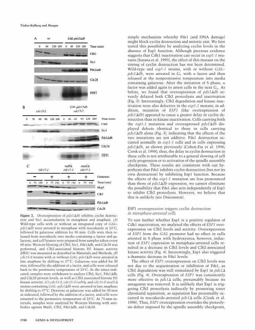
dent destruction. A wild-type strain and a strain carryingGAL–pds1Ddb were arrested in metaphase with noco-dazole, incubated in galactose medium to induce expres-sion of pds1Ddb, and released from nocodazole into ga-lactose medium containing a factor. Overexpression ofpds1Ddb prevented Clb2 proteolysis and significantlydelayed Clb2-associated kinase inactivation (Fig. 2A).We observed consistently that kinase activity decreasedmore rapidly than Clb2 levels, presumably because ofthe increase in Sic1 protein levels at later time points.After 4–5 hr, Cdk1 inactivation was complete and 80%–87% of cells underwent cytokinesis without properlysegregating their sister chromatids (data not shown).
We also analyzed the effects of Pds1Ddb in a late ana-phase arrest. cdc15-2 strains, with or without GAL–pds1Ddb, were arrested in late anaphase by growth at therestrictive temperature. Pds1Ddb production was in-duced and cells were returned to the permissive tempera-ture in the presence of a factor. Again, the pds1Ddb mu-tant blocked the destruction of Clb2 and delayed kinaseinactivation (Fig. 2B); after 4 hr, Cdk1 inactivation wascomplete and mitotic exit occurred in 88% of cells (datanot shown). This result clearly demonstrates that Pds1blocks cyclin destruction by a mechanism that is inde-pendent of sister chromatid separation. It is also consis-tent with our observation that DNA damage can inhibitcyclin destruction in late anaphase (Fig. 1B).
Although the Pds1Ddb protein lacks the amino acidsequence required for APC recognition, it is still possiblethat its overexpression results simply in competitive in-hibition of APC activity. However, we found thatpds1Ddb overexpression did not inhibit the activity ofthe APC in G1 cells and did not cause stabilization ofClb2 (data not shown). It is also known that overexpres-sion of pds1Ddb does not inhibit endogenous Pds1 de-struction (Cohen-Fix et al. 1996; see Fig. 3 below). There-fore, we conclude that Pds1Ddb overexpression does notact simply by competitive inhibition of the APC andcannot inhibit the APC when it is already active. In-stead, our results suggest that Pds1 specifically inhibitsthe processes that trigger APC activation toward cyclinsin late mitosis.
It is possible that overproduction of Pds1Ddb blockscyclin destruction by stimulating the DNA damage re-sponse pathway. However, we found that the cyclin-sta-bilizing effects of Pds1Ddb were unaffected by mutationsin RAD9 or RAD53 (Fig. 2C; data not shown). We alsotested whether the inhibition of Clb2 proteolysis is de-pendent on the spindle assembly checkpoint, as overex-pression of pds1Ddb in metaphase causes spindle abnor-malities. Inhibition of Clb degradation by Pds1Ddb didnot depend on the function of Mad1 (Fig. 2C).
Esp1 is required for normal cyclin destruction
Pds1 is thought to inhibit anaphase by forming a com-plex with Esp1, whose release after Pds1 destruction ini-tiates sister chromatid separation (Ciosk et al. 1998).Therefore, we tested the possibility that Esp1 promotescyclin destruction as well as anaphase, providing a
Control of cyclin destruction by Pds1 and Esp1
GENES & DEVELOPMENT 1939
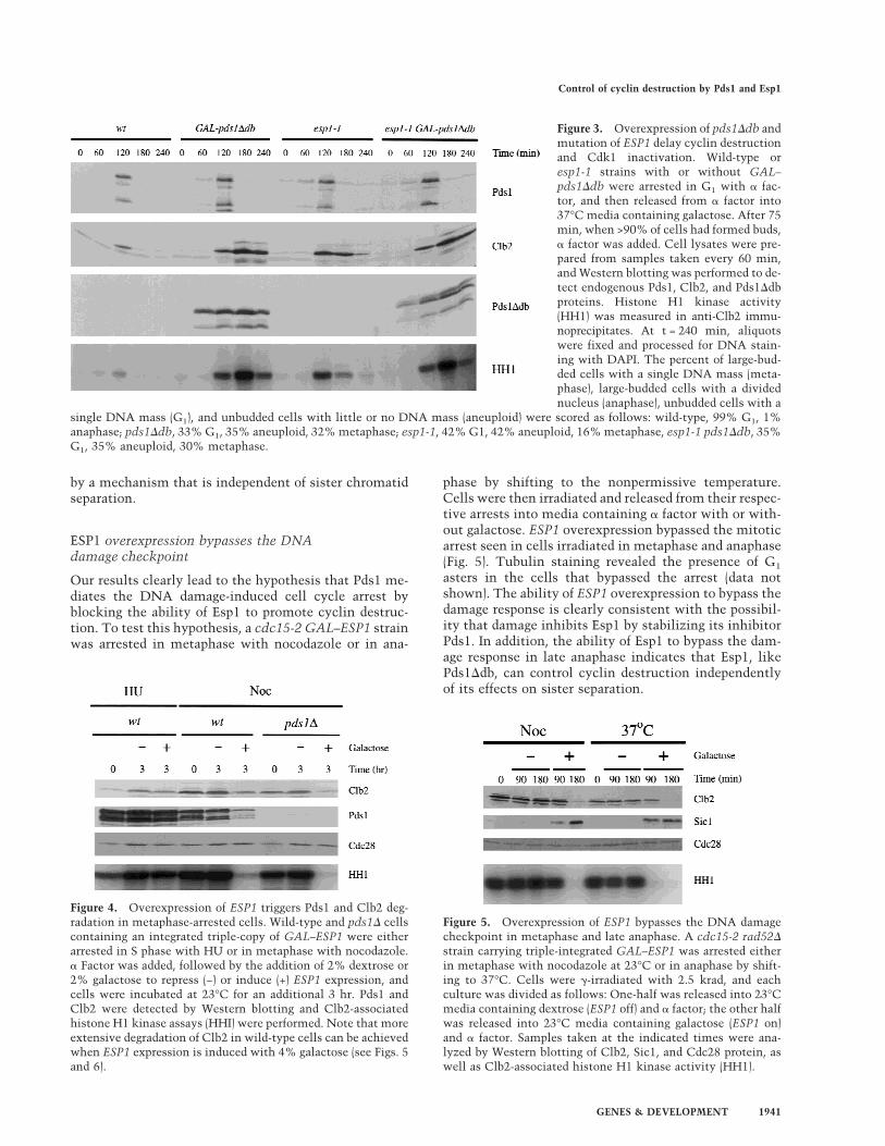
simple mechanism whereby Pds1 (and DNA damage)might block cyclin destruction and mitotic exit. We firsttested this possibility by analyzing cyclin levels in theabsence of Esp1 function. Although previous evidencesuggests that Cdk1 inactivation can occur in esp1-1 mu-tants (Surana et al. 1993), the effect of this mutant on thetiming of cyclin destruction has not been determined.Wild-type and esp1-1 strains, with or without GAL–pds1Ddb, were arrested in G1 with a factor and thenreleased at the nonpermissive temperature into mediacontaining galactose. After the initiation of S phase, afactor was added again to arrest cells in the next G1. Asbefore, we found that overexpression of pds1Ddb se-verely delayed both Clb2 proteolysis and inactivation(Fig. 3). Interestingly, Clb2 degradation and kinase inac-tivation were also defective in the esp1-1 mutant; in ad-dition, mutation of ESP1 (like overexpression ofpds1Ddb) appeared to cause a greater delay in cyclin de-struction than in kinase inactivation. Cells carrying boththe esp1-1 mutation and overexpressed pds1Ddb dis-played defects identical to those in cells carryingpds1Ddb alone (Fig. 3), indicating that the effects of thetwo mutations are not additive. Pds1 destruction oc-curred normally in esp1-1 cells and in cells expressingpds1Ddb, as shown previously (Cohen-Fix et al. 1996;Ciosk et al. 1998); thus, the delay in cyclin destruction inthese cells is not attributable to a general slowing of cellcycle progression or to activation of the spindle assemblycheckpoint. These results are consistent with our hy-pothesis that Pds1 inhibits cyclin destruction (but not itsown destruction) by inhibiting Esp1 function. Becausethe effects of the esp1-1 mutation are less pronouncedthan those of pds1Ddb expression, we cannot eliminatethe possibility that Pds1 also acts independently of Esp1to inhibit Clb2 proteolysis. However, we believe thatthis is unlikely (see Discussion).
ESP1 overexpression triggers cyclin destructionin metaphase-arrested cells
To test further whether Esp1 is a positive regulator ofCdk1 inactivation, we analyzed the effects of ESP1 over-expression on Clb2 levels and activity. Overexpressionof ESP1 from the GAL promoter had no effect in cellsarrested in S phase with hydroxyurea; however, induc-tion of ESP1 expression in metaphase-arrested cells re-sulted in a decrease in Clb2 levels and Clb2-associatedkinase activity (Fig. 4). Interestingly, Esp1 also triggereda dramatic decrease in Pds1 levels.
The effect of ESP1 overexpression on Clb2 levels wasnot due to the sequestration or inhibition of Pds1, asClb2 degradation was still stimulated by Esp1 in pds1Dcells (Fig. 4). Overexpression of ESP1 was consistentlymore effective in pds1D cells, presumably because itsantagonist was removed. It is unlikely that Esp1 is trig-gering Clb2 proteolysis indirectly by promoting sisterchromatid separation, as separation has presumably oc-curred in nocodazole-arrested pds1D cells (Ciosk et al.1998). Thus, ESP1 overexpression overrides the proteoly-sis defect imposed by the spindle assembly checkpoint,
Figure 2. Overexpression of pds1Ddb inhibits cyclin destruc-tion and Sic1 accumulation in metaphase and anaphase. (A)Wild-type cells with or without an integrated copy of GAL–pds1Ddb were arrested in metaphase with nocodazole at 23°C,followed by galactose addition for 30 min. Cells were then re-leased from nocodazole into media containing a factor and ga-lactose, and cell lysates were prepared from samples taken every60 min. Western blotting of Clb2, Sic1, Pds1Ddb, and Cdc28 wasperformed, and Clb2-associated histone H1 kinase activity(HH1) was measured as described in Materials and Methods. (B)cdc15-2 strains with or without GAL–pds1Ddb were arrested inlate anaphase by shifting to 37°C. Galactose was added for 30min, followed by the addition of a factor, and cells were releasedback to the permissive temperature of 23°C. At the times indi-cated, samples were withdrawn to analyze Clb2, Sic1, Pds1Ddb,and Cdc28 protein levels, as well as Clb2-associated histone H1kinase activity. (C) cdc15-2, cdc15-2 rad9D, and cdc15-2 mad1D
strains containing GAL–pds1Ddb were arrested in late anaphaseby shifting to 37°C. Dextrose or galactose was added for 30 minas indicated, followed by the addition of a factor, and cells werereturned to the permissive temperature of 23°C. At 75-min in-tervals, samples were analyzed by Western blotting with anti-bodies against Mad1, Clb2, Pds1Ddb, and Cdc28.
Tinker-Kulberg and Morgan
1940 GENES & DEVELOPMENT
by a mechanism that is independent of sister chromatidseparation.
ESP1 overexpression bypasses the DNAdamage checkpoint
Our results clearly lead to the hypothesis that Pds1 me-diates the DNA damage-induced cell cycle arrest byblocking the ability of Esp1 to promote cyclin destruc-tion. To test this hypothesis, a cdc15-2 GAL–ESP1 strainwas arrested in metaphase with nocodazole or in ana-
phase by shifting to the nonpermissive temperature.Cells were then irradiated and released from their respec-tive arrests into media containing a factor with or with-out galactose. ESP1 overexpression bypassed the mitoticarrest seen in cells irradiated in metaphase and anaphase(Fig. 5). Tubulin staining revealed the presence of G1
asters in the cells that bypassed the arrest (data notshown). The ability of ESP1 overexpression to bypass thedamage response is clearly consistent with the possibil-ity that damage inhibits Esp1 by stabilizing its inhibitorPds1. In addition, the ability of Esp1 to bypass the dam-age response in late anaphase indicates that Esp1, likePds1Ddb, can control cyclin destruction independentlyof its effects on sister separation.
Figure 3. Overexpression of pds1Ddb andmutation of ESP1 delay cyclin destructionand Cdk1 inactivation. Wild-type oresp1-1 strains with or without GAL–pds1Ddb were arrested in G1 with a fac-tor, and then released from a factor into37°C media containing galactose. After 75min, when >90% of cells had formed buds,a factor was added. Cell lysates were pre-pared from samples taken every 60 min,and Western blotting was performed to de-tect endogenous Pds1, Clb2, and Pds1Ddbproteins. Histone H1 kinase activity(HH1) was measured in anti-Clb2 immu-noprecipitates. At t = 240 min, aliquotswere fixed and processed for DNA stain-ing with DAPI. The percent of large-bud-ded cells with a single DNA mass (meta-phase), large-budded cells with a dividednucleus (anaphase), unbudded cells with a
single DNA mass (G1), and unbudded cells with little or no DNA mass (aneuploid) were scored as follows: wild-type, 99% G1, 1%anaphase; pds1Ddb, 33% G1, 35% aneuploid, 32% metaphase; esp1-1, 42% G1, 42% aneuploid, 16% metaphase, esp1-1 pds1Ddb, 35%G1, 35% aneuploid, 30% metaphase.
Figure 4. Overexpression of ESP1 triggers Pds1 and Clb2 deg-radation in metaphase-arrested cells. Wild-type and pds1D cellscontaining an integrated triple-copy of GAL–ESP1 were eitherarrested in S phase with HU or in metaphase with nocodazole.a Factor was added, followed by the addition of 2% dextrose or2% galactose to repress (−) or induce (+) ESP1 expression, andcells were incubated at 23°C for an additional 3 hr. Pds1 andClb2 were detected by Western blotting and Clb2-associatedhistone H1 kinase assays (HHI) were performed. Note that moreextensive degradation of Clb2 in wild-type cells can be achievedwhen ESP1 expression is induced with 4% galactose (see Figs. 5and 6).
Figure 5. Overexpression of ESP1 bypasses the DNA damagecheckpoint in metaphase and late anaphase. A cdc15-2 rad52D
strain carrying triple-integrated GAL–ESP1 was arrested eitherin metaphase with nocodazole at 23°C or in anaphase by shift-ing to 37°C. Cells were g-irradiated with 2.5 krad, and eachculture was divided as follows: One-half was released into 23°Cmedia containing dextrose (ESP1 off) and a factor; the other halfwas released into 23°C media containing galactose (ESP1 on)and a factor. Samples taken at the indicated times were ana-lyzed by Western blotting of Clb2, Sic1, and Cdc28 protein, aswell as Clb2-associated histone H1 kinase activity (HH1).
Control of cyclin destruction by Pds1 and Esp1
GENES & DEVELOPMENT 1941

Esp1-induced Cdk1 inactivation requires Cdc20and the mitotic exit network
Our observation that Esp1 triggers Pds1 destruction (seeFig. 4) raised the intriguing possibility that Esp1 actsprimarily through the activation of Cdc20. Consistentwith this possibility, we found that Cdc20 is required forEsp1 to induce cyclin degradation and accumulation ofSic1 in nocodazole-arrested cells (Fig. 6A). Cdc16 func-tion was also required, further suggesting that Esp1 isacting through activation of APC-dependent proteolysis(Fig. 6A). Thus, we conclude that overexpression of ESP1promotes cyclin destruction and Cdk1 inactivationthrough a Cdc20-dependent pathway.
Deletion of HCT1 did not affect the ability of ESP1overexpression to trigger Pds1 destruction and Sic1 ac-cumulation in nocodazole-arrested cells (Fig. 6B), furtherdemonstrating that Esp1 acts primarily through stimu-lation of Cdc20–APC activity. Deletion of HCT1 did re-duce partially the effects of ESP1 on the levels of Clb2and Clb3 (Fig. 6B). We also found that ESP1 expression inwild-type cells leads to activation of the Hct1–APC (datanot shown). Therefore, we suspect that the effects ofoverexpressed Esp1 on Clb2 and Clb3 levels are due inpart to the direct action of the Cdc20–APC and in part to
Cdc20-dependent destruction of an inhibitor of Hct1–APC (see Discussion).
We also analyzed the effects of CDC20 overexpressionon Clb2 and Clb3 levels in nocodazole-arrested cells. Asin our experiments with GAL–ESP1, GAL–CDC20 ex-pression led to a decrease in Clb2 and Clb3 levels thatwas partially dependent on the presence of Hct1 (Fig.6C). The ability of Esp1 and Cdc20 to induce partiallyHct1-independent cyclin destruction and Cdk1 inactiva-tion further supports the notion that Esp1 acts by stimu-lating Cdc20.
Because the mitotic exit network is known to be re-quired for Hct1–APC activation and Sic1 accumulation,we tested its role in the effects of Esp1 in nocodazole-arrested cells. We found that Esp1-induced Clb2 destruc-tion and Sic1 accumulation were blocked in cdc14 andtem1 mutants (Fig. 6D). The degradation of Pds1 andClb3 was also reduced partially in these mutants, raisingthe possibility that Cdc14 and Tem1 are required forcomplete activation of the Cdc20–APC. We also foundthat the effects of ESP1 overexpression on Clb2 levelswere blocked in cdc5 and cdc15 mutants arrested in lateanaphase (data not shown). Thus, components of the mi-totic exit network are required for Esp1 to exert its in-hibitory effects on Clb2 levels and activity.
Figure 6. Esp1-induced cyclin destruction andSic1 accumulation are dependent on Cdc20 andthe mitotic exit network. (A) Wild-type, cdc20-1,and cdc16-1 strains containing GAL–ESP1 werearrested in metaphase with nocodazole for 3 hr.Cells were then shifted to 35°C for 30 min. Dex-trose or galactose was added, along with a factor,and cells were incubated at the nonpermissivetemperature for an additional 3 hr. Cell lysateswere analyzed by Western blotting of Clb3, Clb2,Sic1, and Cdc28, and Clb2-associated histone H1kinase (HH1) activity was measured. AlthoughGAL–ESP1 had no effect on Clb levels in thecdc16 mutant, it did trigger spindle elongationand nuclear division (data not shown; see also Ci-osk et al. 1998). (B) Wild-type and hct1D strainscontaining GAL–ESP1 were arrested in meta-phase with nocodazole at 23°C. a Factor was thenadded, followed by the addition of dextrose or ga-lactose, and incubation continued for 3 hr. Celllysates were analyzed by Western blotting ofPds1, Clb3, Clb2, and Sic1, and Clb2-associatedhistone H1 kinase activity was measured. (C)Wild-type and hct1D cells containing GAL–CDC20 on a CEN/ARS plasmid were arrested inmetaphase with nocodazole at 23°C. Dextrose orgalactose was added, in addition to a factor, andcells were incubated for an additional 3 hr. Clb2,Clb3, Cdc20, and Cdc28 levels were analyzed byWestern blotting, and Clb2-associated histone H1kinase activity was measured. In control experi-ments (data not shown), addition of galactosecaused cyclin destruction only in cells carrying GAL–ESP1 or GAL–CDC20 and not in cells lacking these plasmids. (D) Wild-type,cdc14-1, and tem1-3 strains containing GAL–ESP1 were treated and analyzed as described in A. Cell lysates were analyzed by Westernblotting of Pds1, Clb3, Clb2, and Sic1, and Clb2-associated histone H1 kinase activity was measured.
Tinker-Kulberg and Morgan
1942 GENES & DEVELOPMENT
Cdc20 activity is required for exit from a lateanaphase cdc15 arrest
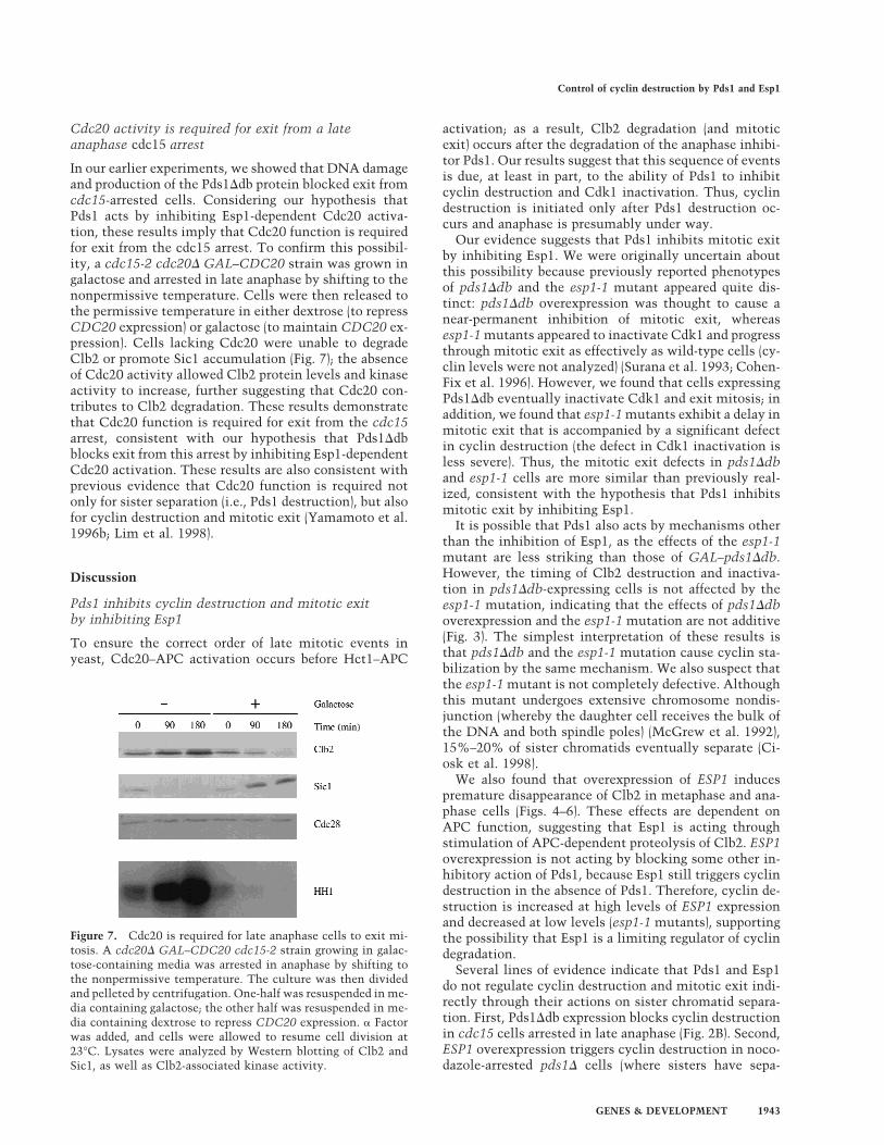
In our earlier experiments, we showed that DNA damageand production of the Pds1Ddb protein blocked exit fromcdc15-arrested cells. Considering our hypothesis thatPds1 acts by inhibiting Esp1-dependent Cdc20 activa-tion, these results imply that Cdc20 function is requiredfor exit from the cdc15 arrest. To confirm this possibil-ity, a cdc15-2 cdc20D GAL–CDC20 strain was grown ingalactose and arrested in late anaphase by shifting to thenonpermissive temperature. Cells were then released tothe permissive temperature in either dextrose (to repressCDC20 expression) or galactose (to maintain CDC20 ex-pression). Cells lacking Cdc20 were unable to degradeClb2 or promote Sic1 accumulation (Fig. 7); the absenceof Cdc20 activity allowed Clb2 protein levels and kinaseactivity to increase, further suggesting that Cdc20 con-tributes to Clb2 degradation. These results demonstratethat Cdc20 function is required for exit from the cdc15arrest, consistent with our hypothesis that Pds1Ddbblocks exit from this arrest by inhibiting Esp1-dependentCdc20 activation. These results are also consistent withprevious evidence that Cdc20 function is required notonly for sister separation (i.e., Pds1 destruction), but alsofor cyclin destruction and mitotic exit (Yamamoto et al.1996b; Lim et al. 1998).
Discussion
Pds1 inhibits cyclin destruction and mitotic exitby inhibiting Esp1
To ensure the correct order of late mitotic events inyeast, Cdc20–APC activation occurs before Hct1–APC
activation; as a result, Clb2 degradation (and mitoticexit) occurs after the degradation of the anaphase inhibi-tor Pds1. Our results suggest that this sequence of eventsis due, at least in part, to the ability of Pds1 to inhibitcyclin destruction and Cdk1 inactivation. Thus, cyclindestruction is initiated only after Pds1 destruction oc-curs and anaphase is presumably under way.
Our evidence suggests that Pds1 inhibits mitotic exitby inhibiting Esp1. We were originally uncertain aboutthis possibility because previously reported phenotypesof pds1Ddb and the esp1-1 mutant appeared quite dis-tinct: pds1Ddb overexpression was thought to cause anear-permanent inhibition of mitotic exit, whereasesp1-1 mutants appeared to inactivate Cdk1 and progressthrough mitotic exit as effectively as wild-type cells (cy-clin levels were not analyzed) (Surana et al. 1993; Cohen-Fix et al. 1996). However, we found that cells expressingPds1Ddb eventually inactivate Cdk1 and exit mitosis; inaddition, we found that esp1-1 mutants exhibit a delay inmitotic exit that is accompanied by a significant defectin cyclin destruction (the defect in Cdk1 inactivation isless severe). Thus, the mitotic exit defects in pds1Ddband esp1-1 cells are more similar than previously real-ized, consistent with the hypothesis that Pds1 inhibitsmitotic exit by inhibiting Esp1.
It is possible that Pds1 also acts by mechanisms otherthan the inhibition of Esp1, as the effects of the esp1-1mutant are less striking than those of GAL–pds1Ddb.However, the timing of Clb2 destruction and inactiva-tion in pds1Ddb-expressing cells is not affected by theesp1-1 mutation, indicating that the effects of pds1Ddboverexpression and the esp1-1 mutation are not additive(Fig. 3). The simplest interpretation of these results isthat pds1Ddb and the esp1-1 mutation cause cyclin sta-bilization by the same mechanism. We also suspect thatthe esp1-1 mutant is not completely defective. Althoughthis mutant undergoes extensive chromosome nondis-junction (whereby the daughter cell receives the bulk ofthe DNA and both spindle poles) (McGrew et al. 1992),15%–20% of sister chromatids eventually separate (Ci-osk et al. 1998).
We also found that overexpression of ESP1 inducespremature disappearance of Clb2 in metaphase and ana-phase cells (Figs. 4–6). These effects are dependent onAPC function, suggesting that Esp1 is acting throughstimulation of APC-dependent proteolysis of Clb2. ESP1overexpression is not acting by blocking some other in-hibitory action of Pds1, because Esp1 still triggers cyclindestruction in the absence of Pds1. Therefore, cyclin de-struction is increased at high levels of ESP1 expressionand decreased at low levels (esp1-1 mutants), supportingthe possibility that Esp1 is a limiting regulator of cyclindegradation.
Several lines of evidence indicate that Pds1 and Esp1do not regulate cyclin destruction and mitotic exit indi-rectly through their actions on sister chromatid separa-tion. First, Pds1Ddb expression blocks cyclin destructionin cdc15 cells arrested in late anaphase (Fig. 2B). Second,ESP1 overexpression triggers cyclin destruction in noco-dazole-arrested pds1D cells (where sisters have sepa-
Figure 7. Cdc20 is required for late anaphase cells to exit mi-tosis. A cdc20D GAL–CDC20 cdc15-2 strain growing in galac-tose-containing media was arrested in anaphase by shifting tothe nonpermissive temperature. The culture was then dividedand pelleted by centrifugation. One-half was resuspended in me-dia containing galactose; the other half was resuspended in me-dia containing dextrose to repress CDC20 expression. a Factorwas added, and cells were allowed to resume cell division at23°C. Lysates were analyzed by Western blotting of Clb2 andSic1, as well as Clb2-associated kinase activity.
Control of cyclin destruction by Pds1 and Esp1
GENES & DEVELOPMENT 1943
rated) (Fig. 4) and in cells subjected to DNA damage inlate anaphase (Fig. 5).
Esp1 triggers mitotic exit by a Cdc20-dependent mechanism
Our results indicate that the effects of Esp1 on cyclindestruction are dependent on CDC20. We found thatEsp1 promotes Pds1 destruction as well as that of Clb2,and that these effects were blocked in the cdc20-1 mu-tant. We conclude that Esp1 somehow promotes Cdc20-dependent APC activity, which then leads to Hct1–APCactivation and Sic1 accumulation.
Overexpression of CDC20 in cells arrested in S phasewith hydroxyurea is sufficient to induce Pds1 degrada-tion (Visintin et al. 1997), whereas we did not see anyeffect of ESP1 overexpression in S phase (Fig. 4). Becausethe actions of Esp1 require Cdc20, these results are con-sistent with previous evidence that Cdc20 protein is ab-sent in S phase (Prinz et al. 1998). In nocodazole-arrestedcells, we found that Esp1 triggers the destruction of Pds1,Clb3, and Clb2; here again, our results are consistentwith previous experiments showing that CDC20 overex-pression in these cells causes degradation of both Pds1and Clb2 (Visintin et al. 1997). Interestingly, we alsofound that the effects of overexpressed ESP1 and CDC20on Clb3, Clb2, and Sic1 levels were affected similarly bythe deletion of HCT1, further suggesting that Esp1 actsthrough Cdc20 (Fig. 6).
We do not believe that Esp1 is normally required forthe initial activation of the Cdc20-dependent APC, asPds1 destruction occurs normally in the esp1-1 mutantand in cells expressing pds1Ddb (Fig. 3). Nor do we be-lieve that Esp1-dependent stimulation of Cdc20 nor-mally causes Hct1-independent Clb2 destruction, as themajority of Clb2 destruction in vivo is dependent onHct1-APC activity (Schwab et al. 1997; Visintin et al.1997). Instead, we suspect that Cdc20 has a low level ofClb2-targeting activity that can cause a reduction inClb2 levels when CDC20 or ESP1 are overexpressed. In anormal cell cycle, it seems likely that Cdc20, with apush from Esp1, promotes the destruction of some cyc-lins and causes a slight decrease in Cdk1 activity, whichmight be sufficient to trigger the Hct1–Sic1 activationswitch (Fig. 8). In the absence of Esp1 (or in the presenceof Pds1Ddb), this activation may be delayed until the
accumulation of Cdc20 activity overcomes the require-ment for Esp1.
Esp1-induced cyclin destruction and Sic1accumulation are dependent on the mitoticexit network
We also found that the ability of Esp1 to trigger Clb2proteolysis and Sic1 accumulation is dependent on com-ponents of the mitotic exit network, including Tem1,Cdc14, Cdc5, and Cdc15 (Fig. 6; data not shown). ESP1-induced degradation of Pds1 was also slightly reduced inlate mitotic mutants. Combined with our evidence thatthe effects of Esp1 require Cdc20, these results point tothe possibility that the mitotic exit network is also re-quired for complete activation of the Cdc20–APC (Fig. 8).
Therefore, our results are consistent with several pre-vious lines of evidence that the mitotic exit networkdoes not act simply by promoting Hct1–APC activity buthas more general actions that include activation of theCdc20–APC as well. First, the late mitotic mutants allarrest with low but still significant levels of Pds1 pro-tein; complete destruction of Pds1 does not occur untilmitotic exit (Jaspersen et al. 1998). Second, the ability ofoverexpressed CDC5 and CDC14 to trigger cyclin de-struction is dependent on CDC20 (Charles et al. 1998;Visintin et al. 1998). There is also biochemical evidencethat the vertebrate Cdc5 homolog phosphorylates theAPC core and promotes its activity, and in yeast there isevidence that Cdc5 and Cdc15 are required for somemodification of the APC that increases its responsive-ness to Hct1 in vitro (Kotani et al. 1998; Jaspersen et al.1999). One explanation for these results (but not the onlyone) is that some components of the mitotic exit net-work act by promoting the activity of the Cdc20–APC aswell as that of the Hct1–APC. Clearly, they are not re-quired for the initial burst of Cdc20 activity that triggersmost Pds1 destruction and causes anaphase, but theymay be required for complete Cdc20-dependent destruc-tion of Pds1 and other inhibitors of cyclin destruction.
The role of Pds1 in the DNA damage response
In most eukaryotic cells, DNA damage blocks entry intomitosis by inhibiting mitotic Cdk activation (Elledge
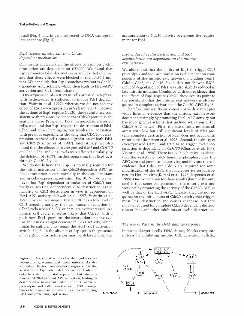
Figure 8. A speculative model of the regulatory re-lationships governing exit from mitosis. As de-scribed in the text, our evidence suggests that theactivation of Esp1 after Pds1 destruction leads notonly to sister chromatid separation but also en-hances Cdc20-dependent APC activation, leading todestruction of an unidentified inhibitor (‘X’) of cyclinproteolysis and Cdk1 inactivation. DNA damageblocks both anaphase and mitotic exit by stabilizingPds1 and preventing Esp1 action.
Tinker-Kulberg and Morgan
1944 GENES & DEVELOPMENT
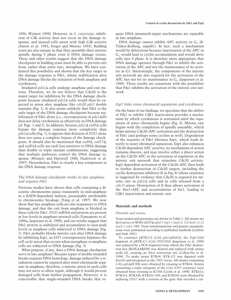
1996; Weinert 1998). However, in S. cerevisiae, inhibi-tion of Cdk activity does not occur in the damage re-sponse, and instead cells arrest with high Cdk activity(Amon et al. 1992; Sorger and Murray 1992). Buddingyeast are also unique in that they assemble their mitoticspindle during S phase even if DNA damage occurs.These and other results suggest that the DNA damagecheckpoint in budding yeast must be able to prevent exitfrom, rather than entry into, metaphase. We have con-firmed this possibility and shown that the key target inthe damage response is Pds1, whose stabilization afterDNA damage blocks the initiation of both anaphase andcytokinesis.
Irradiated pds1D cells undergo anaphase and exit mi-tosis. Therefore, we do not believe that Cdc20 is themajor target for inhibition by the DNA damage check-point because irradiated pds1D cells would then be ex-pected to arrest after anaphase like cdc20 pds1 doublemutants (Fig. 1). It also seems unlikely that Pds1 is theonly target of the DNA damage checkpoint because sta-bilization of Pds1 alone (i.e., overexpression of pds1Ddb)does not delay cytokinesis as effectively as DNA damage(cf. Figs. 1 and 2). In addition, because pds1D rad9D cellsbypass the damage response more completely thanpds1D cells (Fig. 1), it appears that deletion of PDS1 alonedoes not cause a complete bypass of the damage check-point. It should also be mentioned that rad9D, rad17D,and rad24D cells are each less sensitive to DNA damagethan double or triple mutant combinations, suggestingthat multiple pathways control the DNA damage re-sponse (Weinert and Hartwell 1990; Paulovich et al.1997). Nevertheless, Pds1 is clearly a key component inthe DNA damage response.
The DNA damage checkpoint works in late anaphaseand requires Pds1
Previous studies have shown that cells containing a di-centric chromosome pause transiently in mid-anaphasein a RAD9-dependent fashion, presumably attributableto chromosome breakage (Yang et al. 1997). We nowshow that late anaphase cells are also responsive to DNAdamage, and that the exit from anaphase is blocked inthese cells by Pds1. PDS1 mRNA and protein are presentat low levels in anaphase-arrested cells (Yamamoto et al.1996a; Jaspersen et al. 1998), and our results suggest thatPds1 protein is stabilized and accumulates to near-peaklevels in anaphase cells subjected to DNA damage (Fig.1). Pds1 probably blocks mitotic exit after DNA damageby inhibiting Esp1, as ESP1 overexpression bypasses thecell cycle arrest that occurs when metaphase or anaphasecells are subjected to DNA damage (Fig. 5).
What purpose, if any, does a DNA damage checkpointserve in late anaphase? Because repair of double-strandedbreaks requires DNA homology, damage induced by g-ir-radiation cannot be repaired after sister chromatids sepa-rate in anaphase; therefore, a mitotic arrest in such cellsmay not serve to allow repair, although it would preventdamaged cells from further propagation. However, it isconceivable that single-stranded DNA breaks that re-
quire DNA mismatch-repair mechanisms are reparablein late anaphase.
DNA damage cannot inhibit APC activity in G1 (R.Tinker-Kulberg, unpubl.). In fact, such a mechanismwould be deleterious because inactivation of the APC inG1 would lead to cyclin accumulation and would drivecells into S phase. It is therefore more appropriate thatDNA damage operates through Pds1 to inhibit the acti-vation of the APC and not the maintenance of its activ-ity in G1. Interestingly, the components of the mitoticexit network are also required for the activation of theAPC but not for its maintenance in G1 (Jaspersen et al.1998). These results are consistent with the possibilitythat Pds1 inhibits the activation of the mitotic exit net-work.
Esp1 links sister chromatid separation and cytokinesis
On the basis of our findings, we speculate that the abilityof Pds1 to inhibit Cdk1 inactivation provides a mecha-nism by which cytokinesis is restrained until the sepa-ration of sister chromatids begins (Fig. 8). Mitotic exitbegins with the completion of spindle assembly, whichhelps initiate Cdc20–APC activation and the destructionof Pds1 (and perhaps some cyclins as well). Degradationof the majority of Pds1 liberates Esp1, which leads di-rectly to sister chromatid separation. Esp1 also enhancesCdc20-dependent APC activity; its mechanism of actionremains obscure, and may involve either a direct actionon the Cdc20–APC or the activation of regulators in themitotic exit network that stimulate Cdc20 activity.Esp1-dependent activation of the Cdc20–APC then leadsto further destruction of Cdc20 targets, including thecyclin destruction inhibitor (X in Fig. 8) whose existenceis suggested by evidence that Cdc20 is required for mi-totic exit in pds1D cells and in cells released from acdc15 arrest. Destruction of X then allows activation ofthe Hct1–APC and accumulation of Sic1, leading toCdk1 inactivation and mitotic exit.
Materials and methods
Plasmids and strains
Yeast strains and genotypes are shown in Table 1. All strains arederivatives of W303 (MATa ade2-1 trp1-1 leu2-3, 112 his3-11,15ura3-1 can1-100). Yeast transformations and genetic manipula-tions were performed according to published methods (Guthrieand Fink 1991).
To construct pRTK-C2 (GAL–pds1DdbHA), the FspI–NdeIfragment of pRTK-C1 (GAL–PDS1HA) (Jaspersen et al. 1998)was replaced by a PCR fragment from which the Pds1 destruc-tion box (RLPLAAKDN) was deleted and replaced with aminoacids LE, creating an XhoI restriction site (Cohen-Fix et al.1996). To make strain RTK44, RTK–C2 was digested withBsu36I and integrated at the TRP1 locus. All strains containingGAL–pdsDdb HA were obtained by crossing to RTK44. Strainscontaining a triple integrant of the GAL–ESP1 construct wereobtained from crossing to K7346 (Ciosk et al. 1998). RTK313,RTK314, RTK330, RTK335–339, and RTK344 were obtained byreplacing PDS1 with a version of the gene that encoded a car-
Control of cyclin destruction by Pds1 and Esp1
GENES & DEVELOPMENT 1945
boxy-terminally myc18-epitope-tagged Pds1 protein as de-scribed (Shirayama et al. 1998). Deletion of RAD9 was per-formed as described (Weinert and Hartwell 1990) and confirmedby Southern blot hybridization. PDS1 was deleted as described(Yamamoto et al. 1996a) and deletions were identified byscreening for temperature sensitivity (i.e., at 30°C), sensitivityto the microtubule poison benomyl, and the inability tocomplement a pds7A-1F temperature-sensitive mutant (giftfrom Sue Biggins, UCSF). Deletion of RAD52 was carried out asdescribed (Kaytor and Livingston 1994) and confirmed byscreening for hydroxyurea and radiation sensitivity. Deletion ofMAD1 was performed as described (Hardwick and Murray 1995)and confirmed by PCR and Western blot analysis. All otherstrains were derived from crosses using standard methods(Guthrie and Fink 1991). RTK223 and RTK211 were derived bytransformation with the URA3-based centromeric plasmidpLH68, which contains the CDC20 gene fused to a triple hem-agglutinin (HA) tag and a six-histidine tag at its carboxyl termi-nus (Hwang et al. 1998).
Cell-cycle synchronization
Standard protocols were used for cell propagation (Guthrie andFink 1991). To arrest at G1, S phase, or metaphase, cells weregrown at 23°C (or at 19°C in case of pds1D strains) to mid-logphase (OD600 of 0.35) and arrested with 1.5 µg/ml a-factor, 0.1M hydroxyurea (HU), or 15 µg/ml nocodazole at 23°C for 3.5 hr,respectively (unless otherwise indicated). To release from a G1,S phase, or metaphase arrest, cells were pelleted by centrifuga-tion and washed with the appropriate media three times, andresuspended in 23°C or 37°C media as indicated. To arrest tem-
perature-sensitive strains, cells were grown to mid-log phase at23°C and arrested by shifting cells to 37°C for 3.5 hr. Cells werereleased from the arrest by returning them to 23°C. In all cases,90%–95% of cells displayed the correct arrest morphology. Allstrains were either grown in YEP media (Rose et al. 1990)supplemented with 2% dextrose (YEPD) or 2% raffinose(YEPraf) if galactose induction was going to take place. To ex-press from the GAL promoter, cells were grown in YEPraf andexpression was induced by the addition of galactose to 4% (un-less otherwise indicated). Transcription was repressed by theaddition of 4% dextrose (unless otherwise indicated). Strainscontaining the centromeric plasmid with CDC20 were grown tomid-log phase in minimal medium lacking uracil and contain-ing 2% raffinose. Strains were then pelleted and resuspended inYEPraf to perform the experiment described.
Irradiation
After the indicated cell cycle arrest, 20 OD600 units were pel-leted by centrifugation and resuspended in 1 ml of media. Cellswere placed in one well of a 3043-microtiter dish (Fisher) andg-irradiated at 2.5 krads using a cesium source at 360 rads/min(1 rad = 0.1 Gy). Cells were resuspended in the appropriate me-dia as described.
Lysate preparation and Western blot analysis
Protein extracts were prepared by resuspending cells in 3 to4-pellet volumes of ice-cold LLB [50 mM HEPES-NaOH (pH 7.4),75 mM KCl, 50 mM NaF, 50 mM b-glycerophosphate, 1 mM
EGTA, 0.1% NP40, 1 mM DTT, 1 mM phenylmethylsulfonyl-
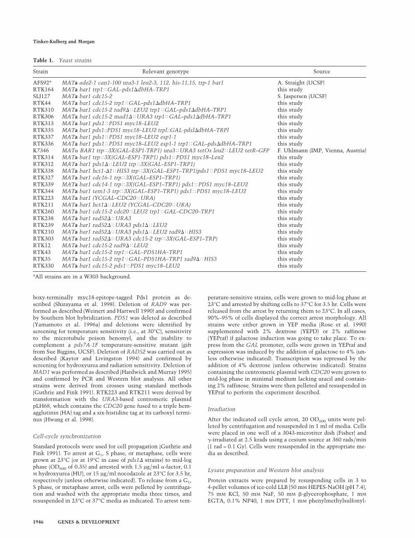
Table 1. Yeast strains
Strain Relevant genotype Source
AFS92a MATa ade2-1 can1-100 ura3-1 leu2-3, 112, his-11,15, trp-1 bar1 A. Straight (UCSF)RTK164 MATa bar1 trp1<GAL–pds1DdbHA–TRP1 this studySLJ127 MATa bar1 cdc15-2 S. Jaspersen (UCSF)RTK44 MATa bar1 cdc15-2 trp1<GAL–pds1DdbHA–TRP1 this studyRTK310 MATa bar1 cdc15-2 rad9D<LEU2 trp1<GAL–pds1DdbHA–TRP1 this studyRTK306 MATa bar1 cdc15-2 mad1D<URA3 trp1<GAL–pds1DdbHA–TRP1 this studyRTK313 MATa bar1 pds1<PDS1 myc18–LEU2 this studyRTK335 MATa bar1 pds1::PDS1 myc18–LEU2 trpl::GAL-pdslDdbHA–TRPl this studyRTK337 MATa bar1 pds1<PDS1 myc18–LEU2 esp1-1 this studyRTK336 MATa bar1 pds1<PDS1 myc18–LEU2 esp1-1 trp1<GAL–pdsDdbHA–TRP1 this studyK7346 MATa BAR1 trp<3X(GAL–ESP1-TRP1) ura3<URA3 tetOs leu2<LEU2 tetR–GFP F. Uhlmann (IMP, Vienna, Austria)RTK314 MATa bar1 trp<3X(GAL–ESP1-TRP1) pds1<PDS1 myc18–Leu2 this studyRTK312 MATa bar1 pds1D<LEU2 trp<3X(GAL–ESP1–TRP1) this studyRTK338 MATa bar1 hct1-D1<HIS3 trp<3X(GAL–ESP1–TRP1)pds1<PDS1 myc18–LEU2 this studyRTK327 MATa bar1 cdc16-1 trp<3X(GAL–ESP1–TRP1) this studyRTK339 MATa bar1 cdc14-1 trp<3X(GAL–ESP1–TRP1) pds1<PDS1 myc18–LEU2 this studyRTK344 MATa bar1 tem1-3 trp<3X(GAL–ESP1–TRP1) pds1<PDS1 myc18–LEU2 this studyRTK223 MATa bar1 (YCGAL–CDC20<URA) this studyRTK211 MATa bar1 hct1D<LEU2 (YCGAL–CDC20<URA) this studyRTK260 MATa bar1 cdc15-2 cdc20<LEU2 trp1<GAL–CDC20–TRP1 this studyRTK238 MATa bar1 rad52D<URA3 this studyRTK239 MATa bar1 rad52D<URA3 pds1D<LEU2 this studyRTK210 MATa bar1 rad52D<URA3 pds1D<LEU2 rad9D<HIS3 this studyRTK303 MATa bar1 rad52D<URA3 cdc15-2 trp<3X(GAL–ESP1–TRP) this studyRTK12 MATa bar1 cdc15-2 rad9D<LEU2 this studyRTK43 MATa bar1 cdc15-2 trp1<GAL–PDS1HA–TRP1 this studyRTK35 MATa bar1 cdc15-2 trp1<GAL–PDS1HA–TRP1 rad9D<HIS3 this studyRTK330 MATa bar1 cdc15-2 pds1<PDS1 myc18–LEU2 this study
aAll strains are in a W303 background.
Tinker-Kulberg and Morgan
1946 GENES & DEVELOPMENT

fluoride, 2 µg/ml aprotinin, 1 µg/ml leupeptin, and 1 µg/mlpepstatin] and an equal volume of glass beads. Samples werelysed by mechanical disruption in a Beadbeater (Biospec) for 2min. Lysates were clarified by centrifugation at 14,000g for 15min at 4°C and protein concentrations were determined usingthe Bio-Rad protein assay.
For Western blotting, 25 µg of lysate was used for detection ofendogenous Clb2, Cdc28, and Pds1-myc18, whereas 60 µg oflysate was used for detection of endogenous Sic1 and Clb3 andgalactose-overexpressed Pds1HA and Pds1DdbHA proteins.Clb2, Cdc28, Sic1, and Clb3 proteins were detected with affin-ity-purified polyclonal antibodies as previously described (Ger-ber et al. 1995; Charles et al. 1998; Jaspersen et al. 1998). Fordetection of HA-tagged proteins, the mouse monoclonal anti-body 16B12 was used as described (Gerber et al. 1995).Pds1myc18 immunoblots were performed with a 1:1000 dilu-tion of a-myc polyclonal antibodies (Santa Cruz).
Kinase assays
To measure Clb2-associated histone H1 kinase activity, 75 µg ofcell lysate was incubated with 0.3 µg of affinity-purified anti-Clb2 antibody and 20 µl of a 1:1 slurry of protein A–sepharose(Sigma) for 1.5 hr at 4°C. Immune complexes were washed twotimes in LLB and once in Buffer A [50 mM HEPES-NaOH (pH7.4) and 1 µM DTT], and incubated for 15 min at 23°C in a 20-µlreaction mixture containing 100 µM ATP, 1 mM MgCl2, 5 µg ofhistone H1 protein, and 2.5 µCi [g-32P] ATP (3000 mCi/mmole)in buffer A. Reaction products were resolved on 12% SDS-PAGEgels and detected by autoradiography.
Cyclin ubiquitination assay
Cyclin ubiquitin–ligase activity of the APC was measured asdescribed (Charles et al. 1998). Briefly, the APC was immuno-precipitated by incubating 400 µg of yeast lysate with 0.5 µg ofanti-Cdc26 polyclonal antibodies and 20 µl of protein A–Sepha-rose for 1.5 hr at 4°C. Immune complexes were washed threetimes in LLB, twice in High Salt QA [20 mM Tris-HCl (pH 7.6),250 mM KCl, 1 mM MgCl2, 1 mM DTT], and twice in buffer QA[20 mM Tris-HCl (pH 7.6), 100 mM KCl, 1 mM MgCl2, 1 mM
DTT]. A mix (15 µl) containing 3.5 pmoles of Uba1, 47 pmolesof Ubc4, 1 mM ATP, 20 µg of bovine ubiquitin (Sigma), and 0.25µl 125I-labeled sea urchin (13-91) cyclin B1 in buffer QA wasadded and the reaction was allowed to proceed for 20 min at23°C. Reaction products were resolved on 12.5% SDS–poly-acrylamide gels and ubiquitin conjugates were detected by au-toradiography with the BioMaxMS System (Kodak). It should benoted that this in vitro assay detects only Hct1-dependent APCactivity and does not detect Cdc20–APC activity (Charles et al.1998).
Acknowledgments
We thank J. Charles, S. Jaspersen, A. Rudner, H. Funabiki, andA. Murray for advice and helpful discussions during the courseof this work, T. Weinert, L. Hu, and D. Dean for instruction andadvice on g-irradiation, and U. Surana, D. Toczyski, F. Uhl-mann, T. Weinert, and various members of the Morgan andMurray laboratories for plasmids, strains, and antibodies. R.L.T-K. thanks the Cold Spring Harbor Yeast Genetic Course instruc-tors A. Adams, D. Gottschling, and C. Kaiser for their valuableteachings and her family for their endless support. We are alsograteful to A. Rudner, J. Charles, H. Funabiki, A. Szidon, A.Murray, J. Nourse, S. Jaspersen, and S. Biggins for comments onthe manuscript. R. L. T-K. was supported by the Cancer Re-
search Fund of the Damon Runyon-Walter Winchell FoundationFellowship, DRG-1375. This work was supported by fundingfrom the National Institute of General Medical Sciences (toD.O.M.).
The publication costs of this article were defrayed in part bypayment of page charges. This article must therefore be herebymarked ‘advertisement’ in accordance with 18 USC section1734 solely to indicate this fact.
References
Alexandru, G., W. Zachariae, A. Schleiffer, and K. Nasmyth.1999. Sister chromatid separation and chromosome re-dupli-cation are regulated by different mechanisms in response tospindle damage. EMBO J. 18: 2707–2721.
Amon, A., U. Surana, I. Muroff, and K. Nasmyth. 1992. Regu-lation of p34CDC28 tyrosine phosphorylation is not requiredfor entry into mitosis in S. cerevisiae. Nature 355: 368–371.
Charles, J.F., S.L. Jaspersen, R.L. Tinker-Kulberg, L. Hwang, A.Szidon, and D.O. Morgan. 1998. The Polo-related kinaseCdc5 activates and is destroyed by the mitotic cyclin de-struction machinery in S. cerevisiae. Curr. Biol. 8: 497–507.
Ciosk, R., W. Zachariae, C. Michaelis, A. Shevchenko, M.Mann, and K. Nasmyth. 1998. An ESP1/PDS1 complex regu-lates loss of sister chromatid cohesion at the metaphase toanaphase transition in yeast. Cell 93: 1067–1076.
Cohen-Fix, O. and D. Koshland. 1997. The anaphase inhibitor ofSaccharomyces cerevisiae Pds1p is a target of the DNA dam-age checkpoint pathway. Proc. Natl. Acad. Sci. 94: 14361–14366.
Cohen-Fix, O., J.-M. Peters, M.W. Kirschner, and D. Koshland.1996. Anaphase initiation in Saccharomyces cerevisiae iscontrolled by the APC-dependent degradation of the ana-phase inhibitor Pds1p. Genes & Dev. 10: 3081–3093.
Donovan, J.D., J.H. Toyn, A.L. Johnson, and L.H. Johnston.1994. P40SDB25, a putative CDK inhibitor, has a role in theM/G1 transition in Saccharomyces cerevisiae. Genes &Dev. 8: 1640–1653.
Elledge, S.J. 1996. Cell cycle checkpoints: Preventing an iden-tity crisis. Science 274: 1664–1672.
Fang, G., H. Yu, and M.W. Kirschner. 1998. Direct binding ofCDC20 protein family members activates the anaphase-pro-moting complex in mitosis and G1. Mol. Cell 2: 163–171.
Fesquet, D., P.J. Fitzpatrick, A.L. Johnson, K.M. Kramer, J.H.Toyn, and L.H. Johnston. 1999. A Bub2p-dependent spindlecheckpoint pathway regulates the Dbf2 kinase in buddingyeast. EMBO J. 18: 2424–2434.
Funabiki, H., K. Kumada, and M. Yanagida. 1996. Fission yeastCut1 and Cut2 are essential for sister chromatid separation,concentrate along the metaphase spindle and form largecomplexes. EMBO J. 15: 6617–6628.
Gerber, M.R., A. Farrell, R. Deshaies, I. Herskowitz, and D.O.Morgan. 1995. Cdc37 is required for association of the pro-tein kinase Cdc28 with G1 and mitotic cyclins. Proc. Natl.Acad. Sci. 92: 4651–4655.
Glotzer, M., A.W. Murray, and M.W. Kirschner. 1991. Cyclin isdegraded by the ubiquitin pathway. Nature 349: 132–138.
Guthrie, C. and G.R. Fink, ed. 1991. Guide to yeast genetics andmolecular biology. Methods in enzymology. AcademicPress, San Diego, CA.
Hardwick, K.G. and A.W. Murray. 1995. Mad1p, a phosphopro-tein component of the spindle assembly checkpoint in bud-ding yeast. J. Cell Biol. 131: 709–720.
Hartwell, L.H. and T.A. Weinert. 1989. Checkpoints: Controlsthat ensure the order of cell cycle events. Science 246: 629–634.
Control of cyclin destruction by Pds1 and Esp1
GENES & DEVELOPMENT 1947

Hershko, A. 1997. Roles of ubiquitin-mediated proteolysis incell cycle control. Curr. Opin. Cell Biol. 9: 788–799.
Hershko, A., D. Ganoth, V. Sudakin, A. Dahan, L.H. Cohen,F.C. Luca, J.V. Ruderman, and E. Eytan. 1994. Componentsof a system that ligates cyclin to ubiquitin and their regula-tion by the protein kinase cdc2. J. Biol. Chem. 269: 4940–4946.
Holloway, S.L., M. Glotzer, R.W. King, and A.W. Murray. 1993.Anaphase is initiated by proteolysis rather than by the inac-tivation of maturation-promoting factor. Cell 73: 1393–1402.
Hwang, L.H., L.F. Lau, D.L. Smith, C.A. Mistrot, K.G. Hard-wick, E.S. Hwang, A. Amon, and A.W. Murray. 1998. Bud-ding yeast Cdc20: A target of the spindle checkpoint. Science279: 1041–1044.
Irniger, S., S. Piatti, C. Michaelis, and K. Nasmyth. 1995. Genesinvolved in sister chromatid separation are needed for B-typecyclin proteolysis in budding yeast. Cell 81: 269–277.
Jaspersen, S.L., J.F. Charles, R.L. Tinker-Kulberg, and D.O. Mor-gan. 1998. A late mitotic regulatory network controlling cy-clin destruction in Saccharomyces cerevisiae. Mol. Biol.Cell 9: 2803–2817.
Jaspersen, S.L., J.F. Charles, and D.O. Morgan. 1999. Inhibitoryphosphorylation of the APC regulator Hct1 is controlled bythe kinase Cdc28 and the phosphatase Cdc14. Curr. Biol.9: 227–236.
Kaytor, M.D. and D.M. Livingston. 1994. Saccharomyces cer-evisiae RAD52 alleles temperature-sensitive for the repair ofDNA double-strand breaks. Genetics 137: 933–944.
Kim, S.H., D.P. Lin, S. Matsumoto, A. Kitazono, and T. Matsu-moto. 1998. Fission yeast Slp1: An effector of the Mad2-dependent spindle checkpoint. Science 279: 1045–1047.
King, R.W., J.-M. Peters, S. Tugendreich, M. Rolfe, P. Hieter, andM.W. Kirschner. 1995. A 20S complex containing CDC27and CDC16 catalyzes the mitosis-specific conjugation ofubiquitin to cyclin B. Cell 81: 279–288.
Kitada, K., A.L. Johnson, L.H. Johnston, and A. Sugino. 1993. Amulticopy suppressor gene of the Saccharomyces cerevisiaeG1 cell cycle mutant gene dbf4 encodes a protein kinase andis identified as CDC5. Mol. Cell. Biol. 13: 4445–4457.
Kotani, S., S. Tugendreich, M. Fujii, P. Jorgensen, N. Watanabe,C. Hoog, P. Hieter, and K. Todokoro. 1998. PKA and MPF-activated Polo-like kinase regulate anaphase-promotingcomplex activity and mitosis progression. Mol. Cell 1: 371–380.
Lahav-Baratz, S., V. Sudakin, J.V. Ruderman, and A. Hershko.1995. Reversible phosphorylation controls the activity of cy-closome-associated cyclin-ubiquitin ligase. Proc. Natl.Acad. Sci. 92: 9303–9307.
Li, R. 1999. Bifurcation of the mitotic checkpoint pathway inbudding yeast. Proc. Natl. Acad. Sci. 96: 4989–4994.
Li, Y., C. Gorbea, D. Mahaffey, M. Rechsteiner, and R. Benezra.1997. MAD2 associates with the cyclosome/anaphase-pro-moting complex and inhibits its activity. Proc. Natl. Acad.Sci. 94: 12431–12436.
Lim, H.H. and U. Surana. 1996. Cdc20, a b-transducin homo-logue, links RAD9-mediated G2/M checkpoint control tomitosis in Saccharomyces cerevisiae. Mol. & Gen. Genet.253: 138–148.
Lim, H.H., P. Goh, and U. Surana. 1998. Cdc20 is essential forthe cyclosome-mediated proteolysis of both Pds1 and Clb2during M phase in budding yeast. Curr. Biol. 8: 231–234.
McGrew, J.T., L. Goetsch, B. Byers, and P. Baum. 1992. Require-ment for ESP1 in the nuclear division of Saccharomyces cer-evisiae. Mol. Biol. Cell 3: 1443–1454.
Moll, T., G. Tebb, U. Surana, H. Robitsch, and K. Nasmyth.
1991. The role of phosphorylation and the CDC28 proteinkinase in the cell cycle-regulated nuclear import of the S.cerevisiae transcription factor SWI5. Cell 66: 743–758.
Morgan, D.O. 1999. Regulation of the APC and the exit frommitosis. Nature Cell Biol. 1: E47–E53.
Murray, A.W. 1994. The genetics of cell cycle checkpoints.Curr. Opin. Genet. Dev. 5: 5–11.
———. 1995. Cyclin ubiquitination: The destructive end of mi-tosis. Cell 81: 149–152.
New, J.H., T. Sugiyama, E. Zaitseva, and S.C. Kowalczykowski.1998. Rad52 protein stimulates DNA strand exchange byRad51 and rereplication protein A. Nature 391: 401–410.
Paulovich, A.G., R.U. Margulies, B.M. Garvik, and L.H.Hartwell. 1997. RAD9, RAD17, and RAD24 are required forS phase regulation in Saccharomyces cerevisiae in responseto DNA damage. Genetics 145: 45–62.
Peters, J.-M. 1998. SCF and APC: The Yin and Yang of cell cycleregulated proteolysis. Curr. Opin. Cell Biol. 10: 759–768.
Prinz, S., E.S. Hwang, R. Visintin, and A. Amon. 1998. Theregulation of Cdc20 proteolysis reveals a role for the APCcomponents Cdc23 and Cdc27 during S phase and early mi-tosis. Curr. Biol. 8: 750–760.
Rose, M.D., F. Winston, and P. Hieter. 1990. Methods in yeastgenetics—A laboratory course manual. Cold Spring HarborLaboratory Press, Cold Spring Harbor, NY.
Rudner, A.D. and A.W. Murray. 1996. The spindle assemblycheckpoint. Curr. Opin. Cell Biol. 8: 773–780.
Schwab, M., A.S. Lutum, and W. Seufert. 1997. Yeast Hct1 is aregulator of Clb2 cyclin proteolysis. Cell 90: 683–693.
Schweitzer, B. and P. Philippsen. 1991. CDC15, an essential cellcycle gene in Saccharomyces cerevisiae, encodes a proteinkinase domain. Yeast 7: 265–273.
Shirayama, M., Y. Matsui, and A. Toh-e. 1994. The yeast TEM1gene, which encodes a GTP-binding protein, is involved intermination of M phase. Mol. Cell. Biol. 14: 7476–7482.
———. 1996. Dominant mutant alleles of yeast protein kinasegene CDC15 suppress the lte1 defect in termination of Mphase and genetically interact with CDC14. Mol. Gen. &Genet. 251: 176–185.
Shirayama, M., W. Zachariae, R. Ciosk, and K. Nasmyth. 1998.The Polo-like kinase Cdc5p and the WD-repeat proteinCdc20p/fizzy are regulators and substrates of the anaphasepromoting complex in Saccharomyces cerevisiae. EMBO J.17: 1336–1349.
Skowyra, D., K.L. Craig, M. Tyers, S.J. Elledge, and J.W. Harper.1997. F-box proteins are receptors that recruit phosphory-lated substrates to the SCF ubiquitin-ligase complex. Cell91: 209–219.
Sorger, P.K. and A.W. Murray. 1992. S-phase feedback control inbudding yeast independent of tyrosine phosphorylation ofp34cdc28. Nature 355: 365–368.
Sudakin, V., D. Ganoth, A. Dahan, H. Heller, J. Hershko, F.C.Luca, J.V. Ruderman, and A. Hershko. 1995. The cyclosome,a large complex containing cyclin-selective ubiquitin-ligaseactivity, targets cyclins for destruction at the end of mitosis.Mol. Biol. Cell 6: 185–198.
Surana, U., A. Amon, C. Dowzer, J. McGrew, B. Byers, and K.Nasmyth. 1993. Destruction of the CDC28/CLB mitotic ki-nase is not required for the metaphase-to-anaphase transi-tion in budding yeast. EMBO J. 12: 1969–1978.
Taylor, G.S., Y. Liu, C. Baskerville, and H. Charbonneau. 1997.The activity of Cdc14p, an oligomeric dual specificity pro-tein phosphatase from Saccharomyces cerevisiae, is requiredfor cell cycle progression. J. Biol. Chem. 272: 24054–24063.
Toyn, J.H., A.L. Johnson, J.D. Donovan, W.M. Toone, and L.H.Johnston. 1996. The Swi5 transcription factor of Saccharo-
Tinker-Kulberg and Morgan
1948 GENES & DEVELOPMENT

myces cerevisiae has a role in exit from mitosis throughinduction of the Cdk-inhibitor Sic1 in telophase. Genetics145: 85–96.
Verma, R., R.S. Annan, M.J. Huddleston, S.A. Carr, G. Reynard,and R.J. Deshaies. 1997. Phosphorylation of Sic1p by G1 Cdkrequired for its degradation and entry into S phase. Science278: 455–460.
Visintin, R., S. Prinz, and A. Amon. 1997. CDC20 and CDH1: Afamily of substrate-specific activators of APC-dependentproteolysis. Science 278: 460–463.
Visintin, R., K. Craig, E.S. Hwang, S. Prinz, M. Tyers, and A.Amon. 1998. The phosphatase Cdc14 triggers mitotic exit byreversal of Cdk-dependent phosphorylation. Mol. Cell2: 709–718.
Wan, J., H. Xu, and M. Grunstein. 1992. CDC14 of Saccharo-myces cerevisiae. J. Biol. Chem. 267: 11274–11280.
Weinert, T. 1998. DNA damage checkpoints update: Gettingmolecular. Curr. Opin. Genet. Dev. 8: 185–193.
Weinert, T.A. and L.H. Hartwell. 1988. The RAD9 gene controlsthe cell cycle response to DNA damage in Saccharomycescerevisiae. Science 241: 317–322.
———. 1990. Characterization of RAD9 of Saccharomyces cer-evisiae and evidence that its function acts posttranslation-ally in cell cycle arrest after DNA damage. Mol. Cell. Biol.10: 6554–6564.
Yamamoto, A., V. Guacci, and D. Koshland. 1996a. Pds1p isrequired for the faithful execution of anaphase in yeast. J.Cell Biol. 133: 85–97.
———. 1996b. Pds1, an inhibitor of anaphase in budding yeast,plays a critical role in the APC and checkpoint pathway(s). J.Cell Biol. 133: 99–110.
Yang, S.S., E. Yeh, E.D. Salmon, and K. Bloom. 1997. Identifi-cation of a mid-anaphase checkpoint in budding yeast. J. CellBiol. 136: 345–354.
Zachariae, W. and K. Nasmyth. 1996. TPR proteins required foranaphase progression mediate ubiquitination of mitotic B-type cyclins in yeast. Mol. Biol. Cell 7: 791–801.
Zachariae, W., M. Schwab, K. Nasmyth, and W. Seufert. 1998.Control of cyclin ubiquitination by CDK-regulated bindingof Hct1 to the anaphase promoting complex. Science282: 1721–1724.
Control of cyclin destruction by Pds1 and Esp1
GENES & DEVELOPMENT 1949