extreme selection in humans against homeotic ... homeotic transformations of cervical ... extreme...
TRANSCRIPT
Extreme Selection in Humans Against Homeotic Transformations of Cervical Vertebrae
Galis, F., Van Dooren, T.J.M., Feuth, J.D., Metz, J.A.J., Witkam, A., Ruinard, S., Steigenga, M.J. and Wijnaendts, L.C.D.
IIASA Interim ReportDecember 2006

Galis, F., Van Dooren, T.J.M., Feuth, J.D., Metz, J.A.J., Witkam, A., Ruinard, S., Steigenga, M.J. and Wijnaendts, L.C.D.
(2006) Extreme Selection in Humans Against Homeotic Transformations of Cervical Vertebrae. IIASA Interim Report. IR-
06-071 Copyright © 2006 by the author(s). http://pure.iiasa.ac.at/8028/
Interim Report on work of the International Institute for Applied Systems Analysis receive only limited review. Views or
opinions expressed herein do not necessarily represent those of the Institute, its National Member Organizations, or other
organizations supporting the work. All rights reserved. Permission to make digital or hard copies of all or part of this work
for personal or classroom use is granted without fee provided that copies are not made or distributed for profit or commercial
advantage. All copies must bear this notice and the full citation on the first page. For other purposes, to republish, to post on
servers or to redistribute to lists, permission must be sought by contacting [email protected]
International Institute for Applied Systems Analysis Schlossplatz 1 A-2361 Laxenburg, Austria
Tel: +43 2236 807 342Fax: +43 2236 71313
E-mail: [email protected]: www.iiasa.ac.at
Interim Reports on work of the International Institute for Applied Systems Analysis receive onlylimited review. Views or opinions expressed herein do not necessarily represent those of theInstitute, its National Member Organizations, or other organizations supporting the work.
Interim Report IR-06-071
Extreme selection in humans against homeotic transformations of cervical vertebrae Frietson Galis ([email protected]) Tom J.M. Van Dooren ([email protected]) Johan D. Feuth ([email protected]) Johan A.J. Metz ([email protected]) Andea Witkam ([email protected]) Sebastiaan Ruinard ([email protected]) Marc J. Steigenga ([email protected]) Liliane C.D. Wijnaendts ([email protected])
Approved by
Ulf Dieckmann Program Leader, Evolution and Ecology Program
December 2006
Contents
Abstract............................................................................................................................. 2
Introduction ...................................................................................................................... 3
Material and Methods....................................................................................................... 4
Radiographs .................................................................................................................. 4
Diagnosis of abnormalities and diseases ...................................................................... 5
Statistical analysis ........................................................................................................ 6
Incidence of vertebral variations in the general adult population ................................ 7
Selection against vertebral variations ........................................................................... 7
Results .............................................................................................................................. 8
Selection against cervical ribs ...................................................................................... 9
Weaker selection against a change at the thoraco-lumbar boundary.......................... 10
Association of cervical ribs with congenital abnormalities........................................ 10
Unilateral ribs ............................................................................................................. 11
Small and large scale mutations ................................................................................. 12
Homeotic transformations of all thoracic vertebrae ................................................... 12
Discussion....................................................................................................................... 13
Strong selection against changes of the number of cervical vertebrae....................... 14
Selection against pleiotropic effects ........................................................................... 15
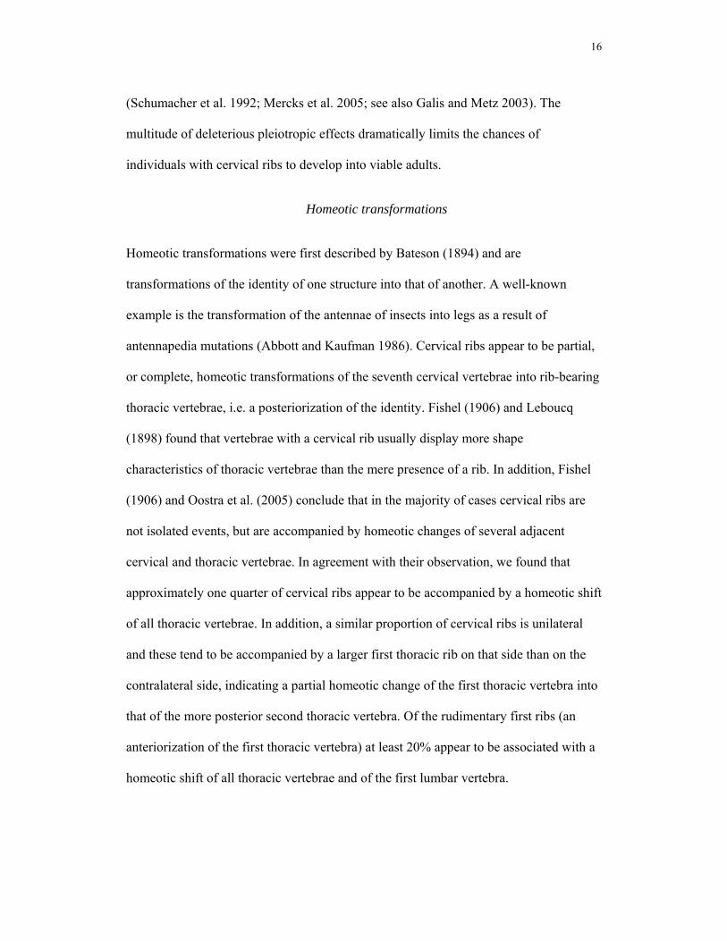
Homeotic transformations .......................................................................................... 16
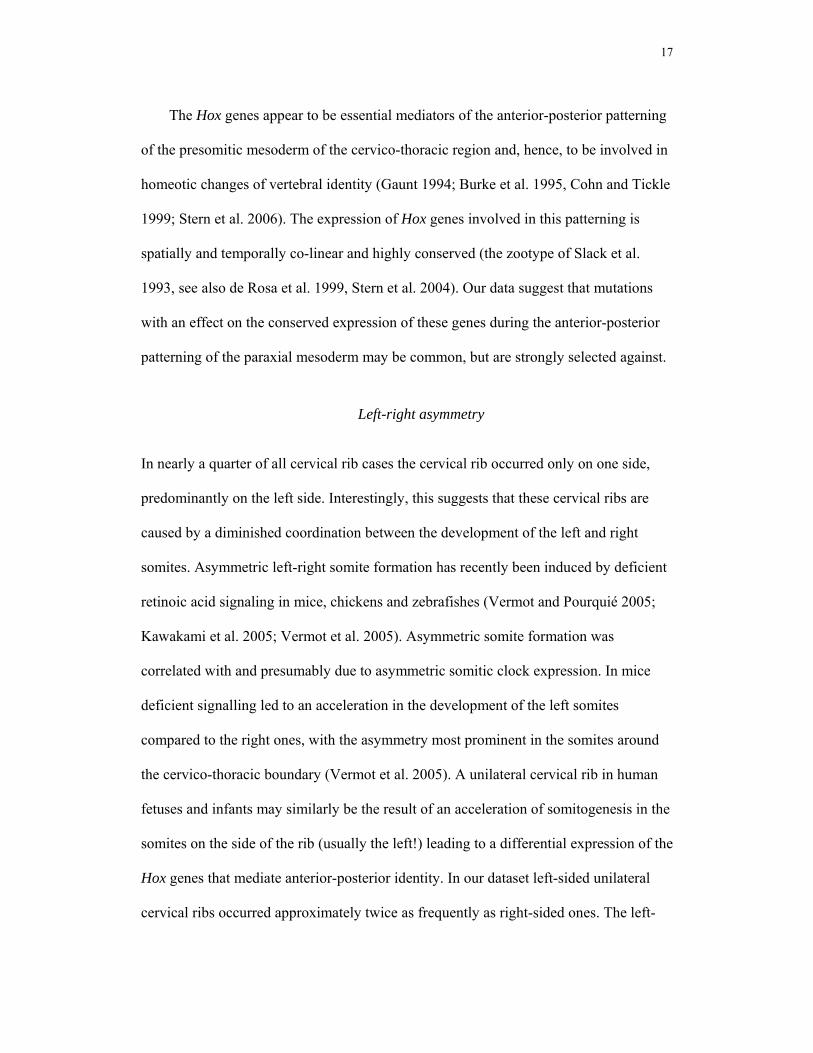
Left-right asymmetry.................................................................................................. 17
Interactivity during the early patterning of the anterior-posterior axis ...................... 18
Modularity, stabilizing selection and conservation .................................................... 20
Acknowledgements ........................................................................................................ 22
Literature cited................................................................................................................ 22
Tables ............................................................................................................................. 33
Figures ............................................................................................................................ 37
1
EXTREME SELECTION IN HUMANS AGAINST HOMEOTIC
TRANSFORMATIONS OF CERVICAL VERTEBRAE
Frietson Galis1, Tom J. M. Van Dooren1, Johan D. Feuth2, Johan A. J. Metz1,3, Andrea
Witkam, Sebastiaan Ruinard1, Marc J. Steigenga1,5, Liliane C. D. Wijnaendts4
1. Institute of Biology, Leiden University, P.O. Box 9516, 2300 RA Leiden, The
Netherlands
2. Department of Surgery, Leiden University Medical Centre, Leiden, The Netherlands
3. International Institute for Applied Systems Analysis, Evolution and Ecology Program
A-2361 Laxenburg, Austria
4. Department of Pathology, Free University Medical Centre (VUMC), 1081 BT,
Amsterdam, The Netherlands
5. Present address: Department of animal ecology I, University of Bayreuth, D-95440,
Bayreuth, Germany
Main Text: 5125 words, abstract 161 words, 3 figures, 4 tables.
LRH: F. Galis et al.
RRH: Extreme selection against homeotic changes
Key words: Body plans, Hox genes, pleiotropy, developmental constraint, modularity, phylotypic stage, evolutionary medicine
2
ABSTRACT
Why do all mammals, except for sloths and manatees, have exactly seven cervical
vertebrae? In other vertebrates and other regions, the vertebral number varies
considerably. We have investigated whether natural selection constrains the number of
cervical vertebrae in humans. To this end, we have determined the incidence of cervical
ribs and other homeotic vertebral changes in radiographs of deceased human fetuses and
infants, and analysed several existing datasets on the incidence in infants and adults.
Our data show that homeotic transformations that change the number of cervical
vertebrae are extremely common in humans, but are strongly selected against: almost all
individuals die before reproduction. Selection is most probably indirect, caused by a
strong coupling of such changes with major congenital abnormalities. Changes in the
number of thoracic vertebrae appear to be subject to weaker selection, in good
correspondence with the weaker evolutionary constraint on these numbers. Our analysis
highlights the role of prenatal selection in the conservation of our common body plan.

3
The exceedingly low level of interspecific variation in the number of cervical vertebrae
of mammals has puzzled biologists for more than 150 years (E.g. Cuvier 1835; Flower
and Lydekker 1891). In birds, reptiles and amphibians, this number varies considerably,
and in mammals the number of vertebrae in other vertebral regions is variable as well
(Fishel 1906; Galis 1999; Narita and Kuratani 2005). Thus, there appears to be an
evolutionary constraint on variations in the mammalian cervical region. Earlier we have
suggested that this constraint is the result of selection against deleterious pleiotropic
effects associated with variations in the number of cervical vertebrae (Galis 1999).
According to this hypothesis selection is, thus, indirect: variations of the number of
cervical vertebrae are not disadvantageous as such, but they are associated with other
disadvantageous changes. Support for the hypothesis comes from the observation that
changes in the number of cervical vertebrae appear to be associated with an increased
susceptibility to pediatric cancers, congenital abnormalities and still births in humans
and mice (Gladstone and Wakeley 1932; Adson and Coffey 1947; Schumacher et al.
1992; Keeling and Kjaer 1999; Galis and Metz 2003; Merks et al. 2005; Steigenga et al.
2006). In addition, the number of cervical vertebrae is determined during the early
organogenesis stage and the conservation of this stage is thought to be the result of
selection against deleterious pleiotropic effects of mutations that have an effect on this
stage (Sander 1983; Raff 1994; Galis and Metz 2001).
To test this hypothesis, one needs a quantitative study in which the strength of
selection against changes of the number of cervical vertebrae is measured and compared
with the strength of selection against a less effectively constrained evolutionary change,
e.g. changes of the number of thoracic vertebrae. In addition, one needs to establish
whether the observed selective disadvantage is indeed the result of deleterious
pleiotropic effects that are associated with changes in the number of cervical vertebrae.
4
To measure the strength of the selection against variations in the number of cervical
and thoracic vertebrae we have analysed in humans to which extent variations in the
number of cervical and thoracic vertebrae are more prevalent in deceased fetuses and
infants than in individuals that survive to a reproductive age. We have screened 598
fetuses and infants that died between 1992 and 1999 in the VU Medical Center in
Amsterdam for variations in cervical and thoracic vertebral number and compared the
results with existing data on prevalence of vertebral variations in the population at large.
Furthermore, to investigate whether selection against vertebral variations is due to
pleiotropic effects, we have tested for significant associations between variations in
vertebral numbers and congenital abnormalities with deleterious effects.
MATERIAL AND METHODS
Radiographs
Since 1980 all fetuses and infants that are presented for autopsy at the VU Medical
Centre in Amsterdam have been standardly radiographed (23mA, 70-90 kV, 4-12 sec,
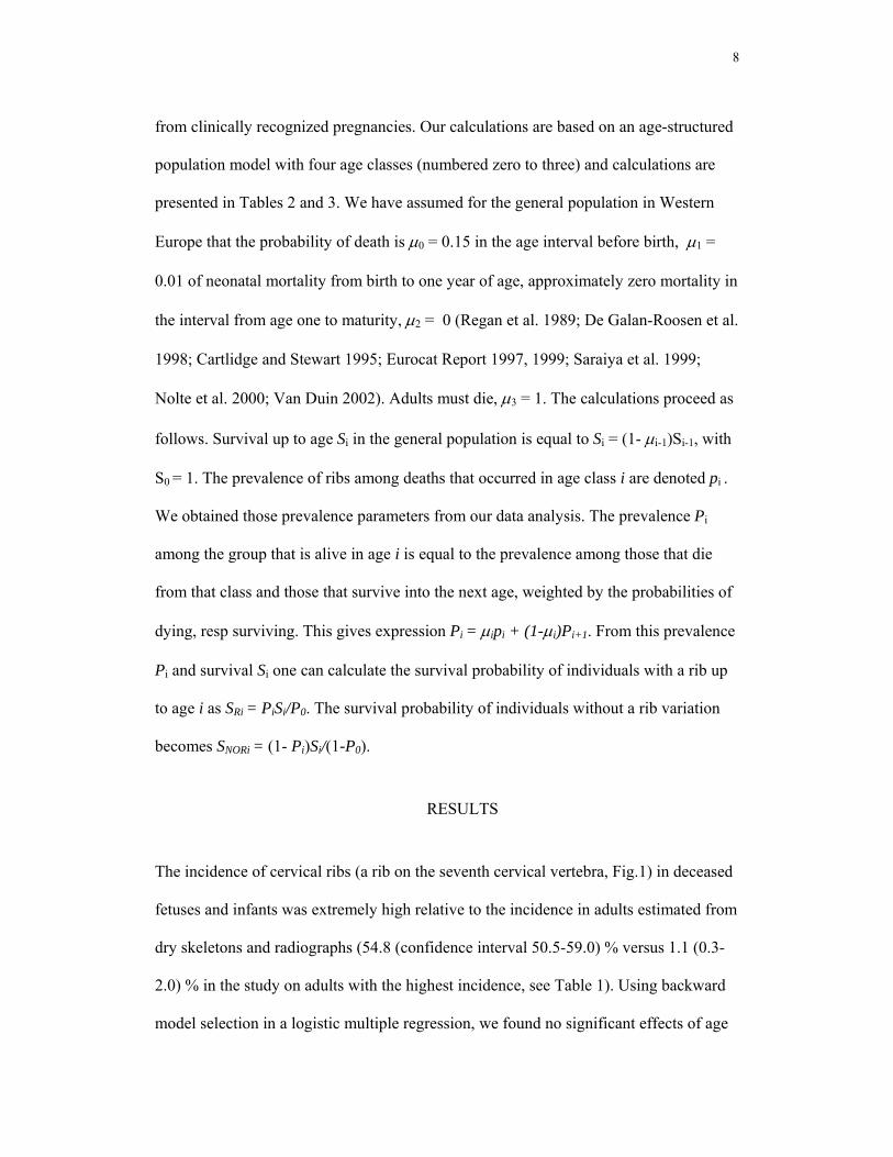
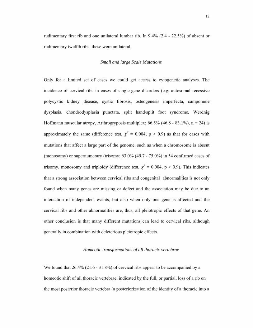
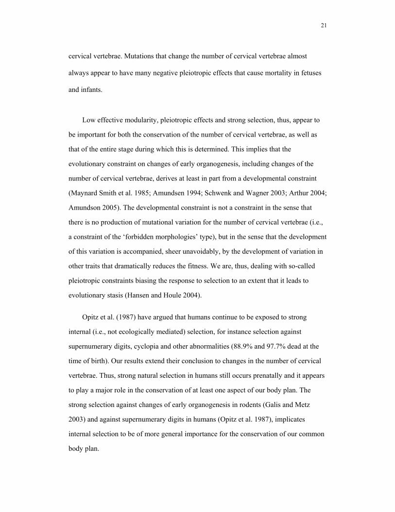
Agfa Gevaert D7DW Structurix films, Figure 1) both ventrally and laterally. We
analysed all radiographs made from 1992-1999 (598 cases). In the analysis, we only
included fetuses older than 14 weeks, since this is the earliest stage at which ossification
centres of cervical ribs can reliably be detected in radiographs (Noback and Robertsen
1951; McNally et al. 1990). ). In total, 30 fetuses were excluded from analysis because
of insufficient ossification. Fetuses from abortions induced for medical reasons (fatal
abnormalities) have been included, except for the calculation of the effect of age at
death. Furthermore, infants were analysed that died before the age of one year. At least
two people independently analysed each radiograph for variations of vertebral numbers,
without prior knowledge of the autopsy reports (however, several congenital anomalies

5
can be seen in radiographs). Radiographs that were difficult to interpret, or where the
interpretation differed between observers, were excluded (44 cases for cervical ribs).
Difficulties in interpretation of vertebral variations were due to either insufficient
contrast, or because the scapula, maxilla or teeth were obstructing the view of potential
cervical ribs. When a cervical rib could only be documented on one side, radiographs
were excluded for the scoring of bilateral symmetry (20 cases) and radiographs with an
obstructed view of the 19th vertebrae were excluded for the scoring of absent or
rudimentary twelfth ribs (4 cases). Absent or rudimentary ribs on the 19th vertebra were
scored as absent or rudimentary 12th ribs, except when a cervical rib was also present
(since somites from which the vertebrae develop arise and are being patterned in rostro-
caudal order). Rudimentary ribs on the 20th vertebra were scored as lumbar ribs except
when a rudimentary first rib was also present. In two cases a cervical rib was present in
combination with the absence of ribs on the 18th vertebra; these cases were scored as a
cervical rib and an absent 12th rib. In two cases a cervical rib was present in
combination with ribs on the 20th vertebra; these cases were scored as a cervical rib and
a lumbar rib.
Diagnosis of abnormalities and diseases
Standard autopsy reports were made by pathologists and filed in a national pathological
archive (PALGA). We searched the reports for single and multiple congenital
abnormalities and, if documented, cytogenetic abnormalities. Furthermore, we
distinguished between minor and major congenital abnormalities following Merks et
al.(2003) for external and skeletal abnormalities and Lancaster and Pedisich (1995) for
further abnormalities. We opted for a classification in terms of minor/major and
single/multiple abnormalities, because it reflects our expectations on the strength of the
deleterious effects of these abnormalities. The condition of the environment (e.g. the
mother) and the condition of the individual both influence the chance of a premature
6
death. Hence, the strength of the deleterious effects of the abnormalities provides
information on the selective disadvantage. Examples of minor congenital abnormalities
are hypertrophy of the heart, spleen, liver or lungs, supernumerary phalanges, club foot,
hydrops foetalis (edema of the whole body), ventricular septal defect (defect in the wall
that separates the left and right ventriculi of the heart), atrial septum defect (defect in the
wall that separates the left and right atria of the heart), patent foramen ovale (persistence
of the fetal opening between left and right atria), patent truncus arteriosus (persistence
of the fetal structure between the left pulmonary artery and the descending aorta),
omphalocele (intestines and other abdominal organs protrude into the base of the
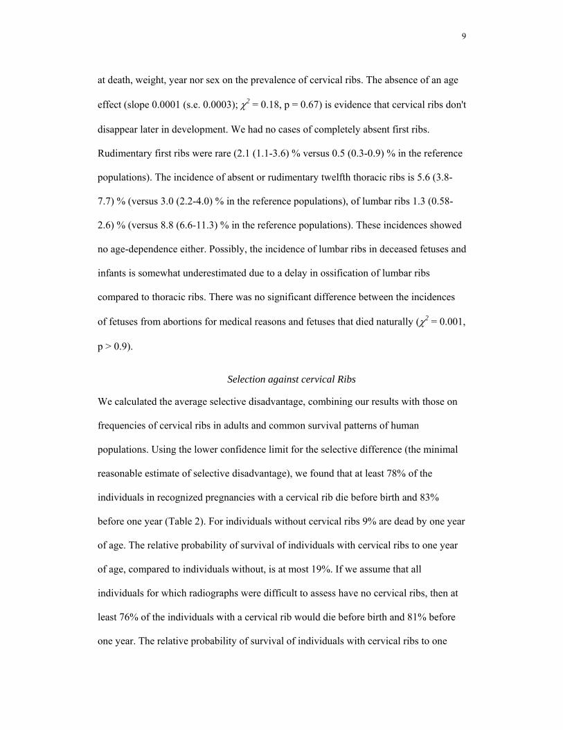
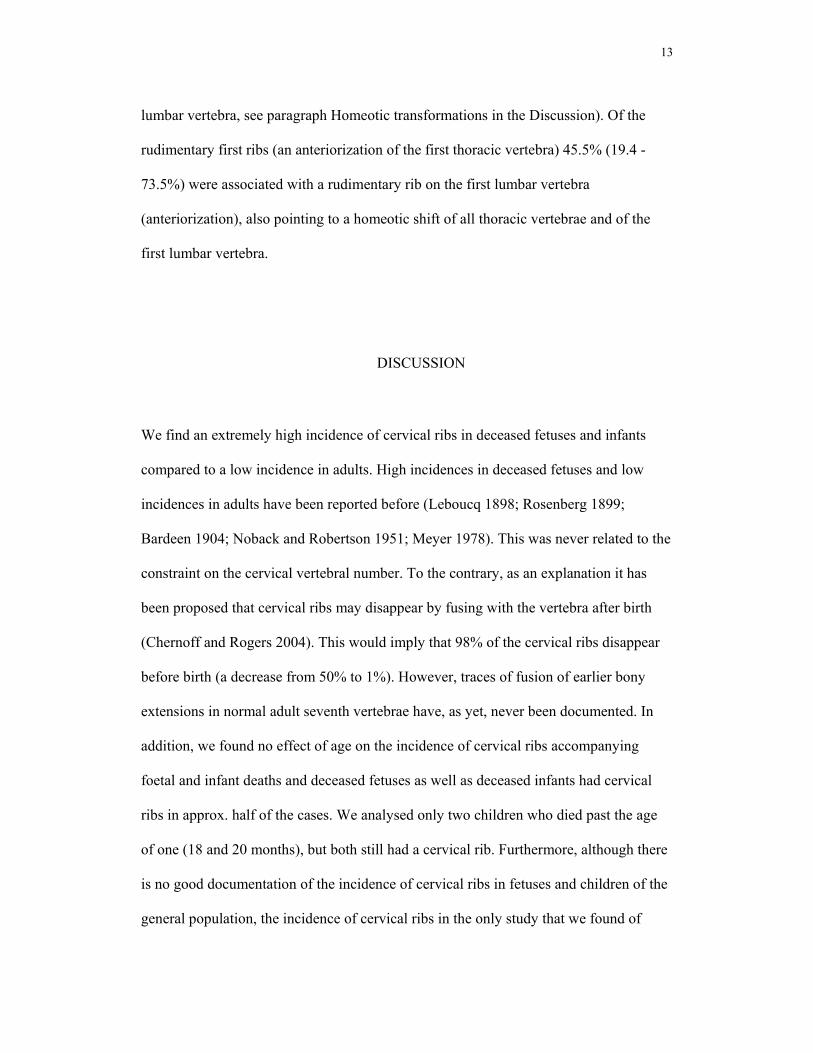
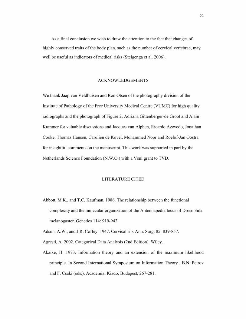
umbilical cord) and slight facial dysmorphologies. Examples of major congenital
abnormalities are the absence of one or both kidneys, anencephaly and other failures of
neural tube closure (Figure 2), absence of the corpus callosum in the brain, cyclops,
cleft lip/palate, tracheo-esophageal fistula (abnormal passage between trachea and
oesophagus), atresia of the aorta, dextroposition of the heart, monoventricular heart,
mono-atrial heart, various chondrodysplasias (abnormally short and deformed limbs)
and sirenomelia (abnormal development of the caudal part of the body with different
degrees of fusion of the legs). In addition we considered dysmaturity and embryonal
tumours to be major abnormalities and prematurity a minor abnormality.
Statistical analysis
We used generalized linear modelling techniques for analysis (Agresti 2002).
Probabilities of occurrence of vertebral variations were studied by means of logistic
regression, associations were investigated using log-linear analysis. In the log-linear
analyses all associations between explanatory variables (types of congenital anomalies)
were fitted in the models, in addition to associations of vertebral variations with
congenital anomalies. This approach was followed because an analysis based on
marginal totals of the contingency table alone can easily lead to incorrect conclusions
7
about an association, i.e., an association between vertebral variations and one
classification variable of congenital abnormalities can reverse sign when the
occurrences in different groups (categories of other classification variables of congenital
anomalies) are pooled (Simpson's paradox, Simpson 1951). In all cases, we checked for
overdispersion of the data relative to the model. We used backward model selection
(from elaborate to simpler) to compare models and test hypotheses. Model comparison
was done using likelihood ratio tests for nested models, and AIC (Akaike Information
criterion, Akaike 1973) for non-nested models. Only test statistics from the selected
logistic regression models and log-linear models are presented. Throughout a
significance level of 95% was used. Confidence intervals reported for probabilities of
occurrence are calculated from logistic regression models (Venables and Ripley 2002).
Incidence of vertebral variations in the general adult population
In order to estimate the prevalence of cervical, rudimentary first, absent or rudimentary
twelfth or lumbar ribs in the general adult population, we analyzed a number of existing
datasets, chosen such that datasets of diseased populations were excluded (see Table 1).
We fitted a binomial regression model for the incidence of each variation, with type of
study (radiographic, dried skeletons) and study as factors. This model was simplified
parameter-by-parameter until all remaining effects were significantly different from
zero. In this manner we lumped studies where differences in incidence were non-
significant, for instance because of relatively low sample sizes of vertebral variations.
The estimates reported for the calculation of the selective disadvantage are the
maximum among the predicted values from the selected model for cervical ribs and the
minimum one for absent or rudimentary twelfth ribs.
Selection against vertebral variations
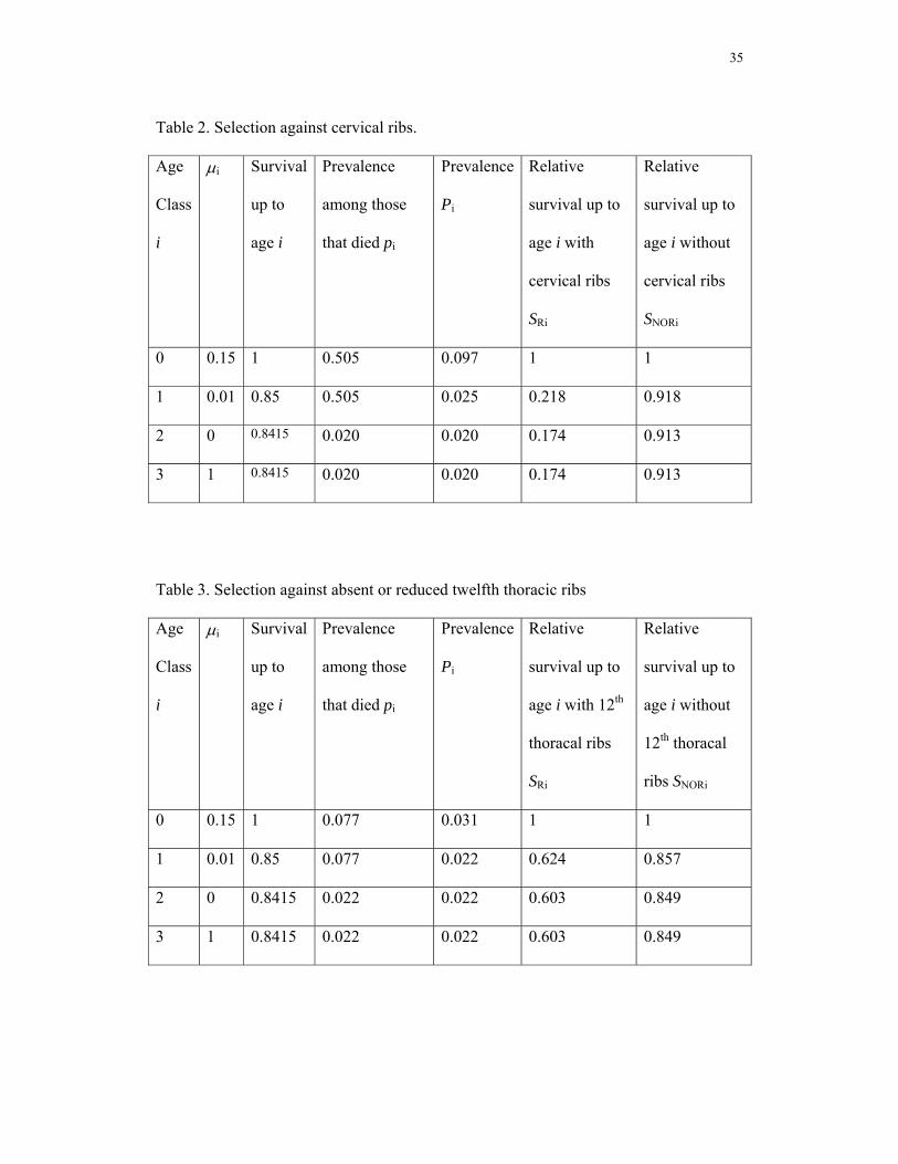
In order to arrive at estimates of selection pressures we calculated the survival pattern of
individuals with or without a cervical rib and an absent or rudimentary 12th rib, starting
8
from clinically recognized pregnancies. Our calculations are based on an age-structured
population model with four age classes (numbered zero to three) and calculations are
presented in Tables 2 and 3. We have assumed for the general population in Western
Europe that the probability of death is μ0 = 0.15 in the age interval before birth, μ1 =
0.01 of neonatal mortality from birth to one year of age, approximately zero mortality in
the interval from age one to maturity, μ2 = 0 (Regan et al. 1989; De Galan-Roosen et al.
1998; Cartlidge and Stewart 1995; Eurocat Report 1997, 1999; Saraiya et al. 1999;
Nolte et al. 2000; Van Duin 2002). Adults must die, μ3 = 1. The calculations proceed as
follows. Survival up to age Si in the general population is equal to Si = (1- μi-1)Si-1, with
S0 = 1. The prevalence of ribs among deaths that occurred in age class i are denoted pi .
We obtained those prevalence parameters from our data analysis. The prevalence Pi
among the group that is alive in age i is equal to the prevalence among those that die
from that class and those that survive into the next age, weighted by the probabilities of
dying, resp surviving. This gives expression Pi = μipi + (1-μi)Pi+1. From this prevalence
Pi and survival Si one can calculate the survival probability of individuals with a rib up
to age i as SRi = PiSi/P0. The survival probability of individuals without a rib variation
becomes SNORi = (1- Pi)Si/(1-P0).
RESULTS
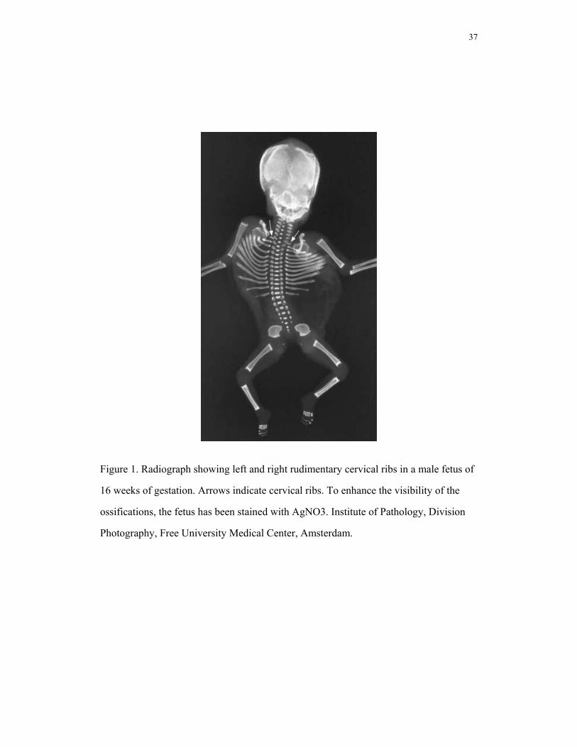
The incidence of cervical ribs (a rib on the seventh cervical vertebra, Fig.1) in deceased
fetuses and infants was extremely high relative to the incidence in adults estimated from
dry skeletons and radiographs (54.8 (confidence interval 50.5-59.0) % versus 1.1 (0.3-
2.0) % in the study on adults with the highest incidence, see Table 1). Using backward
model selection in a logistic multiple regression, we found no significant effects of age
9
at death, weight, year nor sex on the prevalence of cervical ribs. The absence of an age
effect (slope 0.0001 (s.e. 0.0003); χ2 = 0.18, p = 0.67) is evidence that cervical ribs don't
disappear later in development. We had no cases of completely absent first ribs.
Rudimentary first ribs were rare (2.1 (1.1-3.6) % versus 0.5 (0.3-0.9) % in the reference
populations). The incidence of absent or rudimentary twelfth thoracic ribs is 5.6 (3.8-
7.7) % (versus 3.0 (2.2-4.0) % in the reference populations), of lumbar ribs 1.3 (0.58-
2.6) % (versus 8.8 (6.6-11.3) % in the reference populations). These incidences showed
no age-dependence either. Possibly, the incidence of lumbar ribs in deceased fetuses and
infants is somewhat underestimated due to a delay in ossification of lumbar ribs
compared to thoracic ribs. There was no significant difference between the incidences
of fetuses from abortions for medical reasons and fetuses that died naturally (χ2 = 0.001,
p > 0.9).
Selection against cervical Ribs
We calculated the average selective disadvantage, combining our results with those on
frequencies of cervical ribs in adults and common survival patterns of human
populations. Using the lower confidence limit for the selective difference (the minimal
reasonable estimate of selective disadvantage), we found that at least 78% of the
individuals in recognized pregnancies with a cervical rib die before birth and 83%
before one year (Table 2). For individuals without cervical ribs 9% are dead by one year
of age. The relative probability of survival of individuals with cervical ribs to one year
of age, compared to individuals without, is at most 19%. If we assume that all
individuals for which radiographs were difficult to assess have no cervical ribs, then at
least 76% of the individuals with a cervical rib would die before birth and 81% before
one year. The relative probability of survival of individuals with cervical ribs to one
10
year of age, compared to individuals without, would increase to 21%. One can conclude
that our conclusions are at most little biased by the missing observations.
Weaker Selection against a Change at the thoraco-lumbar Boundary
We repeated the above calculations for absent and rudimentary twelfth thoracic ribs,
using the upper confidence limit for the selective difference (the maximal reasonable
estimate of selective disadvantage). We found that at most 38% of the individuals of
recognized pregnancies with an absent or rudimentary twelfth rib die before birth and
40% before age one (Table 3). Hence, the relative probability of survival of individuals
with absent or rudimentary twelfth ribs compared to individuals without this variation is
73%. Assuming that all four individuals for which radiographs were difficult to assess at
the thoraco-lumbar boundary had absent or rudimentary 12th thoracic ribs, this relative
probability decreases to 70%. Thus, this conservative calculation indicates that selection
against a change at the thoraco-lumbar boundary is much weaker than that against a
change at the cervico-thoracic one.
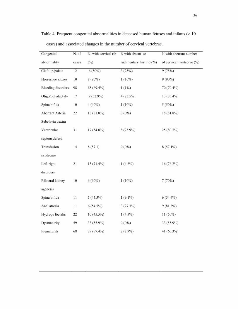
Association of cervical ribs with congenital abnormalities
The incidence of major anomalies in deceased fetuses and infants was 66.7 (62.4 – 70.9)
% . In 64.2 (59.7 – 68.5) % of the cases, multiple abnormalities were present. As a next
step in our analysis, we investigated whether an association with congenital
abnormalities could possibly explain the observed early deaths. The incidence of
cervical ribs was indeed found to be positively associated either with multiple or with
major congenital anomalies (both P[>χ2] = 0.001, see also the raw data in Table 4). A
model including an effect of major anomalies fits the data better than one including
11
multiple anomalies, based on the AIC (AIC major anomalies 611.1, AIC multiple
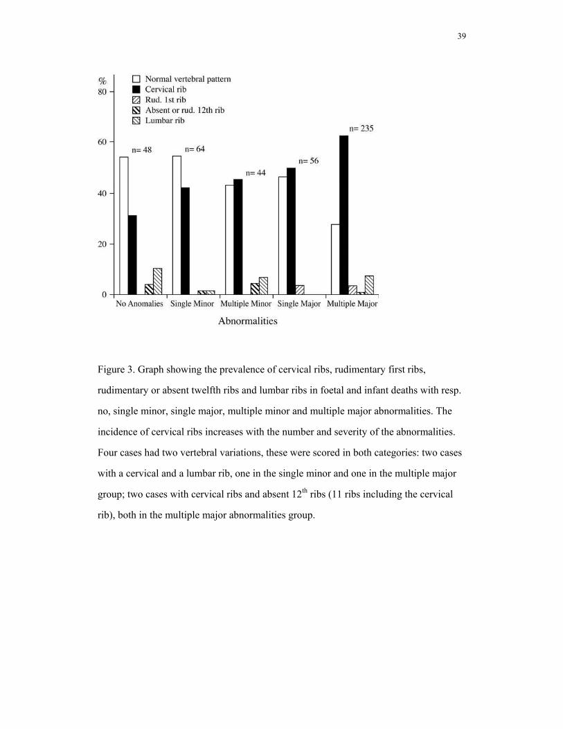
anomalies 613.9) The highest incidence of cervical ribs (63.6% (57.4-69.4%), Fig.3)
was found in fetuses and infants with both multiple and major abnormalities. This
significant association between cervical ribs and the most deleterious class of
abnormalities, and the parallel association between cervical ribs and the total number of
mildly or strongly deleterious effects supports our hypothesis that the presence of
cervical ribs is selectively disadvantageous because of associated negative pleiotropic
effects.
Next to a positive association with major or multiple anomalies, no further significant,
or higher-order, associations of variables with the incidence of cervical vertebrae were
found. We found no effects of age or weight on the association and no association with
sex. No other vertebral variations were significantly associated with any of the
mentioned variables. It is of some interest to note that the few cases of rudimentary first
ribs were always associated with major congenital abnormalities, even though the
incidence was too low for reaching significance.
Unilateral ribs
In 22.8% (18.0 - 28.0 %) of all cervical rib cases the cervical rib occurred only on one
side. In our dataset left-sided unilateral cervical ribs occurred approximately twice as
often as right-sided ones (39 vs. 22, χ2-test, p = 0.02). In the log-linear analysis, the
association between major or multiple congenital abnormalities and cervical ribs was
not different for unilateral and bilateral cervical ribs (e.g., difference unilateral×major
0.17 (s.e. 0.39), χ2 = 0.1, P[>χ2] = 0.75). In addition, we found one unilateral
12
rudimentary first rib and one unilateral lumbar rib. In 9.4% (2.4 - 22.5%) of absent or
rudimentary twelfth ribs, these were unilateral.
Small and large Scale Mutations
Only for a limited set of cases we could get access to cytogenetic analyses. The
incidence of cervical ribs in cases of single-gene disorders (e.g. autosomal recessive
polycystic kidney disease, cystic fibrosis, osteogenesis imperfecta, campomele
dysplasia, chondrodysplasia punctata, split hand/split foot syndrome, Werdnig
Hoffmann muscular atropy, Arthrogryposis multiplex; 66.5% (46.8 - 83.1%), n = 24) is
approximately the same (difference test, χ2 = 0.004, p > 0.9) as that for cases with
mutations that affect a large part of the genome, such as when a chromosome is absent
(monosomy) or supernumerary (trisomy; 63.0% (49.7 - 75.0%) in 54 confirmed cases of
trisomy, monosomy and triploidy (difference test, χ2 = 0.004, p > 0.9). This indicates
that a strong association between cervical ribs and congenital abnormalities is not only
found when many genes are missing or defect and the association may be due to an
interaction of independent events, but also when only one gene is affected and the
cervical ribs and other abnormalities are, thus, all pleiotropic effects of that gene. An
other conclusion is that many different mutations can lead to cervical ribs, although
generally in combination with deleterious pleiotropic effects.
Homeotic transformations of all thoracic vertebrae
We found that 26.4% (21.6 - 31.8%) of cervical ribs appear to be accompanied by a
homeotic shift of all thoracic vertebrae, indicated by the full, or partial, loss of a rib on
the most posterior thoracic vertebra (a posteriorization of the identity of a thoracic into a
13
lumbar vertebra, see paragraph Homeotic transformations in the Discussion). Of the
rudimentary first ribs (an anteriorization of the first thoracic vertebra) 45.5% (19.4 -
73.5%) were associated with a rudimentary rib on the first lumbar vertebra
(anteriorization), also pointing to a homeotic shift of all thoracic vertebrae and of the
first lumbar vertebra.
DISCUSSION
We find an extremely high incidence of cervical ribs in deceased fetuses and infants
compared to a low incidence in adults. High incidences in deceased fetuses and low
incidences in adults have been reported before (Leboucq 1898; Rosenberg 1899;
Bardeen 1904; Noback and Robertson 1951; Meyer 1978). This was never related to the
constraint on the cervical vertebral number. To the contrary, as an explanation it has
been proposed that cervical ribs may disappear by fusing with the vertebra after birth
(Chernoff and Rogers 2004). This would imply that 98% of the cervical ribs disappear
before birth (a decrease from 50% to 1%). However, traces of fusion of earlier bony
extensions in normal adult seventh vertebrae have, as yet, never been documented. In
addition, we found no effect of age on the incidence of cervical ribs accompanying
foetal and infant deaths and deceased fetuses as well as deceased infants had cervical
ribs in approx. half of the cases. We analysed only two children who died past the age
of one (18 and 20 months), but both still had a cervical rib. Furthermore, although there
is no good documentation of the incidence of cervical ribs in fetuses and children of the
general population, the incidence of cervical ribs in the only study that we found of
14
healthy children (25,949 healthy, female child-relatives of Tuberculosis patients,
Menárguez Carretero and Campo Muňoz 1967) is well within the range found for adults
of the general population (0.5% for children versus 0.04-1.07% for adults, see Table 1).
Furthermore, the incidence of cervical ribs in populations of diseased children and
adults also appears to be very similar and varies between one and six percent (mainly
patients with infections and pulmonary diseases, Davis and King 1939; Schumacher et
al. 1992; Mercks et al. 2005 versus Coury and Delaporte 1954; Pionnier and Depraz
1956; Whittaker 1957; Erken 2002). These comparisons do not support a dramatic
decrease in incidence of cervical rib frequency during infanthood or childhood. A
higher incidence of cervical ribs in children was only found in children with embryonal
tumours (Schumacher et al. 1992; Mercks et al. 2005), an association which probably
provides further selection against cervical vertebrae (Galis 1999; Galis and Metz 2003).
Finally, half of the cervical rib cases in our dataset were not isolated, and were
accompanied by posteriorizations of the identity of one or more thoracic vertebrae. This
supports our assumption that cervical ribs in fetuses and infants do not indicate normal
development of a seventh cervical vertebra with a transiently present rib, but instead
represent a homeotic transformation of the seventh cervical vertebra into that of
adjacent, rib-bearing, first thoracic vertebra. We conclude, therefore, that the differences
between adult and prenatal incidence of cervical ribs are mainly due to early differential
mortality of individuals with a cervical rib.
Strong selection against Changes of the Number of cervical Vertebrae
Our data, thus, indicate very strong selection against changes in the number of cervical
vertebrae. The most frequent changes were bilateral and unilateral cervical ribs.
15
Rudimentary first ribs were surprisingly rare, but without exception associated with
major abnormalities. It is possible that we underestimate the incidence of rudimentary
first ribs if they are frequently associated with abnormalities that lead to a very early
death, i.e. before the ribs ossify and we can detect them on radiographs. It is also
possible that rudimentary first ribs are rarer. This needs further investigation.
We found that selection against changes at the thoraco-lumbar boundary is much
weaker than that against a change at the cervico-thoracic one. The number of thoracic
vertebrae indeed varies much more amongst mammals than the number of cervical
vertebrae (Galis 1999; Narita and Kuratani 2005), attesting to a good agreement
between the strength of selection and the apparent evolutionary constraints.
Selection against pleiotropic effects
It is highly improbable that the mere presence of cervical ribs would lead to fetal or
infant deaths. Direct problems related to cervical ribs, compressions of nerves and
arteries (thoracic outlet syndrome), usually only appear in adults (Makhoul and
Machleder 1992; Roos 1996). Apparently, the constraint on the number of cervical
vertebrae should mainly be sought in selection against congenital abnormalities
associated with changes of this number (i.e., pleiotropic effects, Galis 1999). Our
analysis is in agreement with earlier found associations of cervical ribs with
abnormalities (Gladstone and Wakeley 1932; Adson and Coffey 1947; Keeling and
Kjaer 1999; Steigenga et al. 2006) and with the absence in our dataset of such an
association for rudimentary twelfth ribs. In addition, individuals with cervical ribs that
survive infanthood experience on average more cancer than individuals without cervical
ribs. There is a significant association of cervical ribs with specific pediatric cancers
16
(Schumacher et al. 1992; Mercks et al. 2005; see also Galis and Metz 2003). The
multitude of deleterious pleiotropic effects dramatically limits the chances of
individuals with cervical ribs to develop into viable adults.
Homeotic transformations
Homeotic transformations were first described by Bateson (1894) and are
transformations of the identity of one structure into that of another. A well-known
example is the transformation of the antennae of insects into legs as a result of
antennapedia mutations (Abbott and Kaufman 1986). Cervical ribs appear to be partial,
or complete, homeotic transformations of the seventh cervical vertebrae into rib-bearing
thoracic vertebrae, i.e. a posteriorization of the identity. Fishel (1906) and Leboucq
(1898) found that vertebrae with a cervical rib usually display more shape
characteristics of thoracic vertebrae than the mere presence of a rib. In addition, Fishel
(1906) and Oostra et al. (2005) conclude that in the majority of cases cervical ribs are
not isolated events, but are accompanied by homeotic changes of several adjacent
cervical and thoracic vertebrae. In agreement with their observation, we found that
approximately one quarter of cervical ribs appear to be accompanied by a homeotic shift
of all thoracic vertebrae. In addition, a similar proportion of cervical ribs is unilateral
and these tend to be accompanied by a larger first thoracic rib on that side than on the
contralateral side, indicating a partial homeotic change of the first thoracic vertebra into
that of the more posterior second thoracic vertebra. Of the rudimentary first ribs (an
anteriorization of the first thoracic vertebra) at least 20% appear to be associated with a
homeotic shift of all thoracic vertebrae and of the first lumbar vertebra.
17
The Hox genes appear to be essential mediators of the anterior-posterior patterning
of the presomitic mesoderm of the cervico-thoracic region and, hence, to be involved in
homeotic changes of vertebral identity (Gaunt 1994; Burke et al. 1995, Cohn and Tickle
1999; Stern et al. 2006). The expression of Hox genes involved in this patterning is
spatially and temporally co-linear and highly conserved (the zootype of Slack et al.
1993, see also de Rosa et al. 1999, Stern et al. 2004). Our data suggest that mutations
with an effect on the conserved expression of these genes during the anterior-posterior
patterning of the paraxial mesoderm may be common, but are strongly selected against.
Left-right asymmetry
In nearly a quarter of all cervical rib cases the cervical rib occurred only on one side,
predominantly on the left side. Interestingly, this suggests that these cervical ribs are
caused by a diminished coordination between the development of the left and right
somites. Asymmetric left-right somite formation has recently been induced by deficient
retinoic acid signaling in mice, chickens and zebrafishes (Vermot and Pourquié 2005;
Kawakami et al. 2005; Vermot et al. 2005). Asymmetric somite formation was
correlated with and presumably due to asymmetric somitic clock expression. In mice
deficient signalling led to an acceleration in the development of the left somites
compared to the right ones, with the asymmetry most prominent in the somites around
the cervico-thoracic boundary (Vermot et al. 2005). A unilateral cervical rib in human
fetuses and infants may similarly be the result of an acceleration of somitogenesis in the
somites on the side of the rib (usually the left!) leading to a differential expression of the
Hox genes that mediate anterior-posterior identity. In our dataset left-sided unilateral
cervical ribs occurred approximately twice as frequently as right-sided ones. The left-
18
right asymmetries in vertebrae were never accompanied by other left-right asymmetries,
such as asplenia, abnormal lung lobation and intestinal malrotation. This pattern is in
full agreement with the independence found in mice and zebrafishes for the left-right
symmetric development of the somites and the left-right asymmetry of internal organs
(Vermot and Pourquié 2005; Kawakami et al. 2005).
Interactivity during the early patterning of the anterior-posterior axis
The determination of the position of the cervico-thoracic boundary mediated by the Hox
genes forms part of the early anterior-posterior patterning of the presomitic mesoderm
during the early neurula stage (e.g. Gaunt 1994; Burke et al. 1995; Cohn and Tickle
1999; Chernoff and Rogers 2004; Stern et al. 2006). The fact that more than half of all
fetal and infant deaths come with cervical ribs emphasizes once again the vulnerability
of the early organogenesis, or neurula stage (Galis and Metz 2003). The association of
cervical ribs with multiple and with major abnormalities in other parts of the body (see
Table 4) moreover points at the interaction of the early anterior-posterior patterning of
the paraxial mesoderm with many other patterning processes and many morphogenetic
processes. Corroboration for this viewpoint is, firstly, provided by grafting experiments
in which the anterior-posterior position of paraxial mesoderm was changed and which
led to changes in a) the anterior-posterior patterning of the adjacent neuroepithelium
(Bel-Vialar 2002; see also Grapin-Botton et al. 1997; Ensini et al. 1998), b) the timing
of the migration of neural crest cells (Sela-Donenfeld and Kalcheim 2000) and c) the
initiation and outgrowth of the limbs (Saito et al. 2006). Secondly, this viewpoint is
corroborated by experiments in which two processes that are involved in the
determination of the anterior-posterior patterning of paraxial mesoderm were

19
manipulated: the opposing and antagonistic gradient of Fgfs, Wnts and RA and the
oscillatory gene expression (somatic clock) in the paraxial mesoderm. These
experiments have demonstrated couplings of the anterior-posterior patterning of
paraxial mesoderm with morphogenetic processes such as proliferation and axial
lengthening (Dubrulle et al. 2001; Dubrulle and Pourquié 2004), somitogenesis (Zakany
et al. 2001; Dubrulle et al. 2001), convergent extension (Ninomiya et al. 2004, see also
Mathis et al. 2001) and cell migration (Yang et al. 2001), as well as with patterning
along the other embryonic axes, i.e. left-right and midline patterning (Raya et al. 2004,
Krebs et al. 2003 see also Yamamoto et al. 2003 and Latimer et al. 2002) and dorso-
ventral patterning (Diez del corral et al. 2003). There is thus a wealth of data supporting
the strong coordination of the patterning of the three embryonic axes in the three
adjacent germ-layers with a central role of the mesoderm in this process (see also
Kumar et al. 2003) and, additionally, there is strong support for a coupling between
patterning and morphogenetic processes.
The determination of the thoraco-lumbar boundary occurs later than that of the
cervico-thoracic boundary. The lower frequency of shifts of this boundary suggests that
this determination is less vulnerable. A lower interactivity is suggested by the absence
of a significant association between shifts of this boundary and congenital
abnormalities. The supposedly lower interactivity and vulnerability of this later stage at
which the number of thoracic vertebrae is determined is a potential explanation for the
weaker evolutionary constraint on changes of the number of thoracic vertebrae.
20
Modularity, stabilizing Selection and Conservation
The number of cervical vertebrae is determined during the early organogenesis stage
(also referred to as phylotypic stage) that is itself strongly conserved in mammals (for a
review see Hall 1999, but see Richardson et al. 1997). Sander (1983) and Raff (1994)
proposed that the high interactivity between modules is the major cause of conservation
in this stage. This high interactivity causes mutations to have negative pleiotropic
effects that become amplified as development proceeds. Conservation is a consequence
of consistently strong stabilizing selection against mutations via their pleiotropic effects.
We earlier found support for the validity of this hypothesis in an analysis of
teratological studies in rodents (Galis and Metz 2001). We found that chemical and
other disturbances of this stage (phenocopies of mutations) lead to a considerably higher
mortality than disturbances of earlier and later developmental stages. From the pattern
of multiple induced abnormalities (pleiotropic effects), we concluded that it is the high
interactivity and low effective modularity that is the root cause of the vulnerability of
the stage: a particular, potentially useful change almost always will induce lethality
even before the organism is exposed to ecological selection.
During early organogenesis the organ primordia make their first appearance.
Hence the evolutionary conservation of early organogenesis in mammals may well be
implicated in the conservation of the number of repeated organs (eyes, ears, vertebrae,
limbs and digits; for the latter, see e.g. Galis et al. 2002). The present study indeed
strongly suggests that during organogenesis the high interactivity and low modularity of
the patterning of the anterior-posterior axis in the cervical paraxial mesoderm appears to
be the root cause for the selective early deaths of humans with a changed number of
21
cervical vertebrae. Mutations that change the number of cervical vertebrae almost
always appear to have many negative pleiotropic effects that cause mortality in fetuses
and infants.
Low effective modularity, pleiotropic effects and strong selection, thus, appear to
be important for both the conservation of the number of cervical vertebrae, as well as
that of the entire stage during which this is determined. This implies that the
evolutionary constraint on changes of early organogenesis, including changes of the
number of cervical vertebrae, derives at least in part from a developmental constraint
(Maynard Smith et al. 1985; Amundsen 1994; Schwenk and Wagner 2003; Arthur 2004;
Amundson 2005). The developmental constraint is not a constraint in the sense that
there is no production of mutational variation for the number of cervical vertebrae (i.e.,
a constraint of the ‘forbidden morphologies’ type), but in the sense that the development
of this variation is accompanied, sheer unavoidably, by the development of variation in
other traits that dramatically reduces the fitness. We are, thus, dealing with so-called
pleiotropic constraints biasing the response to selection to an extent that it leads to
evolutionary stasis (Hansen and Houle 2004).
Opitz et al. (1987) have argued that humans continue to be exposed to strong
internal (i.e., not ecologically mediated) selection, for instance selection against
supernumerary digits, cyclopia and other abnormalities (88.9% and 97.7% dead at the
time of birth). Our results extend their conclusion to changes in the number of cervical
vertebrae. Thus, strong natural selection in humans still occurs prenatally and it appears
to play a major role in the conservation of at least one aspect of our body plan. The
strong selection against changes of early organogenesis in rodents (Galis and Metz
2003) and against supernumerary digits in humans (Opitz et al. 1987), implicates
internal selection to be of more general importance for the conservation of our common
body plan.
22
As a final conclusion we wish to draw the attention to the fact that changes of
highly conserved traits of the body plan, such as the number of cervical vertebrae, may
well be useful as indicators of medical risks (Steigenga et al. 2006).
ACKNOWLEDGEMENTS
We thank Jaap van Veldhuisen and Ron Otsen of the photography division of the
Institute of Pathology of the Free University Medical Centre (VUMC) for high quality
radiographs and the photograph of Figure 2, Adriana Gittenberger-de Groot and Alain
Kummer for valuable discussions and Jacques van Alphen, Ricardo Azevedo, Jonathan
Cooke, Thomas Hansen, Carolien de Kovel, Mohammed Noor and Roelof-Jan Oostra
for insightful comments on the manuscript. This work was supported in part by the
Netherlands Science Foundation (N.W.O.) with a Veni grant to TVD.
LITERATURE CITED
Abbott, M.K., and T.C. Kaufman. 1986. The relationship between the functional
complexity and the molecular organization of the Antennapedia locus of Drosophila
melanogaster. Genetics 114: 919-942.
Adson, A.W., and J.R. Coffey. 1947. Cervical rib. Ann. Surg. 85: 839-857.
Agresti, A. 2002. Categorical Data Analysis (2nd Edition). Wiley.
Akaike, H. 1973. Information theory and an extension of the maximum likelihood
principle. In Second International Symposium on Information Theory , B.N. Petrov
and F. Csaki (eds.), Academiai Kiado, Budapest, 267-281.
23
Amundson, R. 1994. Two Concepts of Constraint: Adaptationism and the Challenge
from Developmental Biology. Phil. Sci.61: 556-578.
Amundson, R. 2005. The Changing Role of the Embryo in Evolutionary Thought: Roots
of Evo-Devo. Cambridge University Press, Cambridge, U.K.
Ancel, P., and L. Sencert. 1902. De quelques variations dans la nombre des vertèbres
chez l'homme leur interpretation. J. de l’Anat. et Physiol. 38: 218-257.
Arthur, W. 2004. Biased embryos and evolution. Cambridge University Press,
Cambridge, U.K.
Bardeen, Ch.R. 1904. Numerical vertebral variation in the human adult and embryo.
Anat. Anz. 25: 497-519.
Bateson, W. 1894. Materials for the study of variation, MacMillan, London.
Bell-Vialar S., N. Itasaki, and R. Krumlauf. 2002. Initiating Hox gene expression: in the
early chick neural tube differential sensitivity to FGF and RA signalling subdivides
the HoxB genes in two distinct groups. Development 129: 5103-5115.
Berner, F. 1944. über Rippenanomalien auf Grund von 6 Millionen Reihenbildern.
Fortschr. Röntgenstr. 69: 202-221.
Bianchi, S. 1894. Sulla frequenza della anomalie numeriche vertebrali nello scheletro
dei normali e degli alienati. Atti della R.. Accad. Dei Fisiocritici in Siena 7: 21-31.
Burke, A.C., C.E. Nelson, B.A. Morgan, and C. Tabin. 1995. Hox genes and the
evolution of vertebrate axial morphology. Development 121: 333-346.
Cartlidge, P.H.T., and J.H. Stewart. 1995. Effect of changing the stillbirth definition on
evaluation of perinatal mortality rates. Lancet 346: 486-488.
24
Chernoff, N., and J.M. Rogers. 2004. Supernumerary ribs in developmental toxicity
bioassays and in human populations: incidence and biological significance. J.
Toxicol. Environ. Health B. Crit. Rev. 7: 437-49.
Cohn, M.J., and C. Tickle. 1999. Developmental basis of limblessness and axial
patterning in snakes. Nature 399: 474-479.
Coury, C., and J. Delaporte. 1954. Les anomalies congenitales des côtes. Sem. Hop.
Paris: 2656-2673.
Crimm, P.D. 1952. Evaluation of a five year minifilm program. Am. J. Roentg. 68: 240-
246.
Cuvier, G. 1835. Leçons d’ anatomie comparée. Tome premier, 2nd Edn. Crochard,
Paris.
Davidson, A. J., Ernst, P., Wang, Y., Dekens, M. P., Kingsley, P. d., Palis, J.
Korsmeyer, S. J., Daley G. Q., and Zon L. I. 2003. cdx4 mutants fail to specify
blood progenitors and can be rescued by multiple hox genes. Nature 425: 300-306.
Davis, D.B., and J.C. King. 1939. Cervical rib in early life. Am. J. Diseases Children
56: 744-755.
De Galan-Roosen, A.E.M, J.C. Kuijpers, A.P. Meershoek, and D. van Velzen. 1998.
Contribution of congenital malformations to perinatal mortality. A 10 years
prospective regional study in The Netherlands, Eur. J. Obstet. Gyn. Reprod. Biol.
80: 55-61.
De Rosa, R., J. K. Grenier, J. Andreeva, C.E. Cook, A. Adoutte, M. Akam, S. B.
Carroll, and G. Balavoine. 1999. Hox genes in brachiopods and priapulids and
protostome evolution. Nature 399: 772-776.
25
Diez del Corral, R., I. Olivera-Martinez, A. Goriely, E. Gale, M. Maden, and K. Storey
2003. Opposing FGF and retinoid pathways control ventral neural pattern, neuronal
differentiation, and segmentation during body axis extension. Neuron 40: 65-79.
Dubrulle, J., McGrew, M. J., and O. Pourquié. 2001. FGF signaling controls somite
boundary position and regulates segmentation clock control of spatiotemporal Hox
gene activation. Cell 106: 219-232.
Dubrulle J., and O. Pourquié. 2004. fgf8 mRNA decay establishes a gradient that
couples axial elongation to patterning in the vertebrate embryo. Nature 427: 419-
422.
Ensini, M., Tsuchida, T. N., Belting, H.-G., and T. M. Jessell. 1998. The control of
rostrocaudal pattern in the developing spinal cord: specification of motor neuron
subtype identity is initiated by signals from paraxial mesoderm. Development 125:
969-982.
Erken, E., H.T.E. Ozer, B. Gulek, B., and B. Durgun. 2002. The association between
cervical rib and sacralization. Spine 27: 1659-1664
Etter, L.E. 1944. Osseous abnormalities of the thoracic cage seen in forty thousand
consecutive chest photoroentgenograms. Am. J. Roentg. 5: 359-363.
Eurocat Update to Report 7. 1999. Prevalence of Congenital Anomalies in Europe 1995-
6, Brussels.
Fishel, A. 1906. Untersuchungen über die Wirbelsäule und den Brustkorb des
Menschen. Anatomische Hefte 31: 462-588.
Flower, W.H., and R. Lydekker 1891. The study of mammals. Adam and Charles Black,
London.

26
Galis, F. 1999. Why do almost all mammals have seven cervical vertebrae?
Developmental constraints, Hox genes and Cancer. J. exp. Zool. B. (Mol. Dev. Evol.)
285: 19-26.
Galis, F., J.A.J. Metz, and J.J.M. van Alphen 2002. Why five fingers? Evolutionary
constraints on digit numbers. Trends Ecol. Evol. 16: 637-646.
Galis F., and J.A.J. Metz. 2003. Testing the vulnerability of the phylotypic stage.
BioEssays 25: 1035-1039.
Gaunt, S.J. 1994. Conservation in the Hox code during morphological evolution. Int. J.
dev. Biol. 38: 549-552.
Gladstone, R.J., and C.P.G. Wakeley. 1932. Cervical ribs and rudimentary first thoracic
ribs considered from the clinical and etiological standpoints. J. Anat. 66: 334-337.
Grapin-Botton, A., Bonnin, M.-A., and N.M. LeDouarin. 1997. Hox gene induction in
the neural tube depemds on three parameters: competence, signal supply and
paralogue group. Development 124: 849-859.
Hall, B.K. 1999. Evolutionary developmental biology, 2nd edition, Kluwer Academic
Press, Dordrecht.
Hansen, T. F., and D. Houle. 2004. Evolvability, stabilizing selection, and the problem
of stasis. Pp. 130-150 in M. Pigliucci, and K. Preston, eds. Phenotypic integration:
Studying the ecology and evolution of complex phenotypes. Oxford University
Press, Oxford.
Henderson, M.S. 1913. Cervical rib. Report of thirty-one cases. Am. J. Orthop. Surg.
11: 408-430.
27
Kawakami Y., A. Raya, R.M. Raya, C. Rodriguez-Esteban, and J.C. Belmonte. 2005.
Retinoic acid signalling links left-right asymmetric patterning and bilaterally
symmetric somitogenesis in the zebrafish embryo. Nature 435:165-171.
Keating, D.R., and J.R. Amberg.1954. A course of potential error in the roentgen
diagnosis of cervical ribs. Radiology 62: 688-694.
Keeling, J.W., and I. Kjaer. 1999. Cervical ribs: useful marker of monosomy X in fetal
hydrops. Pediat. Devel. Pathol. 2: 119-123.
Krebs, L. T., Iwai, N., Nonaka, S., Welsh, I. C., Lan Y. Jiang, R., Saijoy, Y., O’Brien T.
P., Hamada H., and T. Gridley. 2003. Notch signalling regulates left-right
asymmetry determination by inducing Nodal expression. Genes Dev. 17: 1207-
1212.
Kumar M., N. Jordan, D. Melton, and A. Grapin-Botton. 2003. Signals from lateral
plate mesoderm instruct endoderm toward a pancreatic fate. Dev Biol. 259: 109-22.
Lancaster, P. and E. Pedisich. 1995. Congenital Malformations Australia 1981-1992.
Australian Institute of Health and Welfare, National Perinatal Statistics Unit.
Lanier, R.R.1944. Length of first, twelfth, and accessory ribs in american whites and
negroes; their relationship to certain vertebral variations. Am. J. Physical.
Anthropol. Eurocat Report 7. 1997. 15 Years of Surveillance of Congenital
Anomalies in Europe 1980-1994”, Scientific Institute of Public Health, Brussels.
Latimer, A. J., Dong, X., Markov, Y., and Appel, B. 2002. Delta-Notch signaling
induces hypochord development in zebrafish, Development 129: 2555-2563.
Leboucq, H. 1898. Recherches sur les variations anatomiques de la première côte chez
l’homme. Arch. Biol. 15: 9-178.
28
Makhoul, R.G., and H.I. Machleder. 1992. developmental anomalies at the thoracic
outlet: an analysis of 2000 consecutive cases. J. Vasc. Surg. 16, 534-545.
Mathis, L., Kulesa, P. M. and S. E. Fraser. 2001. FGF receptor signalling is required to
maintain neural progenitors during Hensen's node progression. Nat. Cell Biol. 3:
559–566.
Maynard Smith, J., R. Burian, S. Kauffman, P. Alberch, J. Campbell, B. Goodwin, R.
Lande, D. Raup, and L. Wolpert. 1985. Developmental constraints and evolution.
Quart. Rev. Biol, 60:265-287.
McNally, E., B. Sandin, and R.A. Wilkins. 1990. The ossification of the costal element
of the seventh cervical vertebra with particular reference to cervical ribs. J. Anat.
170: 125-129.
Menárguez Carretero, L., and M. Campo Muňoz.1967. Estudio radiologico y tipos
morfologicos de costillas cervicales en el sexo femenino. Enferm. Torax 16: 285-
308.
Merks, J.H.M., C.D. van Karnebeek, H.N. Caron, and R.C. Hennekam. 2003.
Phenotypic abnormalities: terminology and classification. Am. J. Med. Genet.
123A: 211-230.
Merks, J.H.M., A.M. Smets, R.R. van Rijn, J. Kobes, H.N. Caron, M. Maas, R.C.
Hennekam 2005. Prevalence of rib anomalies in normal Caucasian children and
childhood cancer patients. Eur. J. Med. Genet. 48:113-129.
Meyer D.B. 1978. The appearance of ‘cervical ribs’ during early human fetal
development. Anat. Rec. 190: 481.
29
Narita,Y, and S. Kuratani. 2005. Evolution of the vertebral formulae in mammals: a
perspective on developmental constraints.. J Exp Zool B Mol Dev Evol. 304: 91-
106.
Ninomiya, H., Elinson, R.P., and Winklbauer, R. 2004. Antero-posterior tissue polarity
links mesoderm convergent extension to axial patterning. Nature 430: 364-367.
Noback C.R., and G.G. Robertson. 1951. Sequences of appearance of ossification
centers in the human skeleton during the first five prenatal months. Am. J. Anat. 89:
1-28.
Nolte, E., A. Brand, I. Koupilova, and M. McKee. 2000. Neonatal and postneonatal
mortality in Germany since unification. J. Epidem. Commun. Health 54: 84-90.
Oostra R.J., R.C. Hennekam L. de Rooij L., and A.F. Moorman. 2005. Malformations
of the axial skeleton in Museum Vrolik I: homeotic transformations and numerical
anomalies. Am. J. Med. Genet. A. 134: 268-281.
Opitz, J.M., J.M. Fitzgerald, J.F. Reynolds, S.O. Lewin, A. Daniel, L.S. Ekblom, and S.
Philips. 1987. The Montana fetal genetic pathology program and a review of
prenatal death in humans. Am J. Med. Genet. Suppl. 3: 93-112.
Paterson, A.M. 1893. in Bardeen (1904).
Pionnier, R. A. Depraz 1956. Les anomalies costales d’origine congénital (Etude
statistique d’après 10000 radiographies). Radiol. clin. 25, 170-186.
Raff, R.A. 1994. Developmental mechanisms in the evolution of animal form: origins
and evolvability of body plans. Pp. 489-500 in S. Bengston, ed. Early life on Earth.
Columbia University Press, New York.
30
Raya A., Y. Kawakami, C. Rodriguez-Esteban, M. Ibanes, et al. 2004. Notch activity
acts as a sensor for extracellular calcium during vertebrate left-right determination.
Nature 427: 121-128.
Richardson, M.K., J. Hanken, M.L. Gooneratne, C. Pieau, A. Raynaud, L. Selwood, and
G.M. Wright 1997. There is no highly conserved embryonic stage in the vertebrates:
implications for current theories of evolution and development. Anat. Embryol. 196:
91-106.
Roos, D.B. 1996. Historical perspectives and anatomic considerations. Semin. Thorac.
Cardiovas. Surg. 8:183-189.
Rosenberg, E. 1899. Über eine primitive From der Wirbelsäule des Menschen. Morphol.
Jahrbuch 27: 1-118.
Regan, L., P.R. Braude, and P.L. Trembath 1989. Influence of past reproductive
performance on risk of spontaneous abortion. British Med. J. 299: 51-545.
Saito, D., Yonei-Tamura, S., Takahashi, Y., and K. Tamura. 2006. Level-specific role of
paraxial mesoderm in regulation of Tbx5/Tbx4 expression and limb initiation. Dev.
Biol. 292: 79-89.
Sander, K. 1983. The evolution of patterning mechanisms: gleanings from insect
embryogenesis and spermatogenesis. Pp. 137-159 in B.C. Goodwin, N. Holder, and,
C.C. Wylie, eds. Cambridge University Press, Cambridge.
Saraiya, M. et al. 1999. Estimates of the annual number of clinically recognized
pregnancies in the United States, 1981-1991. Am. J. Epidemiology 149: 1025-1029.
Schumacher, R., A. Mai and P. Gutjahr. 1992. Association of rib anomalies and
malignancy in childhood. Eur. J. Pediatr. 151: 432-434.
31
Schwenk, K., and G.P. Wagner 2003. Constraint. in Keywords and Concepts in
Evolutionary Developmental Biology. B.K. Hall and W. M. Olson, eds. Harvard
University Press, Cambridge M.A..
Sela-donenfeld, D., and C. Kalcheim 2000. Inhibition of noggin expression in the dorsal
neural tube by somitogenesis: a mechanism for coordinating the timing of neural
crest emigration. Development 127: 4845-4854.
Simpson, E.H. 1951. The interpretation of interaction in contingency tables. J. Royal
Stat. Soc. Series B 13: 238-241.
Slack, J.M., P.M.Holland, C.F. and Graham 1993. The zootype and the phylotypic
stage. Nature 361: 490-492.
Southam, A.H., and O.B.E. Bythell.1924. Cervical ribs in children. Brit. Med. J. Nov.
8: 844–845.
Staderini, R. 1894. In Bardeen (1904).
Steigenga, M.J., F.M. Helmerhorst, J. de Koning, A.M.I. Tijssen, S.A.T. Ruinard, and
F. Galis, 2006. Evolutionary conserved structures as indicators of medical risks:
increased incidence of cervical ribs after ovarian hyper stimulation in mice. J.
Anim. Biol. 56: 63-68.
Steinbach 1889. In Bardeen (1904).
Stern, C.D., et al. 2006. Head-tail patterning of the vertebrate embryo: one, two or many
unresolved problems? Int. J. Dev. Biol. 50:3-15.
Sycamore, L.K. 1944. Common congenital anomalies of the bony thorax. Am. J.
Roentg. 51:593-599.
Topinard, P. 1877. Des anomalies de nombre de la colonne vertébrale chez l'homme.
Rev. de l. Anthropol. 6: 575-659.
32
Van Duin, C. 2002. Hogere zuigelingensterfte in minder welvarende gebieden en onder
niet-westerse allochtonen in Nederland. Maandstatistiek van de bevolking, CBS
(Central Registry for Statistics, the Netherlands) 50: 4-6.
Venables, W. N. and, B.D. Ripley 2002. Modern Applied Statistics with S (4th Edn.).
Springer-Verlag, New York.
Vermot, J., O. Pourquié 2005. Retinoic acid coordinates somitogenesis and left-right
patterning in vertebrate embryos. Nature 435: 215-220.
Vermot J. et al. 2005. Retinoic acid controls the bilateral symmetry of somite formation
in the mouse embryo. Science 308: 563-566.
Whittaker, L.R. 1957. The incidence of cervical rib in African patiens. East Afr. Med. J.
34: 144-147.
Yamamoto, M., Mine, N., Mochida, K., Sakai, Y., Saijoh, Y., Meno, C., and H.
Hamada. 2003. Nodal signaling induces the midline barrier by activating NodalI
expression in the lateral plate. Development 130: 1794-1804.
Yang, X., Dormann, D., Muensterberg, A. E., and C.J. Weijer. 2002. Cell movement
patterns during gastrulation in the chick are controlled by positive and negative
chemotaxis mediated by FGF4 and FGF8. Developmental Cell 3: 425-437.
Zakany, J., Kmita, M., Alarcon, P., de la Pompa J. L., and D. Duboule. 2001. Localized
and transient transcription of Hox genes suggests a link between patterning and the
segmentation clock. Cell 106: 207-217.
33
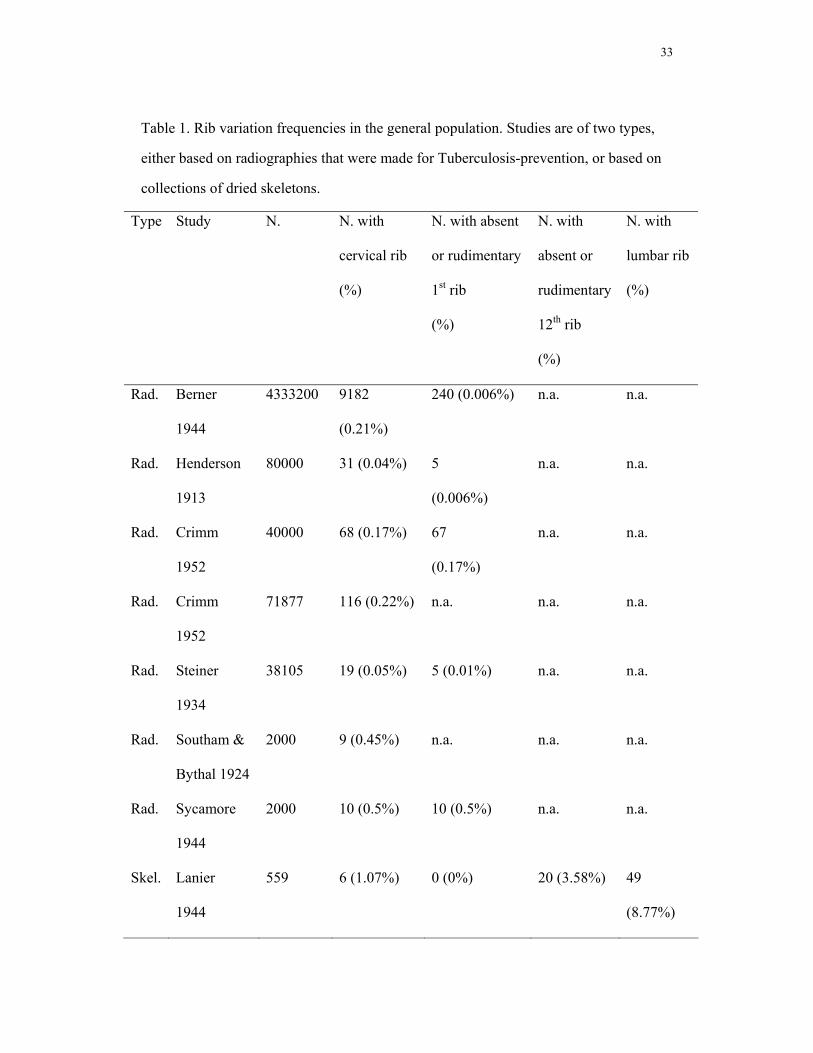
Table 1. Rib variation frequencies in the general population. Studies are of two types,
either based on radiographies that were made for Tuberculosis-prevention, or based on
collections of dried skeletons.
Type Study N. N. with
cervical rib
(%)
N. with absent
or rudimentary
1st rib
(%)
N. with
absent or
rudimentary
12th rib
(%)
N. with
lumbar rib
(%)
Rad. Berner
1944
4333200 9182
(0.21%)
240 (0.006%) n.a. n.a.
Rad. Henderson
1913
80000 31 (0.04%) 5
(0.006%)
n.a. n.a.
Rad. Crimm
1952
40000 68 (0.17%) 67
(0.17%)
n.a. n.a.
Rad. Crimm
1952
71877 116 (0.22%) n.a. n.a. n.a.
Rad. Steiner
1934
38105 19 (0.05%) 5 (0.01%) n.a. n.a.
Rad. Southam &
Bythal 1924
2000 9 (0.45%) n.a. n.a. n.a.
Rad. Sycamore
1944
2000 10 (0.5%) 10 (0.5%) n.a. n.a.
Skel. Lanier
1944
559 6 (1.07%) 0 (0%) 20 (3.58%) 49
(8.77%)
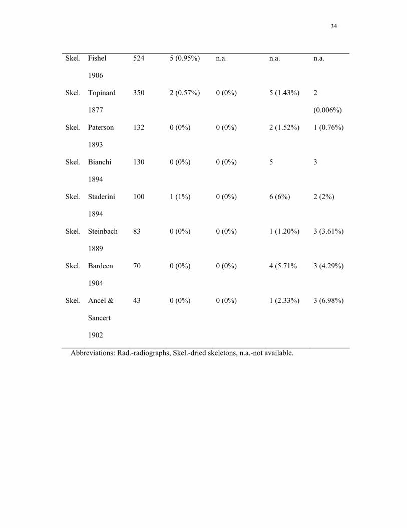
34
Skel. Fishel
1906
524 5 (0.95%) n.a. n.a. n.a.
Skel. Topinard
1877
350 2 (0.57%) 0 (0%) 5 (1.43%) 2
(0.006%)
Skel. Paterson
1893
132 0 (0%) 0 (0%) 2 (1.52%) 1 (0.76%)
Skel. Bianchi
1894
130 0 (0%) 0 (0%) 5 3
Skel. Staderini
1894
100 1 (1%) 0 (0%) 6 (6%) 2 (2%)
Skel. Steinbach
1889
83 0 (0%) 0 (0%) 1 (1.20%) 3 (3.61%)
Skel. Bardeen
1904
70 0 (0%) 0 (0%) 4 (5.71% 3 (4.29%)
Skel. Ancel &
Sancert
1902
43 0 (0%) 0 (0%) 1 (2.33%) 3 (6.98%)
Abbreviations: Rad.-radiographs, Skel.-dried skeletons, n.a.-not available.
35
Table 2. Selection against cervical ribs.
Age
Class
i
μi Survival
up to
age i
Prevalence
among those
that died pi
Prevalence
Pi
Relative
survival up to
age i with
cervical ribs
SRi
Relative
survival up to
age i without
cervical ribs
SNORi
0 0.15 1 0.505 0.097 1 1
1 0.01 0.85 0.505 0.025 0.218 0.918
2 0 0.8415 0.020 0.020 0.174 0.913
3 1 0.8415 0.020 0.020 0.174 0.913
Table 3. Selection against absent or reduced twelfth thoracic ribs
Age
Class
i
μi Survival
up to
age i
Prevalence
among those
that died pi
Prevalence
Pi
Relative
survival up to
age i with 12th
thoracal ribs
SRi
Relative
survival up to
age i without
12th thoracal
ribs SNORi
0 0.15 1 0.077 0.031 1 1
1 0.01 0.85 0.077 0.022 0.624 0.857
2 0 0.8415 0.022 0.022 0.603 0.849
3 1 0.8415 0.022 0.022 0.603 0.849
36
Table 4. Frequent congenital abnormalities in deceased human fetuses and infants (> 10
cases) and associated changes in the number of cervical vertebrae.
Congenital
abnormality
N. of
cases
N. with cervical rib
(%)
N with absent or
rudimentary first rib (%)
N with aberrant number
of cervical vertebrae (%)
Cleft lip/palate 12 6 (50%) 3 (25%) 9 (75%)
Horseshoe kidney 10 8 (80%) 1 (10%) 9 (90%)
Bleeding disorders 98 68 (69.4%) 1 (1%) 70 (70.4%)
Oligo/polydactyly 17 9 (52.9%) 4 (23.5%) 13 (76.4%)
Spina bifida 10 4 (40%) 1 (10%) 5 (50%)
Aberrant Arteria
Subclavia dextra
22 18 (81.8%) 0 (0%) 18 (81.8%)
Ventricular
septum defect
31 17 (54.8%) 8 (25.9%) 25 (80.7%)
Transfusion
syndrome
14 8 (57.1) 0 (0%) 8 (57.1%)
Left-right
disorders
21 15 (71.4%) 1 (4.8%) 16 (76.2%)
Bilateral kidney
agenesis
10 6 (60%) 1 (10%) 7 (70%)
Spina bifida 11 5 (45.5%) 1 (9.1%) 6 (54.6%)
Anal atresia 11 6 (54.5%) 3 (27.3%) 9 (81.8%)
Hydrops foetalis 22 10 (45.5%) 1 (4.5%) 11 (50%)
Dysmaturity 59 33 (55.9%) 0 (0%) 33 (55.9%)
Prematurity 68 39 (57.4%) 2 (2.9%) 41 (60.3%)
37
Figure 1. Radiograph showing left and right rudimentary cervical ribs in a male fetus of
16 weeks of gestation. Arrows indicate cervical ribs. To enhance the visibility of the
ossifications, the fetus has been stained with AgNO3. Institute of Pathology, Division
Photography, Free University Medical Center, Amsterdam.
38
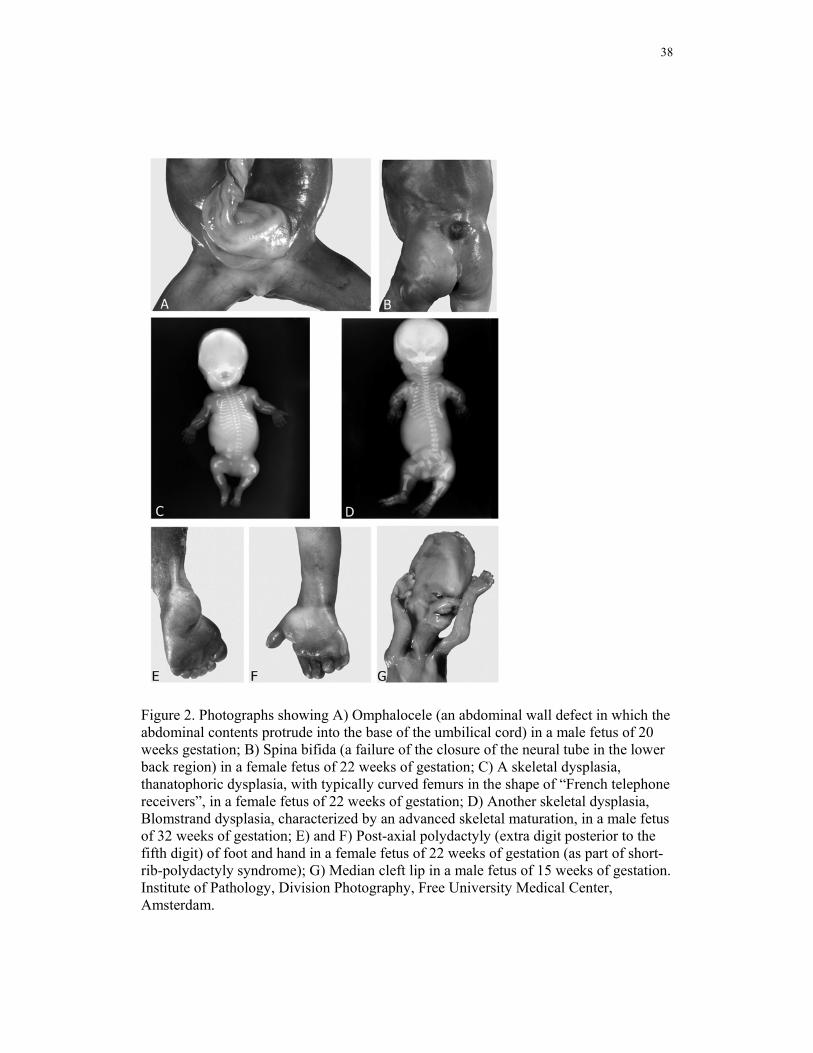
Figure 2. Photographs showing A) Omphalocele (an abdominal wall defect in which the abdominal contents protrude into the base of the umbilical cord) in a male fetus of 20 weeks gestation; B) Spina bifida (a failure of the closure of the neural tube in the lower back region) in a female fetus of 22 weeks of gestation; C) A skeletal dysplasia, thanatophoric dysplasia, with typically curved femurs in the shape of “French telephone receivers”, in a female fetus of 22 weeks of gestation; D) Another skeletal dysplasia, Blomstrand dysplasia, characterized by an advanced skeletal maturation, in a male fetus of 32 weeks of gestation; E) and F) Post-axial polydactyly (extra digit posterior to the fifth digit) of foot and hand in a female fetus of 22 weeks of gestation (as part of short-rib-polydactyly syndrome); G) Median cleft lip in a male fetus of 15 weeks of gestation. Institute of Pathology, Division Photography, Free University Medical Center, Amsterdam.
39
Figure 3. Graph showing the prevalence of cervical ribs, rudimentary first ribs,
rudimentary or absent twelfth ribs and lumbar ribs in foetal and infant deaths with resp.
no, single minor, single major, multiple minor and multiple major abnormalities. The
incidence of cervical ribs increases with the number and severity of the abnormalities.
Four cases had two vertebral variations, these were scored in both categories: two cases
with a cervical and a lumbar rib, one in the single minor and one in the multiple major
group; two cases with cervical ribs and absent 12th ribs (11 ribs including the cervical
rib), both in the multiple major abnormalities group.