pathways cell oncosis, apoptosis, and necrosis* · 82 the pathways of cell death: oncosis,...
TRANSCRIPT

82
The Pathways of Cell Death: Oncosis, Apoptosis, and Necrosis*
BENJAMIN E TRUMP, IRENE K. BEREZESKY, SEUNG H. CHANG, AND PATRICIA C. PHELPS
Department of Pathology, University of Maryland School of Medicine, Baltimore, Maryland 21201, USA
*Address correspondence to: Dr. Benjamin F Trump, University ofMaryland School of Medicine, 10 S. Pine St., Baltimore, Maryland21201.
ABSTRACT
The pathways and identification of cell injury and cell death are of key importance to the practice of diagnostic and researchtoxicologic pathology. Following a lethal injury, cellular reactions are initially reversible. Currently, we recognize two patterns, oncosisand apoptosis. Oncosis, derived from the Greek word "swelling," is the common pattern of change in infarcts and in zonal killingfollowing chemical toxicity, e.g., centrilobular hepatic necrosis after CC14 toxicity. In this common reaction, the earliest changesinvolve cytoplasmic blebbing, dilatation of the endoplasmic reticulum (ER), swelling of the cytosol, normal or condensed mitochondria,and chromatin clumping in the nucleus. In apoptosis, the early changes involve cell shrinkage, cytosolic shrinkage, more markedchromatin clumping, cytoplasmic blebbing, swollen ER on occasion, and mitochondria that are normal or condensed. Following celldeath, both types undergo postmortem changes collectively termed "necrosis." In the case of oncosis, this typically involves broadzones of cells while, in the case of apoptosis, the cells and/or the fragments are often phagocytized prior to their death by adjacentmacrophages or parenchymal cells. In either case, the changes converge to a pattern that involves mitochondrial swelling, mitochondrialflocculent densities and/or calcification, karyolysis, and disruption of plasmalemmal continuity. The biochemical mechanisms of celldeath are currently under intense study, particularly concerning the genes involved in the process. Pro-death genes include p53, theced-3/ICE proteases, and the Bax family. Anti-death genes include ced-9/Bcl-2 and the adenovirus protein E1B. It is clear that ion
deregulation, particularly that of [Ca2+]1 plays an important role in cell death following either apoptosis or oncosis. Genetic evidencestrongly indicates that activation of proteases is an important step, possibly very near to the point where cell death occurs.
Keywords. Cell injury; intracellular calcium; ion regulation; volume regulation; gene expression; proteases
INTRODUCTION
The purpose of this paper is to analyze and interpretthe cellular reactions to lethal injury, including thosechanges that occur prior to cell death and those that occurfollowing cell death. Detection and accurate identificationof such changes is important to toxicologic pathologybecause they may represent the earliest indicators of im-portant toxic reactions to a variety of injuries includingdrugs, anti-microbiologic agents, toxic chemicals, and in-fectious agents. Definition of the criteria and the nomen-clature of these processes is extremely important for pur-poses of data comparison and for experiments designedto elucidate the mechanisms of injury.
RESPONSES TO CELLULAR INJURY
Events Following Cell Injury: Terminology and HistoryThe terminology in this field has had an ancient and
colorful history and evolution in medicine. The past 10yr has seen remarkable progress and, understandably, alsosome confusion in the literature concerning the termi-
nology of the processes of cell injury and cell death
(1,9,14,15,31).Following injury, cells undergo a series of responses
that collectively form what we recognize as a diseaseprocess. Many injuries to cells are sublethal and result inaltered or new steady states in which the cells are ableto survive in some altered or new steady state (26,29).Examples include vacuolization from dilatation of lyso-somes and triglyceride accumulation. On the other hand,certain injuries, e.g., complete ischemia, are often lethalto cells and following a period of reversible reactions, thecell dies and then undergoes a series of degradative re-actions that ultimately restore it to equilibrium with theenvironment. We can thus see that following a lethal in-jury there are a series of 3 phases: (1) changes that weterm &dquo;prelethal,&dquo; which are often reversible; (2) the
&dquo;point-of-no-return&dquo; or cell death; and (3) postmortemautolytic and degradative changes. These postmortemcellular changes are termed &dquo;necrosis,&dquo; which is an an-cient word used to describe the changes that cells andtissues undergo following their death within a living or-ganism. Not all types of death are followed by necrosis,e.g., tissues fixed in formaldehyde are the classic exampleof cells that are killed very rapidly but which subsequent-ly do not undergo the degradative changes of necrosis.

83
FIG. Phase micrograph of a JB6-clone 41 cell (promotable mousekeratinocyte cell line) following treatment with 10-4 M sodium laurylsulfate for 45 min. Note the many blebs, marked vacuolization, andnuclear chromatin clumping all of which are reversible changes.X 1,700. [Reprinted with permission from Jain et al (12).]
Clearly then, death and necrosis must be separated. Atthe same time, it is evident that it is illogical to speak ofdeath by necrosis when in fact there are at least 2 differ-ent forms of necrosis occurring following death.The terminology for the stages of cell reaction to in-
jury prior to cell death has also undergone an interestingevolution over time. It turns out that there are 2 majorcategories consisting of the prelethal reaction which havecome to be known as &dquo;apoptosis&dquo; and &dquo;oncosis.&dquo; Theseare distinguished mainly on the basis of cell volume andnuclear morphology.
In apoptosis, the cells shrink, show multiple cytoplas-mic protrusions or blebs, marked nuclear chromatin con-densation, and ultimately fragmentation. Before this termwas introduced by Kerr et al (13), many terms such askaryorrhexis, nuclear fragmentation, and individual cellnecrosis were used to describe the phenomenon. On theother hand, another common prelethal type of reactioninvolves swelling of the cells prior to death. This hadmany terms over the years including vacuolization andcloudy swelling. Recently, Majno and Joris (15) proposedthat we return to the term &dquo;oncosis,&dquo; the term originallyused in 1910 by von Recklinghausen (20). This, there-fore, gives terms for both types of prelethal injury.
Another issue is the term &dquo;programmed cell death.&dquo;
During embryologic development in multicellular organ-isms, many cells die during the remodeling that occursduring the development of a variety of organs. Investi-gations of the genes involved in such cell death haverevealed the existence of both pro- and anti-cell death
genes. The mechanism of function of these genes is the
subject of many current studies (4,7,10,11,23,30,32,33).However, at the same time, what is often overlooked is
FIG. 2.-Transmission electron micrograph (TEM) of an NRK-52Ecell (normal rat kidney cell line) treated with I mM KCN + 1 mM IAA
(iodoacetic acid) for 3 hr, 45 min. Marked cytoplasmic swelling (ar-rows) can be seen in a large bleb region in which organelles and freeribosomes are absent. However, organelles are present in the nuclearregion of the cell where most mitochondria are swollen (arrowheads).Cytoskeletal elements are not identifiable. X 8,000.
that every cell is totally programmed for death if deprivedof a source of ATP or if the plasma membrane is violated.
The Concept of Programmed vs Accidental Cell Death
Recently, the concept of &dquo;accidental&dquo; vs &dquo;pro-grammed&dquo; cell death has appeared often in the literatureand, also, in our thinking. As far as we can determine,the concept of programmed cell death was originally de-rived from studies of developmental biology in which thedeath of cells in a particular area must die on schedulefor embryologic development and organ differentiationto occur, such as normal phenotypes. Because this deathin embryos is often (but by no means always) the apop-totic pathway, apoptosis has been erroneously equatedwith programmed cell death. It is very important to re-alize that every cell is &dquo;programmed&dquo; to die followingan appropriate stimulus. The pattern, apoptosis or oncos-is, clearly depends on both the cell type and the injury.
ONCOSIS
MorphologyIn oncosis, the early changes include marked altera-
tions in cell shape and volume, frequently occurring with-in seconds to minutes following application of the injury.In monolayer cultures, such cells form cytoplasmic blebs(19); chromatin clumping occurs later, followed by cellspulling apart, rounding up, and often detaching from thesubstrate (Fig. 1). In vivo, the cells often exhibit blebs,

84
FIG. 3.-TEM of a portion of a NRK-52E cell treated with 1 mM
KCN + 1 mM IAA for 3 hr, 45 min. The rough ER (arrow) is markedlydilated and many mitochondria are swollen while others are condensed.
One profile shows both swollen and condensed compartments (arrow-head). On the right side of the micrograph, note the zone of swollencytoplasm with free ribosomes and sparse organelles. At the top of themicrograph, the cell surface shows an irregular contour. X 16,000.
along the luminal borders and the vascular space, whichlater detach and, in the case of the kidney, form casts inthe lumens of the nephrons. By electron microscopy, suchcells show swelling and clearing of the cytosol, dilatationof the ER and Golgi, mitochondrial condensation fol-lowed by swelling, nuclear chromatin clumping, and mul-tiple cytoplasmic blebs that typically are organelle-free(Figs. 2 and 3). During this phase, the elements of thecytoskeleton, including actin and tubulin, are markedlyaltered (8,18). When viewed with the naked eye, tissuesundergoing this change appear opaque and cloudy, prob-ably related to early and reversible denaturation of pro-teins. This is perhaps responsible for the use of earlierterms, such as &dquo;cloudy swelling.&dquo;
Setting/ EtiologyIn vivo, oncosis typically affects broad areas or zones
of cells, e.g., the early changes of cells following totalischemia or the cells in a particular region of the liver orkidney tubule following chemical toxins, such as CC14,or HgC12, respectively. When broad zones of cells areinvolved and following death of the cells, a pronouncedinflammatory reaction typically occurs at the peripheryof the zone. Although oncosis is not typical of pro-grammed cell death in development, it has been describedin some situations.
APOPTOSIS
MorphologyThe earliest light microscopic changes include de-
creased cell volume, marked shape changes with multiple
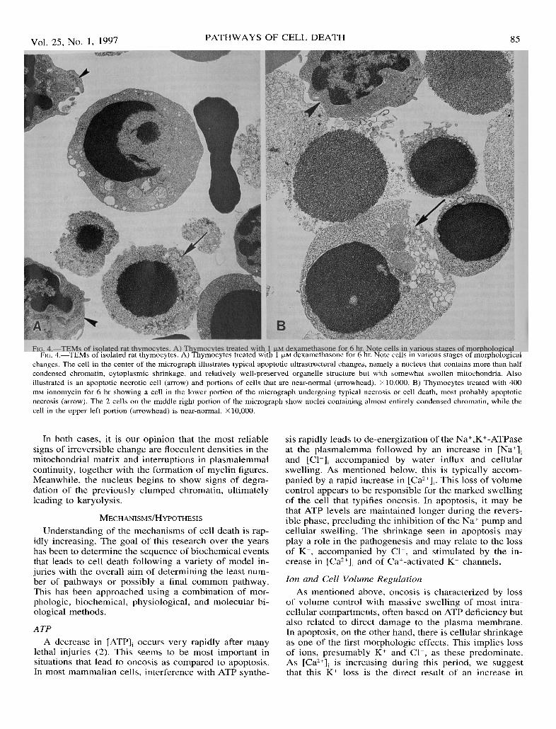
blebs and protrusions typically containing organelles, andmarked nuclear chromatin clumping. By electron micros-copy, the cytosol is dense, the ER may be dilated, themitochondria are condensed and there is marked clump-ing of nuclear chromatin (Fig. 4). The nuclear outlinebecomes quite irregular with multiple protrusions that insection often appear to have detached, though they arefrequently connected to the main nucleoplasm. At thisstage, DNA strand breaks are often occurring that can beseen either in electrophoresis or with the deoxyribonu-cleotidyl transferase (TDT)-mediated dUTP-digoxigeninnick-end labeling (TUNEL) assay, although the latter isnot specific for apoptosis. It is clear that apart from cellshrinkage, the morphologic changes are similar to thoseseen in oncosis. The main difference is shrinkage, ap-parently due to loss of ions, probably mainly K+ and Cl-.The significance of this volume alteration to the otherchanges, e.g., DNA strand breaks, needs further investi-gation ; however, some experiments indicate that the
shrinkage may indeed represent a significant step in themechanism (3).
SettinglEtiology
Apoptosis occurs in a variety of settings. It is wide-
spread during development and it is often in that situationthat it actually represents a prelethal phase of pro-grammed reaction to injury on a schedule set by varioushormonal, nutritional, and micro- and macroenvironmen-tal factors. In adult disease, it is commonly seen in sit-uations with marked atrophy and regression where, infact, the earliest descriptions of the process were made,including atresia of ovarian follicles, atrophy of the pros-tate following castration, and involution of the mammaryepithelium following pregnancy.
Apoptosis also occurs following a variety of chemicaland microbiologic injuries in many different organ sys-tems (6,12,16,21,22,32,33). It is often difficult to estimatethe extent, because of the rapidity with which the apop-totic cells and fragments are taken up by adjacent paren-chymal and/or mesenchymal phagocytes. Although theinflammatory response is uncommon, presumably be-cause the fragments are removed prior to death (24), Maj-no and Joris (15) have pointed out that there are someexceptions.
NECROSIS
MorphologyIn the phase of necrosis, the changes are similar after
either apoptosis or oncosis, though in some cases it ap-pears that the cells showing apoptotic necrosis still re-main somewhat shrunken or more dense, especially invivo. As mentioned above, apoptotic necrosis typicallyoccurs in apoptotic fragments that undergo this changewithin the phagolysosomal system of adjacent phagocy-tizing cells. In vitro, cells undergoing apoptotic necrosis(Fig. 4B) often show marked swelling and may be vir-tually indistinguishable from those showing oncotic ne-crosis. It is important to realize that either oncotic orapoptotic necrosis shows positive results with the TU-NEL assay.
85
FIG. 4.-TEMs of isolated rat thymocytes. A) Thymocytes treated with 1 J..LM dexamethasone for 6 hr. Note cells in various stages of morphologicalchanges. The cell in the center of the micrograph illustrates typical apoptotic ultrastructural changes, namely a nucleus that contains more than halfcondensed chromatin, cytoplasmic shrinkage, and relatively well-preserved organelle structure but with somewhat swollen mitochondria. Alsoillustrated is an apoptotic necrotic cell (arrow) and portions of cells that are near-normal (arrowhead). X 10,000. B) Thymocytes treated with 400mM ionomycin for 6 hr showing a cell in the lower portion of the micrograph undergoing typical necrosis or cell death, most probably apoptoticnecrosis (arrow). The 2 cells on the middle right portion of the micrograph show nuclei containing almost entirely condensed chromatin, while thecell in the upper left portion (arrowhead) is near-normal. x 10,000.
In both cases, it is our opinion that the most reliablesigns of irreversible change are flocculent densities in themitochondrial matrix and interruptions in plasmalemmalcontinuity, together with the formation of myelin figures.Meanwhile, the nucleus begins to show signs of degra-dation of the previously clumped chromatin, ultimatelyleading to karyolysis.
MECHANISMS/HYPOTHESIS
Understanding of the mechanisms of cell death is rap-idly increasing. The goal of this research over the yearshas been to determine the sequence of biochemical eventsthat leads to cell death following a variety of model in-juries with the overall aim of determining the least num-ber of pathways or possibly a final common pathway.This has been approached using a combination of mor-phologic, biochemical, physiological, and molecular bi-ological methods.
ATP
A decrease in [ATP]I occurs very rapidly after manylethal injuries (2). This seems to be most important insituations that lead to oncosis as compared to apoptosis.In most mammalian cells, interference with ATP synthe-
sis rapidly leads to de-energization of the Na+,K+-ATPaseat the plasmalemma followed by an increase in [Na+];and [Cl-]; accompanied by water influx and cellular
swelling. As mentioned below, this is typically accom-panied by a rapid increase in [Caz+];. This loss of volumecontrol appears to be responsible for the marked swellingof the cell that typifies oncosis. In apoptosis, it may bethat ATP levels are maintained longer during the revers-ible phase, precluding the inhibition of the Na+ pump andcellular swelling. The shrinkage seen in apoptosis mayplay a role in the pathogenesis and may relate to the lossof K+, accompanied by Cl-, and stimulated by the in-crease in [Ca2+]; and of Ca+-activated K+ channels.
Ion and Cell Volume RegulationAs mentioned above, oncosis is characterized by loss
of volume control with massive swelling of most intra-cellular compartments, often based on ATP deficiency butalso related to direct damage to the plasma membrane.In apoptosis, on the other hand, there is cellular shrinkageas one of the first morphologic effects. This implies lossof ions, presumably K+ and Cl-, as these predominate.As [Cai2+]; is increasing during this period, we suggestthat this K+ loss is the direct result of an increase in

86
FIG. 5.-Diagram showing our current working hypothesis of the ma-jor events leading from cell injury to cell death, focusing on the roleof [Ca2+],. Some of the major categories of injury are shown at the topof the figure. Decreased and absent [ATP], commonly results from com-plete ischemia, anoxia, or treatment with inhibitors of ATP synthesisincluding potassium cyanide, carbon monoxide, fluoride, and iodoaceticacid. Increased plasma membrane permeability commonly results fromthe immune response, e.g., activation of complement or T-cell releaseof perforin, or from treatment with a variety of toxins including mer-curic salts, ionophores, or mechanical damage. Growth factor depriva-tion is often studied in vitro though, in vivo, it occurs with neuronal
damage. In oncosis, the early events involve cell swelling because ofincreased [Nal]i accompanied by Cl- and water. This can result eitherfrom increased plasmalemmal permeability or decreased [ATP], result-ing in inactivation of the Na+,K+-ATPase. Increased [Na+], can alsocontribute to increased [Ca2+], through decreased Na+/Ca2+ exchange.In apoptosis, the initial events include cellular shrinkage implying lossof ions, in particular K+ and Cl-. The mechanism of the shrinkage isnot presently known although increased [Ca2+], might contribute
through activation of K+ channels. The initial increase of [Ca2+]; canresult from influx from the extracellular space or from redistribution
from intracellular stores. Following the increase, several principal path-ways seem to lead to reversible and irreversible changes. Also illus-trated in the diagram are the events that follow Ca2+-mediated activationof protein kinases, endonucleases, proteases, and phospholipases as dis-cussed in the text. Our current hypothesis of altered gene expressionrelated to cell death is shown near the bottom of the diagram. Abbre-viations : ICE = interleukin-1~ converting enzyme; JNK = jun N-ter-minal kinase; MAP = mitogen-activated protein; PARP = polyadeny-late ribose polymerase [modified from Trump and Berezesky (28)].
[Ca2+]; and stimulation of Ca2+-activated K+ channels. Itmay be that the shrinkage itself has some role in the
pathogenesis of the changes.
Calcium
As indicated in the diagram depicting our current
working hypothesis (Fig. 5), we, as well as other inves-tigators, hypothesize that deregulation of [Ca 2+], plays apivotal role in both oncosis and apoptosis(5,21,22,25,27,28,32,33). Increased [Ca 2+1, stimulates avariety of signals and events that may involve virtuallyall aspects of cell behavior. One unsolved problem is
why, under some conditions in some cells, increased[Ca2+]; results in oncosis while, in other cells, it resultsin apoptosis.
Increasingly, the role of modulation of [Ca 2+], in genetranscription and cytoskeletal function is also being elu-cidated.
TABLE I.-Selected genes and regulators that promote or inhibit celldeath.,
a Modified from Chinnaiyan and Dixit (4). For references, see those in Chin-naiyan and Dixit (4).
Genetic RegulationIn recent years, it has often become easier and more
direct to identify the genes involved in a process than theprecise biochemical mechanisms. This is eminently truefor the changes that lead to cell death. A number of pro-teins have been found that can promote or protect againstcell death [for review, see Chinnaiyan and Dixit (4)] (Ta-ble 1). The issue has evolved into attempts to discoverthe sequence of events and which ones are critical in a
particular example. As knowledge of intracellular signal-ling pathways continues to develop, the pathways leadingto cell death should be elucidated.
At the present time, it is not clear how a cascade of
signalling events and proteins can lead to cell death, noris it clear how some genes lead to apoptosis whereasothers lead to oncosis. For example, in the nematodeCaenorhabditis elegans, the genes ced-3 and ced-4 leadto apoptosis (34,35), whereas mutations in the degenerensgene lead to oncosis.

87
DISCUSSION
The patterns of cellular reaction to injury leading tocell death are important in the understanding and recog-nition of toxic and other types of injury. At the presenttime, two types of prelethal reactions have been charac-terized : apoptosis and oncosis. Both may result from tox-ic injury. The factors that determine which process occursremain to be identified. It does appear, however, thatsome cells such as lymphocytes, are particularly likely toundergo apoptosis while others, such as renal proximaltubular epithelium, seem especially prone to oncosis.
Cellular proteases seem to be involved in both typesof change, particularly in the final stages prior to celldeath (17). It appears that ATP deficiency, especially in-hibition of ATP synthesis, and disruption of membraneion pores or creation of increased plasma membrane per-meability result in oncosis, while alterations that primar-ily affect DNA structure often result in apoptosis possiblythrough activation of p53.
Following cell death, apoptotic or oncotic cells under-go necrosis. At this point, the changes rapidly convergewith loss of volume control in the case of apoptosis andcontinued swelling in the case of oncosis. In the case ofapoptosis, these final stages usually occur within the pha-golysosomal system of adjacent cells that have phago-cytized the apoptotic cells and fragments.The differences between oncosis and apoptosis are
clearly fruitful areas for future research. For example,questions needing answers include: What is the mecha-nism by which p53 induces apoptosis and whether or notin some cases it could induce oncosis? What is the roleof proteases and how does it differ in the 2 cases? Whatis the role of cellular shrinkage in initiating apoptosis?What is the final &dquo;execution step&dquo; and does it differ inthe 2 cases? When these answers become known, themechanisms and pathways of cell injury and cell deathwill then also become known, and an understanding ofthese events will lead us to better methods in diagnosticand research toxicologic pathology.
ACKNOWLEDGMENTS
This is contribution no. 3825 from the Cellular Patho-
biology Laboratory. This work was supported by NIHgrant DK 15440 and Navy N00014-91-J-1863.
REFERENCES
1. Arends MJ and Wyllie AH (1991). Apoptosis: Mechanisms androles in pathology. Int. Rev. Exp. Pathol. 32: 223-254.
2. Bonventre JV (1993). Mechanisms of ischemic renal failure. KidneyInt. 43: 1160-1178.
3. Bortner CD and Cidlowski JA (1997). The absence of regulatorymechanisms contributes to the rapid activation of apoptosis in thy-mocytes. Am. J. Physiol. (in press).
4. Chinnaiyan AM and Dixit VM (1996). The cell-death machine.Curr. Biol. 6(5): 555-562.
5. Clapham DE (1995). Calcium signalling. Cell 80: 259-268.6. Davis MA, Smith MW, Chang SH, and Trump BF (1994). Char-
acterization of a renal epithelial cell model of apoptosis using oka-daic acid and the NRK-52E cell line. Toxicol. Pathol. 22: 595-604.
7. El-Diery WS, Harper JW, O’Connor PM, Veicuiescu VE, CanmanCE, Jackman J, Pietenpol J, Burrell M, Hill DE, Wang Y, WimanKG, Mercer WE, Kastan MB, Kohn KW, Elledge SJ, Kinzier KW,
and Vogelstein B (1994). WAF1/CIP1 is induced in p53-mediatedG1 arrest and apoptosis. Cancer Res. 54: 1-6.
8. Elliget KA, Phelps PC, and Trump BF (1991). HgCl2-induced al-teration of actin filaments in cultured primary rat proximal tubuleepithelial cells labelled with fluorescein phalloidin. Cell Biol. Tox-icol. 7: 263-280.
9. Ellis RE, Yuan Y, and Horvitz HR (1991). Mechanisms and func-tions of cell death. Annu. Rev. Cell Biol. 7: 663-698.
10. Hengartner MO, Ellis RE, and Horvitz HR (1992). Caenorhabditiselegans gene ced-9 protects cells from programmed cell death. Na-ture (Lond.) 356: 494-499.
11. Hockenbery DM, Oltvai ZN, Yin XM, Milliman CL, and Kors-meyer SJ (1993). Bcl-2 functions in an antioxidant pathway to pre-vent apoptosis. Cell 75: 241-251.
12. Jain PT, Fitzpatrick MJ, Phelps PC, Berezesky IK, and Trump BF(1992). Studies of skin toxicity in vitro: Dose-response studies onJB6 cells. Toxicol. Pathol. 20: 394-404.
13. Kerr JFR, Wyllie AH, and Currie AR (1972). Apoptosis: A basicbiological phenomenon with wide-ranging implications in tissue
kinetics. Br. J Cancer 26: 239-257.14. Lints R and Driscoll M (1996). Programmed and pathological cell
death in Caenorhabditis elegans. In: Cellular Aging and Cell
Death, GR Martin and RA Lockshin (eds). Wiley-Liss, Inc, NewYork, pp. 235-253.
15. Majno G and Joris I (1995). Apoptosis, oncosis, and necrosis. Anoverview of cell death. Am. J. Pathol. 146: 3-15.
16. Maki A, Berezesky IK, Fargnoli J, Holbrook NJ, and Trump BF(1992). Role of [Ca2+], in induction of c-fos, c-jun, and c-myc RNAin rat PTE after oxidative stress. FASEB J. 6: 919-924.
17. Nicholson DW (1996). ICE/CED-3-like proteases as therapeutictargets for the control of inappropriate apoptosis. Nature Biotech-nol. 14: 297-301.
18. Phelps PC, Elliget KA, and Trump BF (1996). Cytoskeletal alter-ations following chemical ischemia. FASEB J. 10: A1425.
19. Phelps PC, Smith MW, and Trump BF (1989). Cytosolic ionizedcalcium and bleb formation after acute cell injury of cultured rabbitrenal tubule cells. Lab. Invest. 60: 630-642.
20. Recklinghausen F von (1910). Untersuchungen über Rachitis undOsteomalacie. Verlag Gustav Fischer, Jena.
21. Smith MW, Phelps PC, and Trump BF (1991). Cytosolic Ca2+ de-
regulation and blebbing after HgCl2 injury to cultured rabbit prox-imal tubule cells as determined by digital imaging microscopy.Proc. Natl. Acad. Sci. USA 88: 4926-4930.
22. Smith MW, Phelps PC, and Trump BF (1992). Injury-inducedchanges in cytosolic Ca2+ in individual rabbit proximal tubule cells.Am. J. Physiol. 262 (Renal Fluid Electrolyte 31): F647-F655.
23. Steller H (1995). Mechanisms and genes of cellular suicide. Science267: 1445-1448.
24. Thompson CB (1995). Apoptosis in the pathogenesis and treatmentof disease. Science 267: 1456-1462.
25. Trump BF and Berezesky IK (1992). The role of cytosolic Ca2+ in
cell injury, necrosis and apoptosis. Curr. Opin. Cell Biol. 4: 227-232.
26. Trump BF and Berezesky IK (1994). Cellular and molecular patho-biology of reversible and irreversible injury. In: Methods in Toxi-cology, Vol. 1B, In Vitro Toxicity Indicators, CA Tyson and JMFrazier (eds). Academic Press, New York, pp. 1-22.
27. Trump BF and Berezesky IK (1995). Calcium-mediated cell injuryand cell death. FASEB J. 9: 219-228.
28. Trump BF and Berezesky IK (1996). The mechanisms of calcium-mediated cell injury and cell death. New Horiz. Sci. Pract. AcuteMed. 4: 139-150.
29. Trump BF and Ginn FL (1969). The pathogenesis of subcellularreaction to lethal injury. In: Methods and Achievements in Exper-imental Pathology, Vol. IV, E Bajusz and G Jasmin (eds). Karger,Basel, pp. 1-29.
30. Vaux DL and Strasser A (1996). The molecular biology of apop-tosis. Proc. Natl. Acad. Sci. USA 93: 2239-2244.
31. Wyllie AH, Kerr JFR, and Currie AR (1980). Cell death: The sig-nificance of apoptosis. Int. Rev. Cytol. 68: 251-306.

88
32. Yamamoto N, Maki A, Swann JD, Berezesky IK, and Trump BF(1993). Induction of immediate, early and stress genes in rat prox-imal tubule epithelium following injury: The significance of cyto-solic ionized calcium. Ren. Fail. 15: 163-171.
33. Yamamoto N, Smith MW, Maki A, Berezesky IK, and Trump BF(1994). The role of cytosolic Ca2+ and protein kinases in the in-duction of the hsp70 gene in rat proximal tubular epithelial cells.Kidney Int. 45: 1093-1104.
34. Yuan J and Horvitz H (1992). The C. elegans cell death gene ced-4encodes a novel protein and is expressed during the period of ex-tensive programmed cell death. Development 116: 309-320.
35. Yuan J, Shaham S, Ledoux S, Ellis HM, and Horvitz HR (1993).The C. elegans cell death gene ced-3 encodes a protein similar tomammalian interleukin-1β-converting enzyme. Cell 75: 641-652.