parallel left ventricle simulation using the fenics …ceur-ws.org/vol-1729/paper-05.pdf ·...
TRANSCRIPT
Parallel Left Ventricle Simulation Using theFEniCS Framework
Timofei Epanchintsev1,2,3 and Vladimir Zverev3,1
1 Krasovskii Institute of Mathematics and Mechanics, Yekaterinburg, Russia2 Institute of Immunology and Physiology of the Ural Branch of the
Russian Academy of Sciences, Yekaterinburg, Russia3 Ural Federal University, Yekaterinburg, Russia
Abstract. Heart simulation is complex task that requires multiscalemodeling on cell, tissue and organ levels. Such structure makes difficulthigh performance code development and its maintenance. In this paper,we evaluate how scientific software could be used for heart simulation.An overview of existing frameworks for automated scientific computingis presented. The FEniCS framework was chosen since it supports auto-mated solution of differential equations by the finite element methodof parallel computing systems, provides near-mathematical notation,uses high performance backend and has comprehensive documentation.FEniCS performance was evaluated by simulation the space propaga-tion of membrane potential alternation over a cardiac left ventricle usingthe electrophysiological model of a left ventricle and the Ekaterinburg-Oxford cell model. The FEniCS framework showed good performanceand near-linear scalability up to 240 CPU cores.
Keywords: FEniCS · heart simulation · parallel computing · finite ele-ment method
1 Introduction
Heart simulation is a complex task that requires multiscale modeling on cell,tissue, and organ levels [1]. Such problems are computationally intensive andrequire parallel computing, including the use of modern computational acceler-ators such as GPU and Xeon Phi. However, porting a complicated multilevelsimulation code to a new computational architecture requires a long time (of-ten 3–5 years) during which the architecture may become obsolete. In addition,adaptation for parallel computing architectures often leads to significant changesof code. Consequently, it is very difficult to determine which mathematical mod-els and numerical methods are used in the optimized code. Hence, reflecting thechanges of a mathematical model in the optimized code can be very complicated.As a result, complex multiscale simulation software is rarely adapted to modernparallel computing architectures.
An alternative approach is based on using automated scientific computingframeworks. Such frameworks allow the development of simulation software using
30 Timofei Epanchintsev and Vladimir Zverev
programming languages with near-mathematical notation. Traditionally, a sig-nificant disadvantage of such frameworks was their low simulation performance.However, some modern implementations use advanced tools such as highly ef-ficient mathematical libraries, just-in-time compilers, parallel execution, and soon, which improve their performance. Still, it is not clear if the modern auto-mated scientific computing frameworks are efficient enough for real multiscalesimulation tasks such as heart simulation.
In this paper, we evaluate how the automated scientific computing frame-works could be used for heart simulation on parallel computing systems. Heartsimulation is an attractive task for evaluation of the performance of automatedscientific computing frameworks because numerical simulations can be used as avirtual environment for testing and predicting tissue behavior in the cases whereexperimental techniques can not be applied [1, 2]. As a benchmark problem,we chose the investigation of the space propagation of the membrane potentialalternation over the left ventricle of human heart.
2 Automated Scientific Computing Frameworks
Nowadays, finite element method is the most popular method for numerical in-vestigation of systems with complex geometry, such as human heart. Severalframeworks for automated scientific computing using finite element method ex-ist. The most popular among them are OpenFOAM, OpenCMISS, Chaste, andFEniCS.
OpenFOAM (Open Source Field Operation and Manipulation) [3] is a freeComputational Fluid Dynamics (CFD) software designed for solving problemsin continuum mechanics. OpenFOAM is a C++ library that provides numericalschemes implemented in the traditional finite volume framework with solversthat are known to be efficient for continuum mechanics problems. OpenFOAMuses domain decomposition in order to provide parallel execution based on MPI.
OpenCMISS is a part of the global international project Physiome [4].OpenCMISS is a mathematical modeling environment that enables the applica-tion of finite element analysis techniques to a variety of complex bioengineer-ing problems. For distributed and parallel computing, OpenCMISS uses MPI,OpenMP, and ParMETIS.However, the OpenCMISS project is still under devel-opment, its documentation is incomplete, and it lacks examples.
Chaste (Cancer, Heart, and Soft Tissue Environment) is a general purposesimulation package aimed at multiscale, computationally intensive problems aris-ing in biology and physiology [5, 6]. Current functionality includes the tissueand cell level electrophysiology, the discrete tissue modeling, and the soft tis-sue modeling. Chaste uses solvers from PETSc, mesh distribution algorithmsfrom ParMETIS, and provides the ability to run parallel simulations using MPI.Chaste has already implemented models and methods, but to modify them adeveloper has to deal with sophisticated internal structure. Hence their furtherextension is complicated.

Parallel Left Ventricle Simulation Using the FEniCS Framework 31
FEniCS [7] is a collaborative project for the development of the tools forautomated scientific computing with a particular focus on the automated solu-tion of differential equations by the finite element method. Implementation of afinite element method consists of several stages (obtaining equations’ weak form,discretizing the equations in the weak form, assembling the values calculated oneach element, and solving of the system of algebraic equations). Each stage iscovered by a separate component of the FEniCS framework. It uses third-partyhigh-performance libraries such as PETSc, Trilinos, uBLAS, or Intel MKL. Inaddition, FEniCS allows parallel simulation using MPI.
For our heart simulation task, we chose the FEniCS framework because it pro-vides the automated solution of differential equations by finite element methods,supports automatic parallel execution using MPI, and has a good documenta-tion.
3 Description of the Heart Model
We simulated the electrical activity of a human heart, which is a result of spa-tial and temporal propagation of the electrical signal from each cardiac cell. Weused the Ekaterinburg-Oxford cell model [8] with a detailed description of electri-cal, chemical, and mechanical processes. On the intracellular level, the electricalpotential arises from a very complicated interaction among ionic currents andcell organelles (organized structures in cells), as presented in Fig. 1 (left). Thetrans-membrane potential is shown in Fig. 1 (right). Scaled windows presentsfast processes in period between 0 and 0.015 seconds.
Fig. 1. The schemes of the electric potential and some of the underlying ionic current
The electrical block of the model contains the equations of the membranepotential and dynamic parameters describing the opening and closing of ionchannels. The chemical block describes the kinetics of intracellular concentra-tions of calcium, sodium and potassium, extracellular potassium, the kinetics ofcalcium complexes, and calcium kinetics in the organelles. The mechanical block

32 Timofei Epanchintsev and Vladimir Zverev
of the model includes equations describing the voltage of the cell that depends onits length. The differential equations of the electrical, chemical, and mechanicalprocesses can be presented in the following simplified form:
∂S
∂t= g(V, S), (1)
where V is the electrical potential, S is the vector of model variables that governthe ion currents, and g is the vector-valued function that describes the timeevolution of each variable. The dimension of the vectors S and g is 30. System(1) is defined at each point of the heart tissue, and, consequently, we should solveit for each node of the computational mesh. The space and time propagation ofthe electrical potential is governed by the “reaction-diffusion equation” [9]:
∂V
∂t= D∇2V + Iions(V, S), (2)
where D is the diffusion coefficient, ∇2 is the Laplace operator, and Iions is thesum of the ionic currents related to the capacitance of cell membrane. Boundaryconditions correspond to the condition of electrical isolation.
This model is a nonlinear system of differential equations that can not besolved analytically and is a very computationally intensive task due to the largeamount of variables in the 3D domain.
4 Benchmark Problem
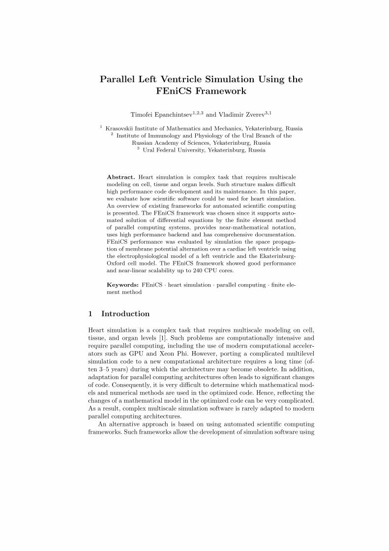
During the experiments, we simulated the electrical activity of the human heartleft ventricle (LV). We used the asymmetric model of LV that was previouslydeveloped in our group [10]. The important feature of the model is the abilityto vary the size of the mesh elements. The personalized model parameters werecaptured with the help of ultrasound imaging. An example of 3D mesh for LVis presented in Fig. 2.
In order to solve model (1)–(2), we use the operator splitting scheme of firstorder (Marchuk–Yanenko method) [11]. Let us consider time domain t ∈ [0, T ]and the uniform grid tn = htn, where ht = T/N and n is an integer that countstime level, 0 <= n <= N . We denote a quantity at time tn as Vn. The scheme ofcomputing Vn and Sn consists of two steps. Let us assume that we have alreadycalculated the values of V (t) and S(t) for t < tn. At the first step, we solve thefollowing partial differential equation:
V ∗n − V ∗
n−1
ht= D∇2V ∗, V ∗(t = tn−1) = V (tn−1), t ∈ [tn−1, tn]. (3)
At the second step, we should solve the following system of ordinary equa-tions:
Parallel Left Ventricle Simulation Using the FEniCS Framework 33
Fig. 2. An example of 3D mesh of left ventricle (asymmetric model)
V ∗∗n − V ∗∗
n−1
ht= Iions, V
∗∗(t = tn−1) = V ∗(tn),
S∗∗n − S∗∗
n−1
ht= g(V ∗, S∗∗), S∗∗(t = tn−1) = S(tn−1).
(4)
The solution of (4) gives us the values of V (tn) and S(tn) according to therules V (tn) = V ∗∗(tn) and S(tn) = S∗∗(tn). This method allows us to tackletask (3) using the implicit method and use the explicit time-scheme for task(4). In addition, we avoid the Newton-like iterations. The disadvantage of suchapproach is that we have to use a very small integration time step, in order tocapture the fast electrochemical processes (Fig. 1).
For testing purposes, we chose activation of an entire LV (the potential isgreater than 40 millivolt) as our initial condition. The simulation duration periodwas 0.3 seconds of physical time because the electrical activity tends to theequilibrium state without an external stimulus approximately after this period.
We use the tetrahedral mesh that was generated by the GMSH software [12].The minimal length of the tetrahedrons was set to 2mm and maximal to 4mm.As a result, the mesh contained 7178 points and 26156 tetrahedrons.
5 Performance Evaluation
In order to estimate the performance and scalability of the LV simulation usingthe FEniCS framework, a series of experiments was performed. We used the Uransupercomputer of the Krasovskii Institute of Mathematics and Mechanics. The

34 Timofei Epanchintsev and Vladimir Zverev
configuration parameters of the computational nodes are presented in Table 1.The FEniCS version 1.6.0 was used.
Table 1. Configuration of computational nodes
Configuration parameter Value
CPU 2 x Intel(R) Xeon(R) CPU X5675 @ 3.07GHzRAM 192 GB
Interconnect Infiniband DDR (20 Gbit)Operating System CentOS Linux 7.2
0
20
40
60
80
100
120
140
160
180
200
8
16
24
32
40
48
56
64
72
80
88
96
104
112
120
132
144
156
168
180
192
204
216
228
240
The
sim
ula
tio
n t
ime,
min
Number of CPU cores
Fig. 3. Simulation time depending on the number of CPU cores
We estimated the scalability by conducting the simulation on varying num-bers of CPU cores, from 1 to 240. Fig. 3 shows the simulation time using differentnumbers of CPU cores and Fig. 4 presents the achieved speedup.
6 Discussion
The FEniCS framework demonstrated good performance and near-linear scal-ability due to the data parallelism. The mesh was distributed among the com-putational nodes before the launch of the simulation. Almost all computationswere performed independently except for the transfer of boundary values of eachmesh fragment between the nodes.

Parallel Left Ventricle Simulation Using the FEniCS Framework 35
0
10
20
30
40
50
60
70
80
90
100
1 8
16
24
32
40
48
56
64
72
80
88
96
104
112
120
132
144
156
168
180
192
204
216
228
240
The
sim
ula
tio
n s
pee
dup
Number of CPU cores
Fig. 4. Simulation speedup depending on the number of CPU cores
Our previous manual implementation of the same model in the LeVen sys-tem [13], which uses the C language and OpenMP for parallelization, providesapproximately the same performance. However, the FEniCS-based solution hasbetter scalability: it scales up to 240 CPU cores while LeVen scales only up to8 cores. In addition, the FEniCS implementation can be easily modified since ithas near–mathematical notation.
Another problem we faced was the import of the model’s description fromCellML [14] into FEniCS. CellML is a language created within the PhysiomeProject to describe mathematical models of cellular biological functions in orderto aid distribution and reuse of the models. We use the CellML description of theEkaterinburg-Oxford model as a basis for our code. However, the standard toolfor converting CellML descriptions to the simulation program from the Phys-iome Project generates non human-readable code and, therefore, is unsuitablefor further use. We found a workaround by using the tools from the GotranProject [15] and converting the CellML model description to the UFL language.However, we had to manually edit output files on each stage due to the com-plexity of models and the limitations of the just-in-time compilation algorithmof FEniCS. For example, we had to replace expressions such as πx by e(ln(π)x)
because FEniCS does not support raising to a fractional power.
Conclusion and Future Work
The FEniCS framework is an efficient tool for heart simulation on parallel com-puting systems because it provides convenient near-mathematical notation, highsimulation performance, and scales well. In comparison to our previous manual
36 Timofei Epanchintsev and Vladimir Zverev
implementation FEniCS provides better scalability and can be easily utilized bybiologists, chemists or physicists.
Possible directions of future work include:
– Testing the scalability of FEniCS on thousands of CPU cores.– Applying FEniCS for real tasks such as simulation of scroll wave dynamics.– Evaluating the ability of FEniCS and other automated scientific computing
frameworks to use modern computational accelerators such as GPU andXeon Phi.
– Developing tools for automatic import of the CellML models to FEniCS.
Acknowledgments. This work was supported by the Russian Science Founda-tion (grant no. 14-35-00005). Our study was performed using the Uran super-computer of the Krasovskii Institute of Mathematics and Mechanics.
References
1. Kerckhoffs, R.C.P., Healy, S.N., Usyk, T.P., McCulloch, A.D.: Computationalmethods for cardiac electromechanics. Proceedings of the IEEE 94 (2006) 769–783
2. Plank, G., Burton, R.A., Hales, P., Bishop, M., Mansoori, T., Bernabeu, M.O.,Garny, A., Prassl, A.J., Bollensdorff, C., Mason, F., et al.: Generation of histo-anatomically representative models of the individual heart: tools and application.Philosophical Transactions of the Royal Society of London A: Mathematical, Phys-ical and Engineering Sciences 367(1896) (2009) 2257–2292
3. Jasak, H., Jemcov, A., Tukovic, Z.: OpenFOAM: a C++ library for complexphysics simulations. In: International workshop on coupled methods in numericaldynamics. Volume 1000. (2007) 1–20
4. Crampin, E.J., Halstead, M., Hunter, P., Nielsen, P., Noble, D., Smith, N., Tawhai,M.: Computational physiology and the physiome project. Experimental Physiology89(1) (2004) 1–26
5. Pitt-Francis, J., Pathmanathan, P., Bernabeu, M.O., Bordas, R., Cooper, J.,Fletcher, A.G., Mirams, G.R., Murray, P., Osborne, J.M., Walter, A., et al.: Chaste:a test-driven approach to software development for biological modelling. ComputerPhysics Communications 180(12) (2009) 2452–2471
6. Mirams, G.R., Arthurs, C.J., Bernabeu, M.O., Bordas, R., Cooper, J., Corrias, A.,Davit, Y., Dunn, S.J., Fletcher, A.G., Harvey, D.G., et al.: Chaste: an open sourceC++ library for computational physiology and biology. PLoS Comput Biol 9(3)(2013) e1002970
7. Alnæs, M.S., Blechta, J., Hake, J., Johansson, A., Kehlet, B., Logg, A., Richardson,C., Ring, J., Rognes, M.E., Wells, G.N.: The FEniCS project version 1.5. Archiveof Numerical Software 3(100) (2015)
8. Solovyova, O., Vikulova, N., Katsnelson, L.B., Markhasin, V.S., Noble, P., Garny,A., Kohl, P., Noble, D.: Mechanical interaction of heterogeneous cardiac musclesegments in silico: effects on Ca 2+ handling and action potential. InternationalJournal of Bifurcation and Chaos 13(12) (2003) 3757–3782
9. Katsnelson, L.B., Vikulova, N.A., Kursanov, A.G., Solovyova, O.E., Markhasin,V.S.: Electro-mechanical coupling in a one-dimensional model of heart musclefiber. Russian Journal of Numerical Analysis and Mathematical Modelling 29(5)(2014) 275–284

Parallel Left Ventricle Simulation Using the FEniCS Framework 37
10. Pravdin, S.: Non-axisymmetric mathematical model of the cardiac left ventricleanatomy. Russian Journal of Biomechanics 17(62) (2013) 75–94
11. Li, Y., Chen, C.: An efficient split-operator scheme for 2-D advection-diffusion sim-ulations using finite elements and characteristics. Applied Mathematical Modelling13(4) (1989) 248–253
12. Geuzaine, C., Remacle, J.F.: Gmsh: a three-dimensional finite element mesh gen-erator with built-in pre- and post-processing facilities. International Journal forNumerical Methods in Engineering 79(11) (2009) 1309–1331
13. Sozykin, A., Pravdin, S., Koshelev, A., Zverev, V., Ushenin, K., Solovyova, O.:LeVen - a parallel system for simulation of the heart left ventricle. 9th InternationalConference on Application of Information and Communication Technologies, AICT2015 Proceedings (2015) 249–252
14. Cuellar, A.A., Lloyd, C.M., Nielsen, P.F., Bullivant, D.P., Nickerson, D.P., Hunter,P.J.: An overview of CellML 1.1, a biological model description language. SIMU-LATION 79(12) (2003) 740–747
15. Hake, J.E.: A general ODE translator (gotran): Towards a versatile tool for generalODEs. CBC and CCI Workshop on Advancing Numerical Technologies in theCardiac Domain, May 15 (May 2013)