paolo carloni - university of california, santa...
TRANSCRIPT
zzFirst Principle Electronic Structure Calculations for Molecular (and Cellular) Biology
Paolo CarloniStatistical and Biological Physics Sector
International School for Advanced Studies (SISSA/ISAS) Triesteand
INFM- DEMOCRITOS Modeling Center for Research in Atomistic Simulation

Ab initio MD Temperature effects are essential
Force-field based molecular dynamics (MD) simulations predict structural dynamical and theormodynamical properties of biomolecules (submicrosecond, 105 particles)
Use of effective potentials may be not appropriate in all cases, and more sophisticated and accurate approaches might be needed.
In ab initio MD, interatomic forces are evaluated from electronic structure calculations (typically DFT) as the simulation proceeds.
This finer level of description demands a much larger computational cost (0.01 ns, 100 atoms). However, several phenomena depend on the electronic states in such an intricate way that they cannot be modeled via effective potentials.

Bond forming and bond breaking phenomena in enzymatic reaction: drug resistance by metallo beta lactamases
Dal Peraro, M., A. J. Vila, P. Carloni and M. L. Klein (2007). Journal of the American Chemical Society 129(10): 2808.
Moran-Barrio, J., J. M. Gonzalez, M. N. Lisa, A. L. Costello, M. Dal Peraro, P. Carloni, B. Bennett, D. L. Tierney, A. S. Limansky, A. M. Viale and A. J. Vila (2007). Journal of Biological Chemistry 282(25): 18286.
Dal Peraro, M., L. I. Llarrull, U. Rothlisberger, A. J. Vila and P. Carloni (2004). Journal of the American Chemical Society 126(39): 12661.
Tomatis PE, Fabiane SM, Simona F, Carloni P, Sutton BJ, Vila AJ. Proc Natl Acad Sci U S A. 2008 105(52):20605.

Chemical shift calculations

Metallic centers in biomolecules
Open issues in force field modeling:-Metal-ligand bond -Ligand field may dictate geometry-In a dynamical environment, the number of ligands and the metal oxidation state can change. -No general consensus on functional form

Molecular Dynamics -I
J. C. Tully, in Classical and Quantum Dynamics in Condensed Phase Simulations, Chapt. 21, p. 489, eds. B. J. Berne, G. Ciccotti, and D. F. Coker (World Scientific, Singapore, 1998).

Molecular Dynamics -II

Molecular Dynamics -III

Molecular Dynamics -IV

Hybrid CP/MM approaches

Hybrid CP/MM approaches

Hybrid CP/MM approaches
Dal Peraro et al., Curr. Op. Str. Biol. 2008

E QM/MM :Non bonded Interactions
1-Electron density is overpolarized at short range: electron spill-out problem
2- # operations ~Nrsgrid x NMM ~1,000 x 10,000

1-Spill out: Replacing the Coulomb potential with an ad hoc function
RCJ=cutoff radii, tested in Laio et al JCP 2002

1-Spill out: Replacing the Coulomb potential with an ad hoc function
RCJ=cutoff radii, tested in Laio et al JCP 2002

2-Computational Cost: Multiple Scheme
1-Calculate integral only for a subset of NN atoms
2- For the MM atoms use multiple expansion

MM
2. Second shell: D-RESP charges of QM system interact with MM charges (within 10.6 Å)
DFT/MM
• Core: QM-system• MM-system: 3 shells• Bonds, angles, torsionals and vdW
are taken into account by the classical force field.
• Electrostatic coupling follows a hierarchical scheme:
1. First shell: explicit coupling between QM charge density and MM point charges (within 5.3Å)
MM
QM
Rothlisberger et al. J Phys. Chem. B 2000Rothlisberger, PC, Erice Lect. Notes Phys. 2006
Dal Peraro et al. Curr. Op. Str. Biol. 2007
3. Third shell: Multipolar expansion of QM system interacts with MM charges
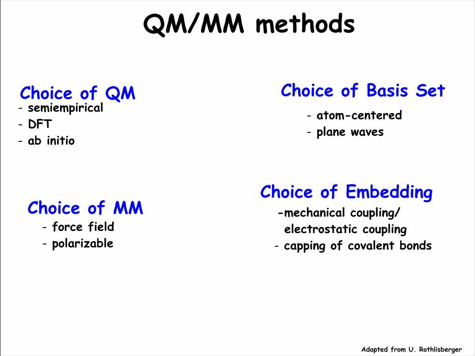
QM/MM methods
Choice of QM Choice of Basis Set
Choice of MMChoice of Embedding
- semiempirical- DFT- ab initio
- atom-centered- plane waves
- force field- polarizable
-mechanical coupling/ electrostatic coupling- capping of covalent bonds
Adapted from U. Rothlisberger

General QM/MM: Some QM/MM Methods
Plane Wave Based QM/MM:
• A. Warshel, M. Levitt, J. Mol. Biol. 103, 227 (1976)• U.C. Singh, P.A. Kollman, J. Comp. Chem. 7, 718 (1986) (AMBER) • M.J. Field, P.A. Bash, M. Karplus, J. Comp. Chem. 11, 700 (1990); P.D. Lyne, M. Hodoscek, M. Karplus, J.
Phys. Chem. A, 103, 3462 (1999) (CHARMM) • review: P. Sherwood, Modern Methods and Algorithms of Quantum Chemistry, Vol. 1, John von Neumann
Institute for Computing, 257 (2000) (www.fz-juelich.de/nic-series/Volume1) (CHEMSHELL)• C.J. Cramer, D. Truhlar, ECCC10 Feature papers: QM/MM: What have we learned, where are we, and where
do we go from here? (QMMM) (comp.chem.umn.edu/Truhlar/hilight/QMMMreview05.htm)• HM Senn, W. Thiel, Angew. Chem. Int Ed Engl. 2009, 48, 1198.
•
• M. Eichinger, P.Tavan, J. Hutter, M. Parrinello, J. Chem. Phys. 110, 10452 (1999) (CPMD)• D.A. Yarne, M.E. Tuckerman, G.J. Martyna, J. Chem. Phys. 115, 3531 (2001) (PINY_MD)•A. Laio, J. VandeVondele, U. Rothlisberger, J. Chem.Phys. 116, 6941 (2002) (CPMD)
Localized Basis Set QM/MM:• F. Maseras, K. Morokuma, J. Comput. Chem. 16, 1170 (1995) (IMOMM, ONIOM@Gaussian) • D. Bakowies, W. Thiel, J. Comp. Chem. 17, 87 (1996) (MNDO/MM)• J. Gao, Rev. Comp. Chem. 7119 (1996) (semiemp/MM)• P.L. Cummins, J.E. Gready, J. Comp. Chem. 18, 1496 (1997) (semiemp/MM@MOZYME)• G. Monnard, K.M. Merz, Acc. Chem. Res. 32, 904 (1999) (semiemp/MM)• T.K. Woo, L. Cavallo, T. Ziegler, Theor. Chem. Acc. 100, 307 (1998) (DFT/MM@ADF)• N. Ferre, M. Olivucci, J. Am. Chem. Soc. 125, 6868 (2003) (CASPT2,CASSCF/MM)
Adapted from U. Rothlisberger

Applications in molecular medicine:An alkylating drug
Pt-based drugs targeting DNACisplatin based therapies are the cornerstone of treatment of many cancers (90 % cure rate for testicular and ovarian cancer)
Unfortunately, its use is severely limited by intrinsic and acquired cell resistance
Resistance mechanism may include cellular uptake and efflux of the drug, increased detoxification of the drug, inhibition of apoptosis and increased DNA repair
Reedijk Proc. Natl. Acad. Sci. U.S.A. 2003, 100, 3611. Wang, Lippard, Nat. Rev. Drug Discovery 2005, 4, 307.Arnesano, Scintilla, Natile, Angew Chem Int Ed Engl. 200746(47):9062-4

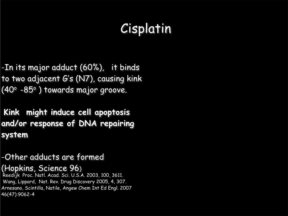
Cisplatin
-In its major adduct (60%), it binds to two adjacent G’s (N7), causing kink (40o -85o ) towards major groove.
Kink might induce cell apoptosis and/or response of DNA repairing system
-Other adducts are formed (Hopkins, Science 96) Reedijk Proc. Natl. Acad. Sci. U.S.A. 2003, 100, 3611. Wang, Lippard, Nat. Rev. Drug Discovery 2005, 4, 307.Arnesano, Scintilla, Natile, Angew Chem Int Ed Engl. 200746(47):9062-4

Computer-aided design of cisplatinand other Pt-based drugs binding to DNA
Molecular simulation play a key role because of DNA flexibility , whilst there are limitations for the docking
Geometrical parameters and energetics depend on electronic structure in a rather intricate way
In Pt-drugs, QM-based methods may be a valuable alternative to parametrizations
Force field methods may require a priori geometry information: limitations for drug design

- -
-
-
Structure of lesion depends on electronic structurePtX4: low spin diamagnetic, square planar d8 metal ion
-

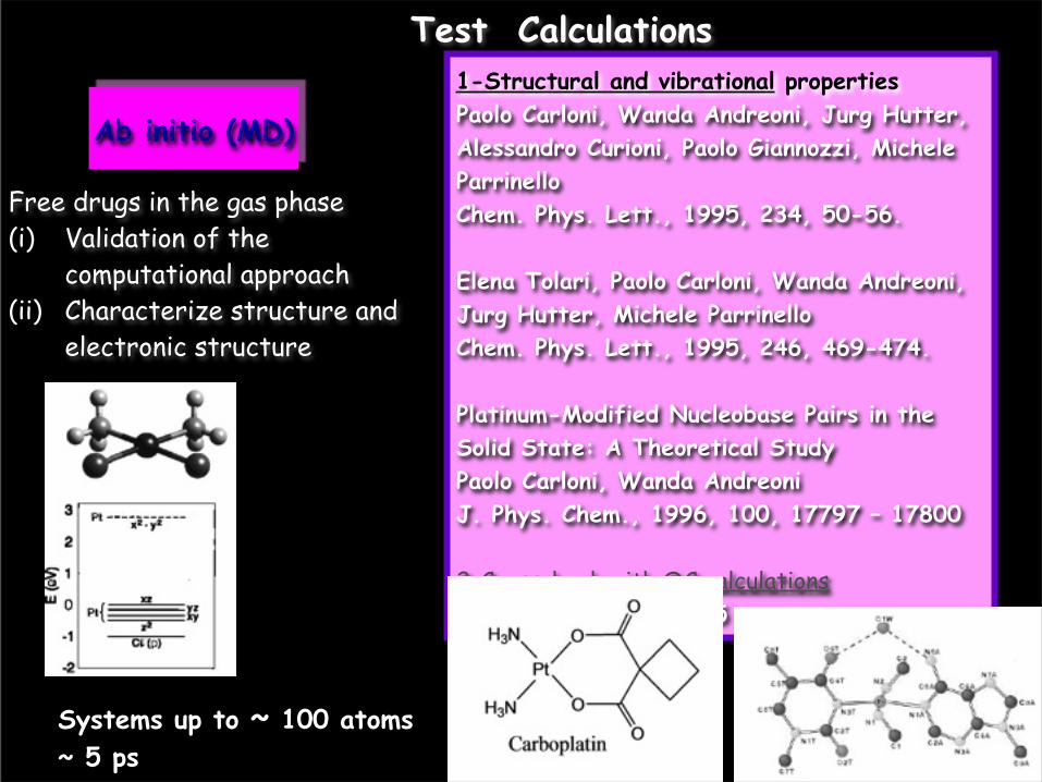
Free drugs in the gas phase(i) Validation of the
computational approach : Pt(ii) Characterize structure and
electronic structure
Ab initio (MD)
Systems up to ~ 100 atoms~ 5 ps
Test Calculations

CPMD ProgramDFT BLYP, BP XC functionalBasis set: PW (70 Ry)Martins Troullier angular momentum dependent scalar-relativistic pseudopotentials18-electron valence shellΔt=4-5 a.u.;µ=800 a.u.;T=300 K
Free drugs in the gas phase(i) Validation of the
computational approach : Pt(ii) Characterize structure and
electronic structure
Ab initio (MD)
Systems up to ~ 100 atoms~ 5 ps
Test Calculations

Free drugs in the gas phase(i) Validation of the
computational approach : Pt(ii) Characterize structure and
electronic structure
Ab initio (MD)
Systems up to ~ 100 atoms~ 5 ps
Test Calculations
1-Structural and vibrational propertiesPaolo Carloni, Wanda Andreoni, Jurg Hutter, Alessandro Curioni, Paolo Giannozzi, Michele ParrinelloChem. Phys. Lett., 1995, 234, 50-56.
Elena Tolari, Paolo Carloni, Wanda Andreoni, Jurg Hutter, Michele ParrinelloChem. Phys. Lett., 1995, 246, 469-474.
Platinum-Modified Nucleobase Pairs in the Solid State: A Theoretical StudyPaolo Carloni, Wanda Andreoni J. Phys. Chem., 1996, 100, 17797 – 17800
2-Crosscheck with QC calculationsSpiegel et al. JPC 2005
Free drugs in the gas phase(i) Validation of the
computational approach (ii) Characterize structure and
electronic structure
Ab initio (MD)
Systems up to ~ 100 atoms~ 5 ps
Test Calculations

3-Energetics of first step of cisplatin hydrolysis by constrained ab initio MD:PtCl2 (NH3)2+H2O [PtCl(NH3)2 H2O)]+ +Cl-
Free drugs in the gas phase(i) Validation of the
computational approach (ii) Characterize structure and
electronic structure
Ab initio (MD)
Systems up to ~ 100 atoms~ 5 ps Paolo Carloni, Michiel Sprik, Wanda Andreoni
J. Phys. Chem. B 2000, 104, 823.
Test Calculations

Ab initio (MD)
~ 100 atoms~ 5 ps
Free drugs in the gas phase(i) Validation of the
computational approach (ii) Characterize structure and
electronic structure
To equilibrate structure
Classical MD
~50,000 atoms~10-50 ns
Cisplatin/DNA in water
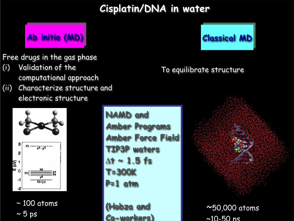
Ab initio (MD)
~ 100 atoms~ 5 ps
Free drugs in the gas phase(i) Validation of the
computational approach (ii) Characterize structure and
electronic structure
To equilibrate structure
NAMD and Amber ProgramsAmber Force FieldTIP3P watersΔt ~ 1.5 fsT=300KP=1 atm
(Hobza and Co-workers)
Classical MD
~50,000 atoms~10-50 ns
Cisplatin/DNA in water

Ab initio (MD)
~ 100 atoms~ 5 ps
Classical MD
~50,000 atoms~10-50 ns
Cisplatin/DNA in water

Ab initio (MD)
~ 100 atoms~ 20 ps
Classical MD
Pt(1)
Pt(2)
QM/MM MD
MM
QM
obtain structural ad spectroscopic information of drug -DNA complexes
~ 50,000 atoms~20 ns
Cisplatin/DNA in water

Similar cisplatin-DNA X-ray and NMR adductsKink decreases on passing from solution to
X-ray (Lippard et al., Nature 1995) NMR (Reedijk et al., Biochemistry 1999)
Lavery, R.; Sklenar, H. Defining the structure of irregular nucleic acids. Conventions and principles. J. Biomol. Struct. Dyn. 1989, 6, 655 667
Structural Information

‘Ab initio’ docking cispt-d(CCTCTG*G*TCTCC)
X-ray NMR QM/MM
Axis bend: 40o 85o 48o
H-bond with phosphate detected in X-ray is broken in NMR and QM/MMoken
Spiegel , Carloni, Rothlisberger J. Phys. Chem. B 2004
Final QM/MM structure
initial X-ray structure

‘Ab initio’ docking cispt-d(CCTCTG*G*TCTCC)
X-ray NMR QM/MM
Axis bend: 40o 85o 48o
H-bond with phosphate detected in X-ray is broken in NMR and QM/MMoken
Spiegel , Carloni, Rothlisberger J. Phys. Chem. B 2004
Final QM/MM structure
initial X-ray structure

‘Ab initio’ docking cispt-d(CCTCTG*G*TCTCC)
X-ray NMR QM/MM
Axis bend: 40o 85o 48o
H-bond with phosphate detected in X-ray is broken in NMR and QM/MMoken
Spiegel , Carloni, Rothlisberger J. Phys. Chem. B 2004
Final QM/MM structure
initial X-ray structure
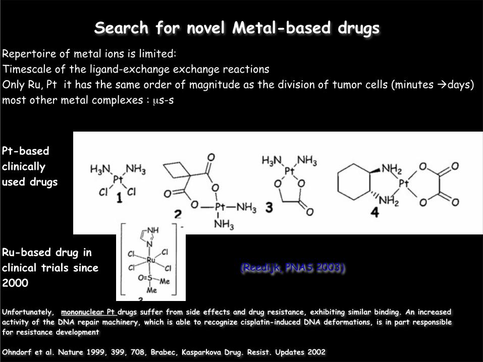
Search for novel Metal-based drugs
(Reedijk, PNAS 2003)
Repertoire of metal ions is limited:Timescale of the ligand-exchange exchange reactions Only Ru, Pt it has the same order of magnitude as the division of tumor cells (minutes days)most other metal complexes : µs-s
Pt-based clinically used drugs
Ru-based drug in clinical trials since 2000
Unfortunately, mononuclear Pt drugs suffer from side effects and drug resistance, exhibiting similar binding. An increased activity of the DNA repair machinery, which is able to recognize cisplatin-induced DNA deformations, is in part responsible for resistance development
Ohndorf et al. Nature 1999, 399, 708, Brabec, Kasparkova Drug. Resist. Updates 2002

Dinuclear compounds
(Reedijk, PNAS 2003)
Cisplatin has severe toxic side effects (such as nausea, ear damage, or vomiting) and intrinsic and acquired cell resistance
Timescale of ligand-exchange reactions:minutes days: Ru, Pt (same order of magnitude as the division of tumor cells)µs-s: most other metal complexes
Pt-based clinically used drugs
Ru-based drug in clinical trials since 2000
second and third generation mononuclear drugs suffer from side effects and drug resistance; They exhibit similar binding
They may cause minor distortions to DNA by alkylating to adjacent G’s, thus lowering the possibility to be recognized by DNA repairing enzymes (Reedijk PNAS 2003)
NMR structures of one complex`1-d(CpTpCpTpG*pG*pTpCpTpCp)
2-3 more active in cisplatin-resistance cell lines 1-3: citoxicity similar to that of cisplatin
Reedjik et al. J. Am. Chem. Soc. 2001, 2002
32 1
1
2
3

5 ps QM/MM simulation of 1-DNA: Comparison with NMR structure
RMSD between Platinated moieties in NMR and QM/MM structures: 0.6 A.

First insights in 2- and 3-DNA structures by QM/MM
Overall axis bend
51 (10) deg19 ± 5 deg 10 ± 3 deg 8 ± 4 deg
1-DNAB-DNA (no drug)
2-DNA3-DNA
NMR pyrazolate
Magistrato et. al J. Phys. Chem. B, 2006Dal Peraro et al. Curr. Op. Str. Biol. 2007,

First insights in 2- and 3-DNA structures by QM/MM
Overall axis bend
51 (10) deg19 ± 5 deg 10 ± 3 deg 8 ± 4 deg
• The overall axis bend of pyrazolate drug is similar to canonical B-DNA
decrease
• The overall axis bend of diazole and triazole bridged decreases monotonically (minimum in 3)
•All of the three drugs do not modify largely DNA structure
1-DNAB-DNA (no drug)
2-DNA3-DNA
NMR pyrazolate
Magistrato et. al J. Phys. Chem. B, 2006Dal Peraro et al. Curr. Op. Str. Biol. 2007,

Extending the time-scale: QM/MM covers timescale of ∼ 0.01 ns Because of limited time-scale, only relative comparisons are useful.
Derive force field which reproduces structural, electrostatic mechanical properties of the QM subsystem based on QM/MM
It takes into account biomolecular frame and temperature effects
Bonded model versus non-bonded model: Highly directional and kinetically inert Pt-X bonds. No risk of ligand exchange.
Calculate structural and thermodynamic averages by MD based on such force field (∼ 0.01 µs)

Force Matching
Ercolessi, Adams, Europhys. Lett. 1994, 26, 583-588
Fe: Laio, A.; Bernard, S.; Chiarotti, G. L.; Scandolo, S.; Tosatti, E. Science 2000, 287,1027 1030
Si/SiO2: Csanyi, G.; Albaret, T.; Payne, M. C.; De Vita, A. Phys. Rev. Lett. 2004, 93, 175503. Lenosky, T. J.; Sadigh, B.; Alonso, E.; Bulatov, V. V.; Diaz de la Rubia, T.; Kim, J.; Voter, A. F.; Kress, J. D. Modelling Simul. Mater. Sci. Eng. 2000, 8, 825
Oxides: Li, Y.; Siegel, D. J.; Adams, J. B.; Liu, X.-Y. Phys. Rev. B 2003, 67, 125101.
Aguado, A.; Madden, P. A. Phys. Rev. B 2004, 70, 245103.
H2O: Izvekov, S.; Parrinello, M.; Burnham, C. J.; Voth, G. A. J. Chem. Phys. 2004, 120,

Force Match ApproachForce field parameters of the platinated site are obtained from QM/MM trajectories via a force matching procedure of the classical forces to ab initio forces.
Maurer et al. JCTC 2007Spiegel et al. J. Comp. Chem 2007

Force Match ApproachForce field parameters of the platinated site are obtained from QM/MM trajectories via a force matching procedure of the classical forces to ab initio forces.
Maurer et al. JCTC 2007Spiegel et al. J. Comp. Chem 2007

Force Match ApproachForce field parameters of the platinated site are obtained from QM/MM trajectories via a force matching procedure of the classical forces to ab initio forces.
Van der Waals parameters are those of the force field.Point Charges are fitted to reproduce both the electrostatic potential and electrostatic field, they are restrained to reference values, and they are averaged over different QM/MM conformations
The classical bonded parameters are obtained through a least squares fit procedure to QM bonded forces.
Equilibrium values for bonds, angles and torsional angles are those of the QM/MM
Maurer et al. JCTC 2007Spiegel et al. J. Comp. Chem 2007

~ 10 ns FM-MD of 1-,2-,3-DNA
QM/MM FM3.3 Å3.4 ± 0.2 Å3.6 ± 0.2 Å 3.5 ± 0.3 Å 3.6 ± 0.2 Å 3.4 ± 0.3 Å 4.1 ± 0.3 Å 4.1 ± 0.5 Å
Rise: G5G6 QM/MM FM29 deg39 ± 4 deg 31 ± 2 deg 30 ± 7 deg32 ± 3 deg 29 ± 5 deg35 ± 3 deg 35 ± 3 deg
Twist: G5G6
1-DNAB-DNA (no drug)
2-DNA3-DNA
NMR pyrazolate
QM/MM FM5 deg-3 ± 7 deg 9 ± 4 deg 6 ± 7 deg 4 ± 4 deg 6 ± 5 deg-5 ± 5 deg -14 ± 9 deg
Roll:G5G6 QM/MM FM10 deg-4 ± 5 deg 8 ± 3 deg 8 ± 5 deg 8 ± 3 deg 4 ± 5 deg 18 ± 4 deg 3 ± 12 deg
Tilt: G5G6axis bendQM/MM FM5 deg19 ± 8 deg19 ± 5 deg 18 ± 9 deg 10 ± 3 deg 18 ± 8 deg 8 ± 4 deg 14 ± 7 deg
1-DNAB-DNA
2-DNA3-DNA
NMR 1-DNA
- QM/MM in qualitative agreement with FM-based MD-1-DNA and 2-DNA almost identical -1-DNA 3-DNA: decrease of axis bend- The decrease of the DNA curvature may be related with the higher cytotoxicity of the drug and to the overcome of cell resistance towards cisplatin.

Addressing resistance issues
Search of alternative drugs
Platinated/DNA-protein interactions

Cellular partners of platinated DNA
DNA (5'-d(CCUCTCTGGACCTTCC)-3')
CisPt
HMG
PHE37
Lippard S.J. et al. Nature 1999
HMG domain proteins bind the minor groove of cisplatin-DNA and bend the protein
Structural differences with 1-3 could affect the binding to HMG domain proteins

MD of platinated (5'-D(CCUCTCTGGACCTTCC)-3')
Complexes with DNA 16mer with the same sequence of cisPt-DNA/HMG protein complex.

MD of platinated (5'-D(CCUCTCTGGACCTTCC)-3')
Complexes with DNA 16mer with the same sequence of cisPt-DNA/HMG protein complex.
Classical MD simulations ~ 10 ns (30,000 atoms)

MD of platinated (5'-D(CCUCTCTGGACCTTCC)-3')
Complexes with DNA 16mer with the same sequence of cisPt-DNA/HMG protein complex.
Classical MD simulations ~ 10 ns (30,000 atoms)
CisPt-DNA:Rise G8-G9: 4.6 ± 0.5Axis bend: 49 ± 15
B-DNARise G8-G9: 3.3 ± 0.2Axis bend: 23 ± 11

Conclusions –I 1- 3 cause small
distortion
Factors might be important for binding of excision repair enzymes and HGM proteins affecting cytoxicity and resistance
Spiegel, Magistrato, Carloni, Reedijk, Klein JPC B 2007

Conclusions –II
Force matching approaches used to describe metal-based drugs and/or drugs forming covalent bonds with their targets
Possible applications include: RNA (Flexibility, Mg2+ , counterions, large electric fields), metal-ions to disordered proteins

Force Matching: Pro’s and Con’s It is a largely automatic procedures
It takes into account implicitly temperature and environment effects
Particularly useful for compounds for which electronic structure dictates stereochemistry and/or stereochemistry is not known
It avoids (i) unwanted solvent-ligand exchanges as non-bonded models (Teletchea at al. Chem. Eur. J. 2006); (ii) closed up’ structures during optimization for RESP charges
-) Expensive

• Biomolecules often do not act alone:
Cellular functions – the decisions to grow and divide, to die by programmed cell death, or to stay static – ultimately lie with macromolecules encoded by DNA.
• Proteins and RNA directly control the cell through the reactions they perform, the conformations they adopt, and the interactions that they make.
• Very heterogeneous systems (cytoplasm as “molecular soup”, cell membrane)
Relevance for in vivo conditions


The cell •Eukaryotic cell: water ∼70% weight, proteins ∼20%, DNA/RNA ∼5% , lipids ∼3%, Polysaccharides 2%Cell Volume ≈10,000 µm3 vs 1 µm3 # proteins ≈4 1010 vs 4 106 [proteins] ≈0.1 pM vs ≈ 1nM Size of genome 3 109 bp and 30,000 genes vs 4.6 106 bp and 4500 genes
The medium: The cytoplasm (saline solution) Compartmentalization: The membrane (made up by lipids)
Endoplasmic reticulum
(Information)
(Wall)(Factory)
Cytoplasm
(Power Generator)
(Garbage disposal)

• Cell stores its own set of instructions for carrying its functions (force or transport, metabolism, protein synthesis, control functions, production of new cells)
• Self-contained and self-maintaining. Structures potentially assemble, perform elaborate biochemical functions, vanish effortlessly when their work is done. Small molecules like drugs may hamper processes such as HIV-1 attack to the cell
• How can all of this work? (concentration of proteins less than nM)

In vitro focus on one
element…
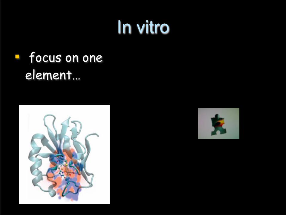
In vitro focus on one
element…

In vivo But processes in real life involve
very complicated pathways in which biomolecules might not act alone
Adapted from U. Rothlisberger


A biased biologist’s viewThe good, the bad (far from biology yet simple) and
the ugly (but the real thing)In silico, in vitro and in vivo
Computational methods

The good, the bad (far from biology yet simple) and the ugly (but the real thing)
In silico, in vitro and in vivo
Drug with high affinity ??
Computational methods

A BIG simplification
An H atom now is not different from a H atom 109 years ago, biological
systems (∼3.5 x 10⁹ years old) are! Darwinian Evolution consists in the survival of the fittest at each new
generation of each organism. It modifies existing mechanisms rather than invent new ones. Thus biological systems are similar (e.g. plants and animal proteins share striking similarities) and they are ‘’robust’’ with respect of small changes. By investigating (structural, functional, cellular) patterns,
we can have clues about these very complicated systems (Bioinformatics)

…a BIG simplification:Use patterns for structural predictions
and for pathways investigations
• The complexity of biological systems is determined not so much by the number of parts they use to carry out their functions, as by the number of interactions involved in the regulation of these functions.
• Thus, although eukaryotes have generally larger genomes than prokaryotes, genome sizes are not correlated with the complexity of the organism.
• Unicelullar eukaryotes have genome sizes that vary 200,000-fold, and the genome of the amoeba is about 200 times greater than that of humans.
• New protein structures reveals motifs already existing in the data banks and that have been used over and over again in related and sometimes even unrelated tasks.


Bioinformatics
• Fundamental features of proteins are not shared by small molecules
• Proteins are the product of natural selection superimposed to random variation:Stability and Reactivity optimized for the biological environment
• Proteins evolving from a common ancestor maintained similar core 3D structures. Structural models of proteins (targets) homologous to other proteins whose 3D structure is known (templates).
• Exploiting the presence of patterns in biology enlarges the predictive power of computational biophysics

ACTTGTAATAAATTTAGT…
From DNA...
A C D E F G
...to the Protein
Proteins that have evolved from a common ancestor may exhibit the same fold and different sequences, except for
functionally important residues
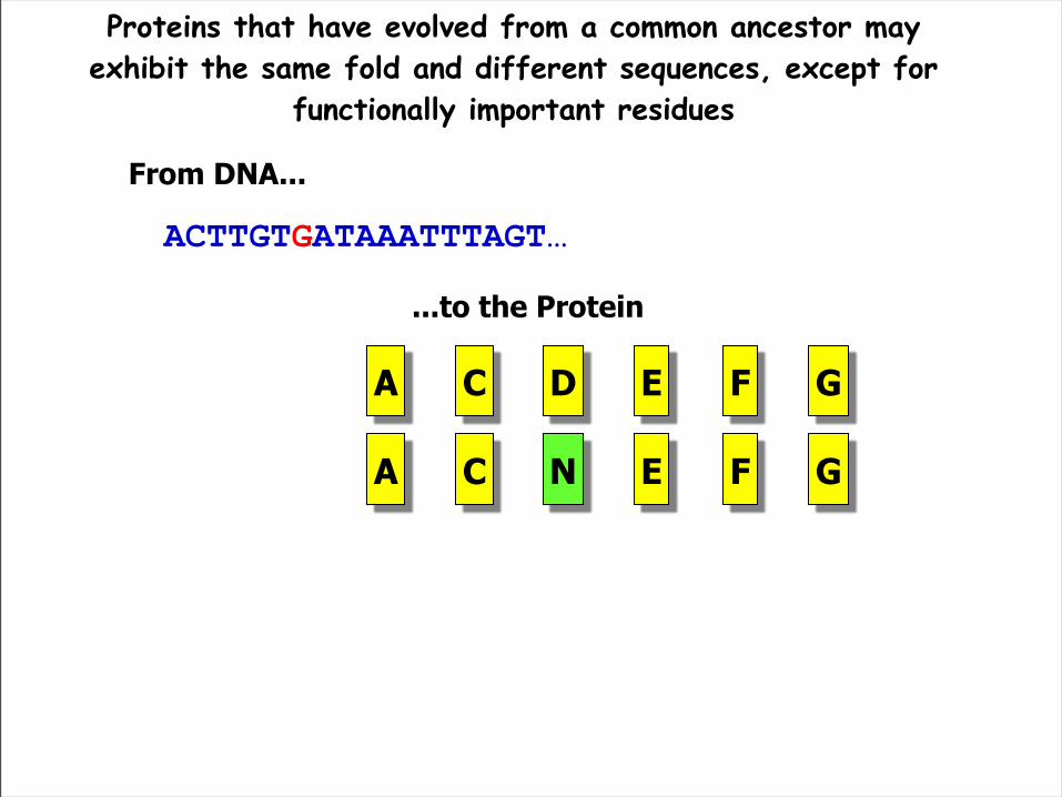
ACTTGTAATAAATTTAGT…ACTTGTGATAAATTTAGT…
From DNA...
A C D E F G
...to the Protein
A C N E F G
Proteins that have evolved from a common ancestor may exhibit the same fold and different sequences, except for
functionally important residues
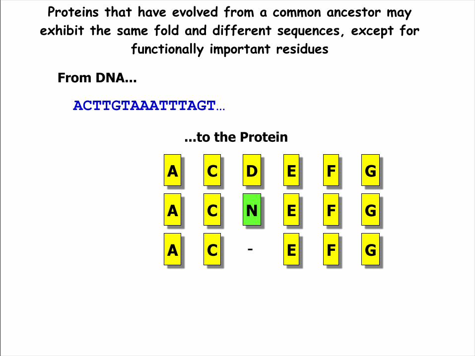
ACTTGTAATAAATTTAGT…ACTTGTGATAAATTTAGT…ACTTGTAAATTTAGT…
From DNA...
A C D E F G
...to the Protein
A C E F G-
A C N E F G
Proteins that have evolved from a common ancestor may exhibit the same fold and different sequences, except for
functionally important residues

ACTTGTAATAAATTTAGT…
From DNA
C D E F G
...To the protein
C E F G-
C N E F G
A
A
A
-
-
-

ACTTGTAATAAATTTAGT…ACTTGTCAAAATAAATTTAGT…
From DNA
C D E F G
...To the protein
C E F G-
C N E F G
A
A
A
C Q D E F GA
-
-
-

ACTTGTAATAAATTTAGT…ACTTGTCAAAATAAATTTAGT…ACTTGTCAAAATAATTTAGT…
From DNA
C D E F G
...To the protein
C E F G-
C N E F G
A
A
A
C Q D E F GA
-
-
-
C N LA - - -Q D


A striking outcome
• Understanding mechanisms. Molecular basis of e.g. enzymatic reactions or receptor signaling in the cell
• Characterizing aberrant processes (e.g. fibrillation) and eventually trying to stop them
• Delivering drugs (nano-biotechnology): e.g. delivering RNA
• Biotechnological applications: effects of mutations, gene knock-out…
• Intervening on mechanisms by improving drug affinity/selectivity?
• Food and agriculture industry: smell, taste…

Membrane signaling confined and lower numbers of players.
GPCR’s are the largest membrane-bound family of receptors (possibly selected by evolution because sensing relies on them)
Ligand binding or light induces cascade of events, in some case relatively well characterized
46 GPCR’s are drug targets
The largest cluster within the family is that
Perspectives of CPMD/MM in molecular systems biology: the case of GPCR membrane signaling
Lagerstrom&Schiott, Nature Drug Disc. 2008

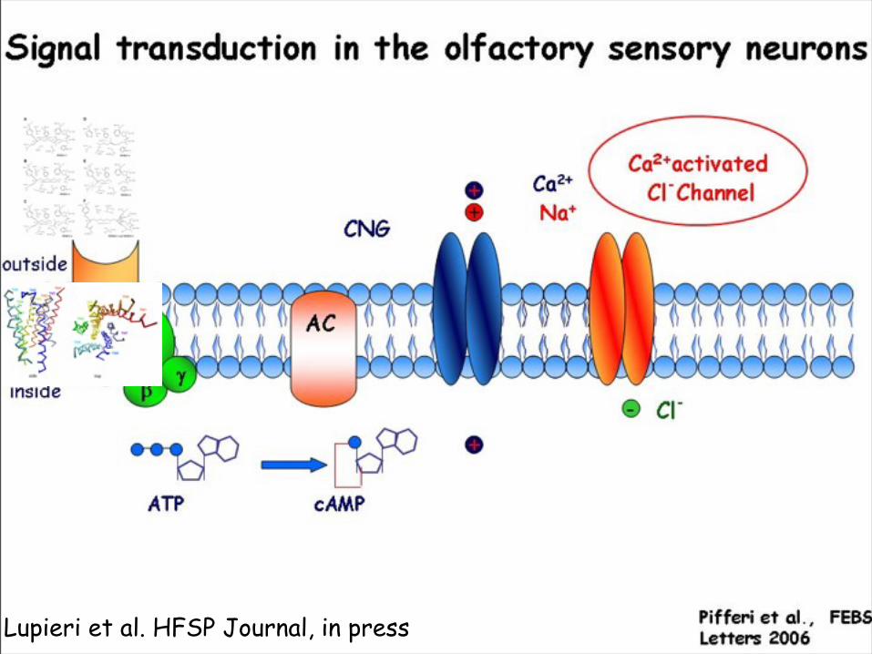
Olfactory signaling cascade
• Discriminate among thousands of structurally diverse odorant molecules, recognizing newly introduced odorants
• Build artificial noses based on biological mechanisms (IIT)
• Food and parfume industry
• Localized in olfactory sensory neurons
Buck, LB. (2005) Unraveling the sense of smell (Nobel lecture). Angew Chem Int Ed Engl. 44(38):6128-40

Signal transduction in the olfactory sensory neurons
Experimental structural information is limited
1 µm
ORα
βγ
outside
inside
Pifferi et al., FEBS Letters 2006
Expressed in the olfactory sensory neurons in the nasal olfactory epithelium
.
Several of these events are not known at the structural and molecular levelPredictions of structure and energetics
Carloni Paolo 9/20/07Odorant binding to an OR causes a conformational change of the receptor. The new conformation has a high affinity for the a subunit of the G protein heterotrimer in complex with GDP (Gα•GDP). As a result, the G-protein releases GDP and it binds to

Signal transduction in the olfactory sensory neurons
Experimental structural information is limited
1 µmAC
OR
αβ
γ
outside
inside
Pifferi et al., FEBS Letters 2006
Expressed in the olfactory sensory neurons in the nasal olfactory epithelium
.
Several of these events are not known at the structural and molecular levelPredictions of structure and energetics
Carloni Paolo 9/20/07Odorant binding to an OR causes a conformational change of the receptor. The new conformation has a high affinity for the a subunit of the G protein heterotrimer in complex with GDP (Gα•GDP). As a result, the G-protein releases GDP and it binds to

Signal transduction in the olfactory sensory neurons
Experimental structural information is limited
1 µmAC
ATP
OR
αβ
γ
cAMP
outside
inside
Pifferi et al., FEBS Letters 2006
Expressed in the olfactory sensory neurons in the nasal olfactory epithelium
.
Several of these events are not known at the structural and molecular levelPredictions of structure and energetics
Carloni Paolo 9/20/07Odorant binding to an OR causes a conformational change of the receptor. The new conformation has a high affinity for the a subunit of the G protein heterotrimer in complex with GDP (Gα•GDP). As a result, the G-protein releases GDP and it binds to

Signal transduction in the olfactory sensory neurons
Experimental structural information is limited
1 µmAC
ATP
OR
αβ
γ
CNG
cAMP
outside
inside
Pifferi et al., FEBS Letters 2006
Expressed in the olfactory sensory neurons in the nasal olfactory epithelium
.
Several of these events are not known at the structural and molecular levelPredictions of structure and energetics
Carloni Paolo 9/20/07Odorant binding to an OR causes a conformational change of the receptor. The new conformation has a high affinity for the a subunit of the G protein heterotrimer in complex with GDP (Gα•GDP). As a result, the G-protein releases GDP and it binds to

Signal transduction in the olfactory sensory neurons
Experimental structural information is limited
1 µmAC
ATP
+
OR
αβ
γ
CNG
cAMP
Ca2+
+
Na+
+
outside
inside
Pifferi et al., FEBS Letters 2006
Expressed in the olfactory sensory neurons in the nasal olfactory epithelium
.
Several of these events are not known at the structural and molecular levelPredictions of structure and energetics
Carloni Paolo 9/20/07Odorant binding to an OR causes a conformational change of the receptor. The new conformation has a high affinity for the a subunit of the G protein heterotrimer in complex with GDP (Gα•GDP). As a result, the G-protein releases GDP and it binds to

Signal transduction in the olfactory sensory neurons
Experimental structural information is limited
1 µmAC
ATP
+
OR
αβ
γ
CNG
cAMP
Ca2+
+
Na+
Ca2+activated
Cl-Channel
+
outside
inside
Pifferi et al., FEBS Letters 2006
Expressed in the olfactory sensory neurons in the nasal olfactory epithelium
.
Several of these events are not known at the structural and molecular levelPredictions of structure and energetics
Carloni Paolo 9/20/07Odorant binding to an OR causes a conformational change of the receptor. The new conformation has a high affinity for the a subunit of the G protein heterotrimer in complex with GDP (Gα•GDP). As a result, the G-protein releases GDP and it binds to

Signal transduction in the olfactory sensory neurons
Experimental structural information is limited
1 µmAC
ATP
+
OR
αβ
γ
-CNG
cAMP
Ca2+
+
Na+
Cl-
Ca2+activated
Cl-Channel
+
outside
inside
Pifferi et al., FEBS Letters 2006
Expressed in the olfactory sensory neurons in the nasal olfactory epithelium
.
Several of these events are not known at the structural and molecular levelPredictions of structure and energetics
Carloni Paolo 9/20/07Odorant binding to an OR causes a conformational change of the receptor. The new conformation has a high affinity for the a subunit of the G protein heterotrimer in complex with GDP (Gα•GDP). As a result, the G-protein releases GDP and it binds to
Lupieri et al. HFSP Journal, in press

Khavizov et al. Proteins, 2008

Lupieri et al. HFSP Journal, in press.

Giorgetti et al. Biophys. J., 2005.Anselmi et al,, Proteins, 2007Giorgetti et al., FEBS letter 2005
Berrera, Pantano, PC , Biophys J. 2006

Kranjic et al. PLOS one, in press.

Peter Mombaerts, Nature Reviews Neuroscience 5, 263-278 (2004)
Assembling the pieces of the puzzle
-Coarse-grained models, protein/protein docking,energetics of binding-Physiological conditions for structure, function and regulation-Spectroscopy-based structural predictions
?

Peter Mombaerts, Nature Reviews Neuroscience 5, 263-278 (2004)
Assembling the pieces of the puzzle
-Coarse-grained models, protein/protein docking,energetics of binding-Physiological conditions for structure, function and regulation-Spectroscopy-based structural predictions
?

Towards a more realistic representation of the cytoplasm
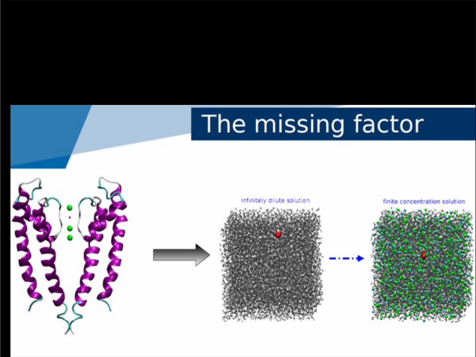


.
What about permeation?Ionic strength

K1=0.91
Wat=0.03
K2=0.87
K3=0.90
Charges averaged over 17 QM/MM conformations
Bucher D et al. Biophys. Chem. 2006

K1=0.91
Wat=0.03
K2=0.87
K3=0.90
Charges averaged over 17 QM/MM conformations
Bucher D et al. Biophys. Chem. 2006

What is the effect of voltage on entire system?

Increase of cytoplasmatic [Ca2+]: excitatory and inhibitory effect by calmodulin (CaM) binding:
(i) Decrease open probability of CNG channels by binding to their N-term (Contessa et al., J. Biom. NMR 2005)
(ii) Cause cAMP AMP by binding and activating CaM-dependent
-cAMP
Shutting down the signal: calmodulin

Peter Mombaerts, Nature Reviews Neuroscience 5, 263-278 (2004)
Assembling the pieces of the puzzle
-Coarse-grained models, protein/protein docking,energetics of binding-Physiological conditions for structure, function and regulation-Spectroscopy-based structural predictions
?

FLUORESCENT PROBES FOR CELL BIOLOGY AND STRUCTURAL BIOLOGY
^ Imaging of mRNA in live cells via nucleic acid reduction of a rhodamine probe (Pianowski et al, JACS 2009)
< Fluorescent BODIPY-based Zn2+ complex to detect neurofibrillary tangles in Alzheimer-affected brains (Ojida et al, JACS 2009)
source: http://www.conncoll.edu/ccacad/zimmer/GFP-ww/GFP-1.htm FLUORESCENCE IN A NUTSHELL:
^ Fibrils formation from prion protein (Baskakov et al, J Biol Chem, 2002)
(Shimomura, Chalfie, Tsien)

Test system
Good probe for Pi: Kd = 70 nM 18-fold change of fluo intensity upon Pi binding Linear increase of emission with increasing of Pi
concentration System suitable for simulation
Protein coordinates known ~ 5,000 atoms Known spectral properties
Experimental support: Webb's group will do mutations designed by myself
and perform NMR Biologically interesting
Pi release is a major assay target (ATPases, GTPase, phosphatases, ...)
Paola Lupieri
KNOWN PROTEIN
STRUCTURE
BIOLOGICALLY RELEVANT
SYSTEM
RemarkableDYE-PROTEIN
INTERACTIONS ≠ with
solution case
Okoh et al., Biochemistry (2006)
(A17C, A197C) PBP from E. coli labeled with 2 rhodamines

The phosphate binding protein (PBP) as a sensor for phosphate (Pi)
PBP from E. coli: FUNCTION: scavenges the periplasm for Pi, transfers it to a membrane protein for transport into the cytoplasm. ACTIVATED and brought to periplasm in Pi-starvation conditions CONFORMATION CHANGE upon Pi binding
Luecke and Quiocho, Nature (1990); Brune et al., Biochemistry (1998)
As a probe for Pi : MONITOR Pi RELEASE Fluorescent labeling exploiting conformation changes: SWITCH INNOVATIVE STRATEGY: 2 DYES
Okoh et al., Biochemistry (2006)

Rhodamines stacking & fluorescenceStructural data about rhodamines from X-ray
(Abrams et al, Inorg Chem (1986))and NMR (Ilich et al, Spectrochimica Acta 1994):
2 x ≈
dimer
6-IATR' rhodamine
DIMER is NON-FLUORESCENT
and has blue-shifted ABSORPTION
w.r.to the monomer
Absorption spectrum: Emission spectrum:
monomer
Ajtai et al, Biochemistry (1992), Van Zandvoort et al, PCCP (1999)
In WATER SOLUTIONKd ~ 10-4 M

Ajtai et al, Biochemistry (1992), Van Zandvoort et al, PCCP (1999)
dimer
Absorption spectrum: Emission spectrum:
monomer
In WATER SOLUTION
In the PROTEIN SCAFFOLD The -Pi complex conformation CAN HAVE a dimeric-like conformation of rhodamines, DISRUPTED in the +Pi conformation The disrupted dimer MUST keep some interaction btw the rhodamines: +Pi and -Pi structures are likely to have dimer character but spectra more complex than "solution" spectra
NO STRUCTURE OF PBP/2-RHOs
COMPLEXES: NEED FOR MOLECULAR
SIMULATION

Validation of protocol
Dock the RHOs on the mutated structures of PBP:monomeric/dimeric-like geometry
Classical MD of the obtained complexes in a simulation box of water, NPT conditions
Extract snapshots clustering structures
QM/MM dynamics
Calculation of optical properties with TD-DFT, GW-BSE, ...
COMPARISON WITH EXPERIMENTAL DATA FOR VALIDATIONspectra in water and on the protein, structure in water, ...

Stable trajectory (~10ns) from classical MD of PBP/2-RHO without Pi from a NON-CONVENTIONAL dimer conformation
The same conformation in solution DID NOT produce a STABLE trajectory: PROTEIN STABILIZATION OF RHODAMINES' CONFORMATION

Expanding the scope of Biomolecular Simulation
Choice of the method, accuracy In vivo conditions
Method development

Acknowledgments
(Previous) Students of Biomolecular Simulation Group in SISSA:
Enzo Carnevale (U. Penn, Philadelhia, US)Cristina Fenollar (MPI, Frankfurt, Germany)Sergio Pantano (Pasteur Inst, Montevideo, Uruguay)Vanessa Leone (SISSA, Trieste, Italy)Katrin Spiegel (Novartis, UK)Attilio Vargiu (U. Cagliari)Stefano Piana (Shaw Company, New York, US)Michele Cascella (U. Bern)Fernando Herrera (Pasteur Inst. Montevideo, Uruguay)
Collaborators:
• Mike Klein (Temple University, Philadelphia,US)• Ursula Rothlisberger (EPFL, Lausanne,CH)• Vincent Torre, A. Menini, S. Gustincich (SISSA, Italy)• Michele Parrinello (ETH Zurich and Lugano,CH)• H. Lashuel (EPFL, Lausanne, CH)• Ian McLay (GSK, UK)• Mauro Giacca (ICGEB, Trieste, Italy)• Paolo Ruggerone (U. Cagliari, Italy)• Simone Raugei (SISSA, Trieste, Italy)• Alessandra Magistrato (Democritos, Trieste, Italy)
SBPsector2008