p215 congenital vertical oculomotor apraxia – due to ante-natal vessel of percheron stroke

1
S88 Posters P215 Congenital vertical oculomotor apraxia due to ante-natal vessel of Percheron stroke R. Jain 1 *, P. Anslow 2 , M.G. Pike 1 . 1 Paediatric Neurology, John Radcliffe Hospital, Oxford, United Kingdom; 2 Neuroradiology, John Radcliffe Hospital, Oxford, United Kingdom We report a case of congenital vertical oculomotor apraxia caused by bilaterally symmetrical inferomedial thalamic infarction in keeping with an occlusion of the vessel of Percheron, presumably of ante-natal timing. Following a normal pre- and peri-natal period and early infancy, a girl presented with squint and head bobbing at the age of 8 months. Horizontal eye movements were full and oculocephalic (doll’s eye) reflex was normal in both vertical and horizontal planes. Her growth and development and her neurological and general examination were normal. The possibility that the head drops might be epileptic was excluded by a normal concurrent EEG, although some epileptiform activity from the left posterior temporal and parieto-occipital region occurred at other times. MRI scan of her brain showed symmetrical high signal in the inferomedial thalamic nuclei − in the artery of Percheron distribution (Figure). Figure: Axial and coronal view of T2 weighted MRI scan showing symmetrical high signal in the inferomedial thalamic nuclei. The presence of an acquired vertical oculomotor abnormality usually suggests a serious condition such as Niemann Pick C. By contrast, congenital vertical oculomotor apraxia is a very rare disorder which becomes evident as the characteristic head bobbing movements become apparent with improving head control in the second half of the first year of life. Head drops in infants and young children may not be epileptic, but may be compensatory movements secondary to vertical oculomotor apraxia as demonstrated in the video. In this child an ante-natal vessel of Percheron stroke is the most likely underlying cause. P216 Investigation of children and young people with transient loss of consciousness: is head-up tilt testing useful? S. Dietz 1 , L. Florence 1 , J. Murfitt 1 , P. Thakker 2 , W.P. Whitehouse 1,3 *. 1 Section of Human Development, University of Nottingham; 2 General Paediatrics & Cardiology, 3 Paediatric Neurology, Nottingham University Hospitals NHS Trust, United Kingdom Objective: Head-Up Tilt Testing (HUTT) is the gold standard investigation for adult patients with Transient Loss of Consciousness (TLOC), but its use in children is controversial. Therefore, we aimed to assess its usefulness in children by a retrospective registered clinical audit at our regional university hospital. Methods: The medical records of 100 consecutive patients aged less that 18 years, undergoing HUTT between October 2001 and December 2008 were reviewed. Information about their episodes, prodromes, triggers, previous tests, indications for the HUTT, the HUTT itself, and clinical outcomes was extracted. Results: Children were 6 to 17 years old (median 14), and there were 32/100 males. Symptom onset was at a median age of 12.5 years. The frequency of TLOC ranged from 1 a year to 4 a day (median 1 a week). Some children had one cluster of episodes. In 32/100 no trigger was reported. Triggers reported included standing up (in 20%) and prolonged standing (in 18%). Dizziness (64%) and altered vision (39%) were the commonest prodromal symptoms. 28/100 had a positive test and 17/100 were negative but symptomatic. 55/100 had a negative asymptomatic test. In 17/28 positive HUTTs, the tilt confirmed the proposed diagnosis (neurally mediated syncope, postural hypotension, or postural tachycardia syndrome). 2/28 were started on medication (fludrocortisone or midodrine). However, in 9/28 neither was the diagnosis clarified nor therapy instigated as a result. Conclusions: Potentially useful information about the TLOC was obtained with reproduction of symptoms in 45/100 cases. The 17/100 with negative but symptomatic results may have had medically unexplained TLOC or emotional attacks, although without concurrent EEG some uncertainty remains. Therefore a new protocol with video-EEG-polygraphy and beat-to-beat finger blood pressure recording and more explicit clinical reporting is being developed. P217 Congenital ataxia and dyskinesia as presenting signs of a de novo CACNA1A mutation L. Blumkin 1,2 *, M. Michelson 2,3 , S. Kivity 1,2 , D. Lev 2,3 , T. Lerman-Sagie 1,2 . 1 Pediatric Neurology Unit, Wolfson Medical Center, Holon, Israel; 2 Metabolic-Neurogenetic Clinic, Wolfson Medical Center, Holon, Israel; 3 Institute of Medical Genetics, Wolfson Medical Center, Holon, Israel Objective: To describe a patient with mental retardation and congenital cerebellar ataxia who developed recurrent episodes of coma from the age of one and a half years and genetic testing revealed a de novo CACNA1A mutation. To describe the literature on recurrent coma as a presentation of a CACNA1A mutation and to define the frequency of a congenital presentation. Methods: We reviewed all the English literature in PubMed using the search terms: CACNA1A, coma. Results: We found 70 cases of recurrent coma related to CACNA1A mutations. In only 13 cases there was data regarding the perinatal history and early development. Early motor development was delayed in 10 cases including our own patient. None of these patients had a significant perinatal history. In seven patients early symptoms were consistent with congenital ataxia. Most patients demon- strated cerebellar atrophy on brain MRI. Some patients did not demonstrate cerebellar atrophy in the initial MRI despite the existence of cerebellar signs. There was no genotype-phenotype correlation. There were no differences in the age of onset of the coma episodes, their severity and frequency, history of previous paroxysmal events, or family history of migraine in patients with and without congenital ataxia. Conclusion: CACNA1A mutations present with a very wide clinical spectrum. Congenital cerebellar ataxia can be a presentation of a CACNA1A mutation especially when associated with recurrent unexplained coma. The combination of paroxysmal episodes including impaired consciousness, autonomic features and mixed motor signs are highly suspicious for CACNA1A mutations. Family or individual history of migraine attacks is not mandatory.

Upload: r-jain

Post on 18-Sep-2016

212 views

Category:

Documents


1 download

TRANSCRIPT

S88 Posters

P215 Congenital vertical oculomotor apraxia − due toante-natal vessel of Percheron stroke

R. Jain1 *, P. Anslow2, M.G. Pike1. 1Paediatric Neurology, JohnRadcliffe Hospital, Oxford, United Kingdom; 2Neuroradiology, JohnRadcliffe Hospital, Oxford, United Kingdom

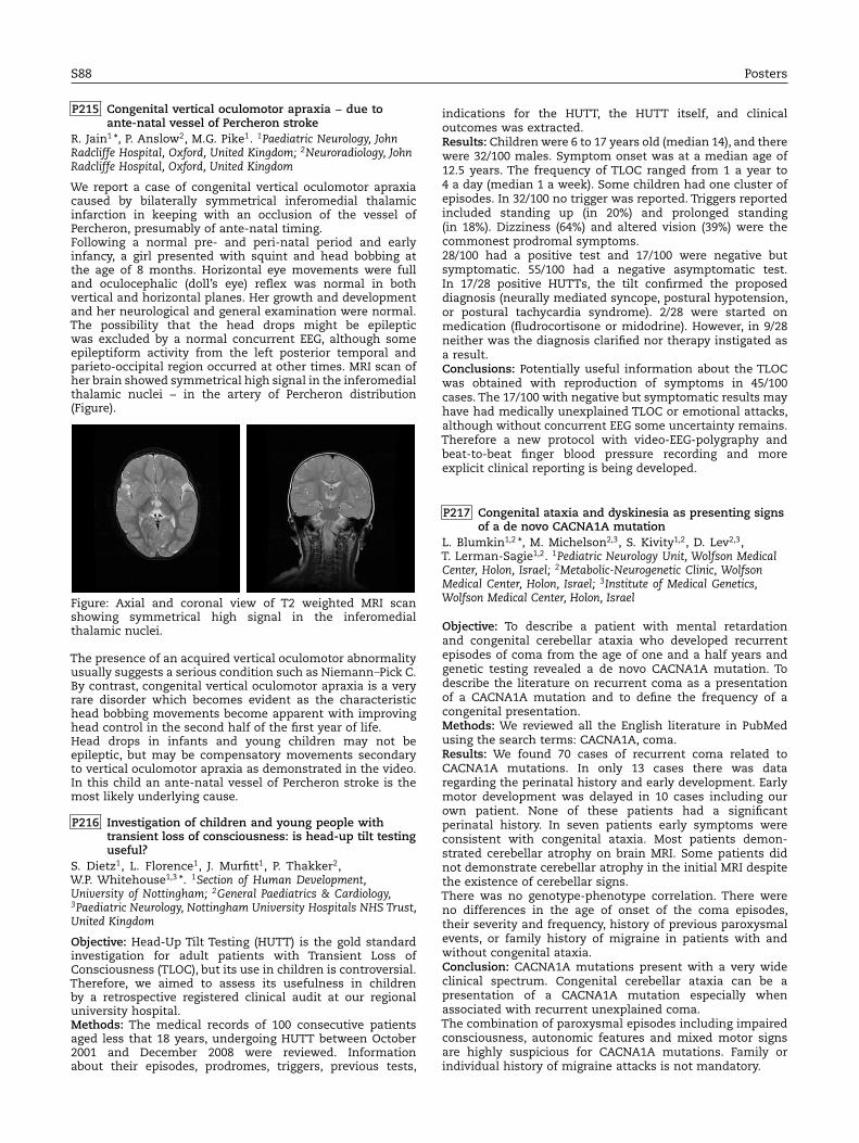
We report a case of congenital vertical oculomotor apraxiacaused by bilaterally symmetrical inferomedial thalamicinfarction in keeping with an occlusion of the vessel ofPercheron, presumably of ante-natal timing.Following a normal pre- and peri-natal period and earlyinfancy, a girl presented with squint and head bobbing atthe age of 8 months. Horizontal eye movements were fulland oculocephalic (doll’s eye) reflex was normal in bothvertical and horizontal planes. Her growth and developmentand her neurological and general examination were normal.The possibility that the head drops might be epilepticwas excluded by a normal concurrent EEG, although someepileptiform activity from the left posterior temporal andparieto-occipital region occurred at other times. MRI scan ofher brain showed symmetrical high signal in the inferomedialthalamic nuclei − in the artery of Percheron distribution(Figure).

Figure: Axial and coronal view of T2 weighted MRI scanshowing symmetrical high signal in the inferomedialthalamic nuclei.

The presence of an acquired vertical oculomotor abnormalityusually suggests a serious condition such as Niemann Pick C.By contrast, congenital vertical oculomotor apraxia is a veryrare disorder which becomes evident as the characteristichead bobbing movements become apparent with improvinghead control in the second half of the first year of life.Head drops in infants and young children may not beepileptic, but may be compensatory movements secondaryto vertical oculomotor apraxia as demonstrated in the video.In this child an ante-natal vessel of Percheron stroke is themost likely underlying cause.

P216 Investigation of children and young people withtransient loss of consciousness: is head-up tilt testinguseful?

S. Dietz1, L. Florence1, J. Murfitt1, P. Thakker2,W.P. Whitehouse1,3 *. 1Section of Human Development,University of Nottingham; 2General Paediatrics & Cardiology,3Paediatric Neurology, Nottingham University Hospitals NHS Trust,United Kingdom

Objective: Head-Up Tilt Testing (HUTT) is the gold standardinvestigation for adult patients with Transient Loss ofConsciousness (TLOC), but its use in children is controversial.Therefore, we aimed to assess its usefulness in childrenby a retrospective registered clinical audit at our regionaluniversity hospital.Methods: The medical records of 100 consecutive patientsaged less that 18 years, undergoing HUTT between October2001 and December 2008 were reviewed. Informationabout their episodes, prodromes, triggers, previous tests,

indications for the HUTT, the HUTT itself, and clinicaloutcomes was extracted.Results: Children were 6 to 17 years old (median 14), and therewere 32/100 males. Symptom onset was at a median age of12.5 years. The frequency of TLOC ranged from 1 a year to4 a day (median 1 a week). Some children had one cluster ofepisodes. In 32/100 no trigger was reported. Triggers reportedincluded standing up (in 20%) and prolonged standing(in 18%). Dizziness (64%) and altered vision (39%) were thecommonest prodromal symptoms.28/100 had a positive test and 17/100 were negative butsymptomatic. 55/100 had a negative asymptomatic test.In 17/28 positive HUTTs, the tilt confirmed the proposeddiagnosis (neurally mediated syncope, postural hypotension,or postural tachycardia syndrome). 2/28 were started onmedication (fludrocortisone or midodrine). However, in 9/28neither was the diagnosis clarified nor therapy instigated asa result.Conclusions: Potentially useful information about the TLOCwas obtained with reproduction of symptoms in 45/100cases. The 17/100 with negative but symptomatic results mayhave had medically unexplained TLOC or emotional attacks,although without concurrent EEG some uncertainty remains.Therefore a new protocol with video-EEG-polygraphy andbeat-to-beat finger blood pressure recording and moreexplicit clinical reporting is being developed.

P217 Congenital ataxia and dyskinesia as presenting signsof a de novo CACNA1A mutation

L. Blumkin1,2 *, M. Michelson2,3, S. Kivity1,2, D. Lev2,3,T. Lerman-Sagie1,2. 1Pediatric Neurology Unit, Wolfson MedicalCenter, Holon, Israel; 2Metabolic-Neurogenetic Clinic, WolfsonMedical Center, Holon, Israel; 3Institute of Medical Genetics,Wolfson Medical Center, Holon, Israel

Objective: To describe a patient with mental retardationand congenital cerebellar ataxia who developed recurrentepisodes of coma from the age of one and a half years andgenetic testing revealed a de novo CACNA1A mutation. Todescribe the literature on recurrent coma as a presentationof a CACNA1A mutation and to define the frequency of acongenital presentation.Methods: We reviewed all the English literature in PubMedusing the search terms: CACNA1A, coma.Results: We found 70 cases of recurrent coma related toCACNA1A mutations. In only 13 cases there was dataregarding the perinatal history and early development. Earlymotor development was delayed in 10 cases including ourown patient. None of these patients had a significantperinatal history. In seven patients early symptoms wereconsistent with congenital ataxia. Most patients demon-strated cerebellar atrophy on brain MRI. Some patients didnot demonstrate cerebellar atrophy in the initial MRI despitethe existence of cerebellar signs.There was no genotype-phenotype correlation. There wereno differences in the age of onset of the coma episodes,their severity and frequency, history of previous paroxysmalevents, or family history of migraine in patients with andwithout congenital ataxia.Conclusion: CACNA1A mutations present with a very wideclinical spectrum. Congenital cerebellar ataxia can be apresentation of a CACNA1A mutation especially whenassociated with recurrent unexplained coma.The combination of paroxysmal episodes including impairedconsciousness, autonomic features and mixed motor signsare highly suspicious for CACNA1A mutations. Family orindividual history of migraine attacks is not mandatory.