p120 catenin is required for normal renal...
TRANSCRIPT
2099RESEARCH ARTICLE
INTRODUCTIONKidney development serves as a model for examining themechanisms that underlie organogenesis, including cellularaggregation, polarization, differentiation and mesenchymal-epithelial interactions. Key processes in kidney development arethe formation of tubules and the regulation of their luminaldiameters. Abnormal regulation of luminal diameter duringdevelopment or repair leads to cystic kidney disease (Harris,2009).
The kidney comprises nephrons and a collecting system, whichare derived from the metanephric mesenchyme and ureteric bud,respectively (Dressler, 2009). During development, inductivesignals from the mesenchyme cause the ureteric bud to undergobranching, whereas signals from the ureteric bud induce theconversion of mesenchyme to epithelium (Saxen and Sariola,1987). Mesenchymal conversion occurs via condensation aroundureteric bud tips, followed by formation of pre-tubular cellaggregates, which then form polarized epithelial renal vesicles. Therenal vesicles elongate into comma-shaped and then s-shapedbodies. Cells in the proximal domain of the s-shaped bodydifferentiate to form the epithelial cells of the renal glomerulus.Cells in the mid- and distal domains differentiate into the tubularportion of the nephron, which is segmented into proximal tubules,loops of Henle and distal tubules.
The differential expression of cadherins is an early aspect oftubule segmentation in the developing nephron (Cho and Dressler,2003). Cadherins play crucial roles in tissue morphogenesis, thecontrol of polarity, proliferation, coordinated cell movements, andtransitions between cellular states (Gumbiner, 2005; Halbleib andNelson, 2006). The cytoplasmic domains of cadherins interact withproteins called catenins. Both p120 catenin (p120ctn; Ctnnd1 –Mouse Genome Informatics) and b-catenin (Ctnnb1) bind todistinct regions of the cytoplasmic tails of cadherins and playimportant roles in organizing the cytoskeleton and regulating cellsignaling (Reynolds, 2007).
Cadherins are differentially expressed within subdomains of thedeveloping kidney (Cho and Dressler, 2003; Cho et al., 1998; Dahlet al., 2002). In the ureteric bud, E-cadherin (cadherin 1) is thepredominant cadherin. In the adjacent mesenchyme, significantcadherin subtype switching occurs. Cadherin 11 is expressed in themesenchyme but is downregulated as the mesenchyme epithelializes.As pre-tubular aggregates form, R-cadherin (cadherin 4) andcadherin 6 are upregulated. As the renal vesicle elongates into thecomma- and s-shaped body, R-cadherin is expressed in the proximaland mid-regions whereas E-cadherin is expressed distally. Cadherin6 is expressed in the mid-region with some overlap with both R-cadherin and E-cadherin. Finally, as the s-shaped body elongates anddifferentiates, E-cadherin becomes the predominant cadherin in theproximal and distal tubules, whereas cadherin 6 is expressed in theloop of Henle and the first portion of the distal tubule.
Despite differential cadherin expression during nephrogenesis,only limited defects are seen in mice lacking single cadherins,perhaps owing to compensation by other cadherins. Mice lackingcadherin 6 or R-cadherin exhibit delayed conversion ofmesenchyme to epithelia (Dahl et al., 2002; Mah et al., 2000). R-cadherin-deficient mice also exhibit mild widening of proximaltubule lumens (Dahl et al., 2002).
Although b-catenin is known to have crucial roles innephrogenesis (Park et al., 2007; Schmidt-Ott and Barasch,2008), the roles of p120ctn family members are not well
Development 138, 2099-2109 (2011) doi:10.1242/dev.056564© 2011. Published by The Company of Biologists Ltd
1Department of Medicine, University of California, San Francisco, CA 94158, USA.2Department of Physiology, University of California, San Francisco, CA 94158, USA.3Department of Pediatrics, University of California, San Francisco, CA 94158, USA.4Department of Anatomy, University of California, San Francisco, CA 94158, USA.5Joint Research Division Vascular Biology, Medical Faculty Mannheim, 68167Mannheim, Germany.
*Present address: Department of Medicine, University of Texas SouthwesternMedical Center, Dallas, TX 75390, USA†Author for correspondence ([email protected])
Accepted 17 February 2011
SUMMARYDefects in the development or maintenance of tubule diameter correlate with polycystic kidney disease. Here, we report thatabsence of the cadherin regulator p120 catenin (p120ctn) from the renal mesenchyme prior to tubule formation leads todecreased cadherin levels with abnormal morphologies of early tubule structures and developing glomeruli. In addition, mutantmice develop cystic kidney disease, with markedly increased tubule diameter and cellular proliferation, and detached luminal cellsonly in proximal tubules. The p120ctn homolog Arvcf is specifically absent from embryonic proximal tubules, consistent with thespecificity of the proximal tubular phenotype. p120ctn knockdown in renal epithelial cells in 3D culture results in a similar cysticphenotype with reduced levels of E-cadherin and active RhoA. We find that E-cadherin knockdown, but not RhoA inhibition,phenocopies p120ctn knockdown. Taken together, our data show that p120ctn is required for early tubule and glomerularmorphogenesis, as well as control of luminal diameter, probably through regulation of cadherins.
KEY WORDS: Cadherins, Glomerulogenesis, Kidney development, p120 catenin, Polycystic disease, Mouse
p120 catenin is required for normal renal tubulogenesis andglomerulogenesisDenise K. Marciano1,2,*,†, Paul R. Brakeman3,4, Chao-Zong Lee1, Natalie Spivak3,4, Dennis J. Eastburn4, David M. Bryant4, Gerard M. Beaudoin III2, Ilse Hofmann5, Keith E. Mostov4 and Louis F. Reichardt2
DEVELO
PMENT

2100
characterized. p120ctn, however, has several essential roles in theembryogenesis of multiple tissues (Davis and Reynolds, 2006;Elia et al., 2006; Oas et al., 2010; Perez-Moreno et al., 2006;Smalley-Freed et al., 2010). The p120ctn family consists of fourproteins: p120ctn, Arvcf, and the more distantly related d-catenin(Ctnnd2 – Mouse Genome Informatics) and p0071 (plakophilin4) (McCrea and Park, 2007). These proteins stabilize cadherinson the cell surface through inhibition of endocytosis (Chiasson etal., 2009; Xiao et al., 2005). Both d-catenin and Arvcf canfunctionally substitute for p120ctn in stabilizing surface E-cadherin in vitro (Davis et al., 2003). To date, p120ctn has beenshown to stabilize many classical cadherins, including E-cadherin, R-cadherin, N-cadherin (cadherin 2 – Mouse GenomeInformatics), P-cadherin (cadherin 3), VE-cadherin (cadherin 5)and cadherin 11 (Reynolds, 2007).
In addition to interacting with cadherins, p120ctn familymembers regulate Rho GTPases. In mice, differential splicinggenerates two major isoforms of p120ctn, isoforms 1 and 3(Montonen et al., 2001), with isoform 1 inhibiting RhoA (Rhoa)and activating Rac1 and Cdc42 (Anastasiadis et al., 2000; Noren etal., 2000; Yanagisawa et al., 2008). Furthermore, Rac1 activationresults in RhoA inhibition through a p120ctn-dependentp190RhoGAP (Grlf1) pathway (Wildenberg et al., 2006).
Given p120ctn’s ability to regulate multiple cadherins, wepostulated that loss of p120ctn would lead to renal defects notobserved in the absence of individual cadherins. Thus, weexamined deficits following specific deletion of p120ctn in theureteric bud or metanephric mesenchyme. Surprisingly, p120ctndeletion from ureteric bud resulted in no obvious phenotype.Deletion from metanephric mesenchyme, however, resulted inrenal hypoplasia, early defects in tubule morphology, and renalcysts in proximal tubules, concurrent with dramatic reductionsin cadherin levels. Glomeruli were abnormal in morphology,displaying deficits in podocyte and endothelial cell organization.Further, p120ctn knockdown in 3D MDCK cell culture led to asimilar cystic phenotype with reduced levels of E-cadherin andactive RhoA. Knockdown of E-cadherin, but not inhibition ofRhoA, phenocopied p120ctn knockdown in cysts. Takentogether, our data show that p120ctn is required for tubule andglomerular formation, probably through control of cadherinlevels.
MATERIALS AND METHODSAnimalsWe crossed Ctnnd1flox/flox (floxed p120ctn) females to Ctnnd1flox/+;Pax3-cretg/+ males (Elia et al., 2006; Li et al., 2000) to obtain Ctnnd1flox/flox;Pax3-cretg/+ mutant embryos or pups. Using the same strategy, we generatedCtnnd1flox/flox;HoxB7-cretg/+ pups (Srinivas et al., 1999). Mice weremaintained on mixed genetic backgrounds and genotyped by PCR.Procedures were performed according to UCSF IACUC-approvedguidelines.
Histology and immunofluorescenceFor paraffin sections, embryonic and neonatal kidneys were fixed in 4%paraformaldehyde in PBS, embedded in paraffin and sectioned at 5 mm.For adult kidneys, mice were perfused with 4% paraformaldehyde inPBS. Antigen retrieval was performed on paraffin sections with Trilogy(Cell Marque). Vibratome sections (70 mm) and cryosections (10 mm)were permeabilized with 0.3% Triton X-100 in PBS (PBST) andblocked with 10% donkey sera in PBST. Sections were incubated withprimary antibodies overnight at 4°C, then with fluorophore-conjugatedsecondary antibodies (Invitrogen) and mounted with Prolong Gold(Invitrogen).
AntibodiesAntibodies used were: pp120 (1:1000; BD Transduction, 2B12), p120 NT(1:1000; Invitrogen, 6H11), p120 F1SH (1:1000; a gift from Al Reynolds,Vanderbilt University, TN, USA), Arvcf (1:100) (Walter et al., 2008), E-cadherin (1:1000; ECCD2), cytokeratin 8 (1:50; Developmental StudiesHybridoma bank, TROMA1), cadherin 6 (1:1000; a gift from G. Dressler,University of Michigan, MI, USA), cadherin 6 (1:1000; a gift from J.Nelson, Stanford University, CA, USA), cadherin 11 (1:100; Zymed, 71-7600), NCAM (1:100; 5B8 a gift from T. Jessell, Columbia University, NY,USA), Na/K ATPase (1:500; 5, a gift from M. Caplan, Yale University, CT,USA), calbindin (1:500; Swant, CB-38a), ZO1 (1:100; a gift from B.Stevenson, University of Alberta, Canada), nephrin (1:150; R&D,AF3159), Ki67 (1:200; Vector, VR-RM04), cleaved caspase 3 (1:100; CellSignaling, 9601), Tamm Horsfall Glycoprotein (1:250; Biogenesis, 8595-0054), NaCl co-transporter (1:500; Chemicon, AB3553), d-catenin (1:100;BD transduction, 611536), PECAM (1:200; ParMingen Int, 1951A), a-E-catenin (1:1000; Sigma, C8114), active-b-catenin (1:1000; Millipore, 8E7),b-catenin (1:1000; Zymed, 13-8400), R-cadherin (1:1000; BD transduction,610414). R-cadherin (1:10; MRCD5, Developmental Studies HybridomaBank), phosphohistone 3 (1:200; Upstate Cell Signaling, 06-570),acetylated tubulin (1:1000; Sigma, 6-11B-1), RhoA (1:250; SCBT, clone26C4), aquaporin-2 (1:100; SCBT 9882), laminin (1:200; Sigma, #9393),podocin (1:100; a gift from P. Mundel, Albert Einstein College ofMedicine, NY, USA), podocalyxin (1:40; Pcx, R&D systems MAB1556)and phalloidin 488 (1:200, Invitrogen).
Imaging and statistical analysisConfocal imaging was performed on a Zeiss LSM5 Pascal microscope.Light and immunofluorescence microscopy were performed with a NikonEclipse E600 (Axiovision HRc camera and Release 4.6 software) or a ZeissAxiovert 200M (Slidebook 4.2 software from Intelligent Imaging). Imageswere analyzed using ImageJ software as indicated. Images were minimallyprocessed with Adobe Photoshop. All data shown are mean ± s.d.Statistical significance was performed using a two-tailed nonparametricMann-Whitney U-test or two-tailed Student’s t-test.
Western blotsE17.5 mouse kidneys were dissected and homogenized in ice cold buffercontaining 1% NP-40, 0.5% sodium deoxycholate, 0.1% SDS, 20 mMsodium phosphate (pH 7.2), 150 mM NaCl, 2 mM Na+EDTA, 50 mM NaF,1 mM Na3VO4, 1 mM PMSF and a protease inhibitor cocktail (Roche).Lysates were centrifuged at 17,000 g for 15 minutes at 4°C to removeinsoluble aggregates, and SDS-PAGE and western blotting wereperformed. Densitometric quantification was performed with Image J andnormalized to GAPDH.
MDCK culture and shRNA knockdownMDCK type II cells were grown and imaged as described previously(Brakeman et al., 2009). Briefly, MDCK cells were trypsinized 1 daybefore plating. On the day of plating, wells were coated with Matrigel (BDBiosciences). Cells were trypsinized and resuspended into a single-cellsuspension containing media and 2% Matrigel prior to plating. Cystswere harvested at day five. The shRNA oligos were designed againstcanine p120 (XM_854180) using the iRNAi program(http://www.mekentosj.com/science/irnai) using guidelines from theAddgene pLKO.1 protocol (www.addgene.org). p120ctn shRNA1 (KD1),CCGGTGACAAGGTGAAGACTGATTTCAAGAGAATCAGTCTTC A -C CTTGTCATTTTTG; p120ctn shRNA2 (KD2), CCGGG -CTGGTGTTGATCAACAAATTCAAGAGATTTGTTGATCA ACACC -AGCTTTTTG; E-cadherin shRNA1 (KD1), CCGGGG -ACGTGGAAGATGTGAATCTCGAGATTCACATCTTCCAC GTCCT -TTTTG; and E-cadherin shRNA2 (KD2), CCGG GTC -TAACAGGGACAAAGAACTCGAGTTCTTTGTCCCTGTTAGAC T -T TTTG were adopted for pLKO.1 (Capaldo and Macara, 2007). shRNAswere cloned into pLKO.1 puro or blast. Viral transduction of MDCK cellswas performed as described previously (Bryant et al., 2010).
RESEARCH ARTICLE Development 138 (10)
DEVELO
PMENT

Measurements of luminal cells and cyst areaTo quantify luminal cells, control and p120ctn shRNA cysts werevisualized by differential interference contrast (DIC) andimmunofluorescence staining. To confirm that intraluminal cells were notattached to the cyst wall, cysts were optically sectioned via confocalmicroscopy. Cysts with a single lumen (as seen by apical markerpodocalyxin staining) were scored for empty lumens or lumenscontaining ≥1 cell. To measure cyst area, we took automated images ofcysts on an IN Cell Analyzer 1000 (GE Healthcare) and converted theimages to binary with ImageJ. The cyst outlines were filled and areacomputed using ImageJ. Cysts contacting other cysts or located at theimage border were excluded. The upper quartile of tabulated cyst areaswere compared.
Measurements of spindle orientationImmunofluorescence was performed with a-tubulin, Hoescht andpodocalyxin to visualize spindles, nuclei and apical membranes of cysts,respectively. Cysts were imaged by confocal microscopy at the largestcross-sectional diameter (equatorial plane). Spindle orientation parallel tothe apical membrane is 0° and perpendicular to the apical membrane is 90°.
Active Rho assaysMDCK cells were lysed in ice cold buffer containing 25 mM Tris-HCl pH7.5, 150 mM NaCl, 10 mM MgCl2, 1% NP-40, 5% glycerol, 1 mM PMSFand protease inhibitor cocktail. Cells were centrifuged to remove insolubleaggregates. Protein concentration was determined by SDS-PAGE and blotwith GAPDH using the Odyssey Infrared Imaging System (Li-CorBiosciences). Equivalent protein amounts were incubated with 20 mgRBD-GST coupled to glutathione-coupled agarose beads (CytoskeletonInc.) for 1 hour at 4°C. After washing, bound material was eluted withLaemmli buffer. Samples were analyzed by blot with anti-RhoA anddensitometry.
RESULTSp120ctn is widely expressed in the developingkidneyp120ctn is widely expressed in many tissues, including adult ratkidney where it localizes to proximal tubules, thick ascendinglimbs, distal tubules, collecting ducts and developing glomeruli(Golenhofen and Drenckhahn, 2000; Usui et al., 2003). Weanalyzed the distribution and isoform expression of p120ctn byexamining sections of embryonic day (E)17.5 kidney, whichcontain nephrons at all stages of development.
Western blot analysis identified two p120ctn isoforms in E17.5kidney lysates (Fig. 1A), isoform 1 and presumptive isoform 3,which is similar to p120ctn isoform expression in other murinetissues (Montonen et al., 2001). Immunofluorescence showed thatp120ctn is broadly expressed in uncondensed mesenchyme,condensed mesenchyme and ureteric bud, with highest levels inureteric bud (Fig. 1C-E). A schematic of the developing renalsegments is depicted in Fig. 1B. p120ctn was also present inderivatives of condensed mesenchyme, including renal vesicles(Fig. 1C), comma-shaped bodies (Fig. 1I) and s-shaped bodies (Fig.1J). Immunofluorescence with the isoform 1-specific antibodydemonstrated that p120ctn isoform 1 is also broadly expressed,albeit at much lower levels in ureteric epithelia than condensedmesenchyme (Fig. 1K,L). Isoform 1 levels decreased asmesenchymal-to-epithelial conversion proceeded, with abundantisoform 1 in condensed mesenchyme and lower levels in renalvesicles, comma bodies (Fig. 1K) and s-shaped bodies (not shown).Very little isoform 1 was detected in proximal tubules (Fig. 1M).Cell culture studies have also shown that isoforms 1 and 3 areexpressed largely in mesenchymal and epithelial cell lines,respectively (Aho et al., 2002; Keirsebilck et al., 1998; Mo andReynolds, 1996).
2101RESEARCH ARTICLEp120ctn regulates tubulogenesis
Fig. 1. p120ctn and Arvcf are broadly expressed duringmetanephric development. (A)Western blot of E17.5 kidney lysatesin control (Ctl; ctnnd1flox/flox) and mutant (Mut; ctnnd1flox/flox; pax3-cretg/0) mice with anti-p120ctn mAb pp120 (2B12; anti-p120ctn C-terminus) shows two isoforms. Western blot with anti-p120ctn isoform1 (6H11, N-terminus mAb) identifies the higher molecular weight bandas isoform 1. The lower molecular weight band co-migrates withp120ctn from MDCK cells, which express only isoform 3 (Ohkubo andOzawa, 2004). (B)Schematic of renal development. The ureteric bud(UB) is surrounded by condensed (CM) and uncondensed (UM)mesenchyme. CM differentiates to form pre-tubular aggregates (PA),which epithelialize to form renal vesicles (RV). These lengthen intocomma-shaped bodies (CB) and subsequently s-shaped bodies (SB). Theconnecting tubule (CT) of the UB fuses with the SB to form acontinuous lumen. (C-H)Localization of p120ctn in E17.5 control showsthat p120ctn (green) is present in UM, CM and UB, and the CMderivative RV (C-E). Mutant kidneys lack p120ctn in UM and CM (F-H).NCAM (red) identifies CM, RV, CB and SB. (I,J)Expression of p120ctn inthe CM derivatives CB (I) and SB (J). (K-M)Downregulation of p120ctnisoform 1 during mesenchymal-to-epithelial differentiation.(K,L)Localization of p120ctn isoform 1 (mAb 6H11) (green) andcytokeratin (red), a UB marker, at E17.5. Low isoform 1 levels arepresent in UB compared with CM. Note high isoform 1 in connectingtubules (CT). (M)There is minimal isoform 1 expression in proximaltubules (PT), identified with lotus tetragonolobus lectin (LTL, notshown). (N-P)Arvcf expression at E17.5. (N-O)Arvcf (green) is expressedin UB, RV (not shown), CB, SB and a developing glomerulus (asterisk,O). (P)Localization of Arvcf (green) and LTL (red) shows little Arvcf ispresent in mature PT. Tubules with low LTL (arrows) express Arvcf andmight be immature PT or descending loops of Henle. (Q,R)Arvcf ispresent in a short loop of Henle, as identified by cadherin 6 (red) andTamm Horsfall protein (THP, blue). (S,T)Arvcf is present in a distaltubule (DT), identified by the sodium chloride co-transporter (NCC), butnot an adjacent PT (LTL, blue). White and red dotted lines delineatestructures. Scale bars: 10mm in I-M,Q-T; 20mm in C-H,N-P. D
EVELO
PMENT

2102
Conditional deletion of p120ctn from renalmesenchyme leads to hypoplastic cystic kidneysTo investigate the functions of p120ctn during nephrogenesis, wedeleted the p120ctn gene (Ctnnd1) in ureteric bud and metanephricmesenchyme using HoxB7-cre and Pax3-cre, respectively (Li et al.,2000; Yu et al., 2002). Deletion from ureteric bud did not induceobvious defects in kidney size or morphology at 4 weeks of age,despite efficient deletion of p120ctn (see Fig. S1 in thesupplementary material). These mice appeared healthy and werefertile.
Pax3-cre has been shown to induce early and efficientrecombination in the metanephric mesenchyme and its epithelialderivatives, including all segments of the nephron (Cheng et al.,2007; Grieshammer et al., 2005). Staining of Ctnnd1flox/flox; Pax3-cretg/0 (hereafter p120ctn mutant) kidneys, showed that p120ctn isabsent in uncondensed and condensed mesenchyme (Fig. 1F-H)and its derivatives (data not shown). In these kidneys, western blotsshowed reduced expression of both isoforms, with almost totalelimination of isoform 1 (Fig. 1A).
Late embryonic p120ctn mutants were present at near Mendelianratios (20%, 27 of 134 embryos at E17.5). Pups were born atsimilar ratios, but p120ctn mutant pups died within 24 hours. Grossanatomical dissection revealed that the mice produced urine (datanot shown), but had significantly smaller kidneys than those ofcontrol littermates at E17.5 (data not shown) and P0 (Fig. 2A-D).
Postnatal day (P)0 p120ctn mutant kidneys exhibited normalcorticomedullary differentiation (Fig. 2F,G). All elements of thenephron were present, including the proximal tubule (Fig. 3B,D),thick ascending limb (Fig. 3J), distal tubule (Fig. 3H) andglomerulus (see below). Heterozygous and wild-type kidneys wereindistinguishable from each other (data not shown). Cysticstructures in the P0 p120ctn mutant kidneys were readily apparentwith 100% penetrance (27/27) (Fig. 2F,G, arrow). At highmagnification the eosinophilic appearance of cysts by Hematoxylinand Eosin (H&E) staining suggests a proximal tubule origin (Fig.2J,K). In addition, nuclear crowding (compare internuclear distancein Fig. 2H with 2J, as indicated by arrows) and areas of multi-layered epithelium (arrowheads in Fig. 2J,K) were observed. Themultilayering persisted in serial sections. Luminal cells wereobserved frequently within cysts (Fig. 2J,K).
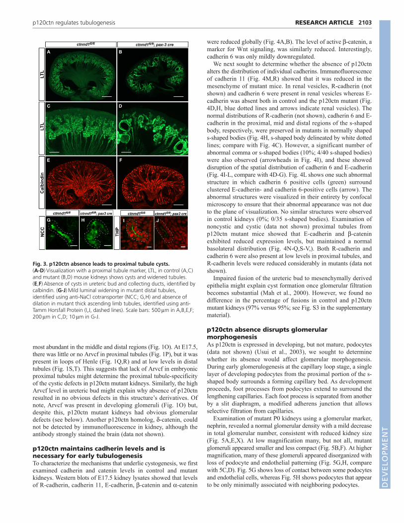
p120ctn regulates tubule diameterImmunofluorescence of sections from E17.5 control and mutantkidneys using a proximal tubule marker, lotus tetragonolobus lectin(LTL) showed that the cystic tubule regions were segments ofproximal tubules (Fig. 3B,D). At higher magnification, there wasincreased diameter and decreased total length of mutant proximaltubules (Fig. 3C,D). Dilated tubules were noted as early as E15.5(data not shown). Even tubules that were not overtly cystic hadincreased diameters. The cysts appeared only in proximal tubules;however, immunofluorescence with a distal tubule marker (sodiumchloride co-transporter, NCC) showed a mild increase in distaltubule diameter (Fig. 3G,H). Visualization of thick ascending limbtubules using Tamm-Horsfall protein showed that they have normaldiameters (Fig. 3I,J). Similarly, ureteric bud epithelia and collectingducts were not dilated (Fig. 3E,F). These results indicate that thecystic defect in mutant kidneys is specific for proximal tubules.
Arvcf expression in developing nephronsLack of redundancy with a p120 homolog in proximal tubules isone possible explanation for the proximal tubule-specificcystogenesis. The localization of Arvcf, the closest p120ctn
homolog, has been characterized in mature tubules (Walter et al.,2008). We extended this work by determining its distribution indeveloping kidneys. At E17.5 Arvcf was strongly expressed inureteric bud, with lower expression in uncondensed and condensedmesenchyme (Fig. 1N; see Fig. S2 in the supplementary material).As the mesenchyme epithelialized, increased Arvcf was present incomma-shaped bodies (Fig. 1N). In the s-shaped bodies, Arvcf was
RESEARCH ARTICLE Development 138 (10)
Fig. 2. p120ctn absence results in hypoplastic cystic kidneys. (A-C)Gross histology of P0 control (ctnnd1flox/flox; A) and mutant(ctnnd1flox/flox; pax3-cretg/0; B,C) mouse kidneys. (D,E)Quantification ofdifferences between control and mutant in (D) renal size [kidney area(mm2) per mouse weight (g)] P<0.003 and (E) P0 animal weightP<0.10. Data are mean ± s.d. Statistical analysis by the Mann-WhitneyU-test. (F,G)Hematoxylin and Eosin (H&E)-stained sections from P0control (F) and mutant (G) kidneys. Panels in G show sections from asingle kidney with a cyst indicated (arrow). (H-K)Higher magnificationimages of H&E-stained cortex in control (H) and mutant (I-K) kidneys. Ishows a cystic tubule involving Bowman’s capsule (arrow) and proximaltubule (PT). J and K illustrate the most common morphologiesobserved: cuboidal epithelium with luminal cells and areas ofmultilayering (arrowheads). Arrows in H,J highlight the nuclearcrowding in J compared with H. Scale bars: 1 mm in A-C; 250mm inF,G; 25mm in H-K.
DEVELO
PMENT
most abundant in the middle and distal regions (Fig. 1O). At E17.5,there was little or no Arvcf in proximal tubules (Fig. 1P), but it waspresent in loops of Henle (Fig. 1Q,R) and at low levels in distaltubules (Fig. 1S,T). This suggests that lack of Arvcf in embryonicproximal tubules might determine the proximal tubule-specificityof the cystic defects in p120ctn mutant kidneys. Similarly, the highArvcf level in ureteric bud might explain why absence of p120ctnresulted in no obvious defects in this structure’s derivatives. Ofnote, Arvcf was present in developing glomeruli (Fig. 1O) but,despite this, p120ctn mutant kidneys had obvious glomerulardefects (see below). Another p120ctn homolog, d-catenin, couldnot be detected by immunofluorescence in kidney, although theantibody strongly stained the brain (data not shown).
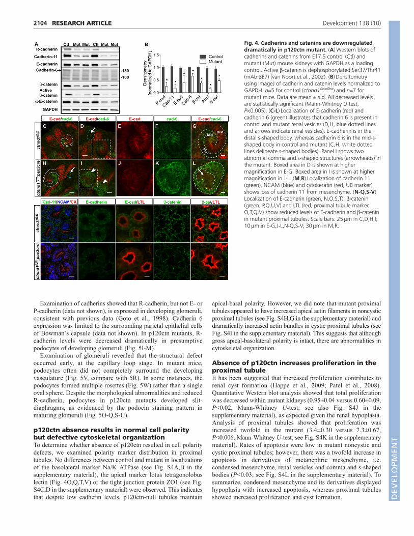
p120ctn maintains cadherin levels and isnecessary for early tubulogenesisTo characterize the mechanisms that underlie cystogenesis, we firstexamined cadherin and catenin levels in control and mutantkidneys. Western blots of E17.5 kidney lysates showed that levelsof R-cadherin, cadherin 11, E-cadherin, b-catenin and a-catenin
were reduced globally (Fig. 4A,B). The level of active b-catenin, amarker for Wnt signaling, was similarly reduced. Interestingly,cadherin 6 was only mildly downregulated.
We next sought to determine whether the absence of p120ctnalters the distribution of individual cadherins. Immunofluorescenceof cadherin 11 (Fig. 4M,R) showed that it was reduced in themesenchyme of mutant mice. In renal vesicles, R-cadherin (notshown) and cadherin 6 were present in renal vesicles whereas E-cadherin was absent both in control and the p120ctn mutant (Fig.4D,H, blue dotted lines and arrows indicate renal vesicles). Thenormal distributions of R-cadherin (not shown), cadherin 6 and E-cadherin in the proximal, mid and distal regions of the s-shapedbody, respectively, were preserved in mutants in normally shapeds-shaped bodies (Fig. 4H, s-shaped body delineated by white dottedlines; compare with Fig. 4C). However, a significant number ofabnormal comma or s-shaped bodies (10%; 4/40 s-shaped bodies)were also observed (arrowheads in Fig. 4I), and these showeddisruption of the spatial distribution of cadherin 6 and E-cadherin(Fig. 4I-L, compare with 4D-G). Fig. 4L shows one such abnormalstructure in which cadherin 6 positive cells (green) surroundclustered E-cadherin- and cadherin 6-positive cells (arrow). Theabnormal structures were visualized in their entirety by confocalmicroscopy to ensure that their abnormal appearance was not dueto the plane of visualization. No similar structures were observedin control kidneys (0%; 0/35 s-shaped bodies). Examination ofnoncystic and cystic (data not shown) proximal tubules fromp120ctn mutant mice showed that E-cadherin and b-cateninexhibited reduced expression levels, but maintained a normalbasolateral distribution (Fig. 4N-Q,S-V,). Both R-cadherin andcadherin 6 were also present at low levels in proximal tubules, andR-cadherin levels were reduced considerably in mutants (data notshown).
Impaired fusion of the ureteric bud to mesenchymally derivedepithelia might explain cyst formation once glomerular filtrationbecomes substantial (Mah et al., 2000). However, we found nodifference in the percentage of fusions in control and p120ctnmutant kidneys (97% versus 95%; see Fig. S3 in the supplementarymaterial).
p120ctn absence disrupts glomerularmorphogenesisAs p120ctn is expressed in developing, but not mature, podocytes(data not shown) (Usui et al., 2003), we sought to determinewhether its absence would affect glomerular morphogenesis.During early glomerulogenesis at the capillary loop stage, a singlelayer of developing podocytes from the proximal portion of the s-shaped body surrounds a forming capillary bed. As developmentproceeds, foot processes from podocytes extend to surround thelengthening capillaries. Each foot process is separated from anotherby a slit diaphragm, a modified adherens junction that allowsselective filtration from capillaries.
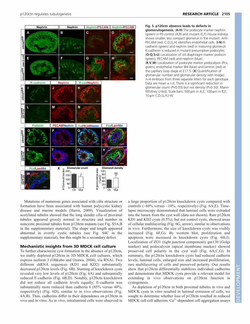
Examination of mutant P0 kidneys using a glomerular marker,nephrin, revealed a normal glomerular density with a mild decreasein total glomerular number, consistent with reduced kidney size(Fig. 5A,E,X). At low magnification many, but not all, mutantglomeruli appeared smaller and less compact (Fig. 5B,F). At highermagnification, many of these glomeruli appeared disorganized withloss of podocyte and endothelial patterning (Fig. 5G,H, comparewith 5C,D). Fig. 5G shows loss of contact between some podocytesand endothelial cells, whereas Fig. 5H shows podocytes that appearto be only minimally associated with neighboring podocytes.
2103RESEARCH ARTICLEp120ctn regulates tubulogenesis
Fig. 3. p120ctn absence leads to proximal tubule cysts. (A-D)Visualization with a proximal tubule marker, LTL, in control (A,C)and mutant (B,D) mouse kidneys shows cysts and widened tubules.(E,F)Absence of cysts in ureteric bud and collecting ducts, identified bycalbindin. (G-J)Mild luminal widening in mutant distal tubules,identified using anti-NaCl cotransporter (NCC; G,H) and absence ofdilation in mutant thick ascending limb tubules, identified using anti-Tamm Horsfall Protein (I,J, dashed lines). Scale bars: 500mm in A,B,E,F;200mm in C,D; 10mm in G-J.
DEVELO
PMENT
2104
Examination of cadherins showed that R-cadherin, but not E- orP-cadherin (data not shown), is expressed in developing glomeruli,consistent with previous data (Goto et al., 1998). Cadherin 6expression was limited to the surrounding parietal epithelial cellsof Bowman’s capsule (data not shown). In p120ctn mutants, R-cadherin levels were decreased dramatically in presumptivepodocytes of developing glomeruli (Fig. 5I-M).
Examination of glomeruli revealed that the structural defectoccurred early, at the capillary loop stage. In mutant mice,podocytes often did not completely surround the developingvasculature (Fig. 5V, compare with 5R). In some instances, thepodocytes formed multiple rosettes (Fig. 5W) rather than a singleoval sphere. Despite the morphological abnormalities and reducedR-cadherin, podocytes in p120ctn mutants developed slit-diaphragms, as evidenced by the podocin staining pattern inmaturing glomeruli (Fig. 5O-Q,S-U).
p120ctn absence results in normal cell polaritybut defective cytoskeletal organizationTo determine whether absence of p120ctn resulted in cell polaritydefects, we examined polarity marker distribution in proximaltubules. No differences between control and mutant in localizationsof the basolateral marker Na/K ATPase (see Fig. S4A,B in thesupplementary material), the apical marker lotus tetragonolobuslectin (Fig. 4O,Q,T,V) or the tight junction protein ZO1 (see Fig.S4C,D in the supplementary material) were observed. This indicatesthat despite low cadherin levels, p120ctn-null tubules maintain
apical-basal polarity. However, we did note that mutant proximaltubules appeared to have increased apical actin filaments in noncysticproximal tubules (see Fig. S4H,G in the supplementary material) anddramatically increased actin bundles in cystic proximal tubules (seeFig. S4I in the supplementary material). This suggests that althoughgross apical-basolateral polarity is intact, there are abnormalities incytoskeletal organization.
Absence of p120ctn increases proliferation in theproximal tubuleIt has been suggested that increased proliferation contributes torenal cyst formation (Happe et al., 2009; Patel et al., 2008).Quantitative Western blot analysis showed that total proliferationwas decreased within mutant kidneys (0.95±0.04 versus 0.60±0.09,P<0.02, Mann-Whitney U-test; see also Fig. S4J in thesupplementary material), as expected given the renal hypoplasia.Analysis of proximal tubules showed that proliferation wasincreased twofold in the mutant (3.4±0.30 versus 7.3±0.67,P<0.006, Mann-Whitney U-test; see Fig. S4K in the supplementarymaterial). Rates of apoptosis were low in mutant noncystic andcystic proximal tubules; however, there was a twofold increase inapoptosis in derivatives of metanephric mesenchyme, i.e.condensed mesenchyme, renal vesicles and comma and s-shapedbodies (P<0.03; see Fig. S4L in the supplementary material). Tosummarize, condensed mesenchyme and its derivatives displayedhypoplasia with increased apoptosis, whereas proximal tubulesshowed increased proliferation and cyst formation.
RESEARCH ARTICLE Development 138 (10)
Fig. 4. Cadherins and catenins are downregulateddramatically in p120ctn mutant. (A)Western blots ofcadherins and catenins from E17.5 control (Ctl) andmutant (Mut) mouse kidneys with GAPDH as a loadingcontrol. Active b-catenin is dephosphorylated Ser37/Thr41(mAb 8E7) (van Noort et al., 2002). (B)Densitometryusing ImageJ of cadherin and catenin levels normalized toGAPDH. n5 for control (ctnnd1flox/flox) and n7 formutant mice. Data are mean ± s.d. All decreased levelsare statistically significant (Mann-Whitney U-test,P<0.005). (C-L)Localization of E-cadherin (red) andcadherin 6 (green) illustrates that cadherin 6 is present incontrol and mutant renal vesicles (D,H, blue dotted linesand arrows indicate renal vesicles). E-cadherin is in thedistal s-shaped body, whereas cadherin 6 is in the mid-s-shaped body in control and mutant (C,H, white dottedlines delineate s-shaped bodies). Panel I shows twoabnormal comma and s-shaped structures (arrowheads) inthe mutant. Boxed area in D is shown at highermagnification in E-G. Boxed area in I is shown at highermagnification in J-L. (M,R)Localization of cadherin 11(green), NCAM (blue) and cytokeratin (red, UB marker)shows loss of cadherin 11 from mesenchyme. (N-Q,S-V)Localization of E-cadherin (green, N,O,S,T), b-catenin(green, P,Q,U,V) and LTL (red, proximal tubule marker,O,T,Q,V) show reduced levels of E-cadherin and b-cateninin mutant proximal tubules. Scale bars: 25mm in C,D,H,I;10mm in E-G,J-L,N-Q,S-V; 30mm in M,R.
DEVELO
PMENT
Mutations of numerous genes associated with cilia structure orformation have been associated with human polycystic kidneydisease and murine models (Harris, 2009). Visualization ofacetylated tubulin showed that the long slender cilia of proximaltubules appeared grossly normal in structure and number innoncystic proximal tubules from p120ctn mutants (see Fig. S5A,Bin the supplementary material). The shape and length appearedabnormal in overtly cystic tubules (see Fig. S4C in thesupplementary material), but this might be a secondary defect.
Mechanistic insights from 3D MDCK cell cultureTo further characterize cyst formation in the absence of p120ctn,we stably depleted p120ctn in 3D MDCK cell cultures, whichexpress isoform 3 (Ohkubo and Ozawa, 2004), via RNAi. Twodifferent shRNA sequences (KD1 and KD2) substantiallydecreased p120ctn levels (Fig. 6B). Staining of knockdown cystsrevealed very low levels of p120ctn (Fig. 6A) and substantiallyreduced E-cadherin (Fig. 6B,D). Notably, p120ctn knockdowndid not reduce all cadherin levels equally; E-cadherin wassubstantially more reduced than cadherin 6 (85% versus 40%,respectively) (Fig. 6D), similar to in vivo observations (Fig.4A,B). Thus, cadherins differ in their dependence on p120ctn invivo and in vitro. As in vivo, intraluminal cells were observed in
a large proportion of p120ctn knockdown cysts compared withcontrols (~60% versus ~10%, respectively) (Fig. 6A,E). Time-lapse microscopy demonstrated that luminal cells were extrudedinto the lumen from the cyst wall (data not shown). Rare p120ctnKD1 and KD2 cysts (0.5%), but not control cysts, showed areasof cellular multilayering (Fig. 6G, arrow), similar to observationsin vivo. Furthermore, the size of knockdown cysts was visiblyincreased (Fig. 6F,G). By western blot, proliferation andapoptosis were increased in knockdown cysts (Fig. 6H,I).Localization of ZO1 (tight junction component), gm130 (Golgimarker) and podocalyxin (apical membrane marker) showedpreserved cell polarity in the cyst wall (Fig. 6A,C,G). Insummary, the p120ctn knockdown cysts had reduced cadherinlevels, luminal cells, enlarged size and increased proliferation,rare multilayering of cells and preserved polarity. Our resultsshow that p120ctn differentially stabilizes individual cadherinsand demonstrate that MDCK cysts provide a relevant model forextending in vivo observations on p120ctn function incystogenesis.
As depletion of p120ctn in both proximal tubules in vivo andMDCK cysts in vitro resulted in luminal extrusion of cells, wesought to determine whether loss of p120ctn resulted in reducedMDCK cell-cell adhesion. Ca2+-dependent cell aggregation assays
2105RESEARCH ARTICLEp120ctn regulates tubulogenesis
Fig. 5. p120ctn absence leads to defects inglomerulogenesis. (A-H)The podocyte marker nephrin(green) in P0 control (A,B) and mutant (E,F) mouse kidneysshows smaller, less compact glomeruli in the mutant. Anti-PECAM (red, C,D,G,H) identifies endothelial cells. (I-N)R-cadherin (green) and nephrin (red) in maturing glomeruli.R-cadherin is reduced in mutant presumptive podocytes.(O-Q,S-U) Localization of slit-diaphragm marker podocin(green), PECAM (red) and nephrin (blue).(R,V,W) Localization of podocyte marker podocalyxin (Pcx,green), endothelial marker IB4 (blue) and laminin (red) atthe capillary loop stage at E17.5. (X)Quantification ofglomerular number and glomerular density with ImageJ.n4 embryos from three separate litters for each genotype.Data are mean ± s.d. There is a significant reduction inglomerular count (P<0.03) but not density (P>0.50). Mann-Whitney U-test. Scale bars: 500mm in A,E; 100mm in B,F;10mm C,D,G,H,I-W.
DEVELO
PMENT

2106
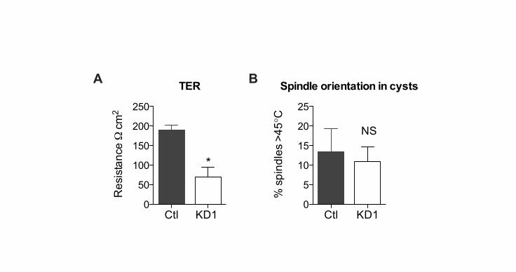
showed that dissociated p120ctn knockdown cells had adramatically reduced ability to aggregate (Fig. 6J). Measurementsof transepithelial resistance, a surrogate of tight junction integrity,in polarized monolayers showed that p120ctn knockdownmonolayers had reduced resistance (see Fig. S6A in thesupplementary material). In addition, confluent p120ctnknockdown cells appeared more elongated and mesenchyme-like(data not shown).
Another potential mechanism that might result in luminal cellsin vivo and in vitro is altered orientation of cell division. The angleof cell division in proximal tubules is difficult to measure given thetubule tortuosity. We measured the spindle angle in the equatorialplane of control and p120ctn knockdown MDCK cysts and found
no statistical differences (see Fig. S6B in the supplementarymaterial). This suggests that misorientation of cell division does notcontribute to the presence of luminal cells.
p120ctn has been shown to regulate the activities of Rho, Racand Cdc42 (Anastasiadis et al., 2000; Noren et al., 2000;Wildenberg et al., 2006; Yanagisawa et al., 2008). In addition,inhibition of Rho kinase, a downstream effector of Rho, has beenshown to cause defects in ureteric bud-derived tubulogenesis(Meyer et al., 2006; Michael et al., 2005). We performed GST-Rhotekin-RBD pulldown assays on MDCK cyst lysates and founda significant decrease in active GTP-bound RhoA in p120ctnknockdown cysts (Fig. 7A). Thus, absence of p120ctn isoform 3appears to reduce RhoA activity in vitro.
RESEARCH ARTICLE Development 138 (10)
Fig. 6. p120 knockdown in 3D cyst culture results in enlarged cysts with reduced E-cadherin. (A,C)p120ctn and E-cadherin in p120ctnknockdown cysts. Panels show staining of phalloidin, ZO1 and gm130 as indicated. Merged images with Hoescht 33342 (blue, nuclei) show thatp120 KD cysts contain intraluminal cells. (B)Western blot of p120ctn. Lanes 1 and 2: E17.5 kidney lysates from control and mutant. Lanes 3-5:MDCK cyst lysates from scramble and two p120 shRNA (KD1 and KD2). (D)Western blots of E-cadherin and cadherin 6 in control and p120ctnknockdown cysts. Percentages are E-cadherin and cadherin 6 levels normalized to GAPDH. (E)Quantification of percentage of cysts containing ≥1luminal cell(s). Results are mean ± s.d. from three independent experiments with >300 cysts each (P<0.001 for scramble versus p120 KD1, andscramble versus p120 KD2). (F)Quantification of cyst area using DIC images analyzed in ImageJ. Results are mean ± s.d. of the upper quartile ofcyst areas measured from three independent experiments done in triplicate with >300 cysts each (P<0.01 for scramble versus p120 KD1).(G)Localization of podocalyxin (Pcx, an apical membrane marker), b-catenin and Hoescht shows enlarged cysts and multilayered epithelium (arrow).(H,I)Densitometry of western blots of cyst lysates with phosphohistone-3 and active caspase-3 normalized to GAPDH. For phosphohistone-3,P<0.04 for scramble versus p120 KD1. For caspase-3, P<0.001 for scramble versus p120 KD2; P<0.09 scramble versus p120 KD1. (J)Cellularaggregation in scramble- and p120ctn-knockdown MDCK cells. Cells dissociated with EDTA were added to wells containing 1 mM CaCl2 or 1 mMEGTA, and cellular aggregation assessed at times 0, 30 and 60 minutes. Percentage aggregation(Na/N)�100 where Nanumber of cells inaggregates ≥3 cells and Ntotal number of cells. Graph shows one of two similar experiments with three replicates for each time. Aggregation(mean ± s.d.) in p120ctn knockdown was reduced compared with control at 30 minutes (P<0.003) and 60 minutes (P<0.02). All statistical analysiswas carried out using unpaired, two-tailed Student’s t-test.
DEVELO
PMENT

To determine whether reduced active RhoA plays a role in thep120ctn-knockdown phenotype, we treated control cysts with aRho kinase (ROCK) inhibitor Y-27632. This did not result inincreased cyst size (Fig. 7B) or intraluminal cell number (Fig. 7C).Thus, decreased RhoA signaling is unlikely to underlie the defectsobserved with p120ctn deficiency.
To determine whether the cystic abnormalities resulting fromp120ctn deficiency might be caused by reduced cadherin levels, weexamined the structures of E-cadherin-deficient cysts, prepared bystable E-cadherin knockdown (Fig. 7D,E). These cysts wereenlarged (Fig. 7E,F) and contained many luminal cells (Fig. 7F,G).Furthermore, E-cadherin knockdown also resulted in reducedcellular aggregation (Fig. 7H). Together, these results suggest thatE-cadherin deficiency, and not RhoA inhibition, underlies thedefects in the p120ctn mutant cysts and suggest that cadherin lossis responsible for the phenotypes observed in vivo.
DISCUSSIONOur data demonstrates that p120ctn absence in the metanephricmesenchyme leads to hypoplastic kidneys with defective tubularand glomerular morphogenesis. Furthermore, p120ctn is requiredfor establishing a normal lumen diameter and for preventing cystformation within proximal tubules. Similarly, p120ctn knockdownin 3D MDCK cells results in enlarged and abnormal cysts.Delaminated cells are observed in both the mutant proximal tubulesand p120ctn-knockdown MDCK cysts. p120ctn knockdown inMDCK cells results in decreased Ca2+-dependent cellularaggregation and loss of tight junction integrity, demonstrating invitro that, in renal epithelial cells, p120ctn is required for normalintercellular adhesion. Both in vivo and in vitro, p120ctn deficiencyresults in reduced cadherin levels. This might be the central eventin causing cystogenesis and cell delamination in vivo, because E-cadherin knockdown partially phenocopies loss of p120ctn in vitro.
Although p120ctn deficiency in proximal tubules manifests ascystic tubules, in other tubule segments loss of p120ctn does notlead to a severe phenotype. Several lines of evidence suggest thatfunctional redundancy of p120ctn and Arvcf might account for thisresult. Collectively, p120ctn family members control cadherinlevels in a dose-dependent manner (Xiao et al., 2003), and Arvcfexpression can functionally substitute for p120ctn in vitro (Davis
2107RESEARCH ARTICLEp120ctn regulates tubulogenesis
Fig. 7. E-cadherin knockdown but not RhoA inhibitionphenocopies loss of p120ctn in MDCK cysts. (A) Rho activity assays.Quantification of GTP-Rho pull-downs using GST Rhotekin-RBD. GTP-Rholevels are normalized to total Rho. (B)Quantification of the effect ofROCK inhibition (Y-27632, 10mM) on cyst area using DIC imagesanalyzed in ImageJ. Results are mean ± s.d. of the upper quartile of cystareas. Cysts treated with Y-27632 are smaller than control (P<0.03).(C)Quantification of the effect of ROCK inhibitor (Y-27632, 10mM) onthe percentage of cysts with luminal cells (P<0.4 for scramble versusscramble +Y-27632). (D)Western blot of E-cadherin from MDCK lysateswith scramble or two E-cadherin shRNA (KD1 and KD2).(E)Quantification of cyst area performed as described for panel B(P<0.001 for scramble versus E-cadherin KD1 or KD2). (F)Visualization ofE-cadherin reduction in E-cadherin KD cysts. Panels show b-catenin andphalloidin as indicated. Merged images with Hoescht 33342 (blue, nuclei)show intraluminal cells in E-cadherin KD cysts. (G)Quantification ofpercentage of cysts containing >1 luminal cell (P<0.001 for scrambleversus each KD). (H)Cellular aggregation in scramble, E-cadherin KD andp120 KD cells. Cells dissociated with EDTA were added to wellscontaining 1 mM CaCl2 and cellular aggregation assessed at 0 and 30minutes. Percentage aggregation was determined as described for Fig. 6J.Graph shows one of two similar experiments with four replicates for eachtime (P<0.15 for scramble versus E-cadherin KD1; P<0.03 for scrambleversus E-cadherin KD2; P<0.001 for scramble versus p120 KD1, all at 30minutes). All graphical results are mean ± s.d. and were analyzed with anunpaired two-tailed Student’s t-test. Panels B,C,E and G are from threeindependent experiments done in triplicate with >300 cysts each.
Fig. 8. Proposed model of p120ctn function in renaltubulogenesis. p120ctn maintains cadherin levels for appropriatemigration and/or sorting of cells in renal vesicles into the distinctdomains of comma- and s-shaped bodies. In the absence of p120ctn,cadherin levels are reduced, with a fraction of the vesicles failing toform normal s-shaped bodies. As the s-shaped body elongates intotubules, p120 regulates proliferation and migration.
DEVELO
PMENT

2108
et al., 2003). Thus, differing levels of Arvcf might partially explainwhy absence of p120ctn results in a segment-specific phenotype,in addition to other contributing factors, such as the unique celltype, the individual cadherins expressed and the total cellularcadherin level. We observed that Arvcf levels are high in uretericbuds and loops of Henle, and absence of p120ctn does not result ina discernible phenotype. Likewise, Arvcf is undetectable inproximal tubules and absence of p120ctn results in proximal tubulecytogenesis. In the cap mesenchyme, podocyte precursors anddistal tubules, Arvcf expression is modest, leading to phenotypesof varying severity. Whether this is due to Arvcf substituting forp120ctn or due to complex cellular differences remains unknown.
From our results, we propose a model (Fig. 8) in which theabsence of p120ctn, through reduced cadherin levels, leads to earlydefects in migration and/or aggregation of cell types (cellularsorting) within s-shaped bodies. The presence of ~10% abnormalstructures in mutant s-shaped bodies suggests that there might be acadherin threshold for these defects, with only the lowest levelsresulting in defective s-shaped organization. Although p120ctnfamily members have not yet been shown to play a crucial role incellular segregation and sorting, a role for cadherins in cellsegregation has been well documented in the central nervoussystem (Inoue et al., 2001; Price et al., 2002).
Intriguingly, our data show that some cadherins are moresensitive than others to absence of p120ctn. Data from cell mixingstudies demonstrate that differences in cadherin levels can promotecell sorting (Duguay et al., 2003). Differences in the reduction ofdifferent cadherins might, thus, cause the disorganized s-shapedbodies observed in p120ctn mutants. As the s-shaped bodiessubsequently undergo proliferation, migration and differentiationto form mature tubule segments, p120ctn may additionally regulateboth proliferation and migration with its absence resulting inhyperproliferative cystic proximal tubules.
Our data suggest that increased tubule diameter in the p120ctnmutant might be the result of several defects. We show that absenceof p120ctn results in excessive proliferation in proximal tubularepithelium, despite overall renal hypoplasia. In addition, p120ctndepletion from MDCK cysts also leads to hyperproliferation.Recent data suggests that increased proliferation enhances cystformation (Happe et al., 2009; Patel et al., 2008). Prior work hasshown that p120ctn isoforms 1 and 3 inhibit cell proliferation invarious cell types (Liu et al., 2009; Soto et al., 2008; Wildenberget al., 2006). These increases may be mediated by the reductions incadherins, as suggested by the enlarged E-cadherin knockdowncysts. E-cadherin has been shown to inhibit cell growth (Perrais etal., 2007) and may do so in a p120ctn-dependent manner (Soto etal., 2008).
Absence of p120ctn might cause abnormal tubular andglomerular phenotypes because of aberrant cell movements duringtubule elongation and glomerular morphogenesis. It has beenproposed that convergent extension-like processes regulate tubuleelongation and diameter (Karner et al., 2009). Depletion ofXenopus p120ctn results in defective convergent-extension ofectoderm explants as well as abnormal morphogenetic movementsin gastrulation and axial elongation (Ciesiolka et al., 2004; Fang etal., 2004). Prior studies conducted in mammalian mesenchymal celllines show that p120ctn regulates cell migration (Boguslavsky etal., 2007; Yanagisawa et al., 2008), and that this is dependenton p120ctn’s interactions with cadherins (Yanagisawa andAnastasiadis, 2006).
The presence of luminal cells within p120ctn-deficient proximaltubules and MDCK cysts suggests the possibility of a cell adhesiondefect. Supportive of this, our data show that dissociated cells fromp120ctn knockdown MDCK cells exhibit decreased Ca2+-dependent aggregation. Further, p120ctn knockdown results inreduced integrity of the intercellular barrier formed by tightjunctions, as recently shown also in a colon cancer line (Smalley-Freed et al., 2010). As with proliferation defects, the decreasedcellular adhesion may be due to reductions in surface cadherin(s)as E-cadherin knockdown also results in reduced adhesion. Insummary, we have shown that p120ctn is crucial for normal tubularand glomerular development probably by modulating cell surfacecadherins.
AcknowledgementsWe thank Keling Zang for technical assistance; Michael Shiloh, Rana Datta,James Linton, Tom Carroll and Hui-Teng Cheng for advice; the UCSF SmallMolecule Discovery Center for use of the IN Cell Analyzer 1000; JonathanEpstein and Andrew McMahon for mice; and Gregory Dressler, James Nelson,Mechthild Hatzfeld, Tom Jessell, Bruce Stevenson, Peter Mundel, LawrenceHolzman and Al Reynolds for antibodies. This work was supported by theNational Kidney Foundation Young Investigator Grant (D.K.M.), March ofDimes (P.R.B.) and NIH grants DK081668, DK068358, DK074398, DK067153,and DK064338. Deposited in PMC for release after 12 months.
Competing interests statementThe authors declare no competing financial interests.
Supplementary materialSupplementary material for this article is available athttp://dev.biologists.org/lookup/suppl/doi:10.1242/dev.056564/-/DC1
ReferencesAho, S., Levansuo, L., Montonen, O., Kari, C., Rodeck, U. and Uitto, J.
(2002). Specific sequences in p120ctn determine subcellular distribution of itsmultiple isoforms involved in cellular adhesion of normal and malignantepithelial cells. J. Cell Sci. 115, 1391-1402.
Anastasiadis, P. Z., Moon, S. Y., Thoreson, M. A., Mariner, D. J., Crawford, H.C., Zheng, Y. and Reynolds, A. B. (2000). Inhibition of RhoA by p120 catenin.Nat. Cell Biol. 2, 637-644.
Boguslavsky, S., Grosheva, I., Landau, E., Shtutman, M., Cohen, M., Arnold,K., Feinstein, E., Geiger, B. and Bershadsky, A. (2007). p120 cateninregulates lamellipodial dynamics and cell adhesion in cooperation with cortactin.Proc. Natl. Acad. Sci. USA 104, 10882-10887.
Brakeman, P. R., Liu, K. D., Shimizu, K., Takai, Y. and Mostov, K. E. (2009).Nectin proteins are expressed at early stages of nephrogenesis and play a role inrenal epithelial cell morphogenesis. Am. J. Physiol. Renal Physiol. 296, F564-F574.
Bryant, D. M., Datta, A., Rodriguez-Fraticelli, A. E., Peranen, J., Martin-Belmonte, F. and Mostov, K. E. (2010). A molecular network for de novogeneration of the apical surface and lumen. Nat. Cell Biol. 12, 1035-1045.
Capaldo, C. T. and Macara, I. G. (2007). Depletion of E-cadherin disruptsestablishment but not maintenance of cell junctions in Madin-Darby caninekidney epithelial cells. Mol. Biol. Cell 18, 189-200.
Cheng, H. T., Kim, M., Valerius, M. T., Surendran, K., Schuster-Gossler, K.,Gossler, A., McMahon, A. P. and Kopan, R. (2007). Notch2, but not Notch1,is required for proximal fate acquisition in the mammalian nephron.Development 134, 801-811.
Chiasson, C. M., Wittich, K. B., Vincent, P. A., Faundez, V. and Kowalczyk, A.P. (2009). p120-catenin inhibits VE-cadherin internalization through a Rho-independent mechanism. Mol. Biol. Cell 20, 1970-1980.
Cho, E. A. and Dressler, G. R. (2003). Formation and development of nephrons.In The Kidney: From Normal Development to Congenital Disease (ed. P. D. Vize,A. S. Woolf and J. Bard), pp. 195-208. Amsterdam and Boston: Academic Press.
Cho, E. A., Patterson, L. T., Brookhiser, W. T., Mah, S., Kintner, C. andDressler, G. R. (1998). Differential expression and function of cadherin-6 duringrenal epithelium development. Development 125, 803-812.
Ciesiolka, M., Delvaeye, M., Van Imschoot, G., Verschuere, V., McCrea, P.,van Roy, F. and Vleminckx, K. (2004). p120 catenin is required formorphogenetic movements involved in the formation of the eyes and thecraniofacial skeleton in Xenopus. J. Cell Sci. 117, 4325-4339.
Dahl, U., Sjodin, A., Larue, L., Radice, G. L., Cajander, S., Takeichi, M.,Kemler, R. and Semb, H. (2002). Genetic dissection of cadherin functionduring nephrogenesis. Mol. Cell. Biol. 22, 1474-1487.
RESEARCH ARTICLE Development 138 (10)
DEVELO
PMENT
Davis, M. A. and Reynolds, A. B. (2006). Blocked acinar development, E-cadherin reduction, and intraepithelial neoplasia upon ablation of p120-cateninin the mouse salivary gland. Dev. Cell 10, 21-31.
Davis, M. A., Ireton, R. C. and Reynolds, A. B. (2003). A core function forp120-catenin in cadherin turnover. J. Cell Biol. 163, 525-534.
Dressler, G. R. (2009). Advances in early kidney specification, development andpatterning. Development 136, 3863-3874.
Duguay, D., Foty, R. A. and Steinberg, M. S. (2003). Cadherin-mediated celladhesion and tissue segregation: qualitative and quantitative determinants. Dev.Biol. 253, 309-323.
Elia, L. P., Yamamoto, M., Zang, K. and Reichardt, L. F. (2006). p120 cateninregulates dendritic spine and synapse development through Rho-family GTPasesand cadherins. Neuron 51, 43-56.
Fang, X., Ji, H., Kim, S. W., Park, J. I., Vaught, T. G., Anastasiadis, P. Z.,Ciesiolka, M. and McCrea, P. D. (2004). Vertebrate development requiresARVCF and p120 catenins and their interplay with RhoA and Rac. J. Cell Biol.165, 87-98.
Golenhofen, N. and Drenckhahn, D. (2000). The catenin, p120ctn, is a commonmembrane-associated protein in various epithelial and non-epithelial cells andtissues. Histochem. Cell Biol. 114, 147-155.
Goto, S., Yaoita, E., Matsunami, H., Kondo, D., Yamamoto, T., Kawasaki, K.,Arakawa, M. and Kihara, I. (1998). Involvement of R-cadherin in the earlystage of glomerulogenesis. J. Am. Soc. Nephrol. 9, 1234-1241.
Grieshammer, U., Cebrian, C., Ilagan, R., Meyers, E., Herzlinger, D. andMartin, G. R. (2005). FGF8 is required for cell survival at distinct stages ofnephrogenesis and for regulation of gene expression in nascent nephrons.Development 132, 3847-3857.
Gumbiner, B. M. (2005). Regulation of cadherin-mediated adhesion inmorphogenesis. Nat. Rev. Mol. Cell Biol. 6, 622-634.
Halbleib, J. M. and Nelson, W. J. (2006). Cadherins in development: celladhesion, sorting, and tissue morphogenesis. Genes Dev. 20, 3199-3214.
Happe, H., Leonhard, W. N., van der Wal, A., van de Water, B., Lantinga-vanLeeuwen, I. S., Breuning, M. H., de Heer, E. and Peters, D. J. (2009). Toxictubular injury in kidneys from Pkd1-deletion mice accelerates cystogenesisaccompanied by dysregulated planar cell polarity and canonical Wnt signalingpathways. Hum. Mol. Genet. 18, 2532-2542.
Harris, P. C. (2009). 2008 Homer W. Smith award: insights into the pathogenesisof polycystic kidney disease from gene discovery. J. Am. Soc. Nephrol. 20, 1188-1198.
Inoue, T., Tanaka, T., Takeichi, M., Chisaka, O., Nakamura, S. and Osumi, N.(2001). Role of cadherins in maintaining the compartment boundary betweenthe cortex and striatum during development. Development 128, 561-569.
Karner, C. M., Chirumamilla, R., Aoki, S., Igarashi, P., Wallingford, J. B. andCarroll, T. J. (2009). Wnt9b signaling regulates planar cell polarity and kidneytubule morphogenesis. Nat. Genet. 41, 793-799.
Keirsebilck, A., Bonne, S., Staes, K., van Hengel, J., Nollet, F., Reynolds, A.and van Roy, F. (1998). Molecular cloning of the human p120ctn catenin gene(CTNND1): expression of multiple alternatively spliced isoforms. Genomics 50,129-146.
Li, J., Chen, F. and Epstein, J. A. (2000). Neural crest expression of Crerecombinase directed by the proximal Pax3 promoter in transgenic mice. Genesis26, 162-164.
Liu, Y., Dong, Q. Z., Zhao, Y., Dong, X. J., Miao, Y., Dai, S. D., Yang, Z. Q.,Zhang, D., Wang, Y., Li, Q. C. et al. (2009). P120-catenin isoforms 1A and 3Adifferently affect invasion and proliferation of lung cancer cells. Exp. Cell Res.315, 890-898.
Mah, S. P., Saueressig, H., Goulding, M., Kintner, C. and Dressler, G. R.(2000). Kidney development in cadherin-6 mutants: delayed mesenchyme-to-epithelial conversion and loss of nephrons. Dev. Biol. 223, 38-53.
McCrea, P. D. and Park, J. I. (2007). Developmental functions of the P120-cateninsub-family. Biochim. Biophys. Acta 1773, 17-33.
Meyer, T. N., Schwesinger, C., Sampogna, R. V., Vaughn, D. A., Stuart, R. O.,Steer, D. L., Bush, K. T. and Nigam, S. K. (2006). Rho kinase acts at separatesteps in ureteric bud and metanephric mesenchyme morphogenesis duringkidney development. Differentiation 74, 638-647.
Michael, L., Sweeney, D. E. and Davies, J. A. (2005). A role for microfilament-based contraction in branching morphogenesis of the ureteric bud. Kidney Int.68, 2010-2018.
Mo, Y. Y. and Reynolds, A. B. (1996). Identification of murine p120 isoforms andheterogeneous expression of p120cas isoforms in human tumor cell lines.Cancer Res. 56, 2633-2640.
Montonen, O., Aho, M., Uitto, J. and Aho, S. (2001). Tissue distribution andcell type-specific expression of p120ctn isoforms. J. Histochem. Cytochem. 49,1487-1496.
2109RESEARCH ARTICLEp120ctn regulates tubulogenesis
Noren, N. K., Liu, B. P., Burridge, K. and Kreft, B. (2000). p120 cateninregulates the actin cytoskeleton via Rho family GTPases. J. Cell Biol. 150, 567-580.
Oas, R. G., Xiao, K., Summers, S., Wittich, K. B., Chiasson, C. M., Martin, W.D., Grossniklaus, H. E., Vincent, P. A., Reynolds, A. B. and Kowalczyk, A. P.(2010). p120-catenin is required for mouse vascular development. Circ. Res.106, 941-951.
Ohkubo, T. and Ozawa, M. (2004). The transcription factor Snail downregulatesthe tight junction components independently of E-cadherin downregulation. J.Cell Sci. 117, 1675-1685.
Park, J. S., Valerius, M. T. and McMahon, A. P. (2007). Wnt/beta-cateninsignaling regulates nephron induction during mouse kidney development.Development 134, 2533-2539.
Patel, V., Li, L., Cobo-Stark, P., Shao, X., Somlo, S., Lin, F. and Igarashi, P.(2008). Acute kidney injury and aberrant planar cell polarity induce cystformation in mice lacking renal cilia. Hum. Mol. Genet. 17, 1578-1590.
Perez-Moreno, M., Davis, M. A., Wong, E., Pasolli, H. A., Reynolds, A. B. andFuchs, E. (2006). p120-catenin mediates inflammatory responses in the skin.Cell 124, 631-644.
Perrais, M., Chen, X., Perez-Moreno, M. and Gumbiner, B. M. (2007). E-cadherin homophilic ligation inhibits cell growth and epidermal growth factorreceptor signaling independently of other cell interactions. Mol. Biol. Cell 18,2013-2025.
Price, S. R., De Marco Garcia, N. V., Ranscht, B. and Jessell, T. M. (2002).Regulation of motor neuron pool sorting by differential expression of type IIcadherins. Cell 109, 205-216.
Reynolds, A. B. (2007). p120-catenin: past and present. Biochim. Biophys. Acta1773, 2-7.
Saxen, L. and Sariola, H. (1987). Early organogenesis of the kidney. Pediatr.Nephrol. 1, 385-392.
Schmidt-Ott, K. M. and Barasch, J. (2008). WNT/beta-catenin signaling innephron progenitors and their epithelial progeny. Kidney Int. 74, 1004-1008.
Smalley-Freed, W. G., Efimov, A., Burnett, P. E., Short, S. P., Davis, M. A.,Gumucio, D. L., Washington, M. K., Coffey, R. J. and Reynolds, A. B.(2010). p120-catenin is essential for maintenance of barrier function andintestinal homeostasis in mice. J. Clin. Invest. 120, 1824-1835.
Soto, E., Yanagisawa, M., Marlow, L. A., Copland, J. A., Perez, E. A. andAnastasiadis, P. Z. (2008). p120 catenin induces opposing effects on tumor cellgrowth depending on E-cadherin expression. J. Cell Biol. 183, 737-749.
Srinivas, S., Goldberg, M. R., Watanabe, T., D’Agati, V., al-Awqati, Q. andCostantini, F. (1999). Expression of green fluorescent protein in the ureteric budof transgenic mice: a new tool for the analysis of ureteric bud morphogenesis.Dev. Genet. 24, 241-251.
Usui, J., Kurihara, H., Shu, Y., Tomari, S., Kanemoto, K., Koyama, A., Sakai,T., Takahashi, T. and Nagata, M. (2003). Localization of intercellular adherensjunction protein p120 catenin during podocyte differentiation. Anat. Embryol.(Berl.) 206, 175-184.
van Noort, M., Meeldijk, J., van der Zee, R., Destree, O. and Clevers, H.(2002). Wnt signaling controls the phosphorylation status of beta-catenin. J.Biol. Chem. 277, 17901-17905.
Walter, B., Schlechter, T., Hergt, M., Berger, I. and Hofmann, I. (2008).Differential expression pattern of protein ARVCF in nephron segments of humanand mouse kidney. Histochem. Cell Biol. 130, 943-956.
Wildenberg, G. A., Dohn, M. R., Carnahan, R. H., Davis, M. A., Lobdell, N.A., Settleman, J. and Reynolds, A. B. (2006). p120-catenin and p190RhoGAPregulate cell-cell adhesion by coordinating antagonism between Rac and Rho.Cell 127, 1027-1039.
Xiao, K., Allison, D. F., Buckley, K. M., Kottke, M. D., Vincent, P. A., Faundez,V. and Kowalczyk, A. P. (2003). Cellular levels of p120 catenin function as aset point for cadherin expression levels in microvascular endothelial cells. J. CellBiol. 163, 535-545.
Xiao, K., Garner, J., Buckley, K. M., Vincent, P. A., Chiasson, C. M., Dejana, E.,Faundez, V. and Kowalczyk, A. P. (2005). p120-Catenin regulates clathrin-dependent endocytosis of VE-cadherin. Mol. Biol. Cell 16, 5141-5151.
Yanagisawa, M. and Anastasiadis, P. Z. (2006). p120 catenin is essential formesenchymal cadherin-mediated regulation of cell motility and invasiveness. J.Cell Biol. 174, 1087-1096.
Yanagisawa, M., Huveldt, D., Kreinest, P., Lohse, C. M., Cheville, J. C.,Parker, A. S., Copland, J. A. and Anastasiadis, P. Z. (2008). A p120 cateninisoform switch affects Rho activity, induces tumor cell invasion, and predictsmetastatic disease. J. Biol. Chem. 283, 18344-18354.
Yu, J., Carroll, T. J. and McMahon, A. P. (2002). Sonic hedgehog regulatesproliferation and differentiation of mesenchymal cells in the mouse metanephrickidney. Development 129, 5301-5312.
DEVELO
PMENT
Cortex Outer stripe OM
ctnn
d1fl/
flct
nnd1
fl/fl ;
hoxB
7cre
ctnnd1fl/fl ctnnd1fl/fl;hoxB7 cre
p120
/AQ
2
Inner stripe OM
B C
D E F
HG
A
SB
UBCB
CM
PT
PT
PT
SB
*
UBCB
CM
PT
PT
PT
ARVCF NCAM (B,E) or LTL (H) Merge
A B C
D E F
G H I
*
A
Figure S3
B C
D E F
Fusion
Ctl Mut0
50
100
150
%U
B-SB
with
nose
para
tion
byla
min
in
Gct
nnd1
fl/fl ;
pax
3 cr
e
c
tnnd
1fl/f
lE-cad/cad-6 E-cad/cad-6/lamlaminin

A
B
C
D
ctnn
d1fl/
flct
nnd1
fl/fl ;
pax3
cre
Na/K ATPase ZO1
E
F
Ki67/LTL/DapiPh
allo
idin
DG H Ictnnd1fl/fl ctnnd1fl/fl pax3 cre ctnnd1fl/fl pax3 cre
J K LTotal proliferation
Ctl Mut0.0
0.5
1.0
1.5
p-hi
ston
e-3/
GAP
DH
*
Proliferation in PT
Ctl Mut02468
10
Ki67
cells
/to
talP
Tce
lls(%
)
*Apoptosis
Ctl Mut0.0
0.1
0.2
0.3
0.4
casp
-3ce
lls/
NC
AM+v
est
ruct
ure
*

A
B
C
TER
Ctl KD10
50
100
150
200
250
*
Res
ista
nce
cm2
A B Spindle orientation in cysts
Ctl KD10
5
10
15
20
25
NS
%sp
indl
es>4
5°C

4/26/11 11:37 AMSupplementary Material
Page 1 of 2http://dev.biologists.org/content/suppl/2011/04/23/138.10.2099.DC1
p120 catenin is required for normal renaltubulogenesis and glomerulogenesisDEV056564 Supplementary Material
Files in this Data Supplement:
Supplemental Figure S1 -
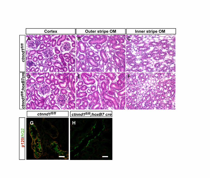
Fig. S1. p120ctn is nonessential in the development of ureteric bud and itsderivatives. (A-F) Hematoxylin and Eosin (H&E)-stained paraffin sections at postnatal day28 from Ctnnd1flox/flox (A-C) and Ctnnd1flox/flox; HoxB7-cretg/0 (D-F) mice in cortex (A,D),outer strip on the outer medulla (B,E) and inner strip of the outer medulla (C,F). (G,H) Co-immunofluorescence with anti-p120ctn (2B12) (red) and anti-aquaporin-2 (green), a markerfor ureteric bud-derived collecting ducts, shows loss of p120ctn from Ctnnd1flox/flox; HoxB7-cretg/0 collecting ducts. n=2 for each genotype. Images A-F are the same magnification.Scale bars: 40 µm in G,H.
Supplemental Figure S2 -
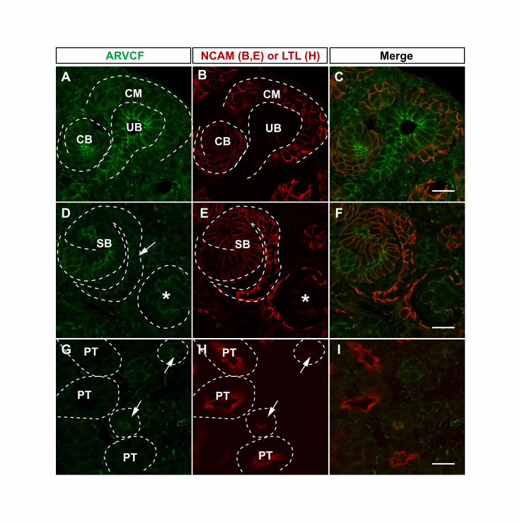
Fig. S2. ARVCF is widely expressed during kidney development. (A-F) Co-immunofluorescence of paraffin sections from E17.5 kidneys with anti-ARVCF (green) andanti-NCAM (red), a marker for condensed mesenchyme (CM), renal vesicles (RV), comma-shaped body (CB) and s-shaped body (SB). ARVCF is highly expressed in ureteric bud (UB)(A-C), RV (not shown), CB (A-C) and SB (D-F). Within the SB, ARVCF is expressed withinthe distal and middle region of the SB (D) as well as in the proximal SB in the precursor cellsthat will differentiate into podocytes (D, arrow). A developing glomerulus (asterisk) also hasARVCF expression in presumptive podocytes (D). (G-I) Co-immunofluorescence with anti-ARVCF (green) and LTL (red), a marker of proximal tubules, shows that little ARVCF ispresent in embryonic proximal tubules. However, tubules with very low levels of LTL expressARVCF (arrows) and might be immature proximal tubules or descending loops of Henle.White dotted lines were added to delineate structures. Scale bars: 20 µm.
Supplemental Figure S3 -
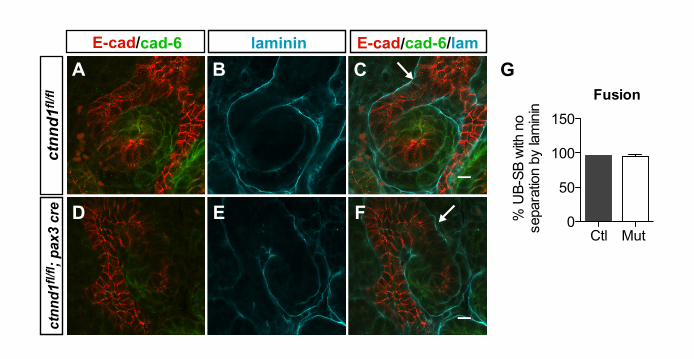
Fig. S3. Fusion of ureteric epithelia to mesenchymally derived epithelia. (A-F)Confocal immunofluorescence of anti-E-cadherin (red), anti-cadherin 6 (green) and anti-laminin (cyan) on P0 sections (70 µm thickness) from control (A-C) and p120ctn mutant (D-F) mice. Ureteric bud (UB) epithelia were identified by morphology and high E-cadherinexpression, whereas adjacent distal s-shaped bodies (SB) were identified by lower levels ofE-cadherin expression contiguous with regions expressing cadherin 6. Sections werevisualized through the z-plane to determine whether the UB and SB were separated bylaminin throughout. The absence of laminin staining at the junction between the ureteric budand distal s-shaped body (arrows) was consistent with fusion of the two (C,F). (G)Quantification of UB-SB fusion. n=2 control and 2 mutant mice. Data represent the averageof the two experiments, and 60 UB-SB fusions were scored. Using this assay, no differencein the percentage of fusions was observed between control and p120ctn mutant kidneys(97% versus 95%). The abnormal appearing structures in p120ctn mutant mice lacking thenormal s-shape were not included in this analysis as their morphologies were difficult tointerpret.
Supplemental Figure S4 -
Fig. S4. Absence of p120ctn leads to defects in proliferation and cytoskeletalrearrangements, but not polarity. (A,B) Localization of Na/K ATPase (green) in proximaltubules shows a basolateral distribution in E17.5 control (A) and mutant (B) kidneys. (C,D)Localization of ZO1 (green), a tight junction component, shows apical distribution. (E,F) Ki67expression (red), a marker of proliferation, in control and mutant kidneys. LTL (green)identifies proximal tubules; Dapi (blue) labels nuclei. (G-I) Immunofluorescence in control(Ctnnd1flox/flox; G) and p120ctn mutant (Ctnnd1flox/flox; Pax3-cretg/0; H,I) kidneys with an F-actin cytoskeletal marker, phalloidin. G and H show proximal tubules of normal caliber,whereas I shows a cystic tubule. The arrows in the inset of G and H highlight differences inthe apical actin cytoskeleton. The inset of I indicates actin bundles in the cystic epithelia,which are absent in control tubules. (J) Densitometry of phosphohistone-3, a marker for cellproliferation, from Western blots of E17.5 lysates was performed using ImageJ and valueswere normalized to GAPDH (P<0.02). n=4 control, n=5 mutant mice. (K) Quantification ofproliferation in proximal tubules. Because levels of proliferation are low in proximal tubules,anti-Ki67 (which labels cells in G2, S and M phase) was used for this immunofluorescence.The percentage of Ki67-positive proximal tubular nuclei as seen in E, F was determined in

4/26/11 11:37 AMSupplementary Material
Page 2 of 2http://dev.biologists.org/content/suppl/2011/04/23/138.10.2099.DC1
three separate experiments (P< 0.006). (L) Quantification of apoptosis. Visualization ofactive caspase-3 (apoptosis marker) and NCAM (marker for condensed mesenchyme, renalvesicles, comma-shaped bodies and s-shaped bodies) was performed and data werequantified as the number of active caspase-3-positive cells per NCAM positive structure (P<0.03). Results are representative of two experiments. More than 200 NCAM positivestructures were counted. All data (J-L) are mean ± s.d. Statistical analysis with the MannWhitney U test. Scale bars: 5 µm in A-D; 20 µm in E-I.
Supplemental Figure S5 -
Fig. S5. Cilia structure in p120ctn mutant mice. (A-C) Immunofluorescence of anti-acetylated tubulin, a component of cilia, in proximal tubules of P0 control (A) and mutant(B,C). Proximal tubules in A and B were identified by colocalization of LTL, a proximal tubulemarker (not shown), and are outlined with white dotted lines. A and B show proximal tubulesof normal caliber, whereas C shows a cystic proximal tubule. Scale bars: 25 µm.
Supplemental Figure S6 -
Fig. S6. p120ctn knockdown results in decreased cellular adhesion and regularspindle orientation. (A) Transepithelial resistance was measured across confluent,polarized MDCK monolayers after 72 hours growth on a polycarbonate transwell.Resistance=measured resistance minus background resistance from transwells without cells.Four replicates for each time point were performed. Graph shows one of two similarexperiments. Resistance is reduced in p120ctn knockdown cells (P< 0.0001, unpaired two-tailed Student�s t test.). (B) Spindle orientation at the equatorial plane of MDCK cysts.Immunofluorescence was performed with α-tubulin, Hoescht and podocalyxin to visualizespindles, nuclei and the apical membrane of the cysts, respectively. Cysts were imaged byconfocal microscopy at the largest cross-sectional diameter (equatorial plane). Spindleorientation was measured as follows: parallel to the apical membrane is 0° and perpendicularto the apical membrane is 90°. The number of spindles counted was n=44 (control) andn=59 (p120ctn KD). Graph shows one of two similar experiments. Results are not statisticallydifferent (P=0.6, unpaired two-tailed Student�s t test).