overview and status of the austrian particle therapy ... · the austrian particle therapy facility...
TRANSCRIPT
Overview and Status of the Austrian Particle Therapy Facility MedAustron Peter Urschütz
MedAustron
Centre for ion beam therapy and non-clinical research
Treatment of 1200 patients/year in full operation
Worldwide the 6th combined centre for ion beam therapy with protons and carbon ions
Founded in 2007 as „EBG MedAustron GmbH“
• in indirect ownership of the federal state of Lower Austria
2
Financing Investment volume: € 200 million
Financing partners:
• Republic of Austria
• Federal State of Lower Austria
• City of Wr. Neustadt
3
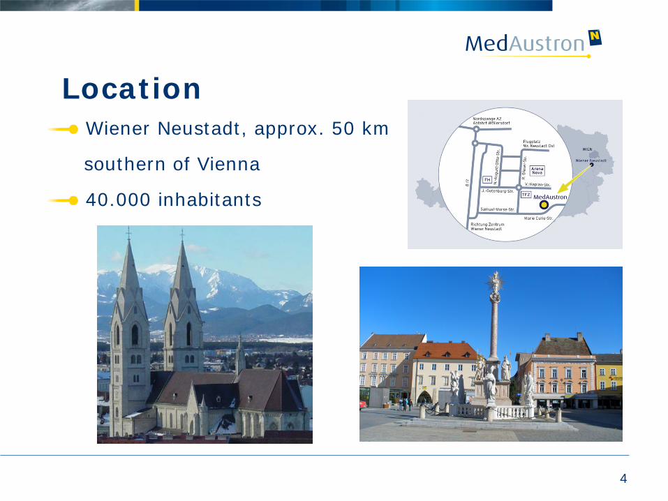
Location
4
Wiener Neustadt, approx. 50 km
southern of Vienna
40.000 inhabitants
5
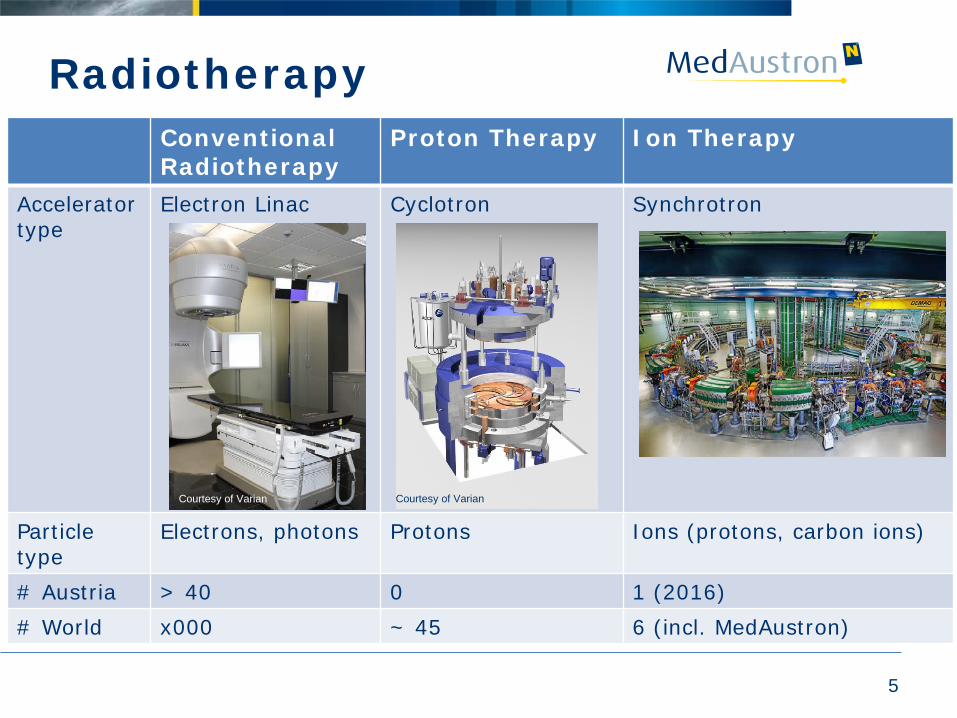
Conventional Radiotherapy
Proton Therapy Ion Therapy
Accelerator type
Electron Linac Cyclotron Synchrotron
Particle type
Electrons, photons Protons Ions (protons, carbon ions)
# Austria > 40 0 1 (2016) # World x000 ~ 45 6 (incl. MedAustron)
Courtesy of Varian Courtesy of Varian
Radiotherapy
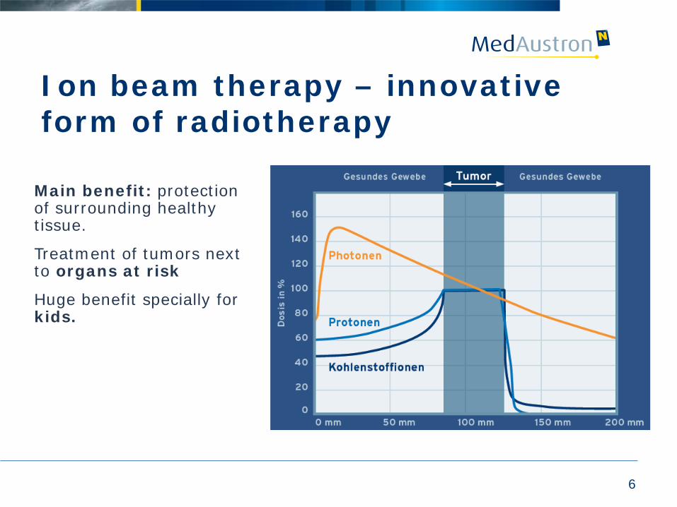
Ion beam therapy – innovative form of radiotherapy
Main benefit: protection of surrounding healthy tissue.
Treatment of tumors next to organs at risk
Huge benefit specially for kids.
6
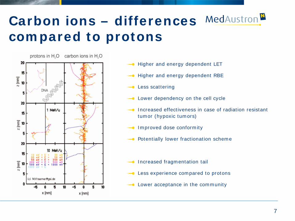
Carbon ions – differences compared to protons
Higher and energy dependent LET
Higher and energy dependent RBE
Less scattering
Lower dependency on the cell cycle
Increased effectiveness in case of radiation resistant tumor (hypoxic tumors)
Improved dose conformity
Potentially lower fractionation scheme
Increased fragmentation tail
Less experience compared to protons
Lower acceptance in the community
7
8
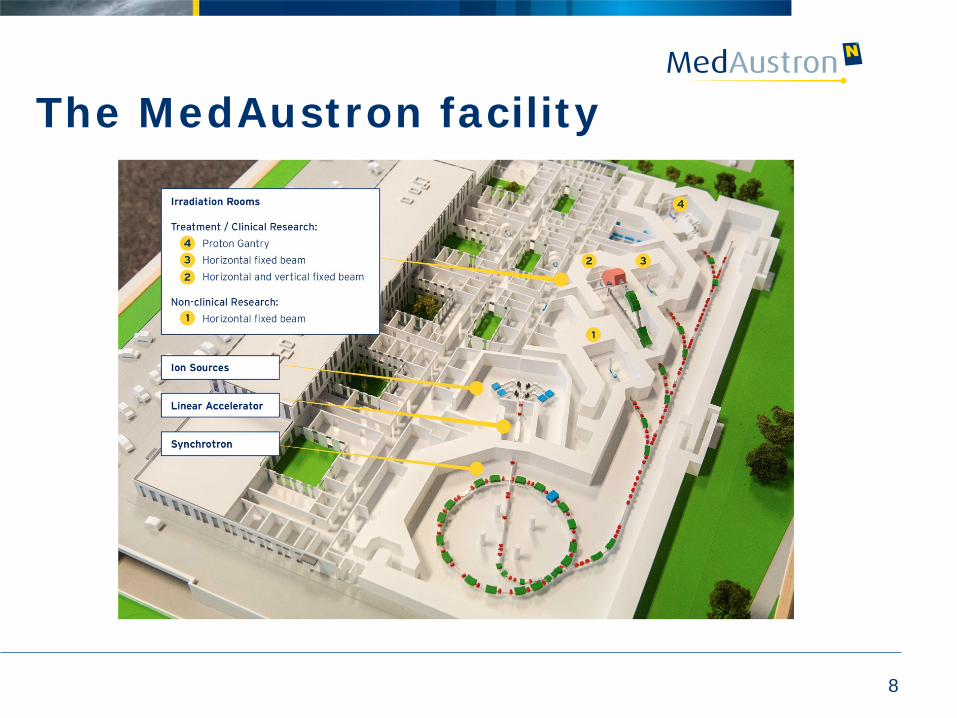
The MedAustron facility
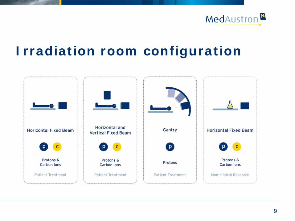
Irradiation room configuration
9
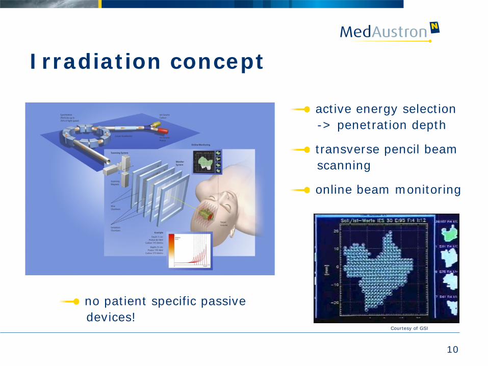
Irradiation concept
active energy selection -> penetration depth
transverse pencil beam scanning
online beam monitoring
10
Courtesy of GSI
no patient specific passive devices!
11
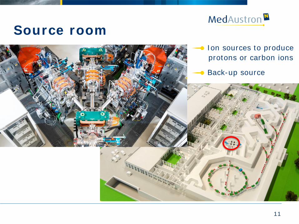
Source room Ion sources to produce protons or carbon ions
Back-up source
12
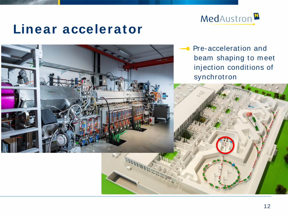
Linear accelerator Pre-acceleration and beam shaping to meet injection conditions of synchrotron
13
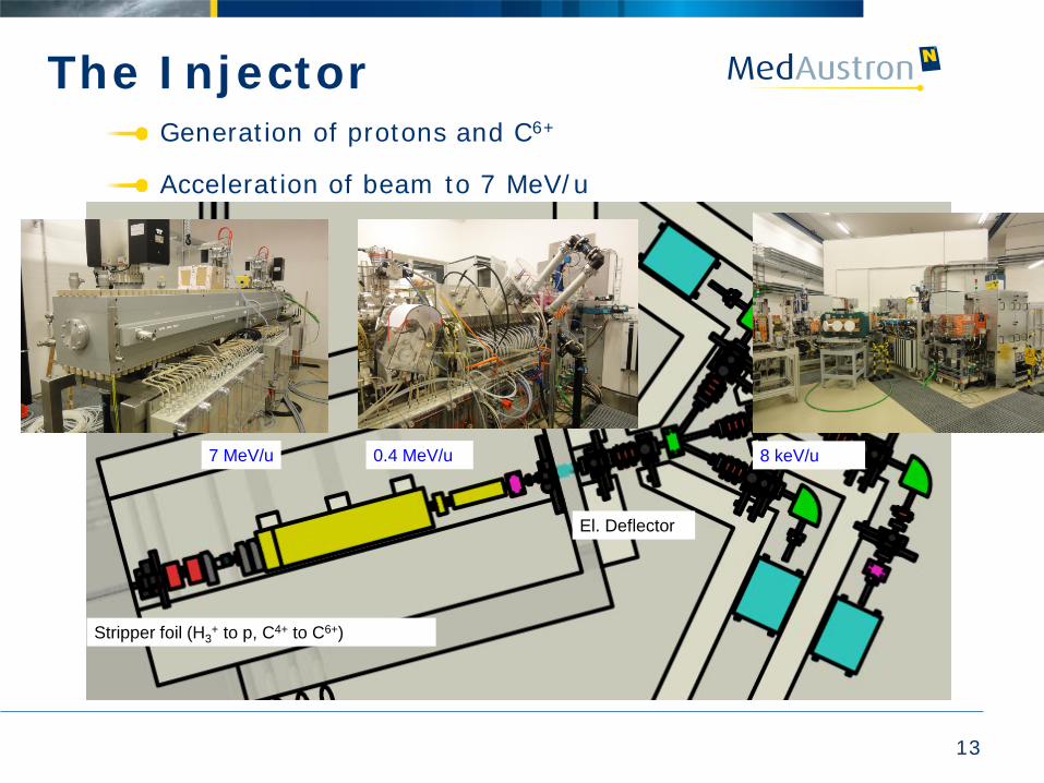
The Injector Generation of protons and C6+
Acceleration of beam to 7 MeV/u
Generation of protons and C6+
8 keV/u
El. Deflector
0.4 MeV/u 7 MeV/u
Stripper foil (H3+ to p, C4+ to C6+)
14
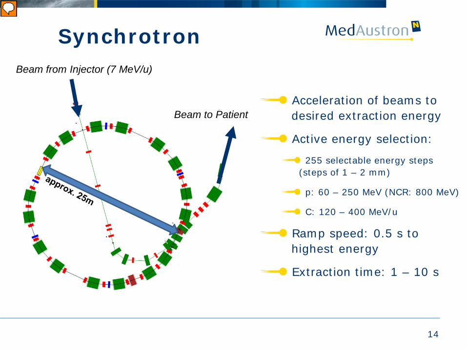
Synchrotron
Beam to Patient
Beam from Injector (7 MeV/u)
Acceleration of beams to desired extraction energy
Active energy selection:
255 selectable energy steps (steps of 1 – 2 mm)
p: 60 – 250 MeV (NCR: 800 MeV)
C: 120 – 400 MeV/u
Ramp speed: 0.5 s to highest energy
Extraction time: 1 – 10 s
15
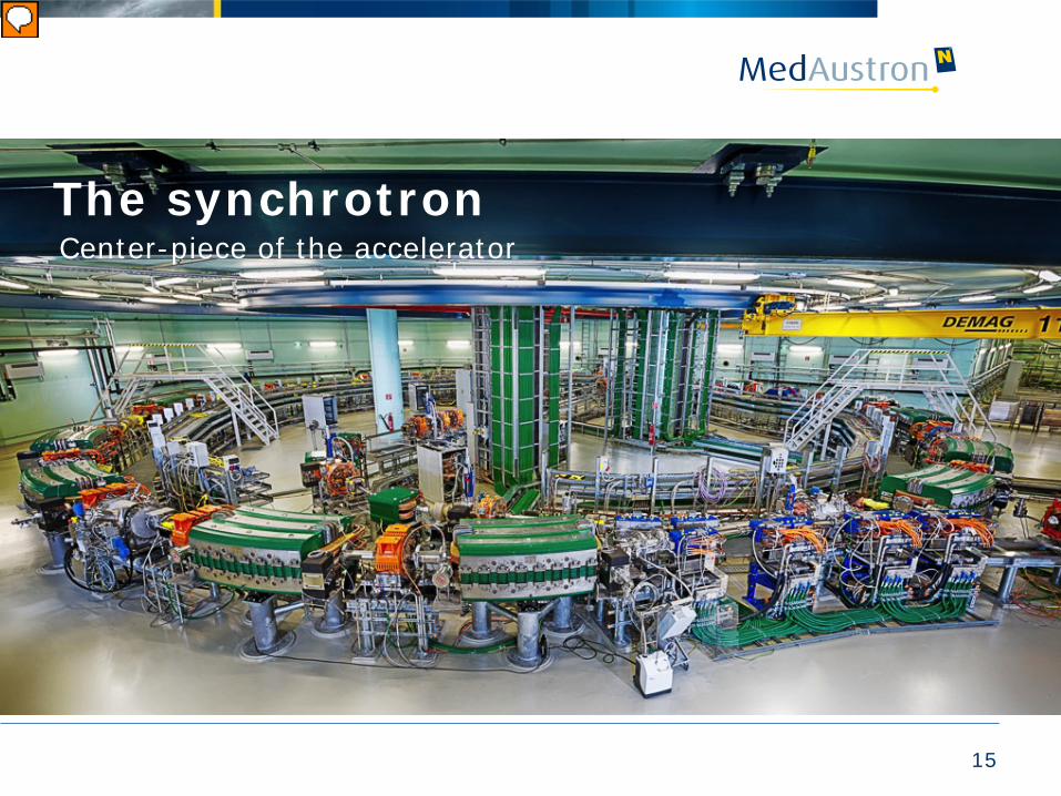
The synchrotron Center-piece of the accelerator
16
Synchrotron
Time
Magnetic field
Bmax B1 B2 Injection Extraction
energy 1 Extraction energy 2
Acceleration
Injection
Acceleration ∼2 s ∼4 s
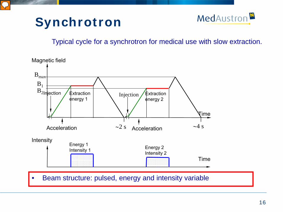
Typical cycle for a synchrotron for medical use with slow extraction.
Intensity
Time
Energy 1 Intensity 1 Energy 2
Intensity 2
• Beam structure: pulsed, energy and intensity variable
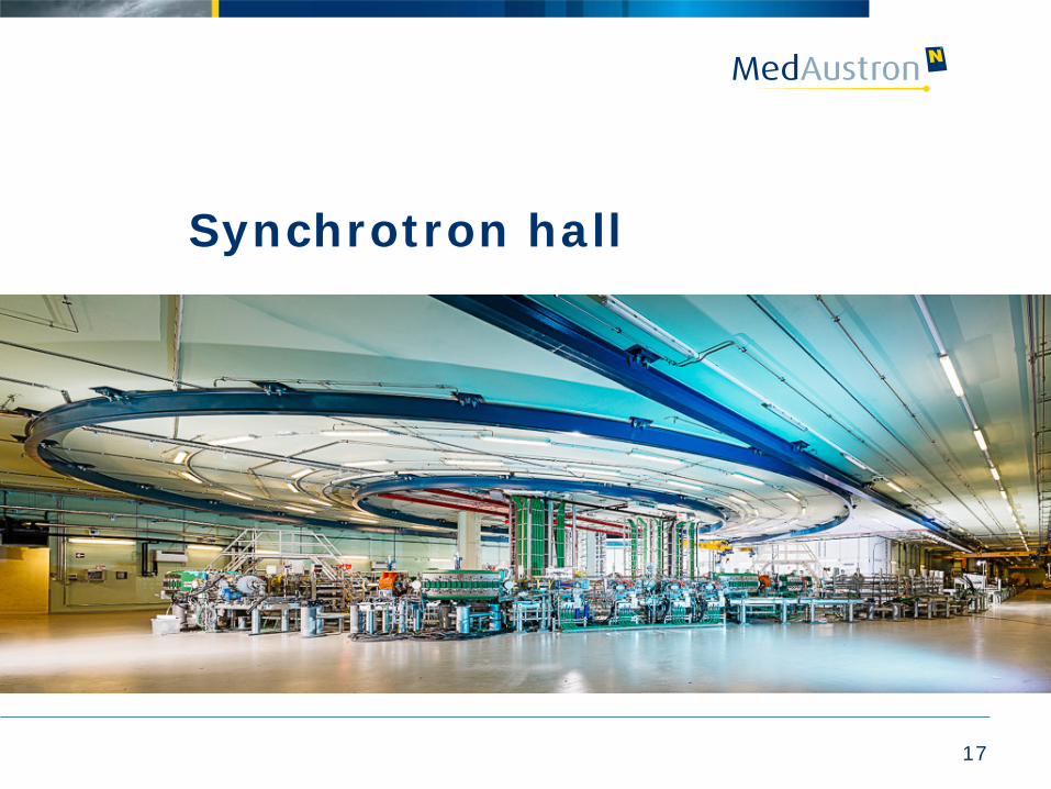
Synchrotron hall
17
18
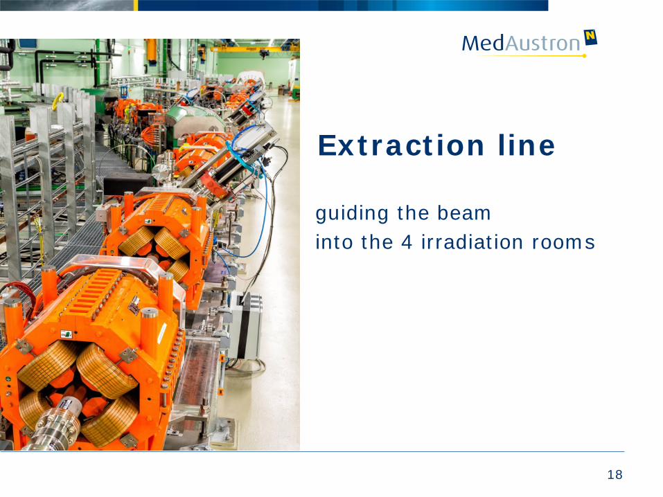
Extraction line
guiding the beam into the 4 irradiation rooms
19
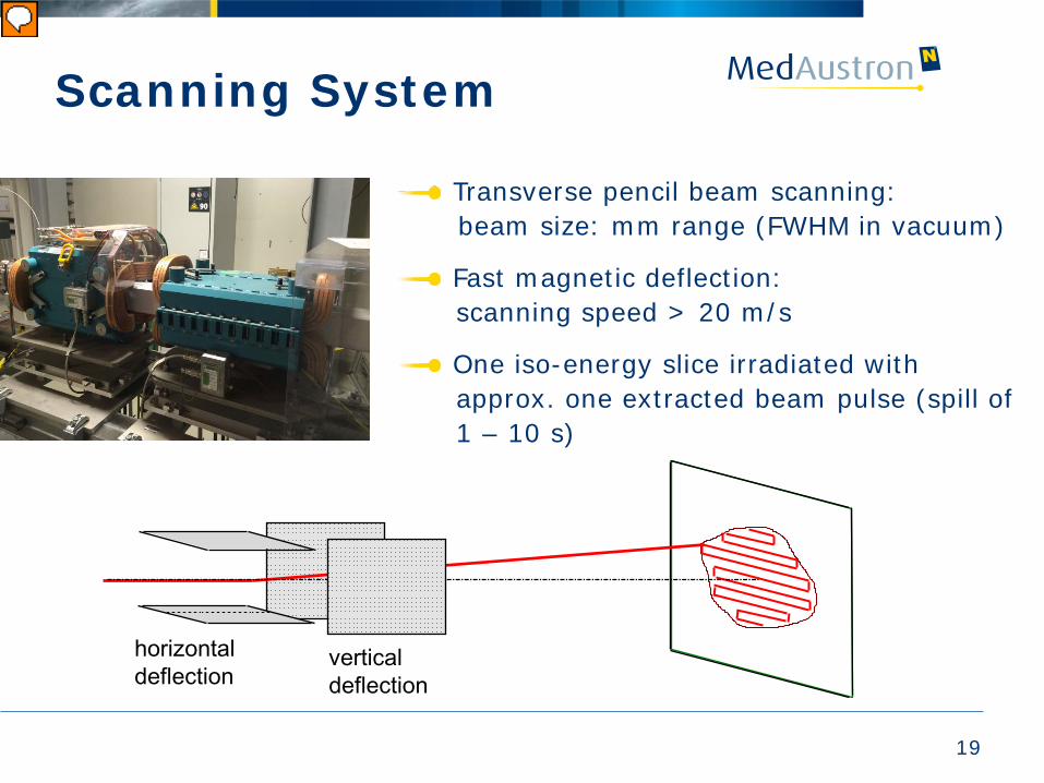
Scanning System
horizontal deflection
vertical deflection
Transverse pencil beam scanning: beam size: mm range (FWHM in vacuum)
Fast magnetic deflection: scanning speed > 20 m/s
One iso-energy slice irradiated with approx. one extracted beam pulse (spill of 1 – 10 s)
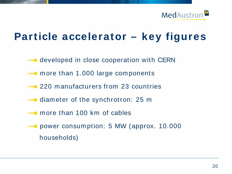
Particle accelerator – key figures
20
developed in close cooperation with CERN
more than 1.000 large components
220 manufacturers from 23 countries
diameter of the synchrotron: 25 m
more than 100 km of cables
power consumption: 5 MW (approx. 10.000
households)
21
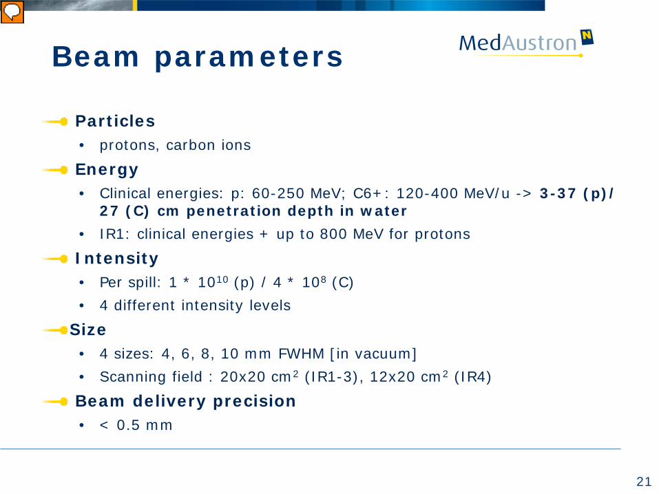
Beam parameters
Particles • protons, carbon ions
Energy • Clinical energies: p: 60-250 MeV; C6+: 120-400 MeV/u -> 3-37 (p)/
27 (C) cm penetration depth in water • IR1: clinical energies + up to 800 MeV for protons
Intensity • Per spill: 1 * 1010 (p) / 4 * 108 (C) • 4 different intensity levels
Size • 4 sizes: 4, 6, 8, 10 mm FWHM [in vacuum] • Scanning field : 20x20 cm2 (IR1-3), 12x20 cm2 (IR4)
Beam delivery precision • < 0.5 mm
22
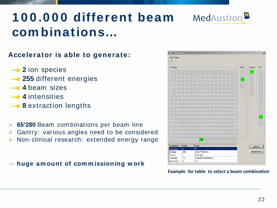
100.000 different beam combinations…
Accelerator is able to generate:
2 ion species 255 different energies 4 beam sizes 4 intensities 8 extraction lengths
65’280 Beam combinations per beam line Gantry: various angles need to be considered Non-clinical research: extended energy range
⇒ huge amount of commissioning work
Example for table to select a beam combination
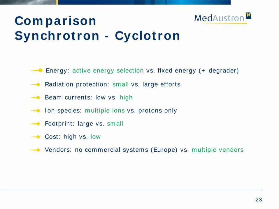
Comparison Synchrotron - Cyclotron
23
Energy: active energy selection vs. fixed energy (+ degrader)
Radiation protection: small vs. large efforts
Beam currents: low vs. high
Ion species: multiple ions vs. protons only
Footprint: large vs. small
Cost: high vs. low
Vendors: no commercial systems (Europe) vs. multiple vendors
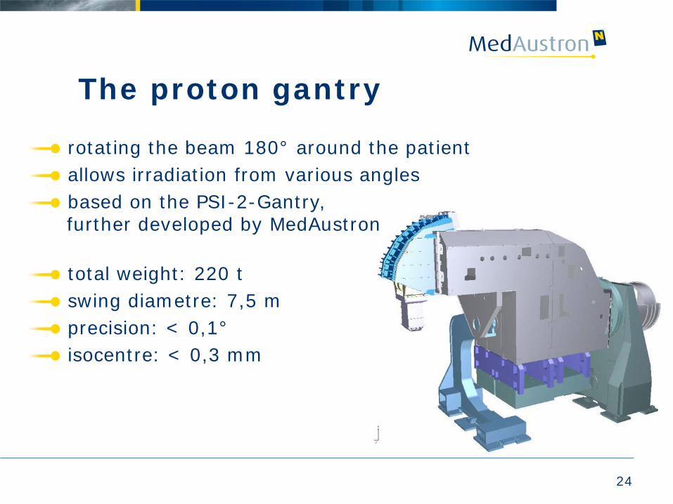
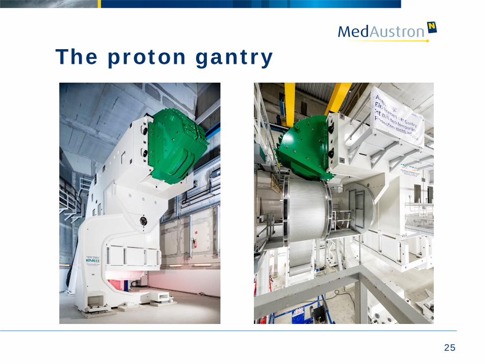
The proton gantry
rotating the beam 180° around the patient allows irradiation from various angles based on the PSI-2-Gantry, further developed by MedAustron total weight: 220 t swing diametre: 7,5 m precision: < 0,1° isocentre: < 0,3 mm
24
25
The proton gantry
26
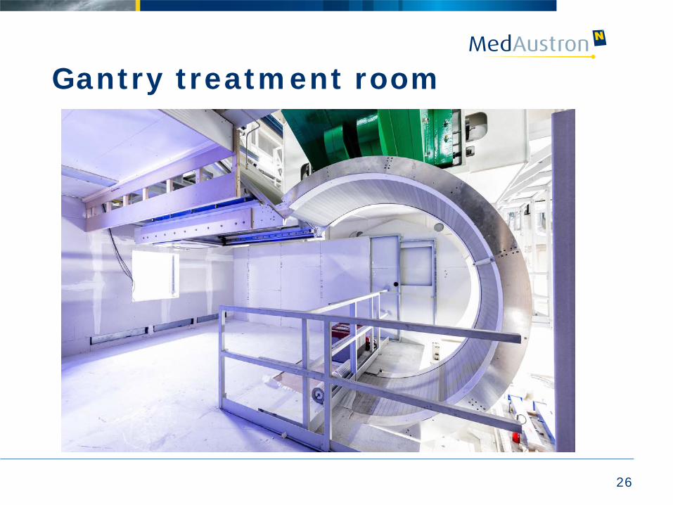
Gantry treatment room
27
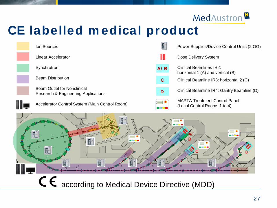
Ion Sources
Linear Accelerator
Synchrotron
Beam Distribution
Beam Outlet for Nonclinical Research & Engineering Applications
Accelerator Control System (Main Control Room)
A/B
C
D
Power Supplies/Device Control Units (2.OG)
Dose Delivery System
Clinical Beamlines IR2: horizontal 1 (A) and vertical (B)
Clinical Beamline IR3: horizontal 2 (C)
Clinical Beamline IR4: Gantry Beamline (D) MAPTA Treatment Control Panel (Local Control Rooms 1 to 4)
A/B
C D
CE labelled medical product
according to Medical Device Directive (MDD)
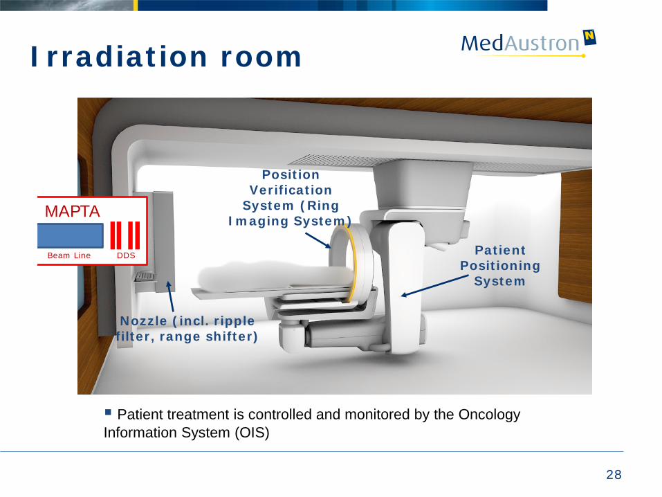
Irradiation room
28
Patient Positioning
System
Position Verification
System (Ring Imaging System)
Nozzle (incl. ripple filter, range shifter)
MAPTA Beam Line DDS
Patient treatment is controlled and monitored by the Oncology Information System (OIS)
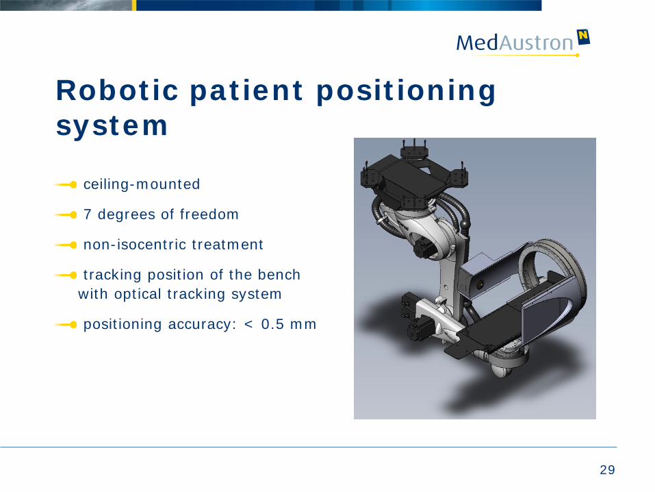
Robotic patient positioning system
ceiling-mounted
7 degrees of freedom
non-isocentric treatment
tracking position of the bench with optical tracking system
positioning accuracy: < 0.5 mm
29
30
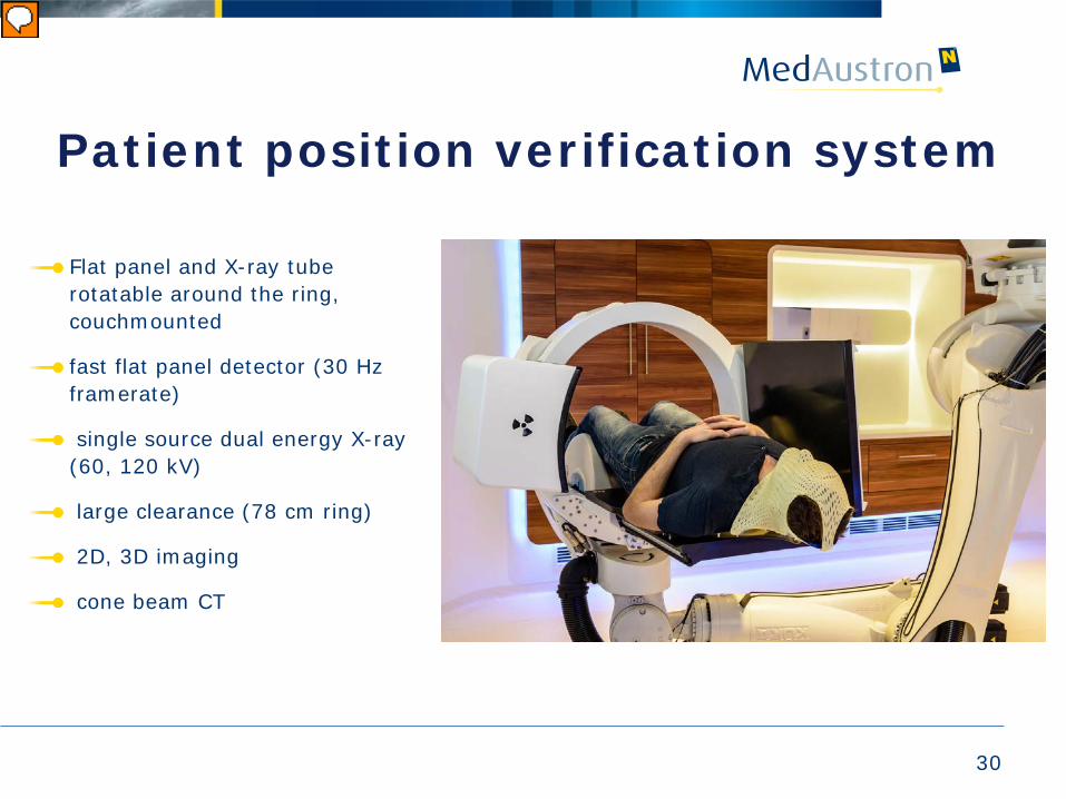
Patient position verification system
Flat panel and X-ray tube rotatable around the ring, couchmounted
fast flat panel detector (30 Hz framerate)
single source dual energy X-ray (60, 120 kV)
large clearance (78 cm ring)
2D, 3D imaging
cone beam CT
31
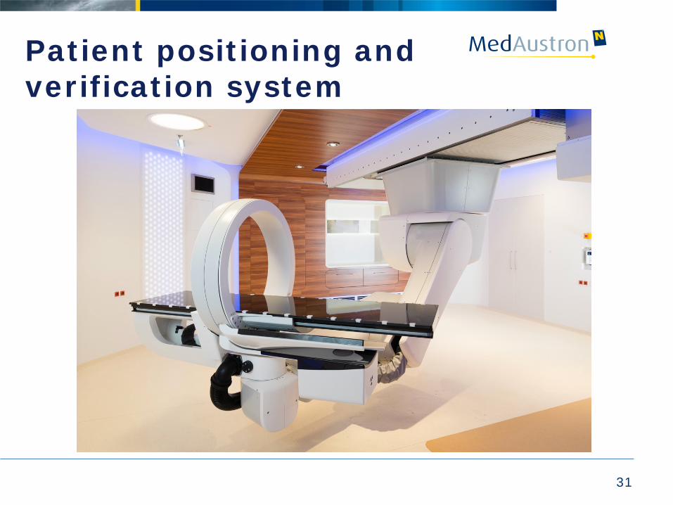
Patient positioning and verification system
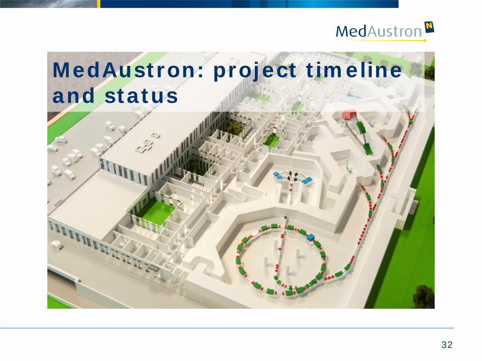
MedAustron: project timeline and status
32
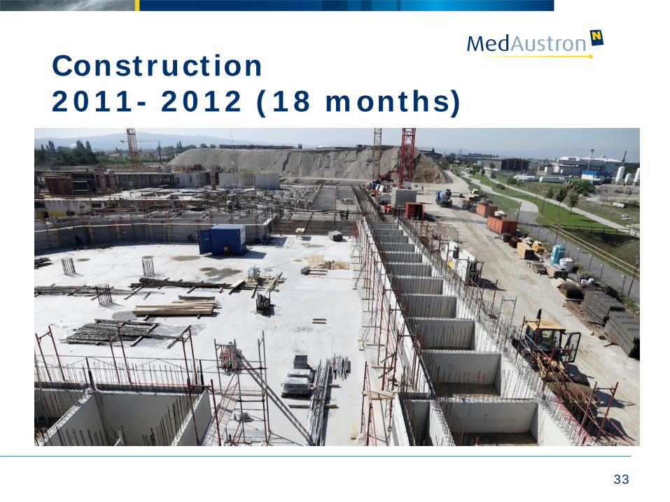
Construction 2011- 2012 (18 months)
33
34
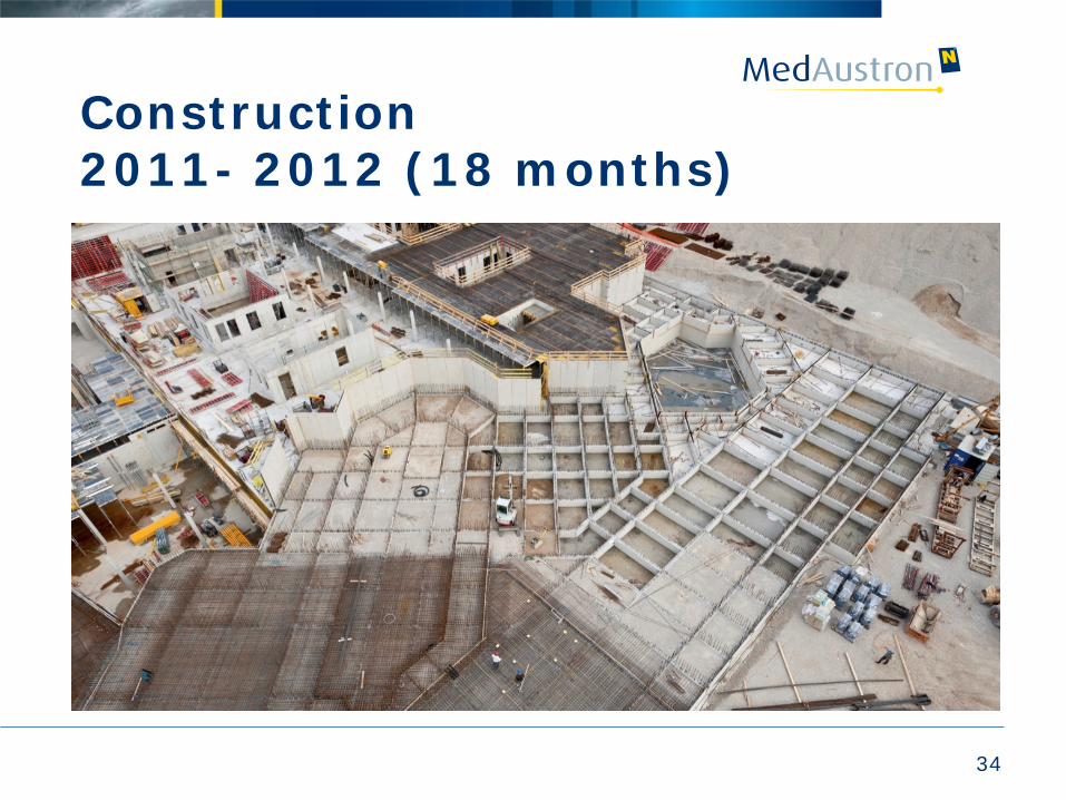
Construction 2011- 2012 (18 months)

35
Oct 2012: Moving in
36
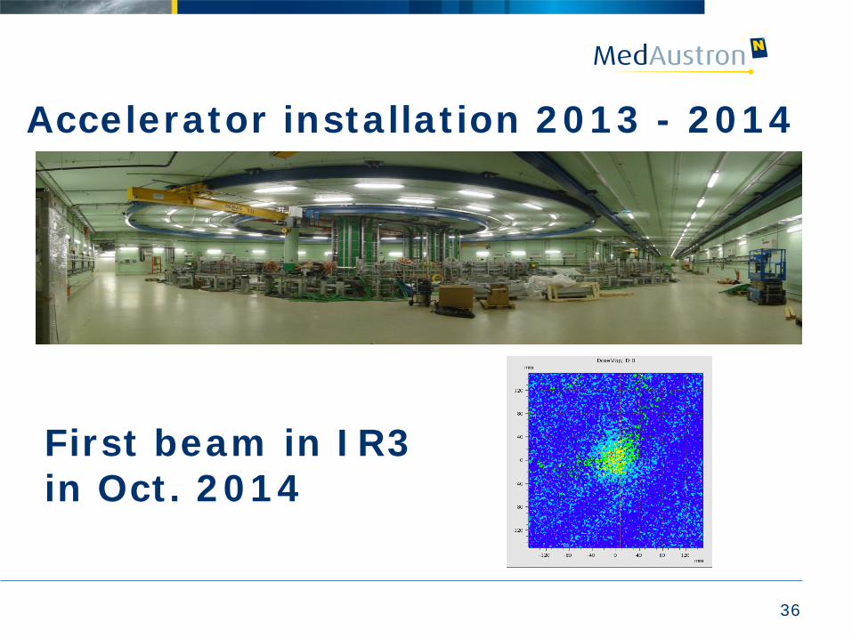
Accelerator installation 2013 - 2014
First beam in IR3 in Oct. 2014
37
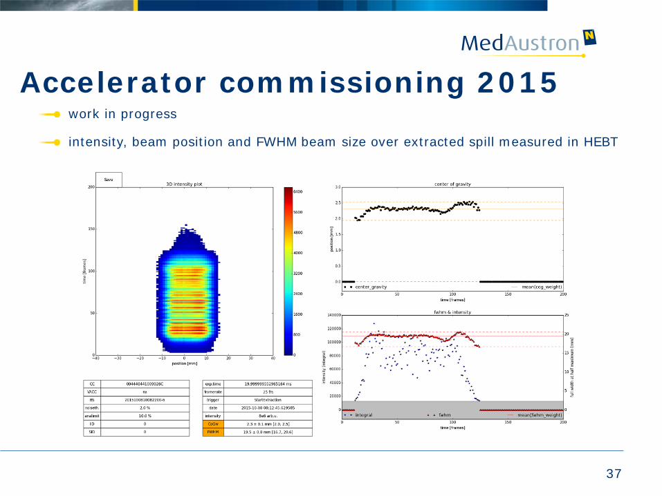
Accelerator commissioning 2015 work in progress
intensity, beam position and FWHM beam size over extracted spill measured in HEBT
38
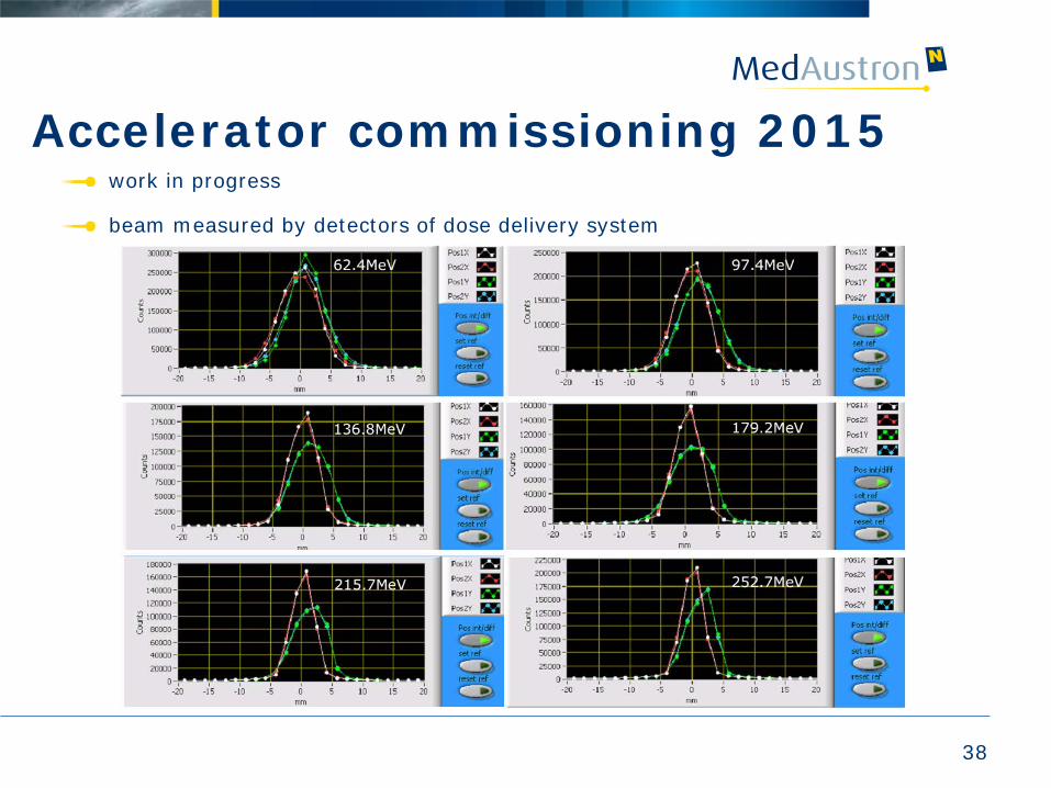
Accelerator commissioning 2015 work in progress
beam measured by detectors of dose delivery system
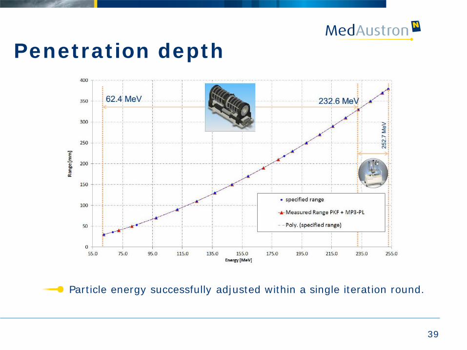
Penetration depth
39
Particle energy successfully adjusted within a single iteration round.
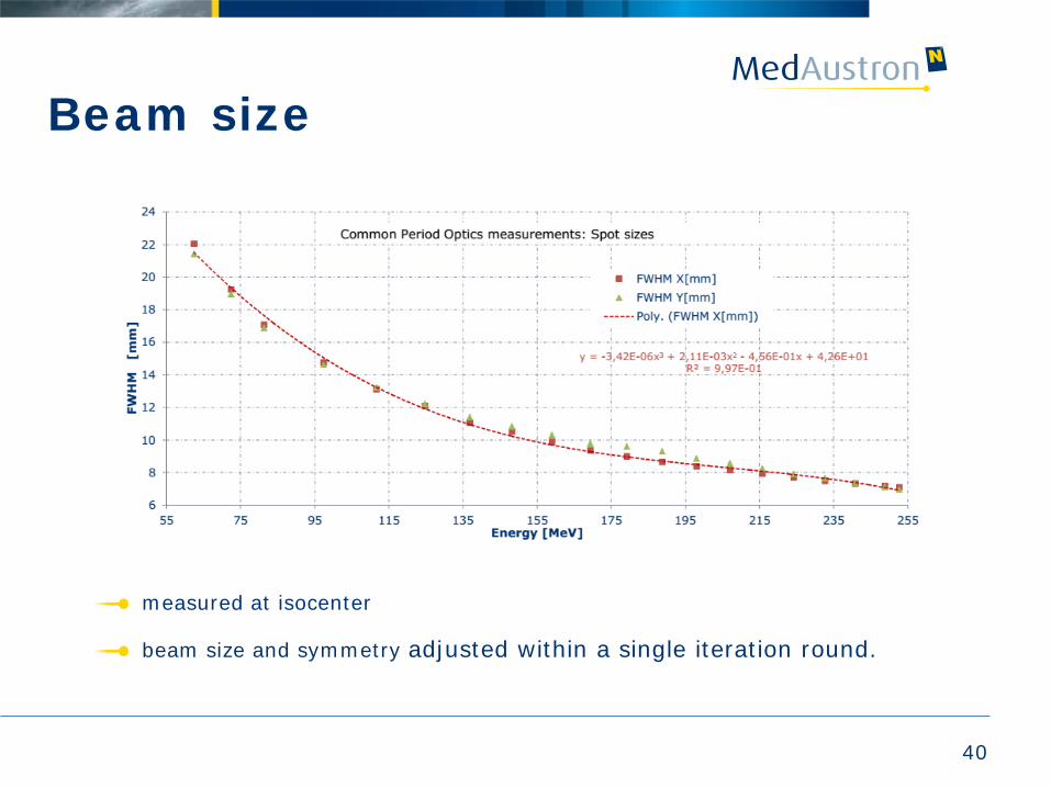
Beam size
40
measured at isocenter
beam size and symmetry adjusted within a single iteration round.
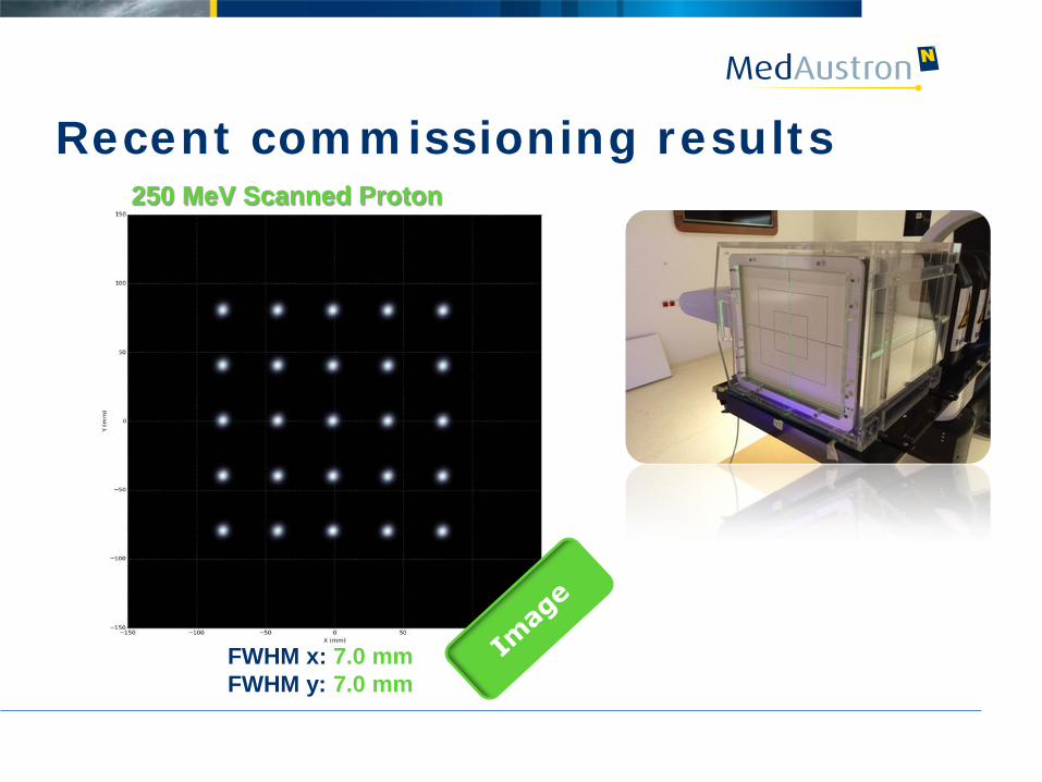
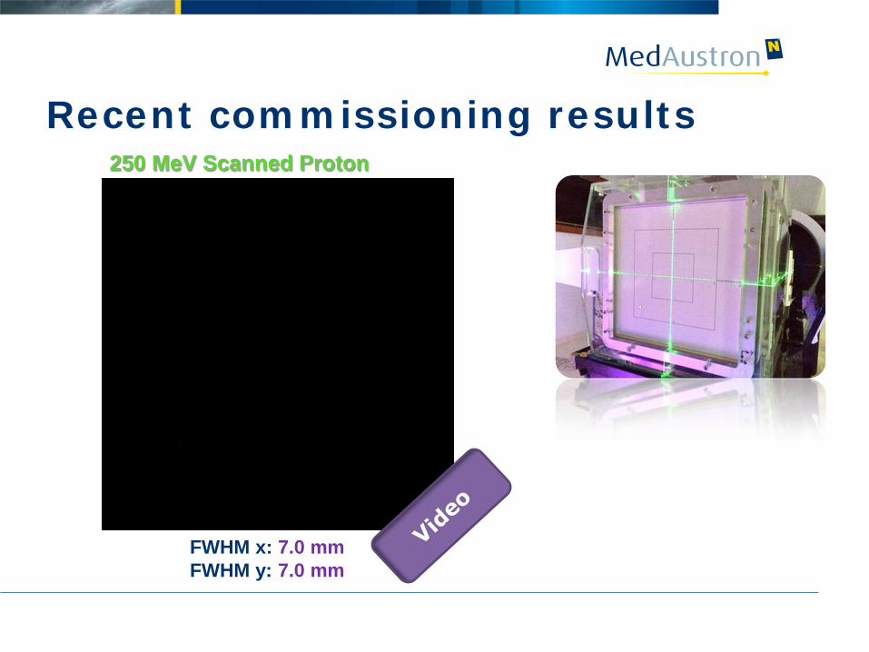
Recent commissioning results
250 MeV Scanned Proton Beam
FWHM x: 7.0 mm FWHM y: 7.0 mm
250 MeV Scanned Proton Beam
FWHM x: 7.0 mm FWHM y: 7.0 mm
Recent commissioning results
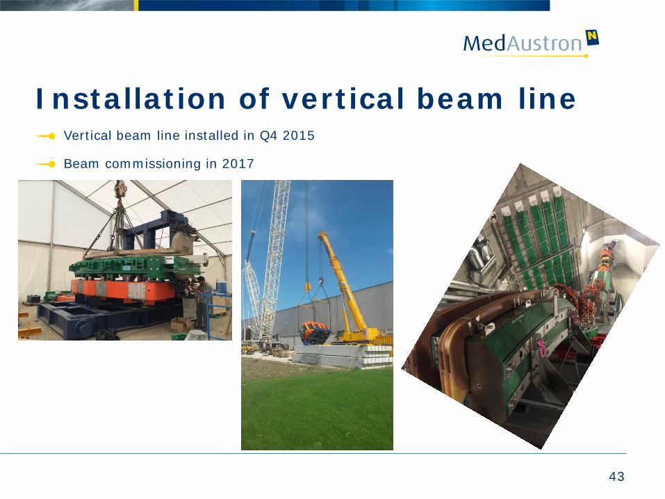
Installation of vertical beam line
43
Vertical beam line installed in Q4 2015
Beam commissioning in 2017
Remaining steps towards 1st patient treatment
Finalisation of the proton beam commissioning for horizontal fixed beam lines
• Fine tuning of beams
• Coverage of full parameter space (different energies, intensities, etc…)
System integration (complete workflow)
Interlock and error management
Verification and validation test phase
Technical documentation and regulatory aspects
Medical physics: base data measurements (TPS)
End to end tests
44
1st patient treatment in 2016
2016: first patient treatment with protons (horizontal fixed beam lines)
2016: provide beams for non clinical research
2017 – 20:
• Commissioning of further beam lines,
• Carbon ions,
• Gantry
• enhance functionalities, performance increase
2020/21: full operation
45

Thank you for your attention!