ovary slide share 2
TRANSCRIPT
OVARIAN TUMORS-II
Dr Aksharaditya ShuklaResident, Department Of Pathology
MGM Medical College & M.Y. Hospital, Indore
Dr Aksharaditya Shukla
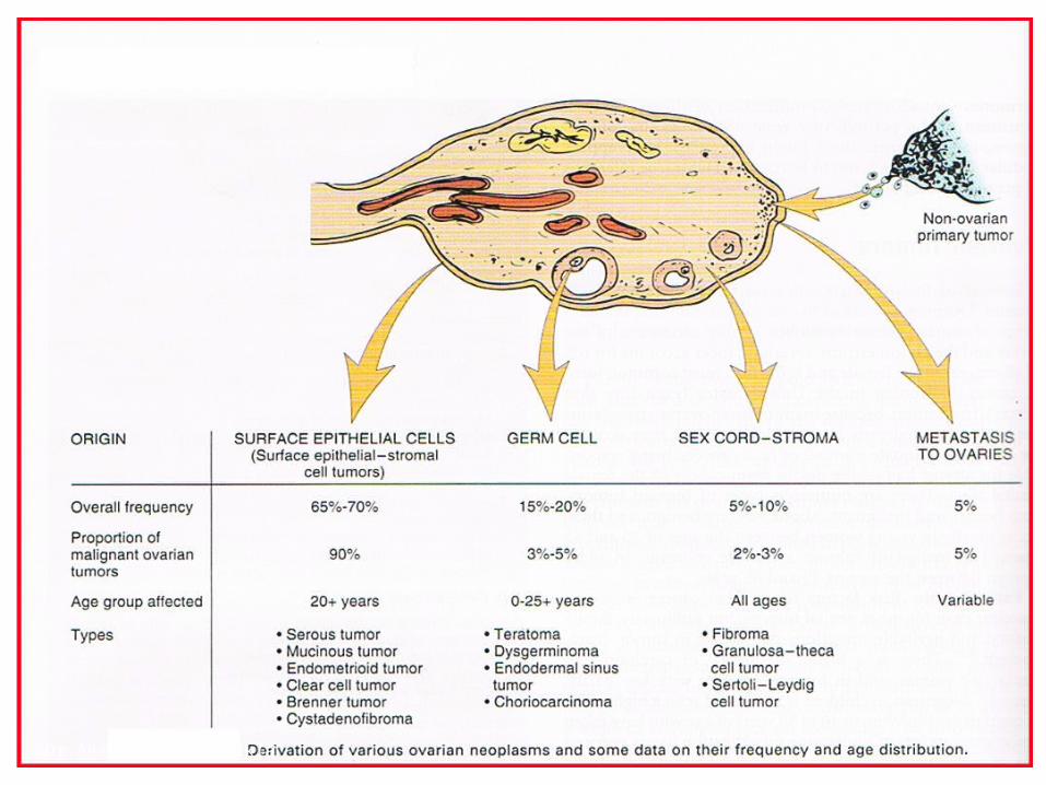
Ovarian tumours
Tumour of the ovary are common form of neoplasia in women
Accounts for 3% of all cancers in females80% are benignMore common in older white women of
northern European ancestry90% of malignancies are carcinoma, 80%
have spread beyond the ovary at diagnosis.
Dr Aksharaditya Shukla
Risk factors for carcinoma
NulliparityFamily historyChildhood gonadal dysgenesisClomipheneHereditary non polyposis colon cancerBRCA1 and BRCA2 mutationsCA-125 present in 80% of serous and
endometrioid tumoursCytogenetics-gain of 12 & 8loss of chr X,22 18,17,14,13,12 & 8 ,benign/borderline tumor exhibit trisomy12

Dr Aksharaditya Shukla
Dr Aksharaditya Shukla
Classification of ovarian tumours
Novak's classification (1967) has advantage of being simple but has certain obvious drawbacks, since it depends primarily on two fundamental factors; benign or malignant and solid or cystic.
Thus the borderline tumors, solid tumors with cystic degeneration and predominantly cystic tumors with solid areas fall into grey zone.
Dr Aksharaditya Shukla
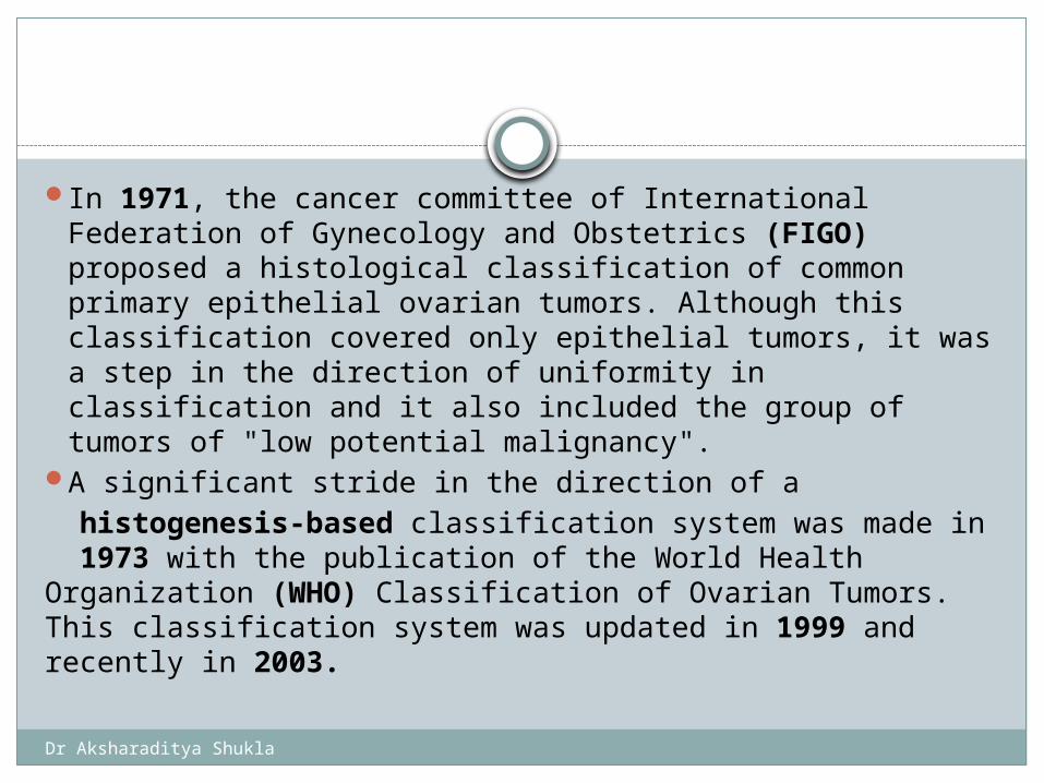
In 1971, the cancer committee of International Federation of Gynecology and Obstetrics (FIGO) proposed a histological classification of common primary epithelial ovarian tumors. Although this classification covered only epithelial tumors, it was a step in the direction of uniformity in classification and it also included the group of tumors of "low potential malignancy".
A significant stride in the direction of a histogenesis-based classification system was made in 1973 with the publication of the World Health Organization (WHO) Classification of Ovarian Tumors. This classification system was updated in 1999 and recently in 2003.
Dr Aksharaditya Shukla
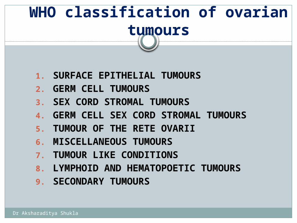
WHO classification of ovarian tumours
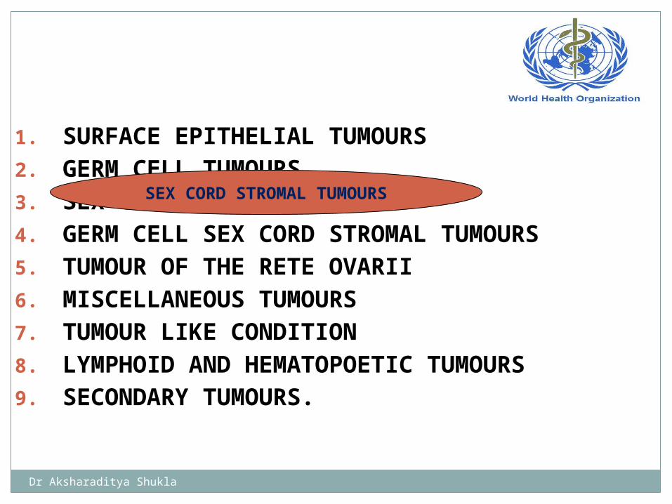
1. SURFACE EPITHELIAL TUMOURS2. GERM CELL TUMOURS3. SEX CORD STROMAL TUMOURS 4. GERM CELL SEX CORD STROMAL TUMOURS 5. TUMOUR OF THE RETE OVARII 6. MISCELLANEOUS TUMOURS 7. TUMOUR LIKE CONDITIONS8. LYMPHOID AND HEMATOPOETIC TUMOURS9. SECONDARY TUMOURS
Dr Aksharaditya Shukla
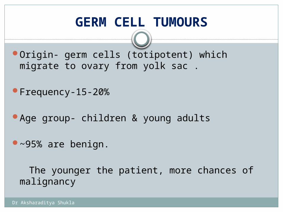
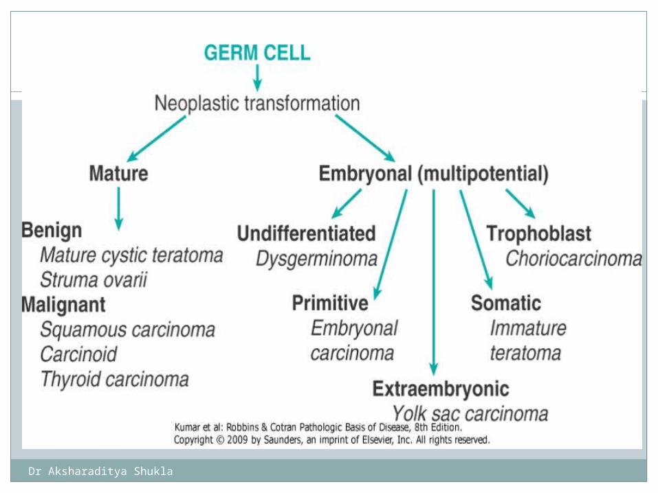
GERM CELL TUMOURS
Origin- germ cells (totipotent) which migrate to ovary from yolk sac .
Frequency-15-20%
Age group- children & young adults
~95% are benign.
The younger the patient, more chances of malignancy
Dr Aksharaditya Shukla

Dr Aksharaditya Shukla
Dysgerminoma
Dysgerminoma is the most common malignant germ cell neoplasia of the ovary, and is similar to seminoma which is the testicular counterpart
<1% of all ovarian tumors ≈5% of malignant ovarian
tumors Usually young <30 years
of age. ≈5% arise in abnormal
gonads: a) pure or mixed
gonadal dysgenesis (from
a gonadoblastoma), b) testicular
feminization (androgen insensitivity) syndrome
More common on right. Bilateral in 15% of cases. Metastases commonly
in:
* contralateral ovary * retroperitoneal nodes * peritoneal cavity: (associated with
decreased survival rate).
Exceptionally associated with hypercalcemia
Extremely radiosenstive.
Dr Aksharaditya Shukla
DYSGERMINOMA
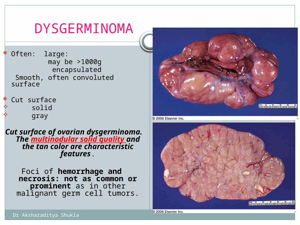
Often: large: may be >1000g encapsulated Smooth, often convoluted surface
Cut surface solid gray
Cut surface of ovarian dysgerminoma. The multinodular solid quality and the tan color are characteristic features.
Foci of hemorrhage and necrosis: not as common or
prominent as in other malignant germ cell tumors.
Typical lobulated outer aspect of ovarian dysgerminoma
Dr Aksharaditya Shukla
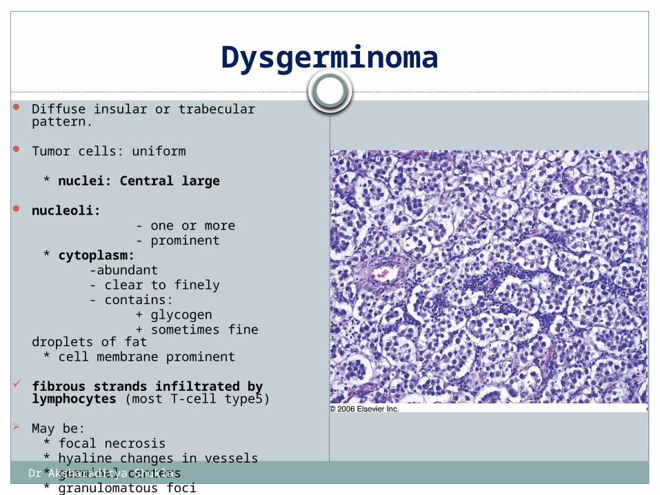
Dysgerminoma Diffuse insular or trabecular pattern.
Tumor cells: uniform
* nuclei: Central large
nucleoli: - one or more - prominent * cytoplasm: -abundant - clear to finely - contains: + glycogen + sometimes fine droplets of fat * cell membrane prominent
fibrous strands infiltrated by lymphocytes (most T-cell type5)
May be: * focal necrosis * hyaline changes in vessels * germinal centers * granulomatous foci
Dr Aksharaditya Shukla
Special Stains and Immunohistochemistryof dysgerminoma
Tumor cells reactive for: -PLAP -CD117 (c-KIT)
Often keratin (erratically and focally)
Sometimes -GFAP -Desmin
Dr Aksharaditya Shukla
Yolk Sac Tumor
Malignant germ cell tumor of the ovary developed as a result of differentiation of primitive malignant germ cell elements in the direction of yolk sac or vitelline structures.
Usually children and young adults:
* Median age 19 years * ≈25% prepubertal at diagnosis
Vaginal bleeding in 1%
Serum AFP level invariably elevatedChorionic gonadotropin levels normal.Highly aggressive, and associated with other germ cell
tumour
Dr Aksharaditya Shukla
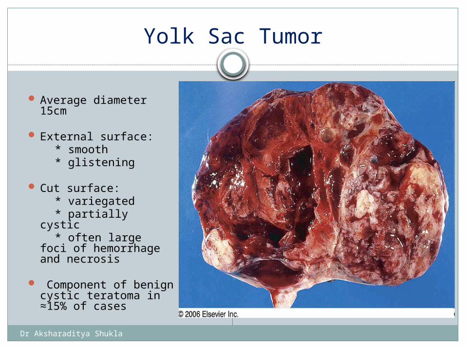
Yolk Sac Tumor
Average diameter 15cm
External surface: * smooth * glistening
Cut surface: * variegated * partially cystic * often large foci of
hemorrhage and necrosis
Component of benign cystic teratoma in ≈15% of cases
Gross appearance of yolk sac tumor. The cut surface is remarkably heterogeneous due to extensive hemorrhage, necrosis, and cystic degeneration
Dr Aksharaditya Shukla
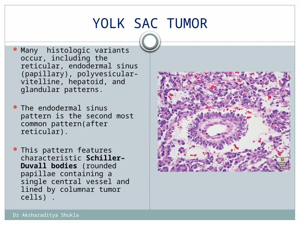
YOLK SAC TUMOR
Many histologic variants occur, including the reticular, endodermal sinus (papillary), polyvesicular–vitelline, hepatoid, and glandular patterns.
The endodermal sinus pattern is the second most common pattern(after reticular).
This pattern features
characteristic Schiller–Duvall bodies (rounded papillae containing a single central vessel and lined by columnar tumor cells) .
Dr Aksharaditya Shukla
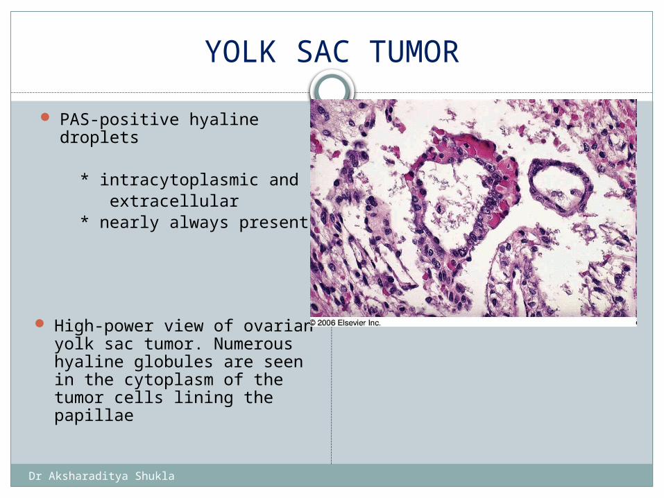
YOLK SAC TUMOR
PAS-positive hyaline droplets
* intracytoplasmic and extracellular * nearly always present
High-power view of ovarian yolk sac tumor. Numerous hyaline globules are seen in the cytoplasm of the tumor cells lining the papillae
Dr Aksharaditya Shukla
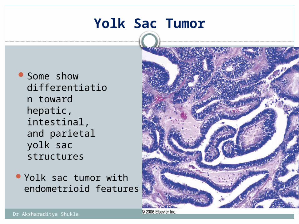
Yolk Sac Tumor
Some show differentiation toward hepatic, intestinal, and parietal yolk sac structures
Yolk sac tumor with endometrioid features
Dr Aksharaditya Shukla
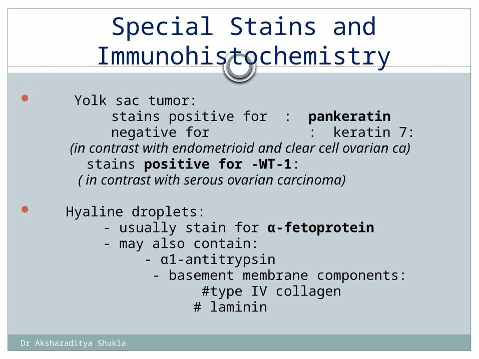
Special Stains and Immunohistochemistry
Yolk sac tumor: stains positive for : pankeratin negative for : keratin 7: (in contrast with endometrioid and clear cell ovarian ca) stains positive for -WT-1: ( in contrast with serous ovarian carcinoma)
Hyaline droplets: - usually stain for α-fetoprotein - may also contain: - α1-antitrypsin - basement membrane components: #type IV collagen # laminin
Dr Aksharaditya Shukla
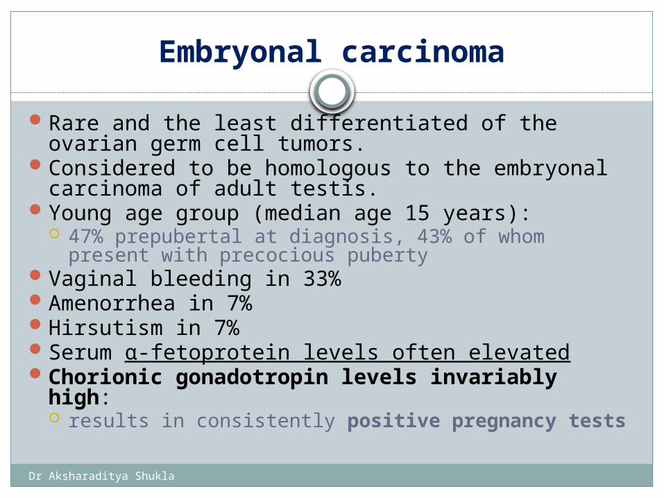
Embryonal carcinoma
Rare and the least differentiated of the ovarian germ cell tumors.
Considered to be homologous to the embryonal carcinoma of adult testis.
Young age group (median age 15 years): 47% prepubertal at diagnosis, 43% of whom present
with precocious pubertyVaginal bleeding in 33%Amenorrhea in 7%Hirsutism in 7%Serum α-fetoprotein levels often elevatedChorionic gonadotropin levels invariably high:
results in consistently positive pregnancy tests
Dr Aksharaditya Shukla
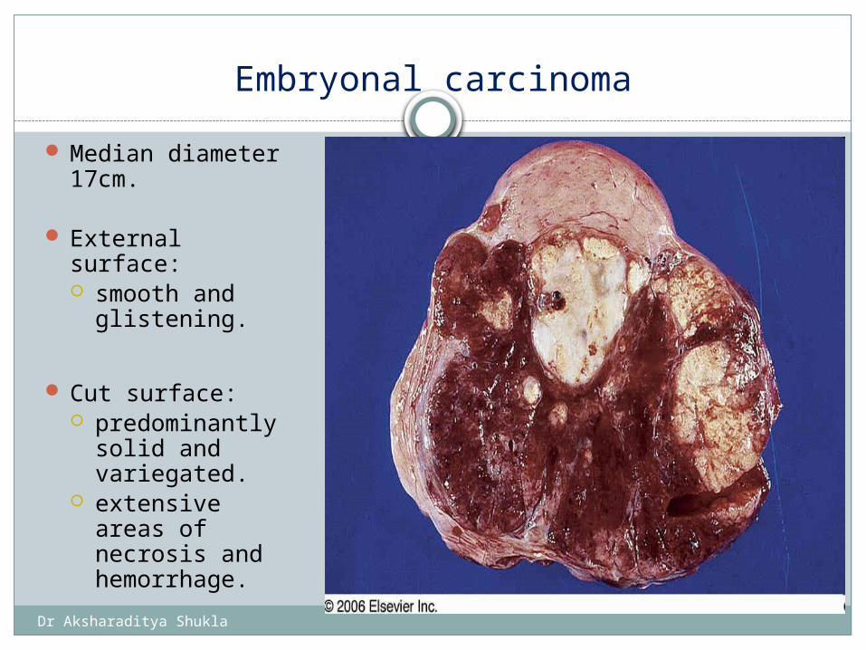
Embryonal carcinoma
Median diameter 17cm.
External surface: smooth and
glistening.
Cut surface: predominantly
solid and variegated.
extensive areas of necrosis and hemorrhage.
Gross appearance of embryonal carcinoma of ovary.
Dr Aksharaditya Shukla
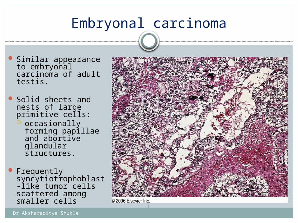
Embryonal carcinoma
Similar appearance to embryonal carcinoma of adult testis.
Solid sheets and nests of large primitive cells: occasionally
forming papillae and abortive glandular structures.
Frequently
syncytiotrophoblast-like tumor cells scattered among smaller cells
Microscopic appearance of embryonal carcinoma of ovary
Dr Aksharaditya Shukla
Special Stains and Immunohistochemistry of embryonal carcinoma
Syncytiotrophoblast-like tumor cells immunoreactive for hCG.
CD30 and cytokeratin
Dr Aksharaditya Shukla
Choriocarcinoma
Malignant tumor of the ovary with trophoblastic differentiation composed of syncytiotrophoblast, cytotrophoblast and intermediate trophoblast.
Exceedingly rare.
May be associated with mature cystic teratoma of contralateral ovary.
Dr Aksharaditya Shukla
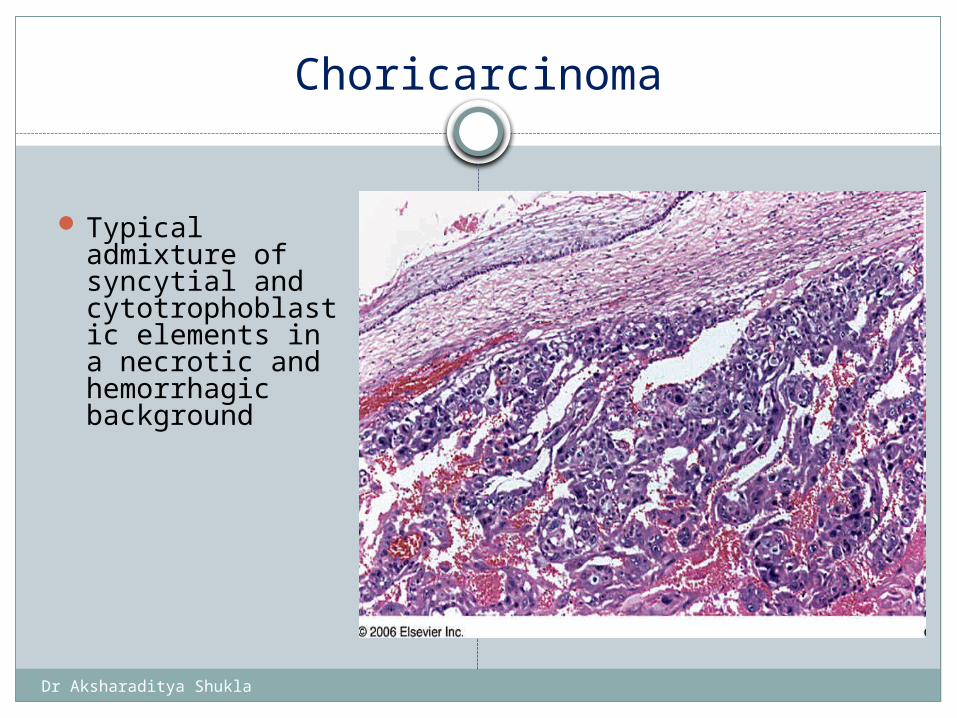
Choricarcinoma
Typical admixture of syncytial and cytotrophoblastic elements in a necrotic and hemorrhagic background
Choriocarcinoma arising in a dermoid cyst
Dr Aksharaditya Shukla
Special Stains and Immunohistochemistry
Usually immunohistochemical reactivity for hCG.
Keratin 7 said to represent a marker for subset of trophoblastic cells.
LK26 (a folate-binding protein): - consistently expressed
Dr Aksharaditya Shukla
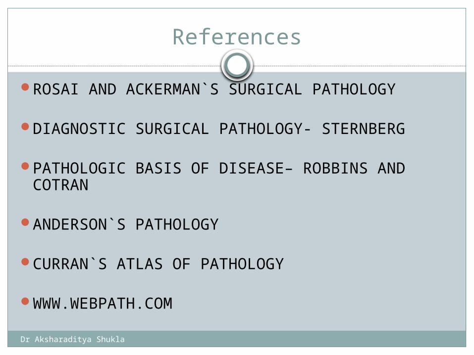
References
ROSAI AND ACKERMAN`S SURGICAL PATHOLOGY
DIAGNOSTIC SURGICAL PATHOLOGY- STERNBERG
PATHOLOGIC BASIS OF DISEASE– ROBBINS AND COTRAN
ANDERSON`S PATHOLOGY
CURRAN`S ATLAS OF PATHOLOGY
WWW.WEBPATH.COM
Dr Aksharaditya Shukla
Thanks Presented By: Dr Aksharaditya Shukla
Resident, Department Of PatholgyMGM Medical College & M.Y. Hospital, Indore
Dr Aksharaditya Shukla
OVARIAN TUMOURS II
Moderator: Dr Poonam Nanwani
Asst. Prof. Department Of PathologyMGM Medical College & M.Y. Hospital, Indore
Dr Aksharaditya Shukla
Dr Aksharaditya Shukla
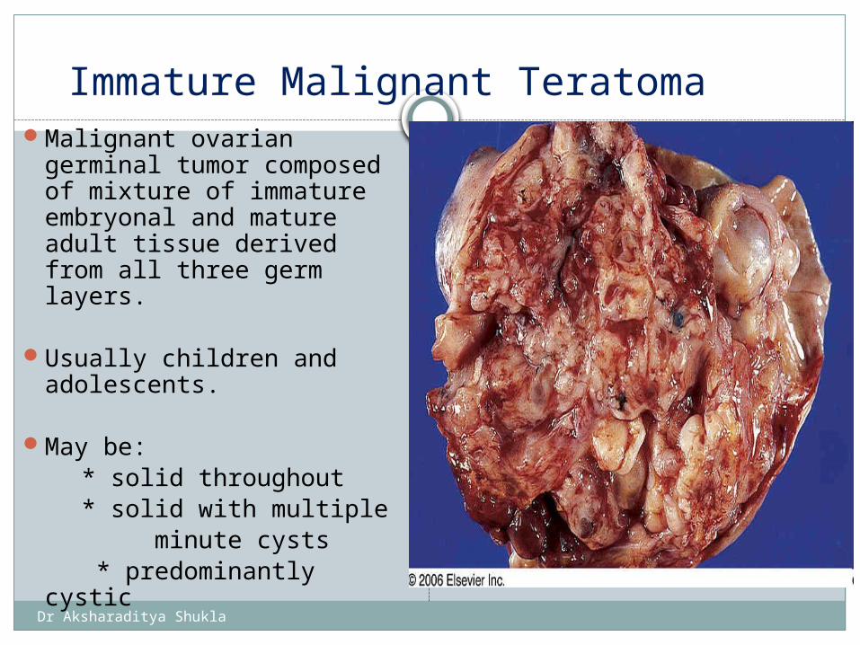
Immature Malignant TeratomaMalignant ovarian
germinal tumor composed of mixture of immature embryonal and mature adult tissue derived from all three germ layers.
Usually children and adolescents.
May be: * solid throughout * solid with multiple minute cysts * predominantly cystic
Gross appearance of ovarian immature teratoma
Dr Aksharaditya Shukla
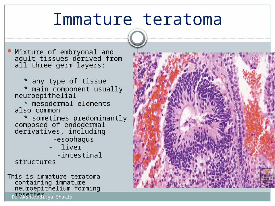
Immature teratoma
Mixture of embryonal and adult tissues derived from all three germ layers:
* any type of tissue * main component usually
neuroepithelial * mesodermal elements also
common * sometimes predominantly
composed of endodermal derivatives, including
-esophagus - liver -intestinal structures
This is immature teratoma containing immature neuroepithelium forming rosettes
immature teratoma containing immature neuroepithelium forming rosettes.
Dr Aksharaditya Shukla
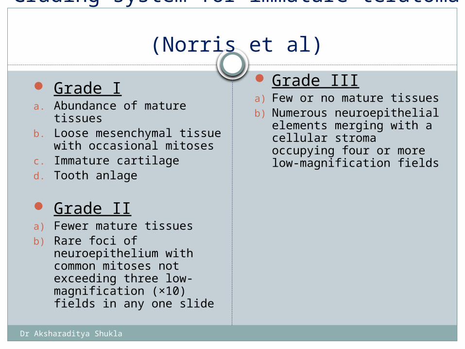
Grading system for immature teratoma (Norris et al)
Grade Ia. Abundance of mature
tissuesb. Loose mesenchymal tissue
with occasional mitosesc. Immature cartilaged. Tooth anlage
Grade IIa) Fewer mature tissuesb) Rare foci of neuroepithelium
with common mitoses not exceeding three low-magnification (×10) fields in any one slide
Grade IIIa) Few or no mature tissuesb) Numerous neuroepithelial
elements merging with a cellular stroma occupying four or more low-magnification fields
Dr Aksharaditya Shukla
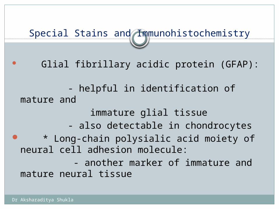
Special Stains and Immunohistochemistry
Glial fibrillary acidic protein (GFAP): - helpful in identification of mature and immature glial tissue - also detectable in chondrocytes * Long-chain polysialic acid moiety of neural cell
adhesion molecule: - another marker of immature and mature
neural tissue
Dr Aksharaditya Shukla
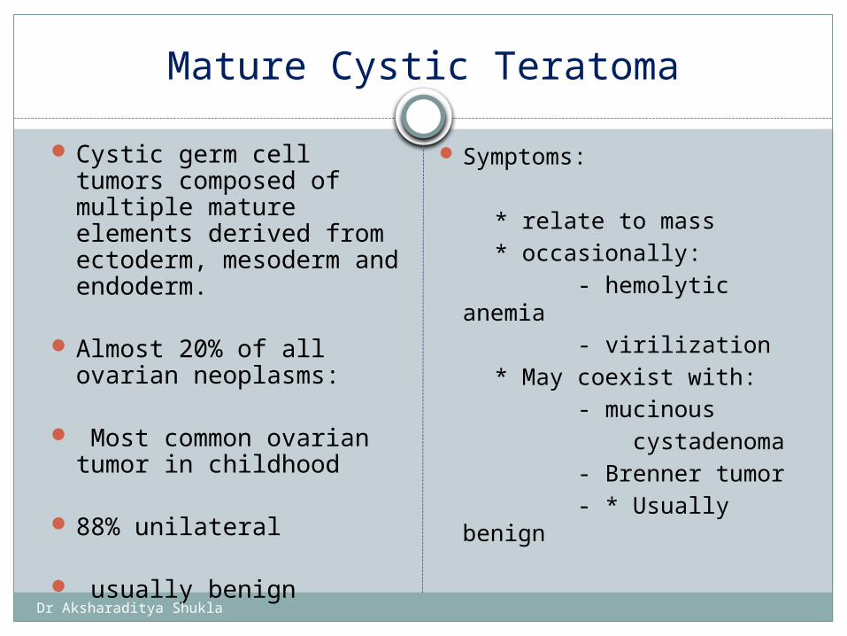
Mature Cystic Teratoma
Cystic germ cell tumors composed of multiple mature elements derived from ectoderm, mesoderm and endoderm.
Almost 20% of all ovarian neoplasms:
Most common ovarian tumor in childhood
88% unilateral
usually benign
Symptoms:
* relate to mass * occasionally: - hemolytic anemia - virilization * May coexist with: - mucinous cystadenoma - Brenner tumor - * Usually benign
Dr Aksharaditya Shukla
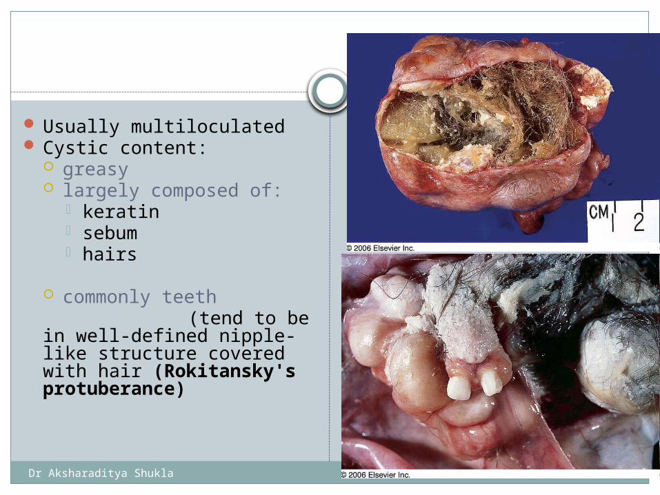
Usually multiloculated Cystic content:
greasy largely composed of:
keratin sebum hairs
commonly teeth (tend to be in well-
defined nipple-like structure covered with hair (Rokitansky's protuberance)
Admixture of sebum and hair within the cavity of an ovarian mature cystic teratoma

Dr Aksharaditya Shukla
Mature cystic teratoma
#Cystic cavities lined by mature epidermis
Extremely common: * skin appendages * neural (particularly
glial) tissue
Also: * cartilage * respiratory tissue * gastrointestinal tract
tissue (- may be peptic ulcer
formation)
Mature cystic teratoma of ovary: gastric mucosa of pyloric type
Dr Aksharaditya Shukla
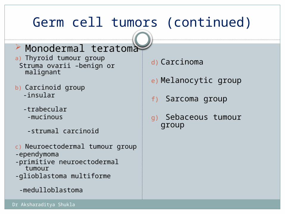
Germ cell tumors (continued)
Monodermal teratoma
a) Thyroid tumour group Struma ovarii –benign or
malignant
b) Carcinoid group -insular -trabecular -mucinous -strumal carcinoid
c) Neuroectodermal tumour group
-ependymoma -primitive neuroectodermal
tumour-glioblastoma multiforme
-medulloblastoma
d) Carcinoma
e) Melanocytic group
f) Sarcoma group
g) Sebaceous tumour group
Dr Aksharaditya Shukla
Struma Ovarii
Ovarian teratoma composed exclusively or predominantly of thyroid tissue
May show any of the pathologic changes seen in a normally placed gland, including:
diffuse or nodular hyperplasia: - may lead to hyperthyroidism - thyroiditis -carcinoma: ( sometimes resulting in metastases)
Thyroid nature fully documented with biologic and immunohistochemical studies for thyroid hormones
Dr Aksharaditya Shukla
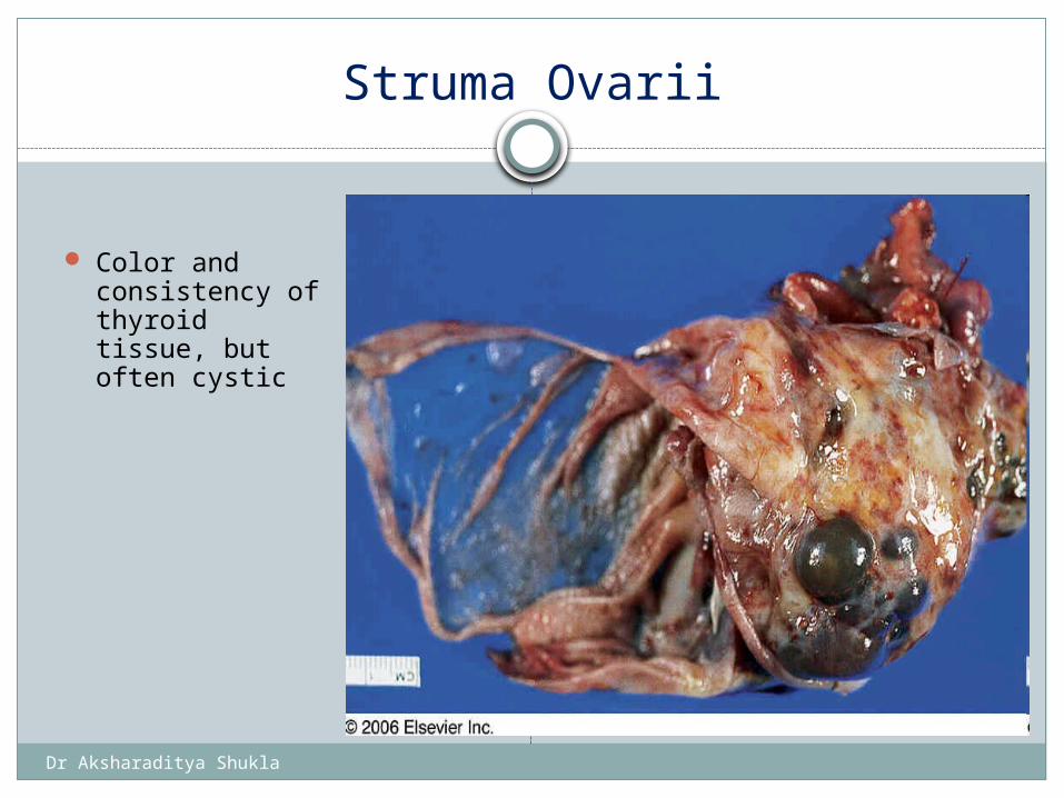
Struma Ovarii
Color and consistency of thyroid tissue, but often cystic
Dr Aksharaditya Shukla
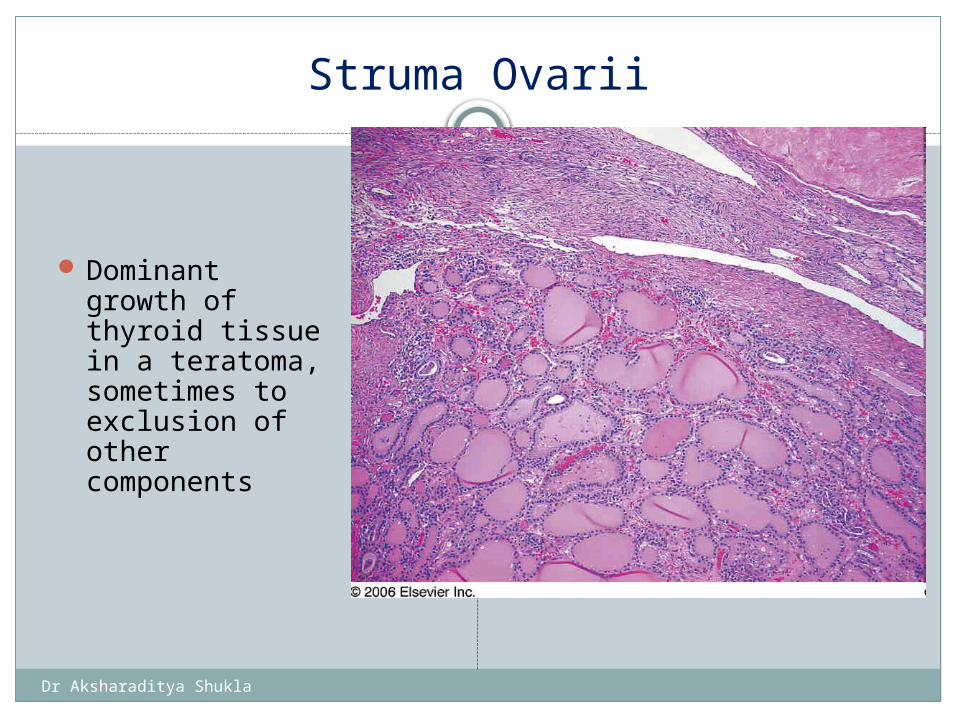
Struma Ovarii
Dominant growth of thyroid tissue in a teratoma, sometimes to exclusion of other components
Struma ovarii. The thyroid tissue, which has a microscopically unremarkable appearance, is sharply delimited from the ovarian stroma
Dr Aksharaditya Shukla
Carcinoid Tumor (monodedrmal teratoma)
Primary well differentiated neuroendocrine tumor of the ovary.
Carcinoid syndrome: * more likely the larger tumor ( > 7 cm)
Sometimes severe constipation: * presumably due to secretion of peptide YY
Dr Aksharaditya Shukla
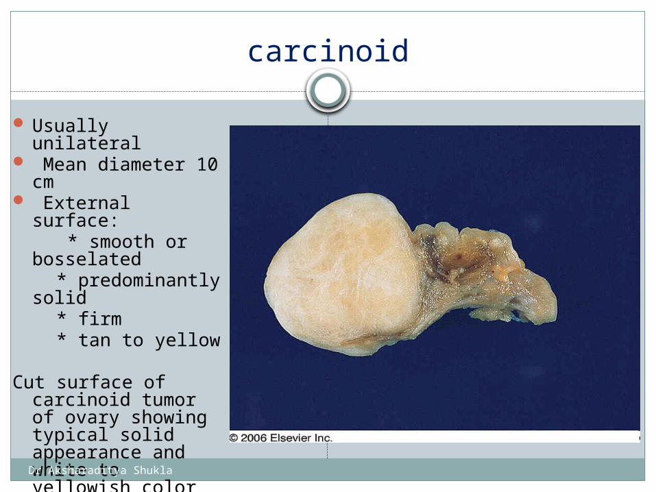
carcinoid
Usually unilateral Mean diameter 10
cm External surface: * smooth or
bosselated * predominantly
solid * firm * tan to yellow
Cut surface of carcinoid tumor of ovary showing typical solid appearance and white to yellowish color
Cut surface of carcinoid tumor of ovary showing typical solid appearance and white to yellowish color
Dr Aksharaditya Shukla
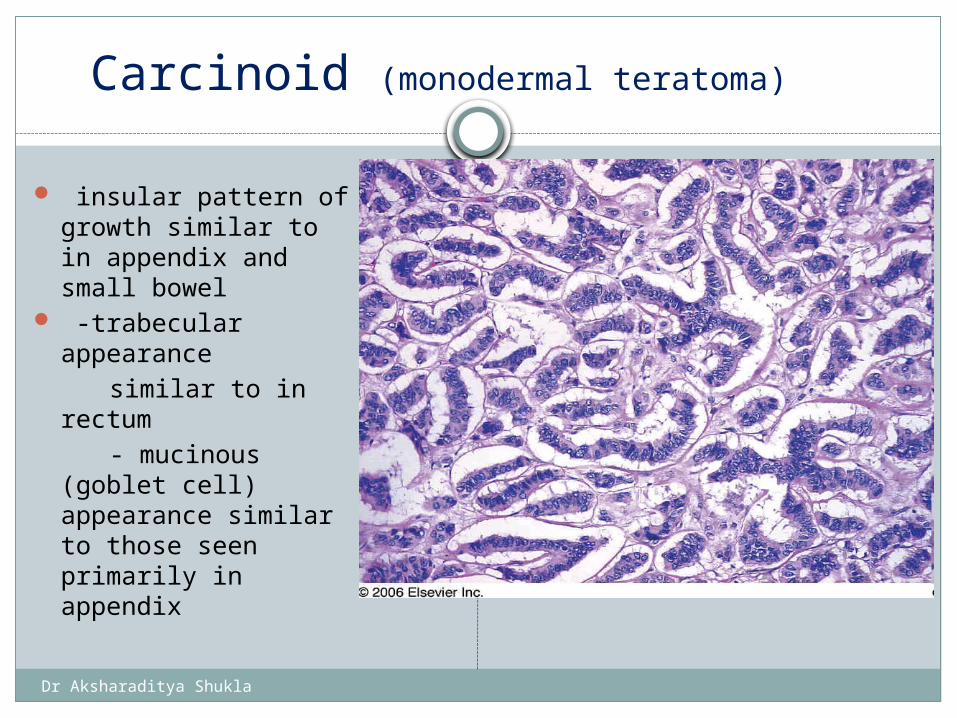
Carcinoid (monodermal teratoma)
insular pattern of growth similar to in appendix and small bowel
-trabecular appearance
similar to in rectum - mucinous (goblet
cell) appearance similar to those seen primarily in appendix
Primary ovarian carcinoid tumor with a trabecular pattern of growth
Dr Aksharaditya Shukla
1. SURFACE EPITHELIAL TUMOURS2. GERM CELL TUMOURS3. SEX CORD STROMAL TUMOURS 4. GERM CELL SEX CORD STROMAL
TUMOURS 5. TUMOUR OF THE RETE OVARII 6. MISCELLANEOUS TUMOURS 7. TUMOUR LIKE CONDITION8. LYMPHOID AND HEMATOPOETIC
TUMOURS9. SECONDARY TUMOURS.
SEX CORD STROMAL TUMOURS
Dr Aksharaditya Shukla
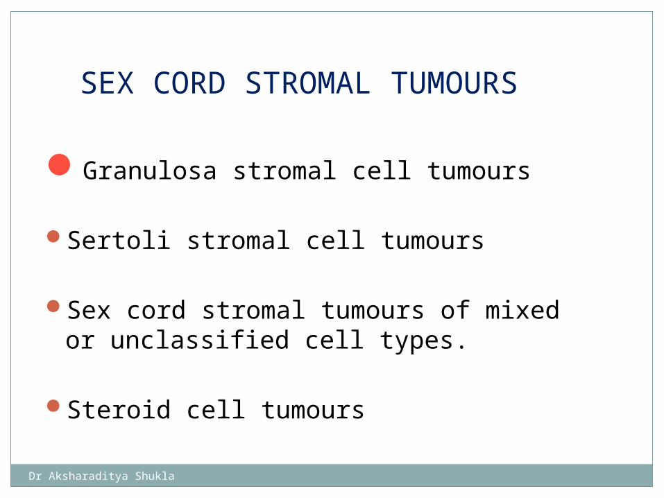
Granulosa stromal cell tumours
Sertoli stromal cell tumours
Sex cord stromal tumours of mixed or unclassified cell types.
Steroid cell tumours
SEX CORD STROMAL TUMOURS
Dr Aksharaditya Shukla
Granulosa stromal cell tumours (SEX CORD STROMAL TUMOURS)
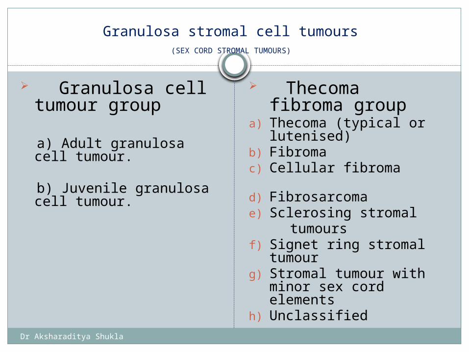
Granulosa cell tumour group
a) Adult granulosa cell tumour.
b) Juvenile granulosa cell
tumour.
Thecoma fibroma group
a) Thecoma (typical or lutenised)
b) Fibromac) Cellular fibroma
d) Fibrosarcomae) Sclerosing stromal tumours f) Signet ring stromal
tumourg) Stromal tumour with
minor sex cord elementsh) Unclassified
Dr Aksharaditya Shukla
Granulosa Cell Tumor group(sex cord stromal tumor)
Differentiation towards follicular granulosa cells that can occur in adults (adult granulosa cell tumor) and in younger patients (juvenile granulosa cell tumor).
Two distinct types: * adult * juvenile
Dr Aksharaditya Shukla
Adult Granulosa Cell Tumor
Usually childbearing age.
- 75% have hyperestrinism, which may result in: +isosexual precocious puberty +metrorrhagia in adults, including postmenopausal women
Elevated serum inhibin and follicle regulatory proteins.
Dr Aksharaditya Shukla
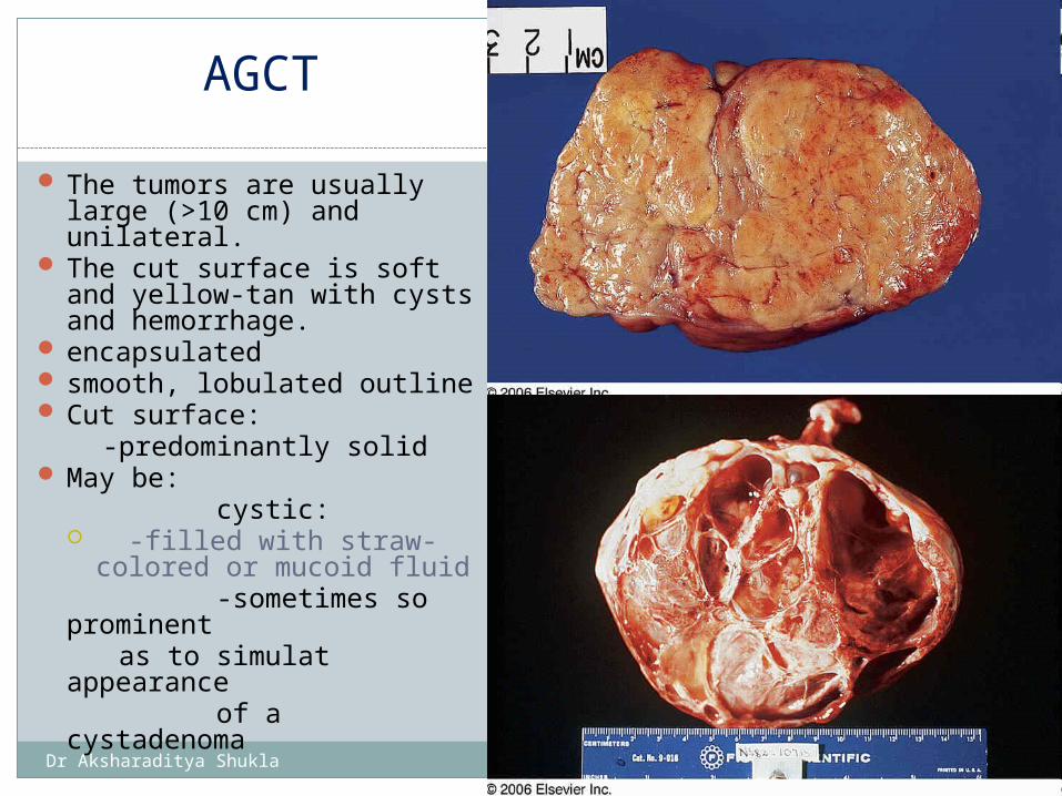
AGCT
The tumors are usually large (>10 cm) and unilateral.
The cut surface is soft and yellow-tan with cysts and hemorrhage.
encapsulated smooth, lobulated outline Cut surface: -predominantly solid May be: cystic:
-filled with straw-colored or mucoid fluid
-sometimes so prominent
as to simulat appearance
of a cystadenoma
Granulosa cell tumor with solid cut surface.
Dr Aksharaditya Shukla
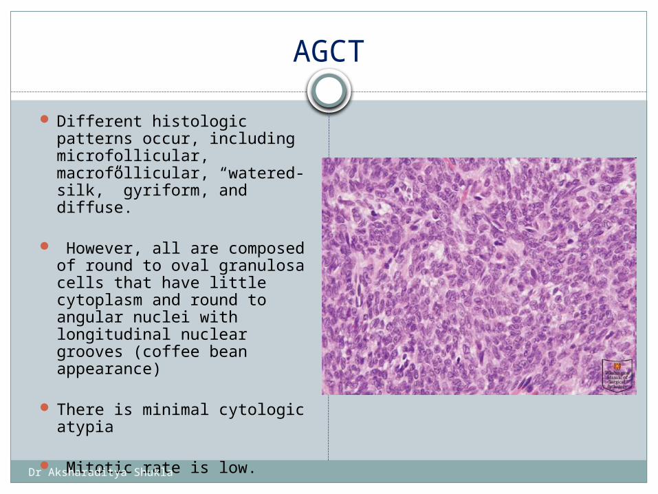
AGCT
Different histologic patterns occur, including microfollicular, macrofollicular, “watered-silk,” gyriform, and diffuse.
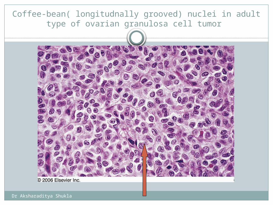
However, all are composed of round to oval granulosa cells that have little cytoplasm and round to angular nuclei with longitudinal nuclear grooves (coffee bean appearance)
There is minimal cytologic atypia
Mitotic rate is low.
Dr Aksharaditya Shukla
Coffee-bean( longitudnally grooved) nuclei in adult type of ovarian granulosa cell tumor
Dr Aksharaditya Shukla
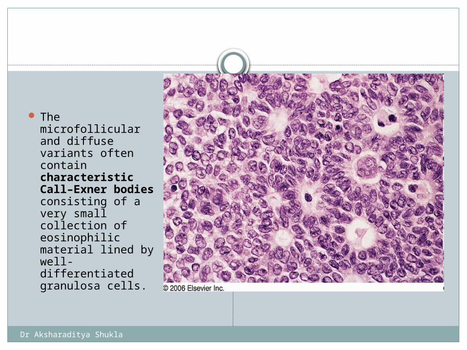
The microfollicular and diffuse variants often contain characteristic Call–Exner bodies consisting of a very small collection of eosinophilic material lined by well-differentiated granulosa cells.
Dr Aksharaditya Shukla
Juvenile Granulosa Cell Tumor
More aggressive than adult
More likely to produce distant metastases
≈80% during first two decades of life * Usually presents with isosexual
precocity * Occasionally associated with: - enchondromatosis (Ollier's disease) - Maffucci's syndrome
Dr Aksharaditya Shukla
Juvenile Granulosa Cell Tumor
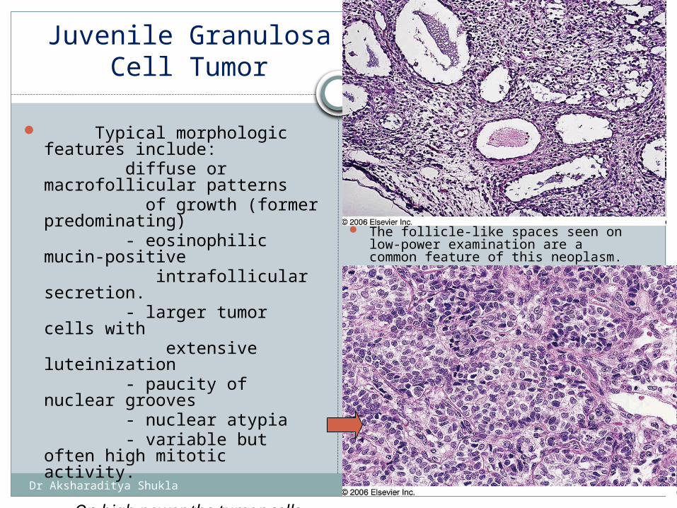
Typical morphologic features include:
diffuse or macrofollicular patterns
of growth (former predominating)
- eosinophilic mucin-positive intrafollicular secretion. - larger tumor cells with extensive luteinization - paucity of nuclear grooves - nuclear atypia - variable but often high
mitotic activity.
On high power the tumor cells lack the coffee-bean nuclei seen in the adult type
The follicle-like spaces seen on low-power examination are a common feature of this neoplasm.
Dr Aksharaditya Shukla
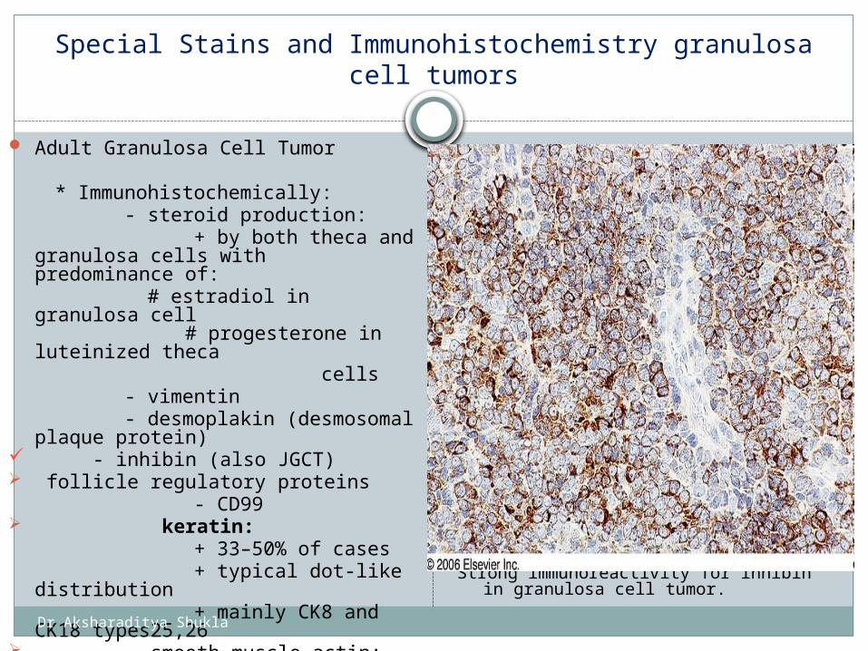
Special Stains and Immunohistochemistry granulosa cell tumors
Adult Granulosa Cell Tumor
* Immunohistochemically: - steroid production: + by both theca and granulosa
cells with predominance of: # estradiol in granulosa cell
# progesterone in luteinized theca
cells - vimentin - desmoplakin (desmosomal plaque
protein) - inhibin (also JGCT) follicle regulatory proteins - CD99 keratin: + 33–50% of cases + typical dot-like distribution + mainly CK8 and CK18
types25,26 smooth muscle actin: + nearly all cases S-100 protein: + ≈50% of cases
Strong immunoreactivity for inhibin in granulosa cell tumor.
Dr Aksharaditya Shukla
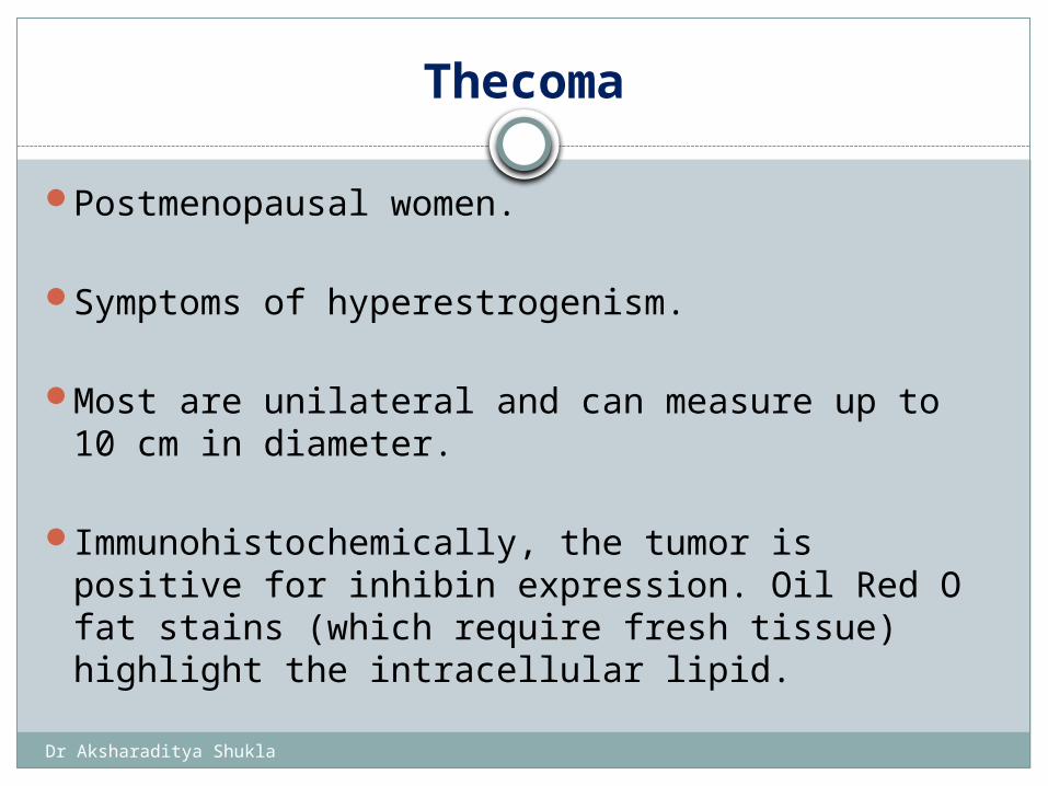
Thecoma
Postmenopausal women.
Symptoms of hyperestrogenism.
Most are unilateral and can measure up to 10 cm in diameter.
Immunohistochemically, the tumor is positive for
inhibin expression. Oil Red O fat stains (which require fresh tissue) highlight the intracellular lipid.
Dr Aksharaditya Shukla
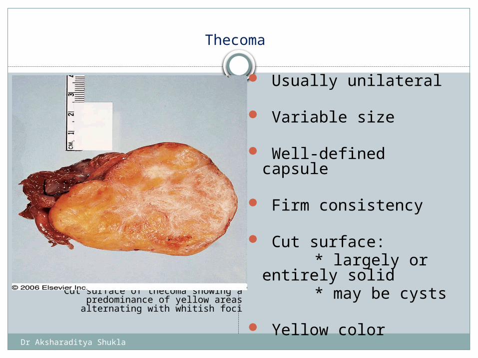
Thecoma
Cut surface of thecoma showing a predominance of yellow areas alternating
with whitish foci
Usually unilateral
Variable size
Well-defined capsule
Firm consistency
Cut surface: * largely or entirely
solid * may be cysts
Yellow color
Dr Aksharaditya Shukla
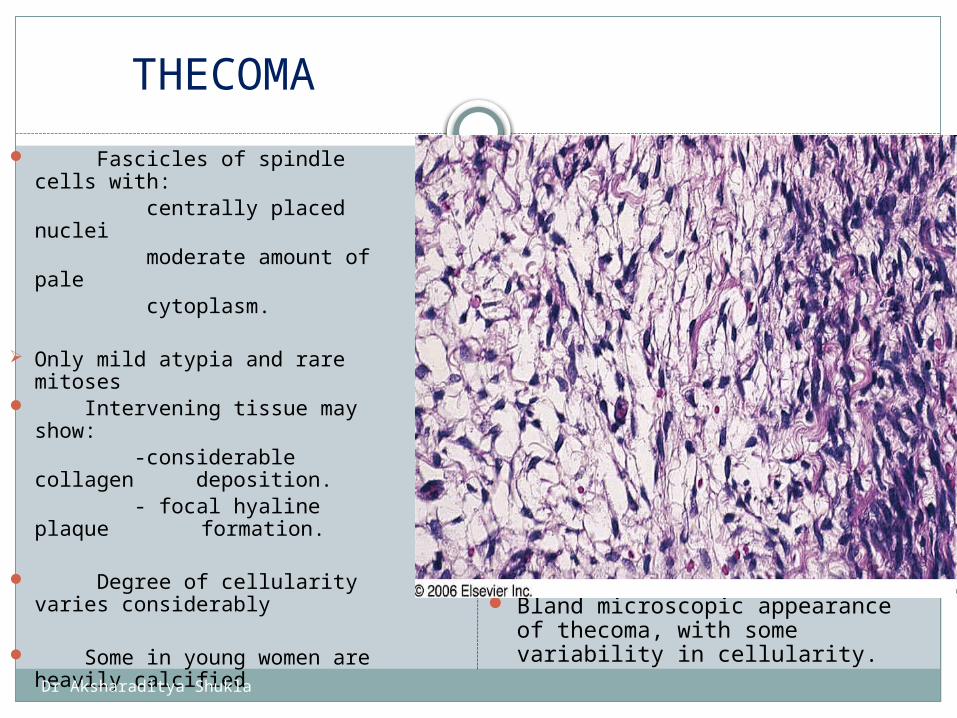
THECOMA
Fascicles of spindle cells with:
centrally placed nuclei moderate amount of pale cytoplasm.
Only mild atypia and rare mitoses
Intervening tissue may show:
-considerable collagen deposition.
- focal hyaline plaque formation.
Degree of cellularity varies considerably
Some in young women are heavily calcified
Bland microscopic appearance of thecoma, with some variability in cellularity.
Dr Aksharaditya Shukla
Special Stains and Immunohistochemistry
Oil red O: (require fresh tissue) - abundant intracytoplasmic neutral fat
Positive for inhibin expression
Silver stains: Estradiol usually limited to a small number of
tumor cell
Dr Aksharaditya Shukla
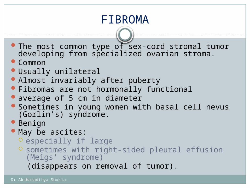
FIBROMA
The most common type of sex-cord stromal tumor developing from specialized ovarian stroma.
CommonUsually unilateralAlmost invariably after puberty Fibromas are not hormonally functionalaverage of 5 cm in diameterSometimes in young women with basal cell nevus
(Gorlin's) syndrome.BenignMay be ascites:
especially if large sometimes with right-sided pleural effusion (Meigs'
syndrome)(disappears on removal of tumor).
Dr Aksharaditya Shukla
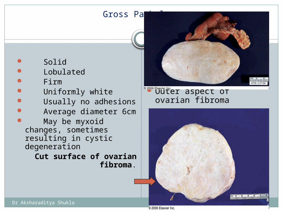
Gross Pathology
Solid Lobulated Firm Uniformly white Usually no adhesions Average diameter 6cm May be myxoid
changes, sometimes resulting in cystic degeneration
Cut surface of ovarian fibroma.
Outer aspect of ovarian fibroma
Dr Aksharaditya Shukla
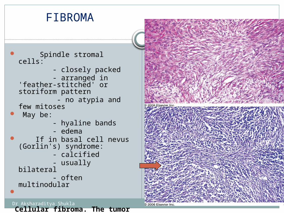
FIBROMA
Spindle stromal cells: - closely packed - arranged in 'feather-
stitched' or storiform pattern - no atypia and few
mitoses May be: - hyaline bands - edema If in basal cell nevus
(Gorlin's) syndrome: - calcified - usually bilateral - often multinodular
Cellular fibroma. The tumor is hypercellular, but
pleomorphism and mitotic activity are minimal
Dr Aksharaditya Shukla
Immunohistochemistry of Fibromas
diffusely positive for vimentin
Dr Aksharaditya Shukla
Small Cell Carcinoma
Poorly differentiated tumors, composed mostly of small cells, and subclassified into hypercalcemic and pulmonary type
Two types: * hypercalcemic: -most common * pulmonary
Hypercalcemic-type Small Cell Carcinoma
Young females (average age 23 years)
Nearly always bilateral
Occasionally familial
Hypercalcemia: - 67% of cases - disappears following
removal * High-grade
malignancy
Dr Aksharaditya Shukla
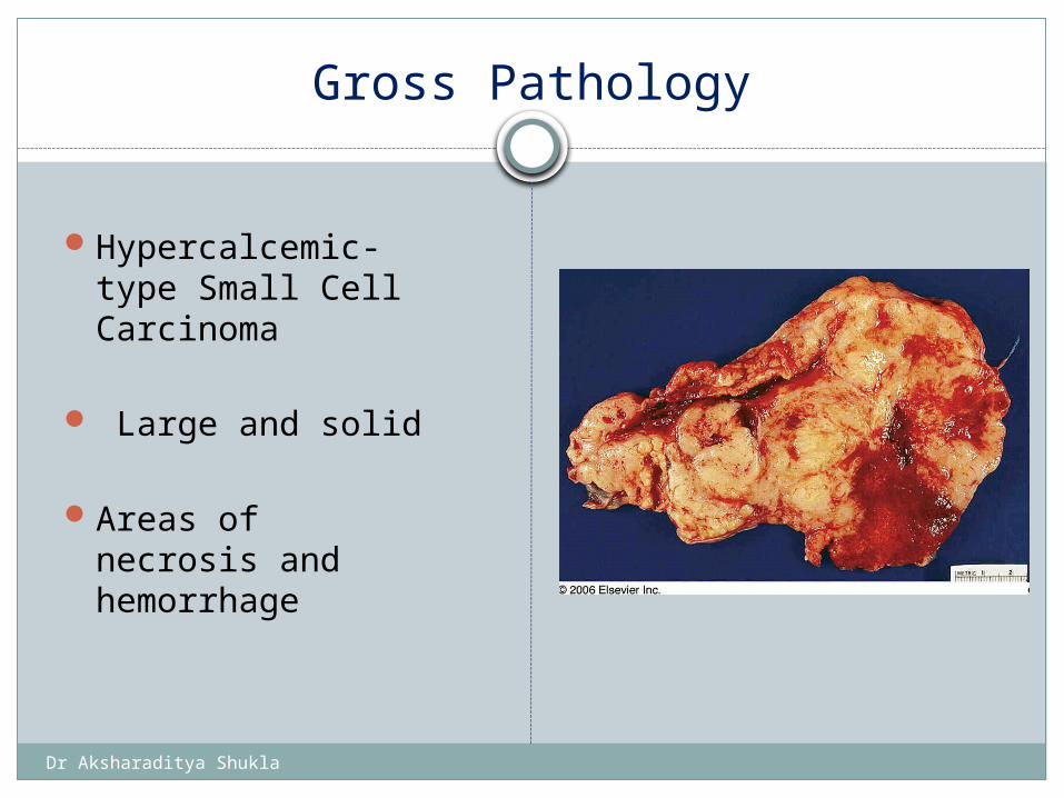
Gross Pathology
Hypercalcemic-type Small Cell Carcinoma
Large and solid
Areas of necrosis and hemorrhage
Dr Aksharaditya Shukla
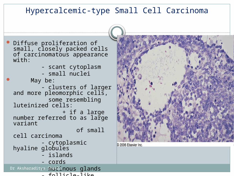
Hypercalcemic-type Small Cell Carcinoma
Diffuse proliferation of small, closely packed cells of carcinomatous appearance with:
- scant cytoplasm - small nuclei May be: - clusters of larger and more
pleomorphic cells, some resembling luteinized
cells: + if a large number
referred to as large variant of small cell carcinoma - cytoplasmic hyaline globules - islands - cords - mucinous glands - follicle-like structures + important clue to
diagnosis
Small cell carcinoma, hypercalcemic type. The presence of follicle-like formations is an important diagnostic feature
Dr Aksharaditya Shukla
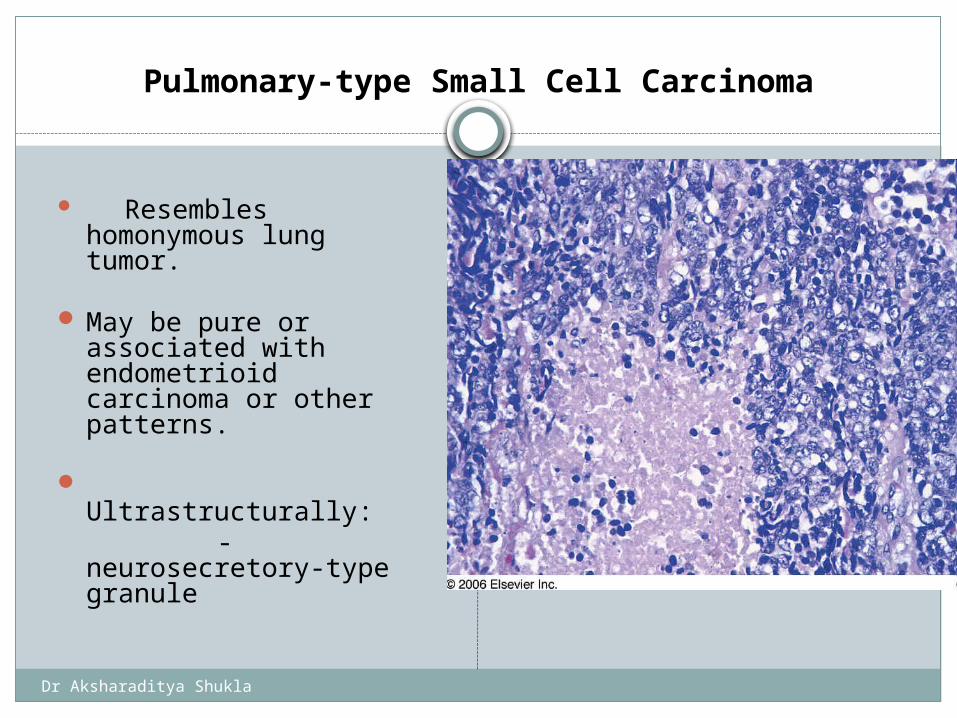
Pulmonary-type Small Cell Carcinoma
Resembles homonymous lung tumor.
May be pure or associated with endometrioid carcinoma or other patterns.
Ultrastructurally: - neurosecretory-
type granule
Dr Aksharaditya Shukla
Thanks Presented By: Dr Aksharaditya Shukla
Resident, Department Of PatholgyMGM Medical College & M.Y. Hospital, Indore