osteoblast behavior on hierarchical micro-/nano-structured titanium surface
TRANSCRIPT
Corresponding author: Yanmin Zhou E-mail: [email protected]
Journal of Bionic Engineering 8 (2011) 234–241
Osteoblast Behavior on Hierarchical Micro-/Nano-Structured Titanium Surface
Weiyan Meng, Yanmin Zhou, Yanjing Zhang, Qing Cai, Liming Yang, Jinghui Zhao, Chunyan Li
School of Stomatology, Jilin University, Changchun 130021, P. R. China
Abstract In the present work, osteoblast behavior on a hierarchical micro-/nano-structured titanium surface was investigated. A hi-
erarchical hybrid micro-/nano-structured titanium surface topography was produced via Electrolytic Etching (EE). MG-63 cells were cultured on disks for 2 h to 7 days. The osteoblast response to the hierarchical hybrid micro-/nano-structured titanium surface was evaluated through the osteoblast cell morphology, attachment and proliferation. For comparison, MG-63 cells were also cultured on Sandblasted and Acid-etched (SLA) as well as Machined (M) surfaces respectively. The results show signifi-cant differences in the adhesion rates and proliferation levels of MG-63 cells on EE, SLA, and M surfaces. Both adhesion rate and proliferation level on EE surface are higher than those on SLA and M surfaces. Therefore, we may expect that, comparing with SLA and M surfaces, bone growth on EE surface could be accelerated and bone formation could be promoted at an early stage, which could be applied in the clinical practices for immediate and early-stage loadings.
Keywords: dental implant, osteoblast, hierarchical micro-/nano-structure, surface treatment, electrolytic etching Copyright © 2011, Jilin University. Published by Elsevier Limited and Science Press. All rights reserved. doi: 10.1016/S1672-6529(11)60031-0
1 Introduction
Commercially pure titanium (Ti) is preferred for dental implants because it has excellent biocompatibility as well as mechanical properties, and it does not induce immunological reactions[1]. Ti implants can strongly osseointegrate, receive a favorable occlusion function, and be stable for a long period of time in the oral cavity. There are many factors affecting the osseointegration of Ti implants, such as implant surface characteristics, bone quality, implant surgery, and other mechanical factors. The micro-structure of an implant surface is one of the key factors affecting the rate and extent of osseointe-gration. Other surface properties of Ti implants (e.g., composition, energy, roughness, and topography) also affect osseointegration[2,3]. Topography has a direct in-fluence on surface characteristics because it can affect the surface energy and roughness. In the early 1980s, topography characteristics were found to determine the initial interactions at the tissue-implant interface. Eventually, the success rate of osseointegration is in-fluenced[4].
Implant surface topography regulates the upgrowth and orientation of osteoblasts on biological materials. It also facilitates contact guidance for epithelial cells, fi-broblasts, and osteoblasts. Cells in deeper and wider grooves grow toward a specific orientation by clasping on tubers. However, cells on a smooth surface grow randomly[5–7]. The surface topography of a micropit or microhole can increase a bone or implant contact area, change the force direction and stress of a bone, as well as reinforce the physical interlocking of the fibrin fibers on the rough surface[8,9]. Topography may also aid in stabi-lizing fibrin clots on an implant surface and fragile ex-tracellular matrix scaffolds. Hence, the micro-environ- ment becomes conducive to osteoblast migration and proliferation. However, several investigations have demonstrated that implant surfaces with microscale roughness depress the proliferation of osteoblasts and reduce the bone formation[10].
Recently, much attention has been focused on the nanoscale topography surface. Nanoscale modifications of an implant surface can contribute to mimicry proc-esses in cellular environments[11,12]. Nano-features can
Meng et al.: Osteoblast Behavior on Hierarchical Micro-/Nano-Structured Titanium Surface 235
alter the conformation of Arginine-Glycine- Aspartic Acid (RGD), increase the protein adhesion, change the cell behaviors by regulating integrin transformation, enhance the attachment and proliferation of osteoblast, as well as promote matrix synthesis. However, Meirelles et al. suggested that only according to the nanoscale topography can not assure a robust osseointegration. Microscale roughness may also be valuable to the process of osseointegration[13,14]. Gao et al. fabricated a micropit and nanostructure on Ti via acid etching and anodization, which increased hydroxyapatite formation in vitro[15]. Kubo et al. created a micropit and nanonod-ule hybrid topography using the self-assembly technique on an acid-etch-created micropit Ti surface. The results were enhanced osteoblast attachment, spreading, adhe-sion, proliferation, and differentiation[16]. Zhao et al. produced micro/nanoscale structures by adding nano-tubes to the acid-etched microstructure surface. They obtained results that could be the first evidence of the synergistic effects of micro- and nano-topographies on osteoblast behavior[10]. However, the mechanism and size-matching of micro- and nano-topographies are still unclear. Producing hierarchical structures that have di-rect clinical applications remains a challenge.
In the present study, reproducible hierarchical hy-brid micro-/nano-structured surfaces were produced by Electrolytic Etching (EE) method. The aim of the in-vestigation was to evaluate osteoblast response to the hierarchical hybrid micro-/nano-topography through the osteoblast cell morphology, attachment, and prolifera-tion. The results of the experiment may provide a foundation for developing new kinds of implant sur-faces.
2 Materials and Methods
2.1 Sample preparation and surface topography examination
Cylindrical Ti samples with diameter of 4 mm were polished with metallographic abrasive paper (300 – 1200 mesh). Each sample was cleaned by ultrasonic rinsing with acetone, dehydrated ethanol, and deionized water for 10 min. Then, hierarchical hybrid micro-/nano- structured surfaces were produced by EE method, in which Ti samples were immersed in an electrolyte solu-tion under a direct current for 2 min. The electrolyte was composed of hydrochloric acid (HCl, 11.5 vol%) and hydrofluoric acid (HF, 6 vol%). The samples were cut at
a low speed using a diamond cutting machine. The thickness of the samples was 2 mm with one side surface being processed by EE. Samples with Sandblasted and Acid-etched (SLA) as well as Machined (M) surfaces were used for comparison. All the samples were washed with distilled water, and then ultrasonically rinsed using acetone, dehydrated ethanol, and deionized water for 10 min respectively. The samples were air-dried before use.
Field-Emission Scanning Electron Microscopy (FESEM) was used to observe the surface topography of the samples. 2.2 MG-63 cell culture
MG-63 cells were cultured in culture flasks using a high-glucose Dulbecco’s Modified Eagle’s Medium (DMEM) supplemented with 10% Fetal Bovine Serum (FBS) as well as the antibiotic antimycotic solution containing 60 U·mL 1 penicillin G sodium and 100 U·mL 1 streptomycin sulfate. The culture conditions were a humidified atmosphere of 95% air, 5% CO2, and 37 C.
2.3 Cell attachment assay
Cells were detached from culture flasks during log phase growth using 0.25% trypsin. They were then seeded into culture plates (1 mL per hole) at a density of 1 × 10⁵ cells·mL 1. After 1 h, 2 h, 6 h, 12 h and 24 h of culture, the cells were gently rinsed three times with 0.01% Phosphate-Buffered Saline (PBS) to remove all unattached cells. The samples were subsequently trans-ferred into another culture plate. For cell detachment, about 0.3 mL of 0.25% trypsin per hole was added, and the reaction was allowed to stand for 3 min. The reaction was terminated using 0.7 mL of DMEM supplemented with 10% FBS. The cell suspension was stained by try-pan blue. The number of detached cells was counted under a microscope to calculate the percentage of cell attachment for each group of samples.
2.4 Cell morphology
The cells were seeded onto the surfaces of Ti sam-ples at a density of 6 × 10⁴ cells·well 1 for the three test groups. On culture day 1, 3, and 5, the cells were gently rinsed three times with 0.01% PBS. They were then fixed with 2.5% glutaraldehyde for 12 h, and with 0.1% osmic acid for 1 h. Then the cultures were washed 3 times with PBS (0.1 mol·L 1) for 15 min each time.
Journal of Bionic Engineering (2011) Vol.8 No.3 236
Dehydration followed using different ethanol concen-trations (50%, 70%, 80%, 90% and 100%) for 15 min. The cultures were then treated with isoamyl acetate for 15 min and air-dried. The cultures were metal-coated before undergoing FESEM examination.
2.5 Cell proliferation
The MG-63 cells at log-phase growth were de-tached using 0.25% trypsin and made into a monocell suspension. The suspension was placed in a culture plate after the cell density was adjusted to 6 × 10⁴ cells·mL 1. At culture day 1, 3, 5, and 7, 10 L MTT (5 mg·mL 1) was added to each hole, and the cultivation was allowed to last for 4 h. The original culture fluid was extracted, and 150 L of DMSO was added per hole. A microplate shaker was used to dissolve the formed crystals for 15 min at room temperature. Finally, the optical density of the solution in each hole was measured using an ELISA at wavelength of 490 nm. 2.6 Statistical analysis
The data were analyzed using SPSS 13.0 software (SPSS, USA). One-way ANOVA was performed to de-termine the level of significance, which was P < 0.05. For cell adhesion tests the sample size n = 3, and for cell proliferation test n = 6. All data were presented as x ± s, x is the mean and s is the standard deviation.
3 Results
3.1 Characteristics of hierarchical micro-/nano- structured Ti surface
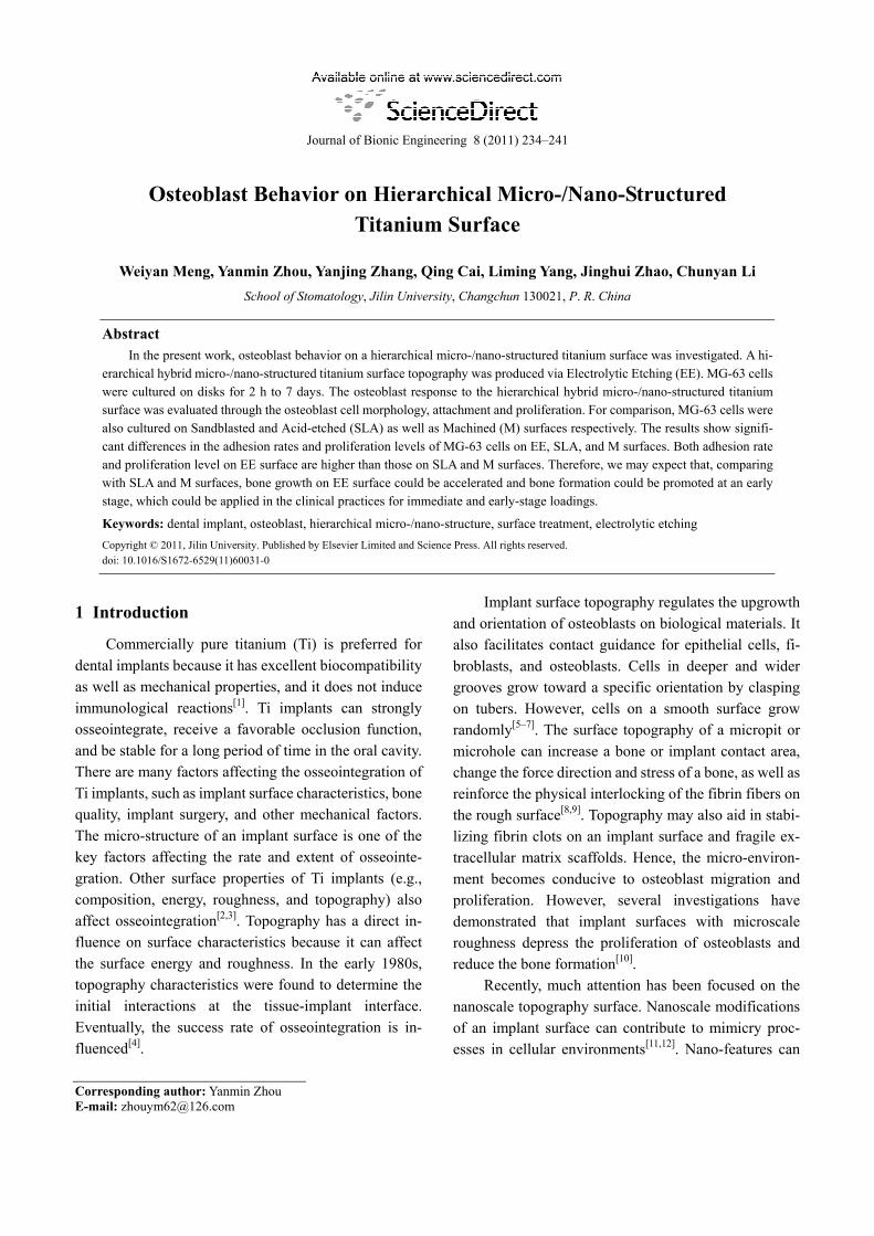
The pure Ti surface treated by EE formed hierar-chical micro-/nano-structures (Fig. 1). Under a high- power electron microscope, homogeneous bowl-shaped micro concaves were observed. The diameter of the concaves was in the range of 30 m – 50 m. Dispersed micropores and gridding structures were also observed. The dispersed micropores consisted of irregular dish-shaped concaves on both side walls and bottom of the bowl-shaped micro concaves. The diameters of the dish-shaped concaves and micropores were 8 m– 10
m and 2 m – 4 m, respectively (Fig. 1b). A great number of submicro and nano pores covered the dish-shaped concaves of the bowl-shaped side walls, as observed under an electron microscope at a 4000 × magnification (Figs. 1c and 1d). Nanosphere structures were clearly seen at the bottom and side walls of the
concaves at 16000 × and 100000 × magnifications, re-spectively (Figs. 1e and 1f). The nano structures were within 30 nm – 50 nm in size. The average roughness Ra of the surface was 5.56 m, and the contact angle was 56.6 . The surface consisted of TiO2, and the oxidation film was thin.
20 m1 mm
5 m 2 m
599 nm
352 nm
2 m 200 nm Fig. 1 FESEM micrographs of the EE surface.
3.2 Cell adhesion
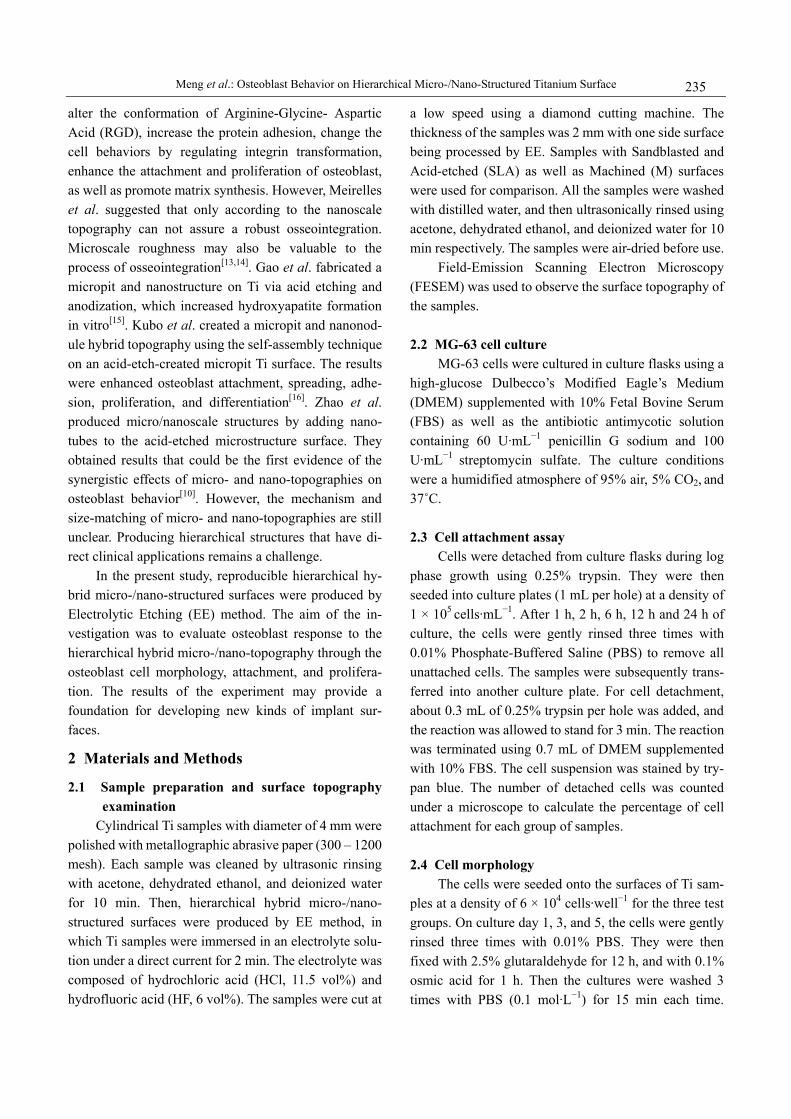
After 1 h of inoculation, differences in MG-63 cell adhesion rates were observed among the EE, SLA, and M groups. From high to low, the adhesion rates were in the order EE, SLA, M (P < 0.05, Fig. 2). At 2 h, 6 h, 12 h, and 24 h of inoculation, the adhesion rates for all groups increased compared with that at 1 h. At all above men-tioned time points, both EE and SLA groups had higher adhesion rates than the M group. At 2 h and 6 h, the rate of the EE group was evidently higher than that of the SLA group. At 12 h and 24 h, the rates of the EE and SLA groups were higher than 100%. This result proved that cell division and multiplication occurred.
EESLA
M
1 2 12 2460
20
60
100120140
40
80
160
Time (h) Fig. 2 Adhesion rates of MG-63 cells of the EE, SLA, and M groups.
Meng et al.: Osteoblast Behavior on Hierarchical Micro-/Nano-Structured Titanium Surface 237
3.3 Cell proliferation The absorbance values of normal osteoblasts,
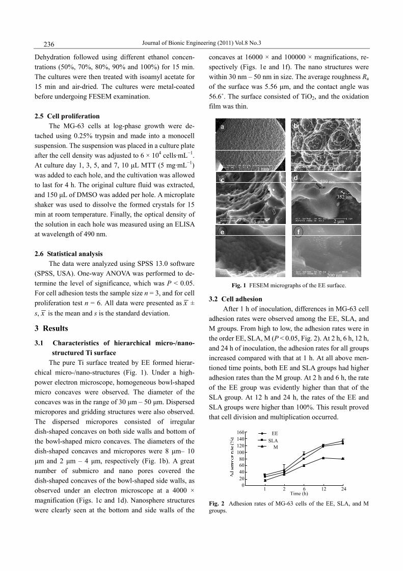
which were immersed and proliferated in liquid culture medium without attaching to any surface, increased with time and showed good proliferation ability (P < 0.05, Fig. 3). The absorbance values of MG-63 cells in all three groups increased with time, which also mean that the cells were proliferating. Group EE had a proliferation ability that was nearly identical with normal cells at day 1 and 3. The proliferation ability of group EE was higher at day 5 and 7 than that of group SLA, and was higher at day 3, 5, and 7 than that of group M. The absorbance values of cells in group SLA gradually increased with time. Compared with normal cells, at day 5 and 7, the absorbance values decreased and indicated that the pro-liferation ability was lower than normal cells. The ab-sorbance value of cells in group M was lower than normal cells at day 3, 5, and 7. This result indicated a lower proliferation. At day 3, 5, and 7, the proliferation rate of the normal, EE, and SLA groups all gradually increased. Normal and EE cells had similar rates, whereas SLA cells had lower rates. The proliferation rate of M cells were restrained on day 1, but increased at day 5 and 7.
Fig. 3 Absorbance values of MG-63 cells of EE, SLA, and M groups. 3.4 Cell morphology
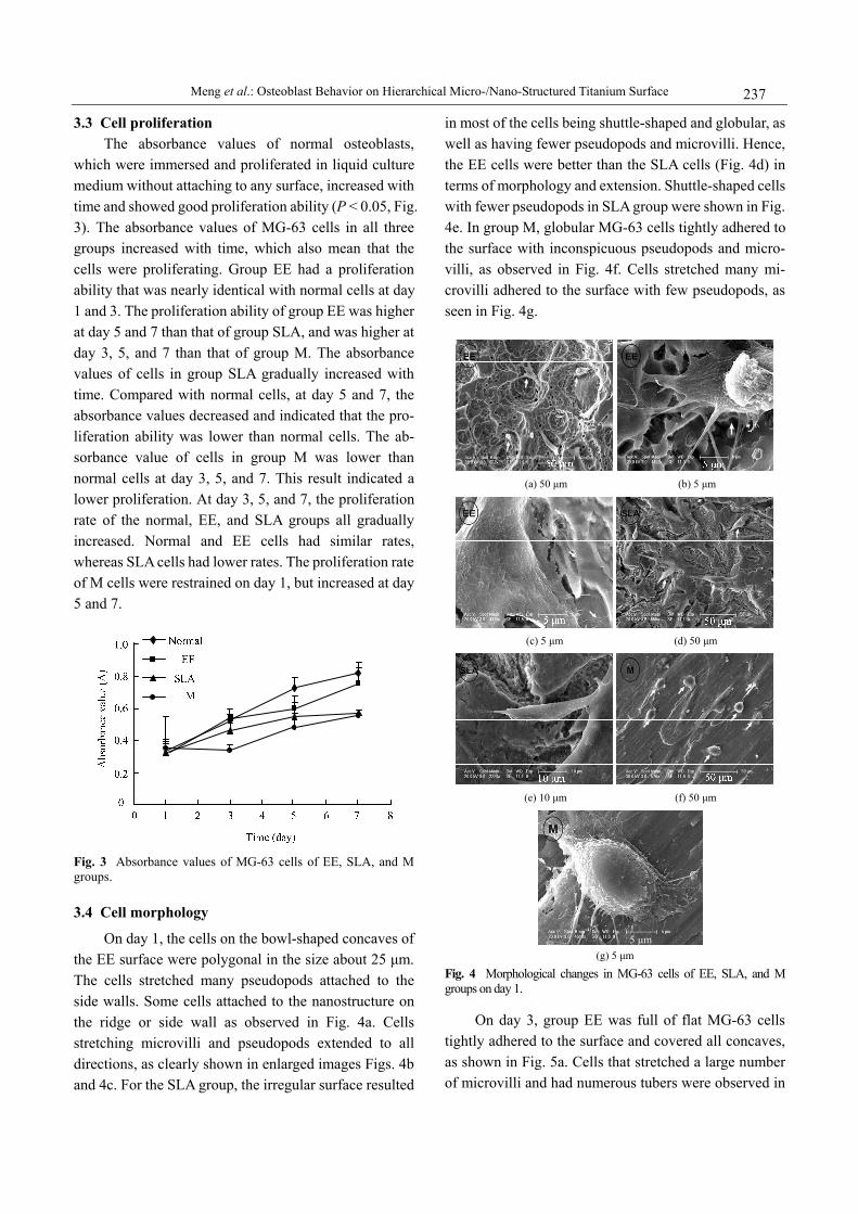
On day 1, the cells on the bowl-shaped concaves of the EE surface were polygonal in the size about 25 m. The cells stretched many pseudopods attached to the side walls. Some cells attached to the nanostructure on the ridge or side wall as observed in Fig. 4a. Cells stretching microvilli and pseudopods extended to all directions, as clearly shown in enlarged images Figs. 4b and 4c. For the SLA group, the irregular surface resulted
in most of the cells being shuttle-shaped and globular, as well as having fewer pseudopods and microvilli. Hence, the EE cells were better than the SLA cells (Fig. 4d) in terms of morphology and extension. Shuttle-shaped cells with fewer pseudopods in SLA group were shown in Fig. 4e. In group M, globular MG-63 cells tightly adhered to the surface with inconspicuous pseudopods and micro-villi, as observed in Fig. 4f. Cells stretched many mi-crovilli adhered to the surface with few pseudopods, as seen in Fig. 4g.
(a) 50 m (b) 5 m
(c) 5 m (d) 50 m
(e) 10 m (f) 50 m
5 m (g) 5 m
Fig. 4 Morphological changes in MG-63 cells of EE, SLA, and M groups on day 1.
On day 3, group EE was full of flat MG-63 cells tightly adhered to the surface and covered all concaves, as shown in Fig. 5a. Cells that stretched a large number of microvilli and had numerous tubers were observed in
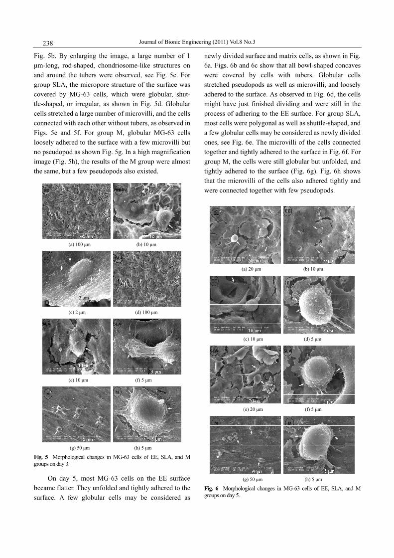
Journal of Bionic Engineering (2011) Vol.8 No.3 238
Fig. 5b. By enlarging the image, a large number of 1 m-long, rod-shaped, chondriosome-like structures on
and around the tubers were observed, see Fig. 5c. For group SLA, the micropore structure of the surface was covered by MG-63 cells, which were globular, shut-tle-shaped, or irregular, as shown in Fig. 5d. Globular cells stretched a large number of microvilli, and the cells connected with each other without tubers, as observed in Figs. 5e and 5f. For group M, globular MG-63 cells loosely adhered to the surface with a few microvilli but no pseudopod as shown Fig. 5g. In a high magnification image (Fig. 5h), the results of the M group were almost the same, but a few pseudopods also existed.
(a) 100 m (b) 10 m
100 m2 m
(c) 2 m (d) 100 m
(e) 10 m (f) 5 m
(g) 50 m (h) 5 m
Fig. 5 Morphological changes in MG-63 cells of EE, SLA, and M groups on day 3.
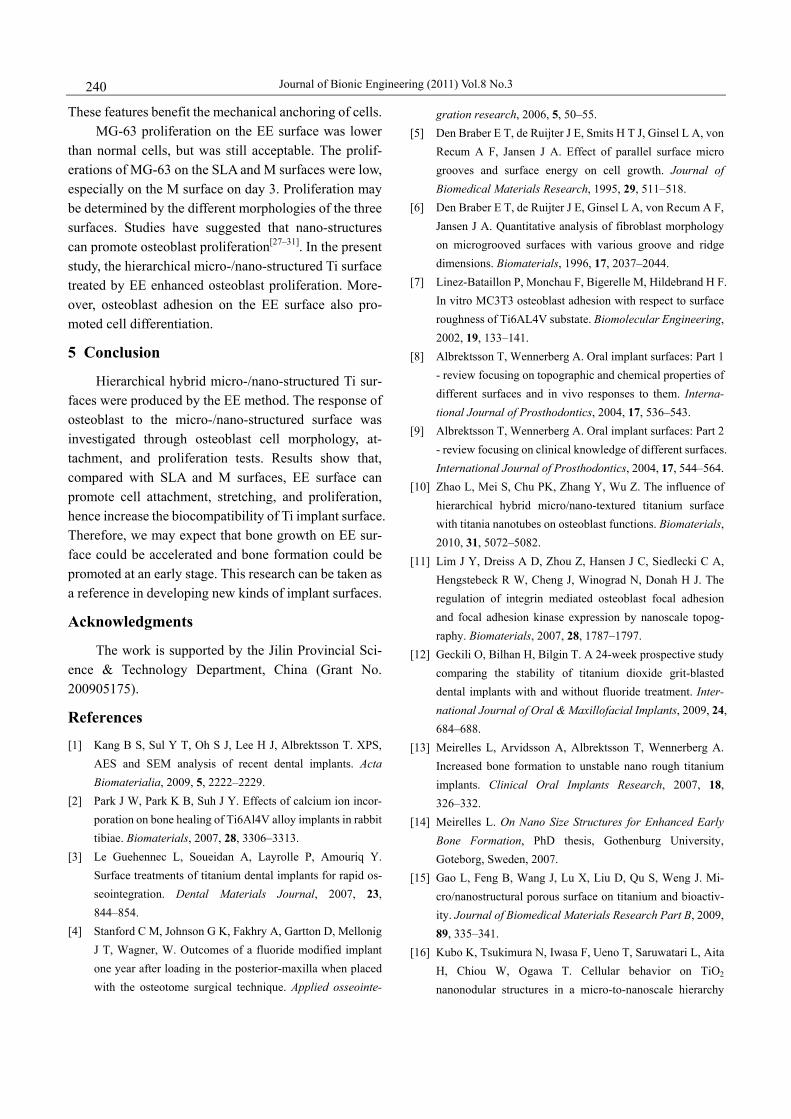
On day 5, most MG-63 cells on the EE surface became flatter. They unfolded and tightly adhered to the surface. A few globular cells may be considered as
newly divided surface and matrix cells, as shown in Fig. 6a. Figs. 6b and 6c show that all bowl-shaped concaves were covered by cells with tubers. Globular cells stretched pseudopods as well as microvilli, and loosely adhered to the surface. As observed in Fig. 6d, the cells might have just finished dividing and were still in the process of adhering to the EE surface. For group SLA, most cells were polygonal as well as shuttle-shaped, and a few globular cells may be considered as newly divided ones, see Fig. 6e. The microvilli of the cells connected together and tightly adhered to the surface in Fig. 6f. For group M, the cells were still globular but unfolded, and tightly adhered to the surface (Fig. 6g). Fig. 6h shows that the microvilli of the cells also adhered tightly and were connected together with few pseudopods.
(a) 20 m (b) 10 m
(c) 10 m (d) 5 m
(e) 20 m (f) 5 m
(g) 50 m (h) 5 m
Fig. 6 Morphological changes in MG-63 cells of EE, SLA, and M groups on day 5.

Meng et al.: Osteoblast Behavior on Hierarchical Micro-/Nano-Structured Titanium Surface 239
4 Discussion
Microstructures affect the osseointegration of im-plants[2]. A rough implant surface can promote osseoin-tegration by mechanical interlocking[8,9]. Currently, studies on implant morphology mainly focus on the micro- and nano-levels[10,15,16]. In the present study, reproducible hierarchical hybrid micro-/nano-structured surfaces were produced by EE.
The micro-structures were primarily bowl-shaped concaves with diameters of 30 m 50 m. Micropores and dish-shaped concaves also existed on both the side walls and bottom of the bowl-shaped concaves. The dish-shaped concaves and micropores were 8 m – 10
m and 2 m 4 m in diameter, respectively. The nanospheres and submicron pores on the side walls and bottom of concaves were clearly observable. Bigerelle et al. considered that cell morphology may be related to a smooth surface smaller than the cell. The morphology may also be related to the high roughness of big and deep concaves. The smooth slopes and steps of the inner walls of the concaves provide a “nest” that surround the cells[17]. When the micropore size is smaller than the cells, osteoblasts more easily attach on a smooth surface. However, when the micropore size is larger, osteoblasts tend to attach to the surface of the bowl-shaped nest. In the present study, the morphologies of hierarchical hy-brid micro-/nano-structured surfaces produced by EE agreed well with the descriptions of Bigerelle and Anselme[18,19]. The micro-structure provides space for osteoblasts, supplying a stable microenvironment. The micro-structure may also be beneficial for the stabiliza-tion of fibrin clots and extracellular matrix scaffolds by increasing osteoblast migration, adhesion, differentia-tion, and matrix secretion[20]. The nanosphere structure provides a similarly stable cell environment that can promote adhesion, activation, and expression. Boyan et al.[21] suggested that larger micro-concaves or -pores can provide the most appropriate compression and stretch-ing signal for the mechanoreceptors of osteoblasts. Mi-cro-morphologies may provide the passage for the mi-gration of cells and the transport of nutrients.
Regarding the adhesion rates of osteoblasts, the EE surface had the highest rate, followed by SLA, and fi-nally by M. The adhesion rate was directly related to the surface topography. EE and SLA surfaces, which were both rough and porous, benefited the mechanical at-
tachment of fibrin. In contrast, the M surface with a groove structure was smooth. Such a structure caused the surface to weakly function in the mechanical inter-locking process of guiding cell adhesion and growth.
The cell attachment, adhesion, and morphology of the three surfaces were all different. Li et al.[22] found that osteoblast attachment and morphology on smooth and rough surfaces were different. In the present study, shuttle-shaped and globular osteoblasts, which were about 10 m – 20 m and had few pseudopods, arranged in a parallel manner on a smooth surface. Most cells on the SLA surface were shuttle-shaped, and the rest of the cells were globular and polygonal. The cells stretched less pseudopods and a large number of microvilli tightly attached to the porous surface. On the EE surface, 25 m – 30 m polygonal cells stretched many pseudopods and microfilaments such that they attached to the ridges and side walls of the bowl-shaped concaves. Some cells across the top of the bowl-shaped concaves also attached to the nanostructures on the ridges and side walls. The microvilli and pseudopods of the osteoblasts extended to all directions, and the osteoblasts became flatter and wider with time. There were also a large number of tu-bers and rod-shaped chondriosome-like structures on the surface.
Research has shown that the distribution of mito-chondria changed the cell morphology to activate the response between the cells and the surface[23]. In the initial stage of the attachment, the rearrangement of the mitochondrial network provided energy. Most mito-chondria are found in the places of adhesion plaque and cell elongation. The cell cytoplasm reorganized to obtain the best morphology and functional status because of adhesion plaque formation. Numerous adhesion plaques and rod-shaped mitochondria-like bodies were presented in cells attached to the EE surface[24]. Therefore, the cells tended to adhere to the nanostructure surface, which was larger than the cell and well differentiated. These results agreed well with those of Bigerelle’s research. Some studies suggested that more obvious peaks and ridges correspond to easier contact induction for osteoblasts. Sub-micron and nano-structures can enhance the adhe-sion, migration, and growth of osteoblasts, hence bene-fiting osseointegration[25,26]. Therefore, the MG-63 cells better attached on the EE surface. This result may be related to the micro-concaves, nano-structures, and in-creased contact area between the rough surface and cells.
Journal of Bionic Engineering (2011) Vol.8 No.3 240
These features benefit the mechanical anchoring of cells. MG-63 proliferation on the EE surface was lower
than normal cells, but was still acceptable. The prolif-erations of MG-63 on the SLA and M surfaces were low, especially on the M surface on day 3. Proliferation may be determined by the different morphologies of the three surfaces. Studies have suggested that nano-structures can promote osteoblast proliferation[27–31]. In the present study, the hierarchical micro-/nano-structured Ti surface treated by EE enhanced osteoblast proliferation. More-over, osteoblast adhesion on the EE surface also pro-moted cell differentiation.
5 Conclusion
Hierarchical hybrid micro-/nano-structured Ti sur-faces were produced by the EE method. The response of osteoblast to the micro-/nano-structured surface was investigated through osteoblast cell morphology, at-tachment, and proliferation tests. Results show that, compared with SLA and M surfaces, EE surface can promote cell attachment, stretching, and proliferation, hence increase the biocompatibility of Ti implant surface. Therefore, we may expect that bone growth on EE sur-face could be accelerated and bone formation could be promoted at an early stage. This research can be taken as a reference in developing new kinds of implant surfaces.
Acknowledgments
The work is supported by the Jilin Provincial Sci-ence & Technology Department, China (Grant No. 200905175).
References
[1] Kang B S, Sul Y T, Oh S J, Lee H J, Albrektsson T. XPS, AES and SEM analysis of recent dental implants. Acta Biomaterialia, 2009, 5, 2222–2229.
[2] Park J W, Park K B, Suh J Y. Effects of calcium ion incor-poration on bone healing of Ti6Al4V alloy implants in rabbit tibiae. Biomaterials, 2007, 28, 3306–3313.
[3] Le Guehennec L, Soueidan A, Layrolle P, Amouriq Y. Surface treatments of titanium dental implants for rapid os-seointegration. Dental Materials Journal, 2007, 23, 844–854.
[4] Stanford C M, Johnson G K, Fakhry A, Gartton D, Mellonig J T, Wagner, W. Outcomes of a fluoride modified implant one year after loading in the posterior-maxilla when placed with the osteotome surgical technique. Applied osseointe-
gration research, 2006, 5, 50–55. [5] Den Braber E T, de Ruijter J E, Smits H T J, Ginsel L A, von
Recum A F, Jansen J A. Effect of parallel surface micro grooves and surface energy on cell growth. Journal of Biomedical Materials Research, 1995, 29, 511–518.
[6] Den Braber E T, de Ruijter J E, Ginsel L A, von Recum A F, Jansen J A. Quantitative analysis of fibroblast morphology on microgrooved surfaces with various groove and ridge dimensions. Biomaterials, 1996, 17, 2037–2044.
[7] Linez-Bataillon P, Monchau F, Bigerelle M, Hildebrand H F. In vitro MC3T3 osteoblast adhesion with respect to surface roughness of Ti6AL4V substate. Biomolecular Engineering, 2002, 19, 133–141.
[8] Albrektsson T, Wennerberg A. Oral implant surfaces: Part 1 - review focusing on topographic and chemical properties of different surfaces and in vivo responses to them. Interna-tional Journal of Prosthodontics, 2004, 17, 536–543.
[9] Albrektsson T, Wennerberg A. Oral implant surfaces: Part 2 - review focusing on clinical knowledge of different surfaces. International Journal of Prosthodontics, 2004, 17, 544–564.
[10] Zhao L, Mei S, Chu PK, Zhang Y, Wu Z. The influence of hierarchical hybrid micro/nano-textured titanium surface with titania nanotubes on osteoblast functions. Biomaterials, 2010, 31, 5072–5082.
[11] Lim J Y, Dreiss A D, Zhou Z, Hansen J C, Siedlecki C A, Hengstebeck R W, Cheng J, Winograd N, Donah H J. The regulation of integrin mediated osteoblast focal adhesion and focal adhesion kinase expression by nanoscale topog-raphy. Biomaterials, 2007, 28, 1787–1797.
[12] Geckili O, Bilhan H, Bilgin T. A 24-week prospective study comparing the stability of titanium dioxide grit-blasted dental implants with and without fluoride treatment. Inter-national Journal of Oral & Maxillofacial Implants, 2009, 24, 684–688.
[13] Meirelles L, Arvidsson A, Albrektsson T, Wennerberg A. Increased bone formation to unstable nano rough titanium implants. Clinical Oral Implants Research, 2007, 18, 326–332.
[14] Meirelles L. On Nano Size Structures for Enhanced Early Bone Formation, PhD thesis, Gothenburg University, Goteborg, Sweden, 2007.
[15] Gao L, Feng B, Wang J, Lu X, Liu D, Qu S, Weng J. Mi-cro/nanostructural porous surface on titanium and bioactiv-ity. Journal of Biomedical Materials Research Part B, 2009, 89, 335–341.
[16] Kubo K, Tsukimura N, Iwasa F, Ueno T, Saruwatari L, Aita H, Chiou W, Ogawa T. Cellular behavior on TiO2 nanonodular structures in a micro-to-nanoscale hierarchy
Meng et al.: Osteoblast Behavior on Hierarchical Micro-/Nano-Structured Titanium Surface 241
model. Biomaterials, 2009, 30, 5319–5329. [17] Bigerelle M, Anselme K, Noël B, Ruderman I, Hardouin P,
Iost A. Improvement in the morphology of Ti-based surfaces: a new process to increase in vitro human osteoblast response. Biomaterials. 2002, 23, 1563–1577.
[18] Bigerelle M, Anselme K. Topography effects of pure tita-nium substates on human osteoblasts long-term adhesion. Acta Biomaterialia, 2005, 1, 211–222.
[19] Bigerelle M, Anselme K. A kinetic approach to osteoblast adhesion on biomaterial surface. Journal of Biomedical Materials Research, 2005, 75, 530–540.
[20] Elter P, Sickel F, Ewald A. Nanoscaled periodic surface structures of medical stainless steel and their effect on os-teoblast cells. Acta Biomaterialia, 2009, 5, 1468–1473.
[21] Boyan B D, Hummer T W, Dean D D, Schwartz Z. Role of material surfaces in regulating bone and cartilage cell re-sponse. Biomaterials, 1996, 17, 137–146.
[22] Li D, Liu B, Wu J, Chen J. Bone interface of dental implants cytologically influenced by a modified sandblasted surface: a preliminary in vitro study. Implant Dentistry, 2001, 10, 132–138.
[23] Uggeri J, Guizzardi S, Scandroglio R, Gatti R. Adhesion of human osteoblasts to titanium: A morpho-functional analy-sis with confocal microscopy. Micron, 2010, 41, 210–219.
[24] Salido M, Vilches-Perez J I, Gonzalez J L, Vilches J. Mitochondrial bioenergetics and distribution in living human osteoblasts grown on implant surfaces. Histology and Histopathology, 2009, 24, 1275–1286.
[25] Anselme K, Bigerelle M. Topography effects of pure tita-nium substates on human osteoblasts long-term adhesion. Acta Biomaterialia, 2005, 1, 211–222.
[26] Brunette D M. The effects of implant surface topography on the behavior of cells. The International Journal of Oral & Maxillofacial Implants, 1988, 3, 231–246.
[27] Meirelles L, Currie F, Jacobsson M, Tomas Albrektsson, Ann Wennerberg. The effect of chemical and nano topog-raphical modifications on early stage of osseointegration. The International Journal of Oral & Maxillofacial Implants, 2008, 23, 641–647.
[28] Meirelles L, Melin L, Peltola T, Kjellin P, Kangasniemi I, Currie F, Andersson M, Albrektsson T, Wennerberg A. Ef-fect of hydroxyapatite and Titania nanostructures on Early in vivo bone response. Clinical Implant Dentistry and Related Research, 2008, 10, 245–254.
[29] Mendes V C, Moineddin R, Davies J E. The effect of dis-crete calcium phosphate nanocrystals on bone-bonding to titanium surfaces. Biomaterials, 2007, 28, 4748–4755.
[30] Li Y, Lee I S, Cui F Z, Choi S H. The biocompatibility of nanostructured calcium phosphate coated on micro-arc oxi-dized titanium. Biomaterials, 2008, 29, 2025–2032.
[31] Goene R, Testori T, Trisi P. Influence of a nanometer-scale surface enhancement on de novo bone formation on titanium implants:a histomorphometric study in human maxillae. The International Journal of Periodontics & Restorative Den-tistry, 2007, 27, 3–11.