ortho eponyms
TRANSCRIPT
819SPECIAL EXHIBIT
Radiologic History ExhibitMusculoskeletal Eponyms:Who Are Those Guys?1
Tim B. Hunter, MD • Leonard F. Peltier, MD, PhD • Pamela J. Lund, MD
IntroductionAccuracy and conciseness are essential elements in medical communication. Accu-racy is achieved through careful and painstaking use of medical nomenclature, whichat times can be cumbersome. This difficulty has been overcome to some extent bythe use of labels, tags, acronyms, and eponyms, which, when precisely defined andaccurately used, convey a great deal of information very concisely. Eponyms are la-bels that provide two kinds of information: the pattern of a complex injury or patho-logic problem and the name of an individual who has been closely identified withthat problem, reminding us that the medicine of today is not entirely the work ofour contemporaries. “The word eponym is derived from the Greek word eponymos,which means named after. An eponym may be defined as the name(s) of one ormore individuals who presumably have devised or described an anatomic structure, aclassification system, a disease, an injury, a principle, a physical sign, or an operativetechnique” (Salter RB, written communication, 1999). Mark Ravitch, in discussingGuillaume Dupuytren’s invention of the Mikulicz enterotome, had this to say abouteponyms: “Given an eponym one may be sure (1) that the man so honored was notthe first to describe the disease, the operation, or the instrument, or (2) that he mis-understood the situation, or (3) that he is generally misquoted, or (4) that (1), (2),and (3) are all simultaneously true.... My own feeling is that whatever their fallibility,eponyms illustrate the lineage of surgery and bring to it the color of old times, distin-guished figures, ancient sieges and pestilences, and continually remind us of the in-ternational nature of science...” (1).
Index terms: Extremities, injuries • Fractures • Radiology and radiologists, history
RadioGraphics 2000; 20:819–836
1From the Department of Radiology, University of Arizona, 1501 N Campbell Ave, Tucson, AZ 85724-5067. Received May 7, 1999; revisionsrequested May 28 and received August 16; accepted August 17. Address reprint requests to T.B.H. (e-mail: [email protected])
©RSNA, 2000
820 May-June 2000 RG ■ Volume 20 • Number 3
All labels, whether they are tags, acronyms, oreponyms, depend for their value on an accurateunderstanding of their meaning. Without this un-derstanding, their use can be confusing, even dan-gerous. When there is an accurate understandingof their meaning, eponyms are valuable shorthandsince they convey a good deal of specific informa-tion in an abbreviated way. Unfortunately, ep-onyms are often not used consistently or accu-rately by all, and, in some instances, there arereasonable differences of opinion concerning theproper descriptive terminology applied to a par-ticular type of injury. This is particularly true inthe case of classifications of ankle fractures.
The classification of ankle fractures has a longand varied history, going back to Paris, France, atthe beginning of the 19th century. With an un-limited supply of fresh cadaver material, Dupuytrenand his students applied controlled mechanicalforces to the ankle and dissected and visualizedthe bone and soft-tissue injuries they had produced.The results of their experiments were surprisinglyconsistent, and they identified categories of frac-tures to which their names were applied (2–5).Over the years, various ankle fracture classifica-tion systems have been developed, including theBosworth, Cotton, Dupuytren, Lauge-Hansen,Le Fort, Pott, and Weber systems. Today, thereare two commonly used classifications, theLauge-Hansen (6–10) and the Weber (11–13)systems. The Lauge-Hansen classification, whichuses the mechanism of injury to classify anklefractures, is confusing. It uses terminology suchas supination and pronation that, when appliedto the foot, is poorly understood, even by experi-enced orthopedic surgeons. The Weber classifica-tion is the most practical system for orthopedicsurgeons. Because of the multiplicity of eponymsand other designations for ankle fractures, thereis great confusion in their usage. For this reason,the terms that are archaic, poorly defined, andpoorly understood should not be used, includingthe Bosworth, Cotton, Dupuytren, Le Fort, andPott classifications. The Lauge-Hansen classifica-tion should be used only if one is very familiarwith its principles.
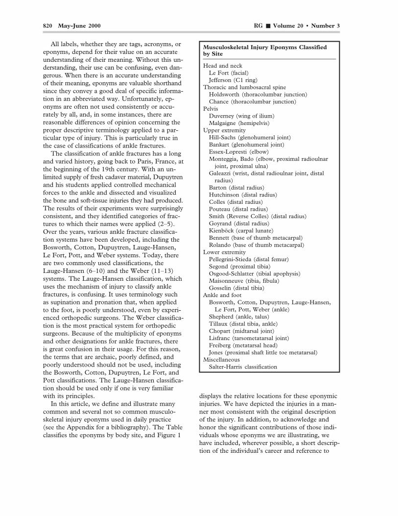
In this article, we define and illustrate manycommon and several not so common musculo-skeletal injury eponyms used in daily practice(see the Appendix for a bibliography). The Tableclassifies the eponyms by body site, and Figure 1
Musculoskeletal Injury Eponyms Classifiedby Site
Head and neckLe Fort (facial)Jefferson (C1 ring)
Thoracic and lumbosacral spineHoldsworth (thoracolumbar junction)Chance (thoracolumbar junction)
PelvisDuverney (wing of ilium)Malgaigne (hemipelvis)
Upper extremityHill-Sachs (glenohumeral joint)Bankart (glenohumeral joint)Essex-Lopresti (elbow)Monteggia, Bado (elbow, proximal radioulnar
joint, proximal ulna)Galeazzi (wrist, distal radioulnar joint, distal
radius)Barton (distal radius)Hutchinson (distal radius)Colles (distal radius)Pouteau (distal radius)Smith (Reverse Colles) (distal radius)Goyrand (distal radius)Kienböck (carpal lunate)Bennett (base of thumb metacarpal)Rolando (base of thumb metacarpal)
Lower extremityPellegrini-Stieda (distal femur)Segond (proximal tibia)Osgood-Schlatter (tibial apophysis)Maisonneuve (tibia, fibula)Gosselin (distal tibia)
Ankle and footBosworth, Cotton, Dupuytren, Lauge-Hansen,
Le Fort, Pott, Weber (ankle)Shepherd (ankle, talus)Tillaux (distal tibia, ankle)Chopart (midtarsal joint)Lisfranc (tarsometatarsal joint)Freiberg (metatarsal head)Jones (proximal shaft little toe metatarsal)
MiscellaneousSalter-Harris classification
displays the relative locations for these eponymicinjuries. We have depicted the injuries in a man-ner most consistent with the original descriptionof the injury. In addition, to acknowledge andhonor the significant contributions of those indi-viduals whose eponyms we are illustrating, wehave included, wherever possible, a short descrip-tion of the individual’s career and reference to
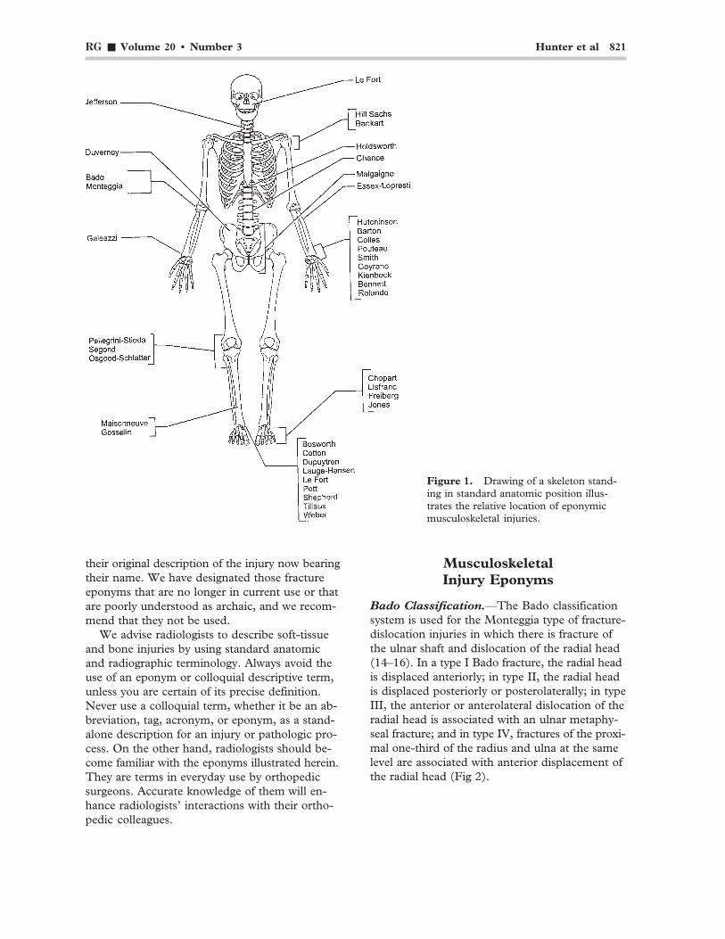
RG ■ Volume 20 • Number 3 Hunter et al 821
Figure 1. Drawing of a skeleton stand-ing in standard anatomic position illus-trates the relative location of eponymicmusculoskeletal injuries.
their original description of the injury now bearingtheir name. We have designated those fractureeponyms that are no longer in current use or thatare poorly understood as archaic, and we recom-mend that they not be used.
We advise radiologists to describe soft-tissueand bone injuries by using standard anatomicand radiographic terminology. Always avoid theuse of an eponym or colloquial descriptive term,unless you are certain of its precise definition.Never use a colloquial term, whether it be an ab-breviation, tag, acronym, or eponym, as a stand-alone description for an injury or pathologic pro-cess. On the other hand, radiologists should be-come familiar with the eponyms illustrated herein.They are terms in everyday use by orthopedicsurgeons. Accurate knowledge of them will en-hance radiologists’ interactions with their ortho-pedic colleagues.
MusculoskeletalInjury Eponyms
Bado Classification.—The Bado classificationsystem is used for the Monteggia type of fracture-dislocation injuries in which there is fracture ofthe ulnar shaft and dislocation of the radial head(14–16). In a type I Bado fracture, the radial headis displaced anteriorly; in type II, the radial headis displaced posteriorly or posterolaterally; in typeIII, the anterior or anterolateral dislocation of theradial head is associated with an ulnar metaphy-seal fracture; and in type IV, fractures of the proxi-mal one-third of the radius and ulna at the samelevel are associated with anterior displacement ofthe radial head (Fig 2).

822 May-June 2000 RG ■ Volume 20 • Number 3
Arthur Sydney Blundell Bankart (1879–1951)was a distinguished British orthopedic surgeon.He was a founding member of the British Ortho-paedic Association and served as its secretary andas its president.
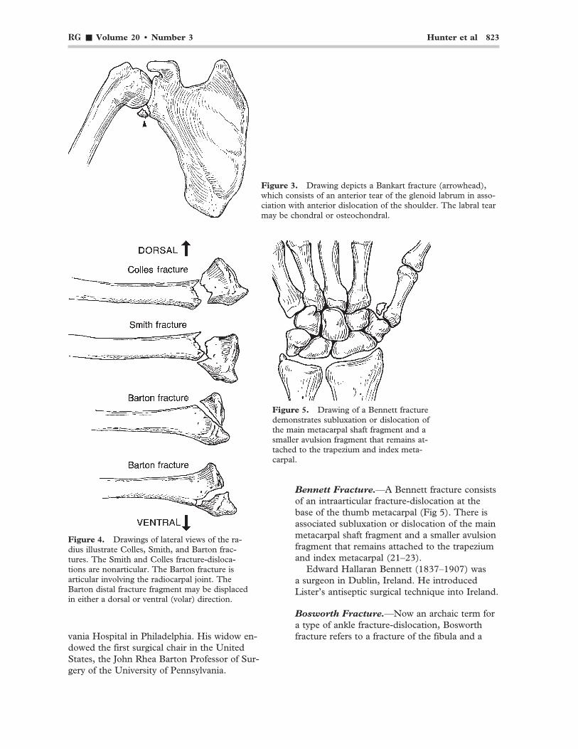
Barton Fracture.—In a Barton fracture-disloca-tion, either the dorsal or ventral (anterior or pos-terior) aspect of the radiocarpal joint is fractured(19,20). The fracture involves the articular sur-face of the radius (Fig 4). (See also Colles Frac-ture and Smith Fracture.)
John Rhea Barton (1794–1871) carried outthe first hip arthroplasty in 1826 at the Pennsyl-
Jose Luis Bado (1903–1977) was a distinguishedorthopedic surgeon from Montevideo, Uruguay.His works have been widely published in LatinAmerica, North America, and Europe. He foundedand headed for many years the Instituto deOrtopedia y Traumatologia in Montevideo.
Bankart Fracture.—A Bankart fracture refersto an anterior tear of the glenoid labrum that isassociated with anterior dislocation of the shoul-der (Fig 3) (17,18).
a. b.
c. d.Figure 2. Drawings illustrate Bado’s classification of Monteggia fractures: type I (a), typeII (b), type III (c), and type IV (d). A type I Bado fracture represents the fracture-disloca-tion originally described by Monteggia, a fracture of the proximal one-third of the ulna withanterior dislocation of the radial head.
RG ■ Volume 20 • Number 3 Hunter et al 823
Figure 3. Drawing depicts a Bankart fracture (arrowhead),which consists of an anterior tear of the glenoid labrum in asso-ciation with anterior dislocation of the shoulder. The labral tearmay be chondral or osteochondral.
Figure 4. Drawings of lateral views of the ra-dius illustrate Colles, Smith, and Barton frac-tures. The Smith and Colles fracture-disloca-tions are nonarticular. The Barton fracture isarticular involving the radiocarpal joint. TheBarton distal fracture fragment may be displacedin either a dorsal or ventral (volar) direction.
Figure 5. Drawing of a Bennett fracturedemonstrates subluxation or dislocation ofthe main metacarpal shaft fragment and asmaller avulsion fragment that remains at-tached to the trapezium and index meta-carpal.
Bennett Fracture.—A Bennett fracture consistsof an intraarticular fracture-dislocation at thebase of the thumb metacarpal (Fig 5). There isassociated subluxation or dislocation of the mainmetacarpal shaft fragment and a smaller avulsionfragment that remains attached to the trapeziumand index metacarpal (21–23).
Edward Hallaran Bennett (1837–1907) wasa surgeon in Dublin, Ireland. He introducedLister’s antiseptic surgical technique into Ireland.
Bosworth Fracture.—Now an archaic term fora type of ankle fracture-dislocation, Bosworthfracture refers to a fracture of the fibula and avania Hospital in Philadelphia. His widow en-
dowed the first surgical chair in the UnitedStates, the John Rhea Barton Professor of Sur-gery of the University of Pennsylvania.
824 May-June 2000 RG ■ Volume 20 • Number 3
posterior dislocation of the talus. This type offracture is frequently irreducible by closed meth-ods (24).
David M. Bosworth (1897–1979) was an im-portant and innovative orthopedic surgeon inNew York. His most important contribution wasthe introduction of streptomycin for the treat-ment of bone and joint tuberculosis (25).
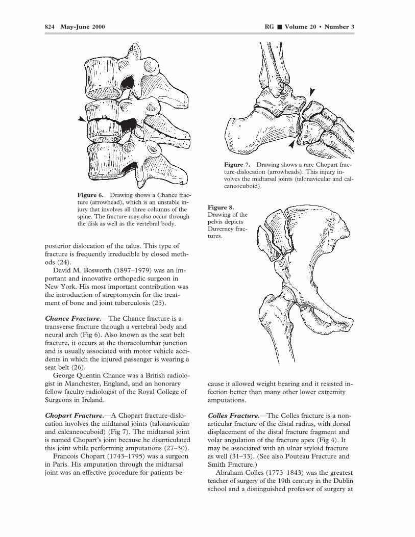
Chance Fracture.—The Chance fracture is atransverse fracture through a vertebral body andneural arch (Fig 6). Also known as the seat beltfracture, it occurs at the thoracolumbar junctionand is usually associated with motor vehicle acci-dents in which the injured passenger is wearing aseat belt (26).
George Quentin Chance was a British radiolo-gist in Manchester, England, and an honoraryfellow faculty radiologist of the Royal College ofSurgeons in Ireland.
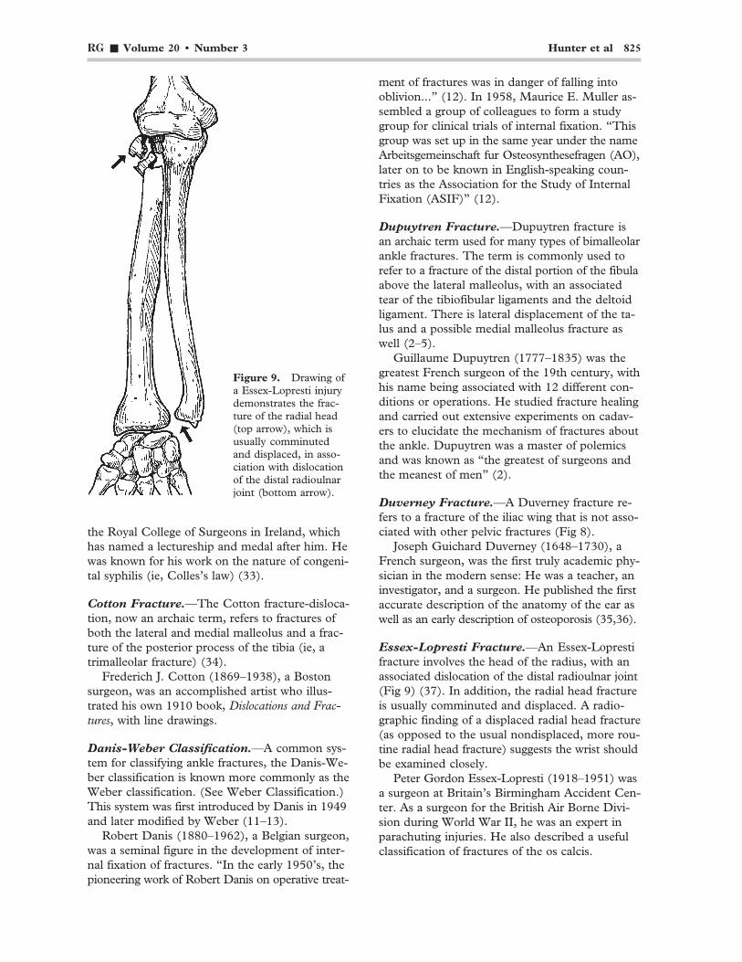
Chopart Fracture.—A Chopart fracture-dislo-cation involves the midtarsal joints (talonavicularand calcaneocuboid) (Fig 7). The midtarsal jointis named Chopart’s joint because he disarticulatedthis joint while performing amputations (27–30).
Francois Chopart (1743–1795) was a surgeonin Paris. His amputation through the midtarsaljoint was an effective procedure for patients be-
cause it allowed weight bearing and it resisted in-fection better than many other lower extremityamputations.
Colles Fracture.—The Colles fracture is a non-articular fracture of the distal radius, with dorsaldisplacement of the distal fracture fragment andvolar angulation of the fracture apex (Fig 4). Itmay be associated with an ulnar styloid fractureas well (31–33). (See also Pouteau Fracture andSmith Fracture.)
Abraham Colles (1773–1843) was the greatestteacher of surgery of the 19th century in the Dublinschool and a distinguished professor of surgery at
Figure 7. Drawing shows a rare Chopart frac-ture-dislocation (arrowheads). This injury in-volves the midtarsal joints (talonavicular and cal-caneocuboid).
Figure 6. Drawing shows a Chance frac-ture (arrowhead), which is an unstable in-jury that involves all three columns of thespine. The fracture may also occur throughthe disk as well as the vertebral body.
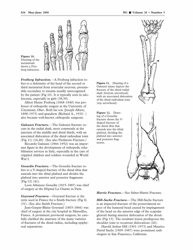
Figure 8.Drawing of thepelvis depictsDuverney frac-tures.
RG ■ Volume 20 • Number 3 Hunter et al 825
the Royal College of Surgeons in Ireland, whichhas named a lectureship and medal after him. Hewas known for his work on the nature of congeni-tal syphilis (ie, Colles’s law) (33).
Cotton Fracture.—The Cotton fracture-disloca-tion, now an archaic term, refers to fractures ofboth the lateral and medial malleolus and a frac-ture of the posterior process of the tibia (ie, atrimalleolar fracture) (34).
Frederich J. Cotton (1869–1938), a Bostonsurgeon, was an accomplished artist who illus-trated his own 1910 book, Dislocations and Frac-tures, with line drawings.
Danis-Weber Classification.—A common sys-tem for classifying ankle fractures, the Danis-We-ber classification is known more commonly as theWeber classification. (See Weber Classification.)This system was first introduced by Danis in 1949and later modified by Weber (11–13).
Robert Danis (1880–1962), a Belgian surgeon,was a seminal figure in the development of inter-nal fixation of fractures. “In the early 1950’s, thepioneering work of Robert Danis on operative treat-
ment of fractures was in danger of falling intooblivion...” (12). In 1958, Maurice E. Muller as-sembled a group of colleagues to form a studygroup for clinical trials of internal fixation. “Thisgroup was set up in the same year under the nameArbeitsgemeinschaft fur Osteosynthesefragen (AO),later on to be known in English-speaking coun-tries as the Association for the Study of InternalFixation (ASIF)” (12).
Dupuytren Fracture.—Dupuytren fracture isan archaic term used for many types of bimalleolarankle fractures. The term is commonly used torefer to a fracture of the distal portion of the fibulaabove the lateral malleolus, with an associatedtear of the tibiofibular ligaments and the deltoidligament. There is lateral displacement of the ta-lus and a possible medial malleolus fracture aswell (2–5).
Guillaume Dupuytren (1777–1835) was thegreatest French surgeon of the 19th century, withhis name being associated with 12 different con-ditions or operations. He studied fracture healingand carried out extensive experiments on cadav-ers to elucidate the mechanism of fractures aboutthe ankle. Dupuytren was a master of polemicsand was known as “the greatest of surgeons andthe meanest of men” (2).
Duverney Fracture.—A Duverney fracture re-fers to a fracture of the iliac wing that is not asso-ciated with other pelvic fractures (Fig 8).
Joseph Guichard Duverney (1648–1730), aFrench surgeon, was the first truly academic phy-sician in the modern sense: He was a teacher, aninvestigator, and a surgeon. He published the firstaccurate description of the anatomy of the ear aswell as an early description of osteoporosis (35,36).
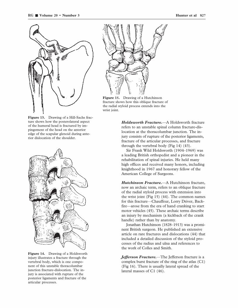
Essex-Lopresti Fracture.—An Essex-Loprestifracture involves the head of the radius, with anassociated dislocation of the distal radioulnar joint(Fig 9) (37). In addition, the radial head fractureis usually comminuted and displaced. A radio-graphic finding of a displaced radial head fracture(as opposed to the usual nondisplaced, more rou-tine radial head fracture) suggests the wrist shouldbe examined closely.
Peter Gordon Essex-Lopresti (1918–1951) wasa surgeon at Britain’s Birmingham Accident Cen-ter. As a surgeon for the British Air Borne Divi-sion during World War II, he was an expert inparachuting injuries. He also described a usefulclassification of fractures of the os calcis.
Figure 9. Drawing ofa Essex-Lopresti injurydemonstrates the frac-ture of the radial head(top arrow), which isusually comminutedand displaced, in asso-ciation with dislocationof the distal radioulnarjoint (bottom arrow).
826 May-June 2000 RG ■ Volume 20 • Number 3
Freiberg Infraction.—A Freiberg infraction re-fers to a deformity of the head of the second orthird metatarsal from avascular necrosis, presum-ably secondary to trauma usually unrecognizedby the patient (Fig 10). It is typically seen in ado-lescents, especially in girls (38,39).
Albert Henry Freiberg (1868–1940) was pro-fessor of orthopedic surgery at the University ofCincinnati, Ohio. Both his son (Joseph Albert,1898–1973) and grandson (Richard A., 1932– )also became well-known orthopedic surgeons.
Galeazzi Fracture.—The Galeazzi fracture oc-curs in the radial shaft, most commonly at thejunction of the middle and distal thirds, with anassociated dislocation of the distal radioulnar joint(Fig 11) (16,40). (See also Piedmont Fracture.)
Riccardo Galeazzi (1866–1952) was an impor-tant figure in the development of orthopedic reha-bilitation services in Italy, especially in the care ofcrippled children and soldiers wounded in WorldWar I.
Gosselin Fracture.—The Gosselin fracture re-fers to a V-shaped fracture of the distal tibia thatextends into the tibial plafond and divides theplafond into anterior and posterior fragments(Fig 12) (41).
Leon Athanese Gosselin (1815–1887) was chiefof surgery at the Hôpital La Charite in Paris.
Goyrand Fracture.—Goyrand fracture is theterm used in France for a Smith fracture (Fig 4)(31). (See also Smith Fracture.)
Jean-Gaspar-Blaise Goyrand (1803–1866) waschief of surgery at the city hospital in Marseilles,France. A prominent provincial surgeon, he care-fully clarified the anatomy of the many varietiesof fractures of the distal radius, including epiphy-seal separations.
Harris Fracture.—See Salter-Harris Fracture.
Hill-Sachs Fracture.—The Hill-Sachs fractureis an impacted fracture of the posterolateral as-pect of the humeral head caused by impingementof the head on the anterior edge of the scapularglenoid during anterior dislocation of the shoul-der (Fig 13). The resultant lesion predisposes theshoulder joint to recurrent dislocations (42).
Harold Arthur Hill (1901–1973) and MauriceDavid Sachs (1909–1987) were prominent radi-ologists in San Francisco, California.
Figure 11. Drawing of aGaleazzi injury depicts thefracture of the distal radialshaft (bottom arrowhead)with an associated dislocationof the distal radioulnar joint(top arrowhead).
Figure 12. Draw-ing of a Gosselinfracture shows the V-shaped fracture ofthe distal tibia thatextends into the tibialplafond, dividing theplafond into anteriorand posterior frag-ments.
Figure 10.Drawing of themetatarsalsshows a Frei-berg infraction.
RG ■ Volume 20 • Number 3 Hunter et al 827
Figure 13. Drawing of a Hill-Sachs frac-ture shows how the posterolateral aspectof the humeral head is fractured by im-pingement of the head on the anterioredge of the scapular glenoid during ante-rior dislocation of the shoulder.
Holdsworth Fracture.—A Holdsworth fracturerefers to an unstable spinal column fracture-dis-location at the thoracolumbar junction. The in-jury consists of rupture of the posterior ligaments,fracture of the articular processes, and fracturethrough the vertebral body (Fig 14) (43).
Sir Frank Wild Holdsworth (1904–1969) wasa leading British orthopedist and a pioneer in therehabilitation of spinal injuries. He held manyhigh offices and received many honors, includingknighthood in 1967 and honorary fellow of theAmerican College of Surgeons.
Hutchinson Fracture.—A Hutchinson fracture,now an archaic term, refers to an oblique fractureof the radial styloid process with extension intothe wrist joint (Fig 15) (44). The common namesfor this fracture—Chauffeur, Lorry Driver, Back-fire—arose from the era of hand cranking to startmotor vehicles (45). These archaic terms describean injury by mechanism (a kickback of the crankhandle) rather than by anatomy.
Jonathan Hutchinson (1828–1913) was a promi-nent British surgeon. He published an extensivearticle on rare fractures and dislocations (44) thatincluded a detailed discussion of the styloid pro-cesses of the radius and ulna and references tothe work of Colles and Smith.
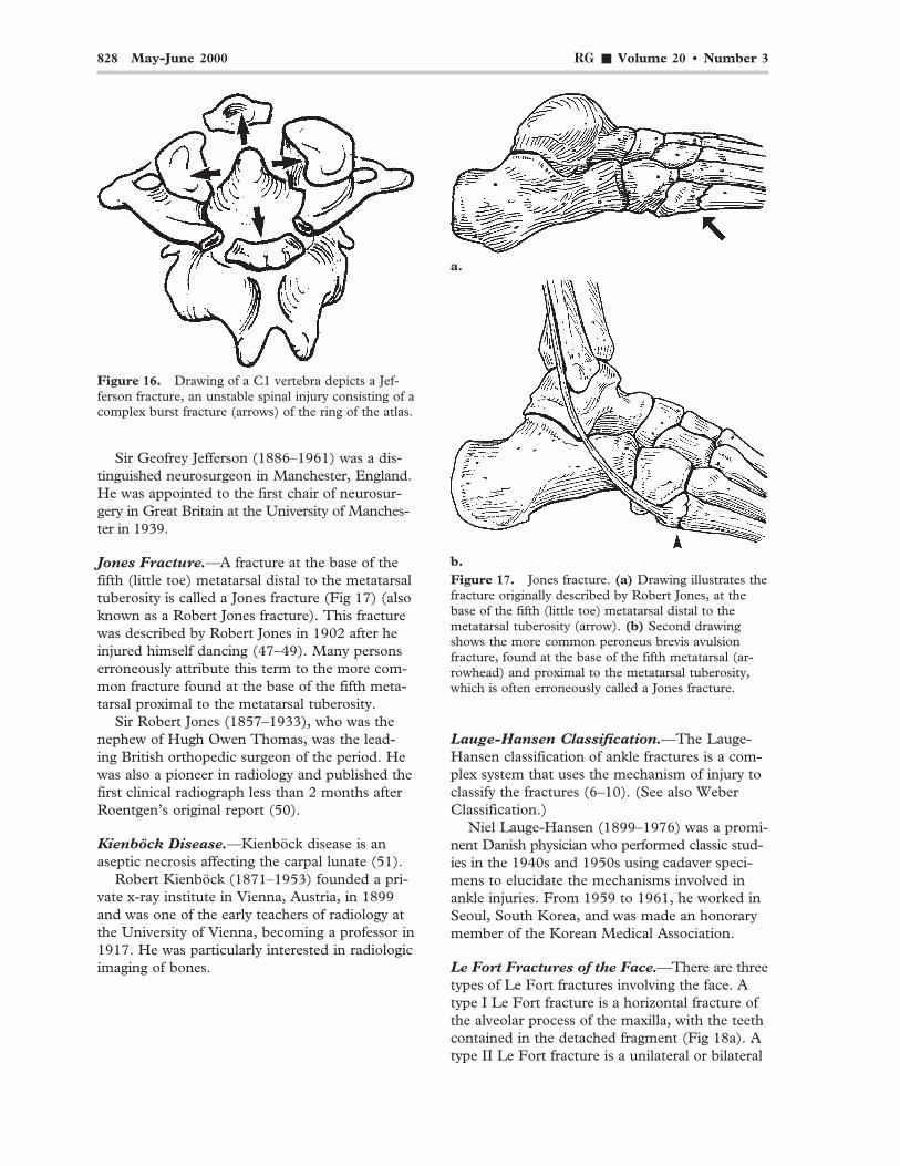
Jefferson Fracture.—The Jefferson fracture is acomplex burst fracture of the ring of the atlas (C1)(Fig 16). There is usually lateral spread of thelateral masses of C1 (46).
Figure 14. Drawing of a Holdsworthinjury illustrates a fracture through thevertebral body, which is one compo-nent of this unstable thoracolumbarjunction fracture-dislocation. The in-jury is associated with rupture of theposterior ligaments and fracture of thearticular processes.
Figure 15. Drawing of a Hutchinsonfracture shows how this oblique fracture ofthe radial styloid process extends into thewrist joint.
828 May-June 2000 RG ■ Volume 20 • Number 3
Sir Geofrey Jefferson (1886–1961) was a dis-tinguished neurosurgeon in Manchester, England.He was appointed to the first chair of neurosur-gery in Great Britain at the University of Manches-ter in 1939.
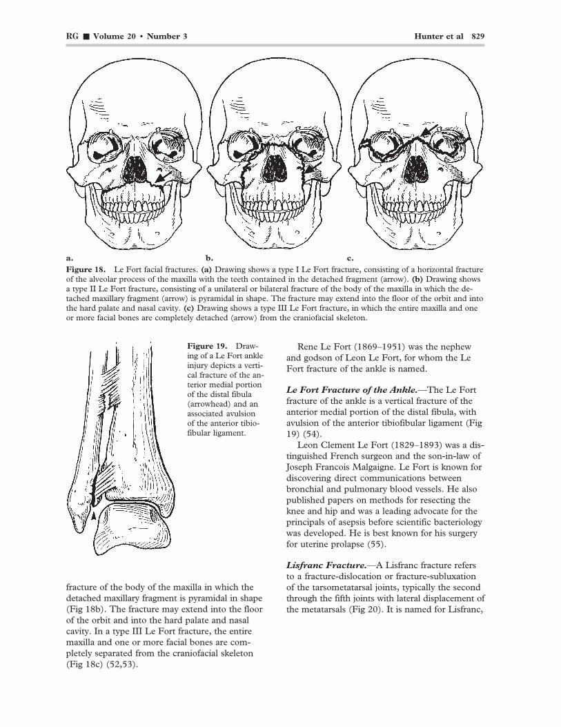
Jones Fracture.—A fracture at the base of thefifth (little toe) metatarsal distal to the metatarsaltuberosity is called a Jones fracture (Fig 17) (alsoknown as a Robert Jones fracture). This fracturewas described by Robert Jones in 1902 after heinjured himself dancing (47–49). Many personserroneously attribute this term to the more com-mon fracture found at the base of the fifth meta-tarsal proximal to the metatarsal tuberosity.
Sir Robert Jones (1857–1933), who was thenephew of Hugh Owen Thomas, was the lead-ing British orthopedic surgeon of the period. Hewas also a pioneer in radiology and published thefirst clinical radiograph less than 2 months afterRoentgen’s original report (50).
Kienböck Disease.—Kienböck disease is anaseptic necrosis affecting the carpal lunate (51).
Robert Kienböck (1871–1953) founded a pri-vate x-ray institute in Vienna, Austria, in 1899and was one of the early teachers of radiology atthe University of Vienna, becoming a professor in1917. He was particularly interested in radiologicimaging of bones.
Lauge-Hansen Classification.—The Lauge-Hansen classification of ankle fractures is a com-plex system that uses the mechanism of injury toclassify the fractures (6–10). (See also WeberClassification.)
Niel Lauge-Hansen (1899–1976) was a promi-nent Danish physician who performed classic stud-ies in the 1940s and 1950s using cadaver speci-mens to elucidate the mechanisms involved inankle injuries. From 1959 to 1961, he worked inSeoul, South Korea, and was made an honorarymember of the Korean Medical Association.
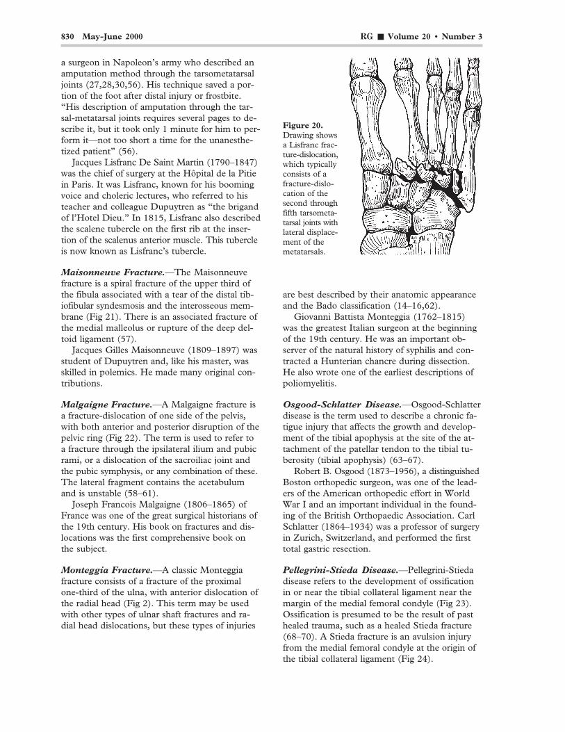
Le Fort Fractures of the Face.—There are threetypes of Le Fort fractures involving the face. Atype I Le Fort fracture is a horizontal fracture ofthe alveolar process of the maxilla, with the teethcontained in the detached fragment (Fig 18a). Atype II Le Fort fracture is a unilateral or bilateral
a.
b.Figure 17. Jones fracture. (a) Drawing illustrates thefracture originally described by Robert Jones, at thebase of the fifth (little toe) metatarsal distal to themetatarsal tuberosity (arrow). (b) Second drawingshows the more common peroneus brevis avulsionfracture, found at the base of the fifth metatarsal (ar-rowhead) and proximal to the metatarsal tuberosity,which is often erroneously called a Jones fracture.
Figure 16. Drawing of a C1 vertebra depicts a Jef-ferson fracture, an unstable spinal injury consisting of acomplex burst fracture (arrows) of the ring of the atlas.
RG ■ Volume 20 • Number 3 Hunter et al 829
fracture of the body of the maxilla in which thedetached maxillary fragment is pyramidal in shape(Fig 18b). The fracture may extend into the floorof the orbit and into the hard palate and nasalcavity. In a type III Le Fort fracture, the entiremaxilla and one or more facial bones are com-pletely separated from the craniofacial skeleton(Fig 18c) (52,53).
a. b. c.Figure 18. Le Fort facial fractures. (a) Drawing shows a type I Le Fort fracture, consisting of a horizontal fractureof the alveolar process of the maxilla with the teeth contained in the detached fragment (arrow). (b) Drawing showsa type II Le Fort fracture, consisting of a unilateral or bilateral fracture of the body of the maxilla in which the de-tached maxillary fragment (arrow) is pyramidal in shape. The fracture may extend into the floor of the orbit and intothe hard palate and nasal cavity. (c) Drawing shows a type III Le Fort fracture, in which the entire maxilla and oneor more facial bones are completely detached (arrow) from the craniofacial skeleton.
Rene Le Fort (1869–1951) was the nephewand godson of Leon Le Fort, for whom the LeFort fracture of the ankle is named.
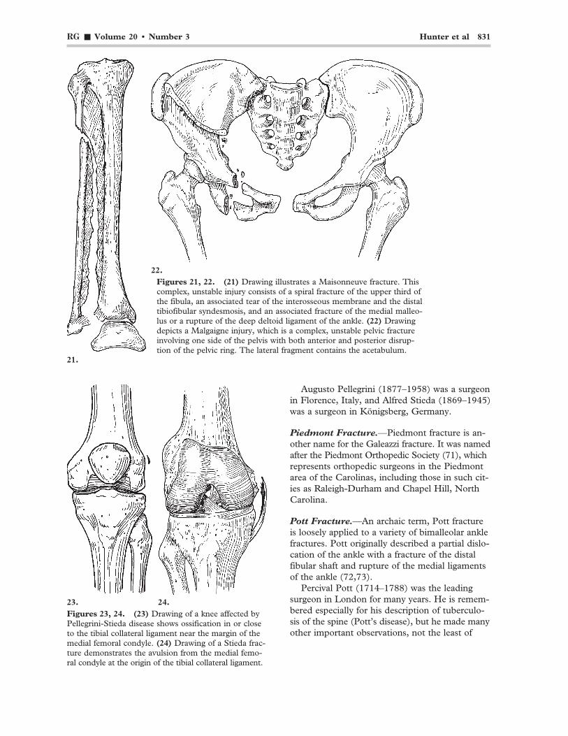
Le Fort Fracture of the Ankle.—The Le Fortfracture of the ankle is a vertical fracture of theanterior medial portion of the distal fibula, withavulsion of the anterior tibiofibular ligament (Fig19) (54).
Leon Clement Le Fort (1829–1893) was a dis-tinguished French surgeon and the son-in-law ofJoseph Francois Malgaigne. Le Fort is known fordiscovering direct communications betweenbronchial and pulmonary blood vessels. He alsopublished papers on methods for resecting theknee and hip and was a leading advocate for theprincipals of asepsis before scientific bacteriologywas developed. He is best known for his surgeryfor uterine prolapse (55).
Lisfranc Fracture.—A Lisfranc fracture refersto a fracture-dislocation or fracture-subluxationof the tarsometatarsal joints, typically the secondthrough the fifth joints with lateral displacement ofthe metatarsals (Fig 20). It is named for Lisfranc,
Figure 19. Draw-ing of a Le Fort ankleinjury depicts a verti-cal fracture of the an-terior medial portionof the distal fibula(arrowhead) and anassociated avulsionof the anterior tibio-fibular ligament.
830 May-June 2000 RG ■ Volume 20 • Number 3
a surgeon in Napoleon’s army who described anamputation method through the tarsometatarsaljoints (27,28,30,56). His technique saved a por-tion of the foot after distal injury or frostbite.“His description of amputation through the tar-sal-metatarsal joints requires several pages to de-scribe it, but it took only 1 minute for him to per-form it—not too short a time for the unanesthe-tized patient” (56).
Jacques Lisfranc De Saint Martin (1790–1847)was the chief of surgery at the Hôpital de la Pitiein Paris. It was Lisfranc, known for his boomingvoice and choleric lectures, who referred to histeacher and colleague Dupuytren as “the brigandof l’Hotel Dieu.” In 1815, Lisfranc also describedthe scalene tubercle on the first rib at the inser-tion of the scalenus anterior muscle. This tubercleis now known as Lisfranc’s tubercle.
Maisonneuve Fracture.—The Maisonneuvefracture is a spiral fracture of the upper third ofthe fibula associated with a tear of the distal tib-iofibular syndesmosis and the interosseous mem-brane (Fig 21). There is an associated fracture ofthe medial malleolus or rupture of the deep del-toid ligament (57).
Jacques Gilles Maisonneuve (1809–1897) wasstudent of Dupuytren and, like his master, wasskilled in polemics. He made many original con-tributions.
Malgaigne Fracture.—A Malgaigne fracture isa fracture-dislocation of one side of the pelvis,with both anterior and posterior disruption of thepelvic ring (Fig 22). The term is used to refer toa fracture through the ipsilateral ilium and pubicrami, or a dislocation of the sacroiliac joint andthe pubic symphysis, or any combination of these.The lateral fragment contains the acetabulumand is unstable (58–61).
Joseph Francois Malgaigne (1806–1865) ofFrance was one of the great surgical historians ofthe 19th century. His book on fractures and dis-locations was the first comprehensive book onthe subject.
Monteggia Fracture.—A classic Monteggiafracture consists of a fracture of the proximalone-third of the ulna, with anterior dislocation ofthe radial head (Fig 2). This term may be usedwith other types of ulnar shaft fractures and ra-dial head dislocations, but these types of injuries
are best described by their anatomic appearanceand the Bado classification (14–16,62).
Giovanni Battista Monteggia (1762–1815)was the greatest Italian surgeon at the beginningof the 19th century. He was an important ob-server of the natural history of syphilis and con-tracted a Hunterian chancre during dissection.He also wrote one of the earliest descriptions ofpoliomyelitis.
Osgood-Schlatter Disease.—Osgood-Schlatterdisease is the term used to describe a chronic fa-tigue injury that affects the growth and develop-ment of the tibial apophysis at the site of the at-tachment of the patellar tendon to the tibial tu-berosity (tibial apophysis) (63–67).
Robert B. Osgood (1873–1956), a distinguishedBoston orthopedic surgeon, was one of the lead-ers of the American orthopedic effort in WorldWar I and an important individual in the found-ing of the British Orthopaedic Association. CarlSchlatter (1864–1934) was a professor of surgeryin Zurich, Switzerland, and performed the firsttotal gastric resection.
Pellegrini-Stieda Disease.—Pellegrini-Stiedadisease refers to the development of ossificationin or near the tibial collateral ligament near themargin of the medial femoral condyle (Fig 23).Ossification is presumed to be the result of pasthealed trauma, such as a healed Stieda fracture(68–70). A Stieda fracture is an avulsion injuryfrom the medial femoral condyle at the origin ofthe tibial collateral ligament (Fig 24).
Figure 20.Drawing showsa Lisfranc frac-ture-dislocation,which typicallyconsists of afracture-dislo-cation of thesecond throughfifth tarsometa-tarsal joints withlateral displace-ment of themetatarsals.
RG ■ Volume 20 • Number 3 Hunter et al 831
22.Figures 21, 22. (21) Drawing illustrates a Maisonneuve fracture. Thiscomplex, unstable injury consists of a spiral fracture of the upper third ofthe fibula, an associated tear of the interosseous membrane and the distaltibiofibular syndesmosis, and an associated fracture of the medial malleo-lus or a rupture of the deep deltoid ligament of the ankle. (22) Drawingdepicts a Malgaigne injury, which is a complex, unstable pelvic fractureinvolving one side of the pelvis with both anterior and posterior disrup-tion of the pelvic ring. The lateral fragment contains the acetabulum.
21.
23. 24.Figures 23, 24. (23) Drawing of a knee affected byPellegrini-Stieda disease shows ossification in or closeto the tibial collateral ligament near the margin of themedial femoral condyle. (24) Drawing of a Stieda frac-ture demonstrates the avulsion from the medial femo-ral condyle at the origin of the tibial collateral ligament.
Augusto Pellegrini (1877–1958) was a surgeonin Florence, Italy, and Alfred Stieda (1869–1945)was a surgeon in Königsberg, Germany.
Piedmont Fracture.—Piedmont fracture is an-other name for the Galeazzi fracture. It was namedafter the Piedmont Orthopedic Society (71), whichrepresents orthopedic surgeons in the Piedmontarea of the Carolinas, including those in such cit-ies as Raleigh-Durham and Chapel Hill, NorthCarolina.
Pott Fracture.—An archaic term, Pott fractureis loosely applied to a variety of bimalleolar anklefractures. Pott originally described a partial dislo-cation of the ankle with a fracture of the distalfibular shaft and rupture of the medial ligamentsof the ankle (72,73).
Percival Pott (1714–1788) was the leadingsurgeon in London for many years. He is remem-bered especially for his description of tuberculo-sis of the spine (Pott’s disease), but he made manyother important observations, not the least of
832 May-June 2000 RG ■ Volume 20 • Number 3
which was his description of cancer of the scro-tum in chimney sweeps. His report was one ofthe first to associate coal tar exposure with devel-opment of a cancer.
Pouteau Fracture.—Pouteau fracture is the nameused in France for a Colles fracture (Fig 4) (74).(See also Colles Fracture.)
Claude Pouteau (1725–1775), a surgeon atthe city hospital in Lyon, France, was an earlyadvocate of cleanliness in surgery, evidenced byhis insistence on hand washing and use of dispos-able paper dressings. He was a premier lithoto-mist (one who removed bladder calculi throughan incision in the perineum while the unanesthe-tized patient was in the “lithotomy” position),achieving a mortality rate of only 3%.
Robert Jones Fracture.—See Jones Fracture.
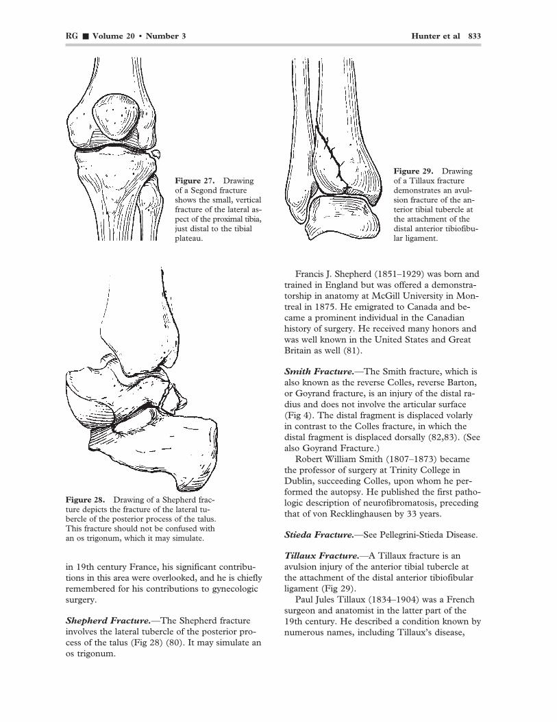
Rolando Fracture.—A Rolando fracture is acomminuted Y- or T-shaped fracture-dislocationat the base of the thumb metacarpal (Fig 25). Thedislocation accompanies the disruption of the ar-ticular surface of the thumb metacarpal (75,76).
Silvio Rolando (?–1941?) was a prominent sur-geon in Genoa, Italy. He practiced in the firstpart of this century and specialized in genitouri-nary tract surgery.
Salter-Harris Classification.—Salter-Harrisclassification is the most commonly used systemfor categorizing growth plate injuries (Fig 26) (77).
Robert Bruce Salter (1924– ), a prominentCanadian surgeon, is now semiretired as a “uni-versity professor” and professor emeritus of or-thopaedic surgery and senior scientist emeritus,University of Toronto. Earlier he was chief of or-
thopedic surgery and later surgeon in chief at theHospital for Sick Children, Toronto, Universityof Toronto. W. Robert Harris (1922– ) is a promi-nent Canadian orthopedic surgeon who is nowretired. He is a professor emeritus of surgery atthe University of Toronto and a fellow of theRoyal College of Physicians and Surgeons.
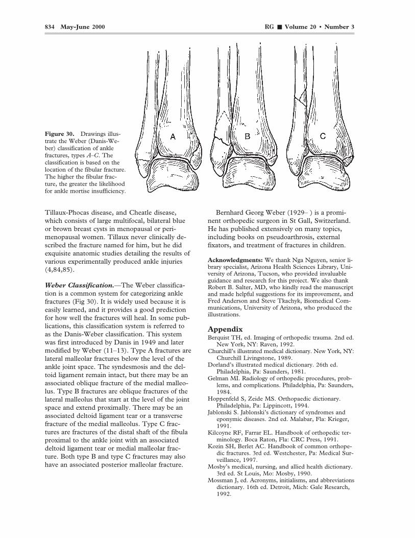
Segond Fracture.—A Segond fracture is a small,vertical fracture of the lateral aspect of the proxi-mal tibia just distal to the tibial plateau (Fig 27).It has a high association with anterior cruciateligament and meniscal injuries (78,79).
Paul Ferdinand Segond (1851–1912) was pro-fessor of surgery at the University of Paris andsurgeon in chief at the Saltpetriere. AlthoughSegond was one of the foremost “knee specialists”
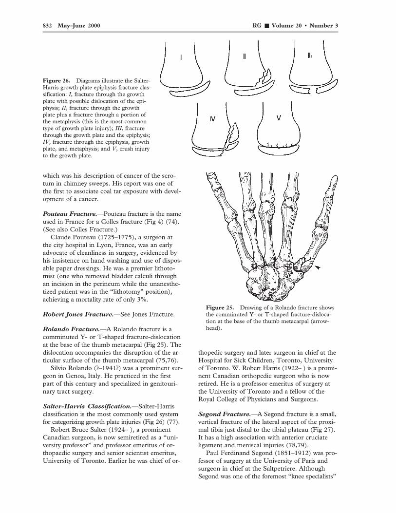
Figure 26. Diagrams illustrate the Salter-Harris growth plate epiphysis fracture clas-sification: I, fracture through the growthplate with possible dislocation of the epi-physis; II, fracture through the growthplate plus a fracture through a portion ofthe metaphysis (this is the most commontype of growth plate injury); III, fracturethrough the growth plate and the epiphysis;IV, fracture through the epiphysis, growthplate, and metaphysis; and V, crush injuryto the growth plate.
Figure 25. Drawing of a Rolando fracture showsthe comminuted Y- or T-shaped fracture-disloca-tion at the base of the thumb metacarpal (arrow-head).
RG ■ Volume 20 • Number 3 Hunter et al 833
in 19th century France, his significant contribu-tions in this area were overlooked, and he is chieflyremembered for his contributions to gynecologicsurgery.
Shepherd Fracture.—The Shepherd fractureinvolves the lateral tubercle of the posterior pro-cess of the talus (Fig 28) (80). It may simulate anos trigonum.
Francis J. Shepherd (1851–1929) was born andtrained in England but was offered a demonstra-torship in anatomy at McGill University in Mon-treal in 1875. He emigrated to Canada and be-came a prominent individual in the Canadianhistory of surgery. He received many honors andwas well known in the United States and GreatBritain as well (81).
Smith Fracture.—The Smith fracture, which isalso known as the reverse Colles, reverse Barton,or Goyrand fracture, is an injury of the distal ra-dius and does not involve the articular surface(Fig 4). The distal fragment is displaced volarlyin contrast to the Colles fracture, in which thedistal fragment is displaced dorsally (82,83). (Seealso Goyrand Fracture.)
Robert William Smith (1807–1873) becamethe professor of surgery at Trinity College inDublin, succeeding Colles, upon whom he per-formed the autopsy. He published the first patho-logic description of neurofibromatosis, precedingthat of von Recklinghausen by 33 years.
Stieda Fracture.—See Pellegrini-Stieda Disease.
Tillaux Fracture.—A Tillaux fracture is anavulsion injury of the anterior tibial tubercle atthe attachment of the distal anterior tibiofibularligament (Fig 29).
Paul Jules Tillaux (1834–1904) was a Frenchsurgeon and anatomist in the latter part of the19th century. He described a condition known bynumerous names, including Tillaux’s disease,
Figure 29. Drawingof a Tillaux fracturedemonstrates an avul-sion fracture of the an-terior tibial tubercle atthe attachment of thedistal anterior tibiofibu-lar ligament.
Figure 27. Drawingof a Segond fractureshows the small, verticalfracture of the lateral as-pect of the proximal tibia,just distal to the tibialplateau.
Figure 28. Drawing of a Shepherd frac-ture depicts the fracture of the lateral tu-bercle of the posterior process of the talus.This fracture should not be confused withan os trigonum, which it may simulate.
834 May-June 2000 RG ■ Volume 20 • Number 3
Tillaux-Phocas disease, and Cheatle disease,which consists of large multifocal, bilateral blueor brown breast cysts in menopausal or peri-menopausal women. Tillaux never clinically de-scribed the fracture named for him, but he didexquisite anatomic studies detailing the results ofvarious experimentally produced ankle injuries(4,84,85).
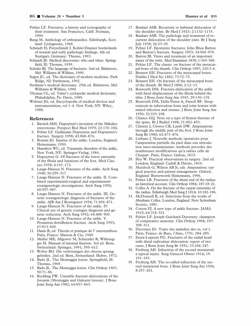
Weber Classification.—The Weber classifica-tion is a common system for categorizing anklefractures (Fig 30). It is widely used because it iseasily learned, and it provides a good predictionfor how well the fractures will heal. In some pub-lications, this classification system is referred toas the Danis-Weber classification. This systemwas first introduced by Danis in 1949 and latermodified by Weber (11–13). Type A fractures arelateral malleolar fractures below the level of theankle joint space. The syndesmosis and the del-toid ligament remain intact, but there may be anassociated oblique fracture of the medial malleo-lus. Type B fractures are oblique fractures of thelateral malleolus that start at the level of the jointspace and extend proximally. There may be anassociated deltoid ligament tear or a transversefracture of the medial malleolus. Type C frac-tures are fractures of the distal shaft of the fibulaproximal to the ankle joint with an associateddeltoid ligament tear or medial malleolar frac-ture. Both type B and type C fractures may alsohave an associated posterior malleolar fracture.
Bernhard Georg Weber (1929– ) is a promi-nent orthopedic surgeon in St Gall, Switzerland.He has published extensively on many topics,including books on pseudoarthrosis, externalfixators, and treatment of fractures in children.
Acknowledgments: We thank Nga Nguyen, senior li-brary specialist, Arizona Health Sciences Library, Uni-versity of Arizona, Tucson, who provided invaluableguidance and research for this project. We also thankRobert B. Salter, MD, who kindly read the manuscriptand made helpful suggestions for its improvement, andFred Anderson and Steve Tkachyk, Biomedical Com-munications, University of Arizona, who produced theillustrations.
AppendixBerquist TH, ed. Imaging of orthopedic trauma. 2nd ed.
New York, NY: Raven, 1992.Churchill’s illustrated medical dictionary. New York, NY:
Churchill Livingstone, 1989.Dorland’s illustrated medical dictionary. 26th ed.
Philadelphia, Pa: Saunders, 1981.Gelman MI. Radiology of orthopedic procedures, prob-
lems, and complications. Philadelphia, Pa: Saunders,1984.
Hoppenfeld S, Zeide MS. Orthopaedic dictionary.Philadelphia, Pa: Lippincott, 1994.
Jablonski S. Jablonski’s dictionary of syndromes andeponymic diseases. 2nd ed. Malabar, Fla: Krieger,1991.
Kilcoyne RF, Farrar EL. Handbook of orthopedic ter-minology. Boca Raton, Fla: CRC Press, 1991.
Kozin SH, Berlet AC. Handbook of common orthope-dic fractures. 3rd ed. Westchester, Pa: Medical Sur-veillance, 1997.
Mosby’s medical, nursing, and allied health dictionary.3rd ed. St Louis, Mo: Mosby, 1990.
Mossman J, ed. Acronyms, initialisms, and abbreviationsdictionary. 16th ed. Detroit, Mich: Gale Research,1992.
Figure 30. Drawings illus-trate the Weber (Danis-We-ber) classification of anklefractures, types A–C. Theclassification is based on thelocation of the fibular fracture.The higher the fibular frac-ture, the greater the likelihoodfor ankle mortise insufficiency.
RG ■ Volume 20 • Number 3 Hunter et al 835
Peltier LF. Fractures: a history and iconography oftheir treatment. San Francisco, Calif: Norman,1990.
Rang M. Anthology of orthopaedics. Edinburgh, Scot-land: Livingstone, 1968.
Schmidt H, Freyschmidt J. Kohler/Zimmer borderlandsof normal and early pathologic findings. 4th ed.Stuttgart, Germany: Thieme, 1993.
Schmidt JE. Medical discoveries: who and when. Spring-field, Ill: Thomas, 1959.
Schultz RJ. The language of fractures. 2nd ed. Baltimore,Md: Williams & Wilkins, 1990.
Segen JC, ed. The dictionary of modern medicine. ParkRidge, NJ: Parthenon, 1992.
Stedman’s medical dictionary. 25th ed. Baltimore, Md:Williams & Wilkins, 1990.
Thomas CL, ed. Taber’s cyclopedic medical dictionary.Philadelphia, Pa: Davis, 1989.
Webster JG, ed. Encyclopedia of medical devices andinstrumentation, vol 1–4. New York, NY: Wiley,1988.
References1. Ravitch MM. Dupuytren’s invention of the Mikulicz
enterotome. Perspect Biol Med 1979; 22:170–184.2. Peltier LF. Guillaume Dupuytren and Dupuytren’s
fracture. Surgery 1958; 43:868–874.3. Bonnin JG. Injuries of the ankle. London, England:
Heinemann, 1950.4. Hamilton WC, ed. Traumatic disorders of the ankle,
New York, NY: Springer-Verlag, 1984.5. Dupuytren G. Of fractures of the lower extremity
of the fibula and luxations of the foot. Med Clas-sics 1939; 4:151–172.
6. Lauge-Hansen N. Fractures of the ankle. Arch Surg1948; 56:259–317.
7. Lauge-Hansen N. Fractures of the ankle. II. Com-bined experimental-surgical and experimental-roentgenologic investigations. Arch Surg 1950;60:957–987.
8. Lauge-Hansen N. Fractures of the ankle. III. Ge-netic roentgenologic diagnosis of fractures of theankle. AJR Am J Roentgenol 1954; 71:456–471.
9. Lauge-Hansen N. Fractures of the ankle. IV.Clinical use of genetic roentgen diagnosis and ge-netic reduction. Arch Surg 1952; 64:488–500.
10. Lauge-Hansen N. Fractures of the ankle. V.Pronation-dorsiflexion fracture. Arch Surg 1953;67:813–820.
11. Danis R, ed. Theorie et pratique de l’ osteosynthese.Paris, France: Masson & Cie, 1949.
12. Muller ME, Allgower M, Schneider R, Willeneg-ger H. Manual of internal fixation. 3rd ed. Bern,Switzerland: Springer, 1991; 595–612.
13. Weber BG. Die verletzungen des oberen sprung-gelenkes. 2nd ed. Bern, Switzerland: Huber, 1972.
14. Bado JL. The Monteggia lesion. Springfield, Ill:Thomas, 1969.
15. Bado JL. The Monteggia lesion. Clin Orthop 1967;50:71–86.
16. Reckling FW. Unstable fracture-dislocations of theforearm (Monteggia and Galeazzi lesions). J BoneJoint Surg Am 1982; 64:857–863.
17. Bankart ASB. Recurrent or habitual dislocation ofthe shoulder joint. Br Med J 1923; 2:1132–1133.
18. Bankart ASB. The pathology and treatment of re-current dislocation of the shoulder joint. Br J SurgJuly 1938; 26:23–29.
19. Peltier LF. Eponymic fractures: John Rhea Bartonand Barton’s fracture. Surgery 1953; 34:960–970.
20. Barton JR. Views and treatment of an importantinjury of the wrist. Med Examiner 1838; 1:365–368.
21. Peltier LF. The classic: on fracture of the metacar-pal bone of the thumb. Clin Orthop 1987; 220:3–6.
22. Bennett EH. Fractures of the metacarpal bones.Dublin J Med Sci 1882; 73:72–75.
23. Bennett EH. On fracture of the metacarpal boneof the thumb. Br Med J 1886; 2:12–13.
24. Bosworth DM. Fracture-dislocation of the anklewith fixed displacement of the fibula behind thetibia. J Bone Joint Surg Am 1947; 29:130–135.
25. Bosworth DM, Della Pietra A, Farrell BF. Strep-tomycin in tuberculous bone and joint lesions withmixed infection and sinuses. J Bone Joint Surg Am1950; 32:103–108.
26. Chance GQ. Note on a type of flexion fracture ofthe spine. Br J Radiol 1948; 21:452–453.
27. Christie J, Clowes CB, Lamb DW. Amputationsthrough the middle part of the foot. J Bone JointSurg Br 1980; 62:473–474.
28. Lisfranc J. Nouvelle methode operatoire pourl’amputation partielle du pied dans son articula-tion tarso-metatarsienne: methode precedee desnombreuses modifications qu’a subies celle deChopart. Paris, France: Gabon, 1815.
29. Hey W. Practical observations in surgery. 2nd ed.London, England: Cadell & Davies, 1810.
30. Murdoch G, Wilson AB Jr, eds. Amputation: sur-gical practice and patient management. Oxford,England: Butterworth Heinemann, 1996.
31. Peltier LR. Fractures of the distal end of the radius:an historical account. Clin Orthop 1984; 187:18–21.
32. Colles A. On the fracture of the carpal extremity ofthe radius. Edinburgh Med Surg J 1814; 10:182–186.
33. McDonnell R, ed. Selections from the works ofAbraham Colles. London, England: New SydenhamSociety, 1881.
34. Cotton FJ. A new type of ankle fracture. JAMA1915; 64:318–321.
35. Peltier LF. Joseph Guichard Duverney: championof comparative anatomy. Clin Orthop 1984; 187:308–311.
36. Duverney JG. Traite des maladies des os, vol 1.Paris, France: de Bure, l’Aine, 1751; 284–285.
37. Essex-Lopresti PG. Fractures of the radial headwith distal radioulnar dislocation: report of twocases. J Bone Joint Surg Br 1951; 33:244–247.
38. Freiberg AH. Infraction of the second metatarsal:a typical injury. Surg Gynecol Obstet 1914; 19:191–193.
39. Freiberg AH. The so-called infraction of the sec-ond metatarsal bone. J Bone Joint Surg Am 1926;8:257–261.
836 May-June 2000 RG ■ Volume 20 • Number 3
40. Reckling FW, Peltier LF. Riccardo Galeazzi andGaleazzi’s fracture. Surgery 1965; 58:453–459.
41. Gosselin LA; Stimson LA, trans. Clinical lectureson surgery. Philadelphia, Pa: Lea, 1878.
42. Hill HA, Sachs MD. The grooved defect of thehumeral head: a frequently unrecognized compli-cation of dislocations of the shoulder joint. Radiol-ogy 1940; 35:690–700.
43. Holdsworth FW. Fractures, dislocations, and frac-ture-dislocations of the spine. J Bone Joint Surg Br1963; 45:6–20.
44. Hutchinson J. Original Lectures: notes on some ofthe more rare forms of fractures and dislocations.Medical Times and Gazette, June 30, 1866; 683–684.
45. Edwards HC. The mechanism and treatment ofbackfire fracture. J Bone Joint Surg Am 1926; 4:701–717.
46. Jefferson G. Fracture of atlas vertebrae: report offour cases and a review of those previously recorded.Br J Surg 1920; 7:407–422.
47. Jones R. Fracture of the fifth metatarsal bone. Liver-pool Med Chir J 1902; 22:103–107.
48. Jones R. Fracture of the base of the fifth metatar-sal bone by indirect violence. Ann Surg 1902; 35:697–700.
49. Peltier LF. Eponymic fractures: Robert Jones andJones’s fracture. Surgery 1972; 71:522–526.
50. Jones R, Lodge O. The discovery of a bullet lost inthe wrist by means of the Roentgen rays. Lancet1896; 1(22 Feb):476–477.
51. Peltier LF. The classic: concerning traumatic ma-lacia of the lunate and its consequences—degen-eration and compression fractures. Clin Orthop1980; 149:4–8.
52. Le Fort R; Tilson HB, McFee AS, Soudah HP,trans. The maxillo-facial works of Rene Le Fort.Houston, Tex: University of Texas Dental Branch,1972.
53. Le Fort R. Etude experimentale sur les fractures dela machoire superieure. Rev Chir 1901; 23:208–227.
54. Le Fort LC. Note sur une variete non-decritede fracture verticale de la malleole externe pararrachement. Bull Gen Ther 1886; 110:193–199.
55. Speert H. The book shelf: Leon Le Fort and hisoperation for uterine prolapse. Surg GynecolObstet 1957; 104:121–124.
56. Cassebaum WH. Lisfranc fracture-dislocations.Clin Orthop 1963; 30:116–128.
57. Maisonneuve JG. Recherches sur la fracture duperone, Paris, France: Loquin & Cie, 1840.
58. Malgaigne JF. Traites des fractures et des luxations,part 1. Paris, France: Bailliere, 1847.
59. Malgaigne JF. Traites des fractures et des luxations,part 2. Paris, France: Bailliere, 1855.
60. Peltier LF. Joseph Francois Malgaigne and Mal-gaigne’s fracture. Surgery 1958; 44:777–784.
61. Bucholz RW. The pathological anatomy of Mal-gaigne fracture-dislocations of the pelvis. J BoneJoint Surg Am 1981; 63:400–404.
62. Peltier LF. Eponymic fractures: Giovanni BattistaMonteggia and Monteggia’s fracture. Surgery 1957;42:585–591.
63. Uhry E. Osgood-Schlatter disease. Arch Surg 1944;48:406–414.
64. Osgood RB. Lesions of the tibial tubercle occur-ring during adolescence. Boston Med Surg J 1903;148:114–117.
65. Schlatter C. Verletzungen der schnabelformigenfortsatzes der obseren tibia epiphyse. Beitr KlinChir 1903; 38:874–887.
66. Cole JP. A study of Osgood-Schlatter disease. SurgGynecol Obstet 1937; 65:55–67.
67. Schlatter C. Unvollstandige abrissfrakturen der tu-berositas tibiae oder wachstumsanomalien? BeitrKlin Chir 1908; 59:518–546.
68. Ritvo M, Resnik J. Pellegrini-Stieda’s disease. AmJ Roentgenol Rad Ther 1934; 32:189–195.
69. Pellegrini A. Osificazione traumatica del ligamen-tocollaterale tibiale dell’articolazione del ginocchiosinistra. Clin Moderna 1905; 11:433–439.
70. Stieda A. Uber eine typische verletzung am unterenfemurende. Arch Klin Chir 1908; 85:815–826.
71. Hughston JD. Fractures of the distal radial shaft. JBone Joint Surg Am 1957; 39:249–264.
72. Peltier LR. Percival Pott and Pott’s fracture. Sur-gery 1962; 58:280–286.
73. Pott P. Some few general remarks on fractures anddislocations. London, England: Hawes, Clarke,Collins; 1768.
74. Pouteau C. Contenant quelques reflexions surquelques fractures de l’avant-bras, sur les luxationsincomplettes du poignet & sur le diastasis. In:Oeuvres posthumes de M. Pouteau, Paris, France:Pierres, 1783.
75. Langhoff O, Andersen K, Kjaer-Petersen K. Ro-lando’s fracture. J Bone Joint Surg Br 1991; 16:454–459.
76. Rolando S. Fracture de la base du premier meta-carpien et principalement sur une variete non en-core decrite. Presse Med 1910; 33:303–304.
77. Salter RB, Harris WR. Injuries involving the epi-physeal plate. J Bone Joint Surg Am 1963; 45:587–622.
78. Paessler HH, Michel D. How new is the Lachmantest. Am J Sports Med 1992; 20:95–98.
79. Segond PF. Recherches cliniques et experimentalessur les epanchements sanguins du genou parentorse. Progres Medical 1879; 16:297–299, 320–321, 340–341, 379–381, 400–401, 419–421.
80. Shepherd FJ. A hitherto undescribed fracture ofthe astralagus. J Anat Physiol 1882; 17:79–81.
81. MacDermot HE. History of Canadian surgery:Francis J. Shepherd. Can J Surg 1957; 1:5–7.
82. Peltier LR. Eponymic fractures: Robert WilliamSmith and Smith’s fracture. Surgery 1959; 45:1035–1042.
83. Smith RW. A treatise on fractures in the vicinity ofjoints, and on certain forms of accidental and con-genital dislocations. Dublin, Ireland: Hodges &Smith, 1847.
84. Tillaux P. Traite de chirurgie clinique. Paris,France: Asselin & Houzeau, 1886–1889.
85. Tillaux P. Traite d’anatomie topographique. 6thed. Paris, France: Asselin, 1890.