original - vrf.iranjournals.ir
TRANSCRIPT
ORIGINAL ARTICLE
Veterinary Research Forum. 2020; 11 (1) 7 – 13
doi: 10.30466/vrf.2018.91253.2047
Journal Homepage: vrf.iranjournals.ir
Protective effect of ethyl pyruvate on testicular histology and fertilization potential in cyclophosphamide treated mice
Zahra Bakhtiary, Rasoul Shahrooz*, Abbas Ahmadi, Farhad Soltanalinejad
Department of Basic Sciences, Faculty of Veterinary Medicine, Urmia University, Urmia, Iran.
Article Info Abstract
Article history: Received: 20 January 2018 Accepted: 22 May 2018 Available online: 15 March 2020
In the present study, we aimed to address the use of ethyl pyruvate (EP) against the harmful effects of cyclophosphamide (CP) treatment. Thirty-nine adult male mice were divided into three groups including control group received normal saline [0.20 mL per day; intraperitoneally (IP)], CP group received CP (15.00 mg kg-1 per week, IP) and CP+EP group received EP (40.00 mg kg-1 per day, IP) along with CP and treated for 35 days. After preparation of paraffin sections and Hematoxylin and Eosin staining, the histomorphometric studies were performed on the testicular tissue. Additionally, the serum superoxide dismutase (SOD) and testosterone level, testis malondialdehyde (MDA) and in vitro fertilization rate were assessed. The results showed an increase in the tubal differentiation index, repopulation index, spermiogenesis index, thickness of testicular capsule, mean distribution of active Sertoli cells, SOD and testosterone levels of the CP+EP group in comparison with the CP group. Moreover, the MDA levels in the CP+EP group were lower than the CP group. An increase occurred in the percentage of fertilization in the CP+EP group compared to the CP group. Results of this study revealed that the EP ameliorates deleterious side effects of CP on testicular histology and in vitro fertility.
© 2020 Urmia University. All rights reserved.
Keywords: Cyclophosphamide Ethyl pyruvate In vitro fertilization Mice Spermatogenesis
Introduction
Cyclophosphamide (CP) with brand names of Revimmune, Procytox, Neosar, Cytoxan, and Endoxan, is an alkylating agent belonging to the oxazophorine group drugs, which has been widely used as a chemotherapy chemical. The CP links the alkyl group to the guanine of DNA in number seven atoms of nitrogen on the imidazole ring. Furthermore, due to its antitumor and immune suppressor properties, it is used for the treatment of various types of cancers and autoimmune disorders.1 Extensive studies have been done in order to identify the side effects of CP, which were confirmed its toxicity in heart,2 reproductive organs,3 liver,4 immune system5, and urinary tract.6 Moreover, the CP is known for its impact on genetic mutations,7 secondary malignancies, particularly cancers of the urinary bladder,8 suppression of hemato-poietic tissue,9 brain oxidative injuries,10 diabetes11 and embryo development as a teratogen.12,13 Phosphoramide mustard and acrolein14 are known as active metabolites of CP. According to previous findings, acrolein is the main
cause of CP toxicity.15 Acrolein generates oxygen free radicals, which are responsible for mutations in mammalian cells.16 Reactive oxygen species (ROS) decrease sperm quality and fertility in atrazine-treated male rats.17 Previous studies have confirmed the role of antioxidants in decreasing sperm damage reduction and pregnancy rate increase.18,19 Half of total referral couples to infertility clinics are observed to be caused by males20 and the most important factors involved in male infertility are drugs and chemical agents.21 Cyclophosphamide as a current chemotherapeutic drug in addition to antimitotic effect causes genital tract toxicity through ROS production and lipid peroxidation elevation.22
Studies have shown that the antioxidants are able to beneficially reduce the toxic effects of hydrogen peroxide on embryos.23 The previous studies have confirmed the role of antioxidants in the amelioration of embryo development impairment by decreasing the ROS production of sperms.24 Using the pyruvate as a drug is limited due to its unstable nature, so it used in the form of ethyl pyruvate (EP) containing pyruvic acid and ethanol.
*Correspondence:
Rasoul Shahrooz. DVM, DVSc Department of Basic Sciences, Faculty of Veterinary Medicine, Urmia University, Urmia, Iran E-mail: [email protected]
Veterinary Research
Forum
This work is licensed under a Creative Commons Attribution-NonCommercial 4.0 International License which allows users to read, copy, distribute and make derivative works for non-commercial purposes from the material, as long as the author of the original work is cited properly.

8 Z. Bakhtiary et al. Veterinary Research Forum. 2020; 11 (1) 7 - 13
Pyruvate as a final metabolite of glycolysis is a substrate with three carboxylic acid cycle and acts as a mediator in metabolism.25 The pyruvate is involved in ROS elimination in cell.26 This study designed to evaluate the effects of EP in moderating CP side effects on the reproductive tract. Materials and Methods
Chemicals. Cyclophosphamide (Baxter Oncology GmbH, Halle, Germany) was prepared from a local drugstore and ethyl pyruvate was purchased from Sigma (St. Luis, USA).
Animals and treatment groups. In this experimental study, 39 adult male mice (6-8 weeks; 20-25 g) were kept in a standard condition of 22.00 ± 2.00 ˚C and humidity of 30.00-60.00% and a period of 14 hr light and 10 hr darkness. They were randomly divided into three groups as follows: Control group receiving normal saline [0.20 mL per day, intraperitoneally (IP)], CP group receiving CP (15.00 mg kg-1 per week, IP)27 and CP+EP group receiving CP plus EP (40.00 mg kg-1 per day, IP).28 All procedures for animal care and use were carried out in accordance with the guidelines approved by the Animal Ethics Committee of Urmia University (IR-UU-AEC-1210/AD3/).
Sampling. At the end of the treatment period (35 days), all animals were euthanized by intraperitoneal injection of ketamine (300 mg kg-1; Alfasan, Woerden, The Netherlands) and right testes from 5 mice in each group were sampled for histomorphometric study and malon-dialdehyde (MDA) evaluation. The epididymides from eight mice were also sampled in each group for the evaluation of the in vitro fertilization (IVF) rate. Super-oxide dismutase (SOD) and testosterone concentrations have been measured in the serum of animals.
Histomorphometric study. After opening the abdominal cavity of animals, testes were harvested. The right testes were fixed in 10% formal saline for 72 hr. The tissues were processed through paraffin embedding and cut at 6.00 to 7.00 µm thicknesses. Then, the slides were stained using Hematoxylin and Eosin method. Using optical microscopy (model BH-2; Olympus, Tokyo, Japan), the morphometrical analyses were conducted. The mean thickness of the testicular capsule was measured by a graded objective lens calibrated previously and mean distribution of active Sertoli cells (the number of accumulating locations of maturating sperms in the apical portion of Sertoli cells) was counted by the latticed objective lens in 0.25 mm2 of testicular tissue. The percentage of active to resting spermatogonia with dark or pale nuclei was calculated as a repopulation index (RI), the percentage of seminiferous tubules with three or more to tubules with less than three germinal epithelial cell layers was estimated as a tubal differentiation index (TDI) and the spermiogenesis index (SI) was conducted by calculating the percentage of seminiferous tubules with or without maturing sperm.27
Hormonal assessment. The serum testosterone level was evaluated using immune radiometric methods using the kits of WHO/Sigma Asso-RTGC-768/98 (Abbott Laboratories, Abbott Park, USA). The intra-assay coefficients variance and inter-assay coefficient variances (for 10 times) for testosterone were 3.56 and 8.98, respectively.
Superoxide dismutase determination. The serum level of SOD was evaluated using specific kits (Randox Laboratories, Crumlin, UK). Serum was incubated in 1 M TRIS buffer and pirogalol (1.20 mg mL-1) and auto-oxidation inhibition was measured at 420 nm in a spectrophotometer (UV-1650 PC; Shimadzu, Tokyo, Japan) as described by Marklund and Marklund.29 The results were expressed as SOD U nL-1 of erythrocytes.
Determination of MDA levels. For determining of lipid peroxidation rate, the MDA content of the testis samples was measured using thiobarbituric acid (TBA). In brief, 0.30-0.40 g of the testis samples were homogenized in ice-cold KCL (150 mM), then the mixture was centrifuged at 3,000 g for 10 min; 0.50 mL of the supernatant was added to 3.00 mL phosphoric acid (1.00%; v/v), and after that followed vortex mixing, 2.00 mL of 6.70 g L-1 TBA was added to the samples. The samples were boiled at 100 ˚C for 45 min and chilled on ice. Finally, 3.00 mL N-butanol was added and the samples were further centrifuged at 3,000 g for 10 min. The absorbance of the supernatant was measured with a spectrophotometer at 532 nm and the MDA concentration was calculated according to the simultaneously prepared calibration curves using MDA standards. The rate of MDA was expressed as nmol per mg protein of the samples.28 The protein composition of the sample was assayed according to the Lowry method.30
The methodology of IVF. For IVF and sperm DNA damage evaluation, eight animals in each group were euthanized and sperms were collected from the tail of the epididymis. Sperm DNA damage was evaluated by acridine orange staining method. For IVF method, hCG (Intervet, Boxmeer, The Netherlands) (0.10 mL, IP) was injected to 72 female adult mice, 10-12 hr later all animals were euthanized and their oviduct was dissected in human tubal fluid (HTF; Sigma) medium in 37.00 ˚C The ovulated oocytes were collected and after washing, they were transferred to drop under the mineral oil contained HTF medium and bovine serum albumin (4.00 mg mL-1; Sigma). Then, a suspension was prepared from motile and capacitated sperms (106 mL-1) in a medium and 3 to 5 hr after adding the sperms to the drop containing oocyte, the fertilization was evaluated by two pro-nuclei observation. After that, following washing of all prepared zygotes, they were transferred to a new medium that was previously prepared for all groups. The rate of cleavage was evaluated after 24 hr and embryo development was analyzed five days after fertilization.
9 Z. Bakhtiary et al. Veterinary Research Forum. 2020; 11 (1) 7 - 13
Statistical analysis. The data were analyzed by SPSS (version 20.0; SPSS Inc., Chicago, USA) and Bonferroni post hoc test was also applied after one way ANOVA. The IVF data were analyzed using two ways ANOVA, by Minitab (version 16.0; Minitab Inc., Boston, USA). A p-value of less than 0.05 considered a significant difference. Results
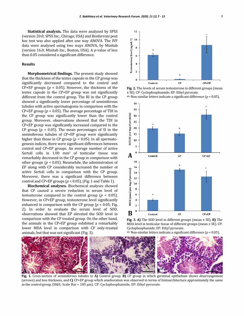
Morphometrical findings. The present study showed that the thickness of the testes capsule in the CP group was significantly decreased compared to the control and CP+EP groups (p < 0.05). However, the thickness of the testes capsule in the CP+EP group was not significantly different from the control group. The RI in the CP group showed a significantly lower percentage of seminiferous tubules with active spermatogonia in comparison with the CP+EP group (p < 0.05). The average percentage of TDI in the CP group was significantly lower than the control group. Moreover, observations showed that the TDI in CP+EP group was significantly increased compared to the CP group (p < 0.05). The mean percentages of SI in the seminiferous tubules of CP+EP group were significantly higher than those in CP group (p < 0.05). In all spermato-genesis indices, there were significant differences between control and CP+EP groups. An average number of active Sertoli cells in 1.00 mm2 of testicular tissue was remarkably decreased in the CP group in comparison with other groups (p < 0.05). Meanwhile, the administration of EP along with CP considerably increased the number of active Sertoli cells in comparison with the CP group. Moreover, there was a significant difference between control and CP+EP groups (p < 0.05), (Fig. 1 and Table 1).
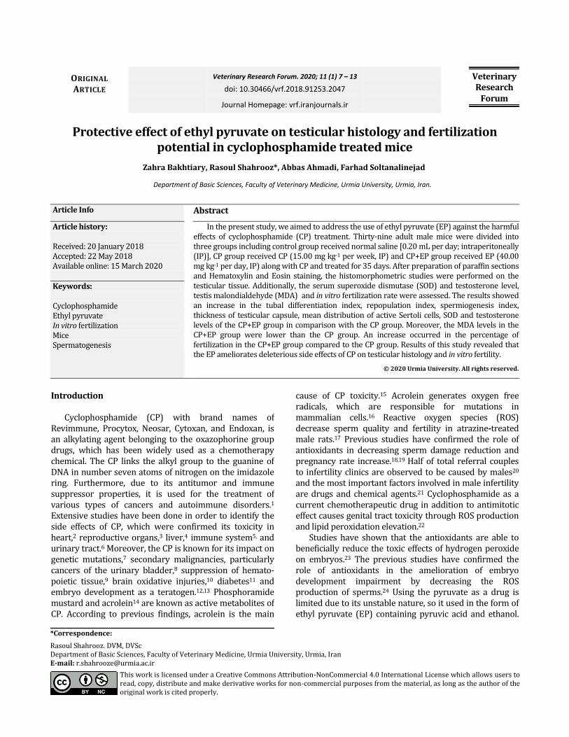
Biochemical analyses. Biochemical analyses showed that CP caused a severe reduction in serum level of testosterone compared to the control group (p < 0.05). However, in CP+EP group, testosterone level significantly enhanced in comparison with the CP group (p < 0.05; Fig. 2). In order to evaluate the serum level of SOD, observations showed that EP elevated the SOD level in comparison with the CP treated group. On the other hand, the animals in the CP+EP group exhibited a remarkably lower MDA level in comparison with CP only-treated animals, but that was not significant (Fig. 3).
Fig. 2. The levels of serum testosterone in different groups (mean ± SE). CP: Cyclophosphamide; EP: Ethyl pyruvate. abc Non-similar letters indicate a significant difference (p < 0.05).
Fig. 3. A) The SOD level in different groups (mean ± SE), B) The MDA level in testicular tissue of different groups (mean ± SE). CP: Cyclophosphamide; EP: Ethyl pyruvate. abc Non-similar letters indicate a significant difference (p < 0.05).
Fig. 1. Cross-section of seminiferous tubules in A) Control group, B) CP group in which germinal epithelium shows disarrangement (arrows) and less thickness, and C) CP+EP group which amelioration was observed in terms of histoarchitecture approximately the same as the control group, (H&E; Scale Bar = 185 µm). CP: Cyclophosphamide; EP: Ethyl pyruvate.
10 Z. Bakhtiary et al. Veterinary Research Forum. 2020; 11 (1) 7 - 13
In vitro fertilization. The IVF results showed a notable
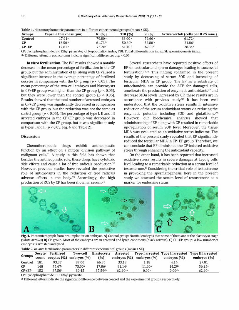
decrease in the mean percentage of fertilization in the CP group, but the administration of EP along with CP caused a significant increase in the average percentage of fertilized oocytes in comparison with the CP group (p < 0.05). The mean percentage of the two-cell embryos and blastocysts in CP+EP group was higher than the CP group (p < 0.05), but they were lower than the control group (p < 0.05). Results showed that the total number of arrested embryos in CP+EP group was significantly decreased in comparison with the CP group, but the reduction was not the same as control group (p < 0.05). The percentage of type I, II and III arrested embryos in the CP+EP group was decreased in comparison with the CP group, but it was significant only in types I and II (p < 0.05; Fig. 4 and Table 2).
Discussion
Chemotherapeutic drugs exhibit antineoplastic function by an effect on a mitotic division pathway of malignant cells.31 A study in this field has shown that besides the antineoplastic role, these drugs have cytotoxic side effects and cause a lot of free radicals production.32 However, previous studies have revealed the protective role of antioxidants in the reduction of free radicals adverse effects in the body.33 Accordingly, the high production of ROS by CP has been shown in serum.34
Several researchers have reported positive effects of EP on testicular and sperm damages leading to successful fertilization.35,36 This finding confirmed in the present study by decreasing of serum SOD and increasing of testicular MDA in CP group. The EP as a substrate of mitochondria can provide the ATP for damaged cells, ameliorate the production of enzymatic antioxidants37 and decrease MDA levels increased by CP, these results are in accordance with previous study.38 It has been well understood that the oxidative stress results in intensive reduction of the serum antioxidant status via reducing the enzymatic potential including SOD and glutathione.39 However, our biochemical analyses showed that administrating of EP along with CP resulted in remarkable up-regulation of serum SOD level. Moreover, the tissue MDA was evaluated as an oxidative stress indicator. The results of the present study revealed that EP significantly reduced the testicular MDA in CP+EP group. Therefore, we can conclude that EP diminished the CP-induced oxidative stress through enhancing the antioxidant capacity.
On the other hand, it has been reported that increased oxidative stress results in severe damages at Leydig cells level leading to a remarkable reduction at a serum level of testosterone.40 Considering the critical role of testosterone in provoking the spermatogenesis, here in the present study we assessed the serum level of testosterone as a marker for endocrine status.
Table 1. Histomorphometric parameters in different experimental groups (mean ± SE).
Groups Capsule thickness (µm) RI (%) TDI (%) SI (%) Active Sertoli (cells per 0.25 mm2)
Control 17.91a 79.80 a 83.00 a 79.60 a 61.72 a CP 13.50 b 61.73 b 50.80b 52.80 b 21.86b CP+EP 17.61 a 75.26c 61.40 c 67.00c 28.34 c
CP: Cyclophosphamide; EP: Ethyl pyruvate; RI: Repopulation index; TDI: Tubal differentiation index; SI: Spermiogenesis index. abc Different letters in each column indicate significant differences at p < 0.05.
Fig. 4. Photomicrograph from pre-implantation embryos. A) Control group: Normal embryos that some of them are at the blastocyst stage (white arrows) B) CP group: Most of the embryos are in arrested and lysed conditions (black arrows). C) CP+EP group: A low number of embryos is arrested and lysed.
Table 2. In vitro fertilization parameters in different experimental groups (mean ± SE).
Groups Oocyte count
Fertilized oocytes (%)
Two-cell embryos (%)
Blastocysts (%)
Arrested embryos (%)
Type I arrested embryos (%)
Type II arrested embryos (%)
Type III arrested embryos (%)
Control 181 93.37 87.00 66.86 33.13 1.18 4.14 27.81 CP 148 75.67a 75.00a 17.86a 82.14a 11.60a 14.29a 56.25a
CP+EP 152 87.50b 80.45 37.59ab 62.40ab 0.00b 0.00ab 62.40a
CP: Cyclophosphamide; EP: Ethyl pyruvate. ab Different letters indicate the significant difference between control and the experimental groups, respectively.
11 Z. Bakhtiary et al. Veterinary Research Forum. 2020; 11 (1) 7 - 13
This study showed that the thickness of the testicular capsule decreased in CP group. Oxidative stress may induce inflammatory mechanisms such as mast cells number increase leading to testicular capsular changes due to proteases secretion by these cells.41 Observations showed that CP significantly down-regulated the serum level of testosterone and reduced the percentage of tubules with positive TDI, RI, and SI. Meanwhile, EP remarkably inhibited CP-induced derangements. Thus, it can be concluded that EP improved the testicular endocrine potential via antioxidant status up-regulation. Ultimately, it could be able to enhance the spermatogenesis through Leydig cells salvation from CP-induced oxidative stress. Higher numbers of active Sertoli cells in EP-treated animals confirm this theory. Considering the essential and key roles of Sertoli cells in structural evolution and maturation of germ cells in testis42 as well as their physiological dependency for testosterone,43 we can come close to this fact that CP-induced damages are partly dependent to Sertoli cells derangement. Accordingly, the spermatogenesis parameters (TDI, RI, and SI) were enhanced via Sertoli cells survival up-regulation in EP-treated animals.
The ROS production increase causes changes in all bases, removal, and uncoupling of complement bases, deformation and cross-linking of DNA strands and re-arrangement of chromosomes44 and administration of CP in long-duration causes a significant breaking in DNA strands and cross-linking of DNA-DNA in the mice sperm.45 It is also attendant with breaking of one or both strands of DNA.46 The previous studies on human beings have confirmed the negative effect of sperm DNA damage on in vitro embryo development.46 On the other hand, some reports have emphasized the role of antioxidants in DNA damage reduction and increasing fertility and implantation.36,47 Hence, it could be concluded that the decrease in the rate of fertilization, two-cell embryos and blastocysts and generally low-level fertility in the CP group in comparison with other groups can be due to the function of CP metabolites such as phosphoramide mustard causing cross-linking between DNA strands.18 In spite of that, EP had protective effects by decreasing the percentage of sperm DNA damage and caused a significant increase in the percentage of fertility in the CP+EP group in comparison with the CP group. The major reason for the low percentage of embryo production in the IVF is the high level of ROS leading to the arrest in oocytes meiotic division48 and embryo development along with apoptosis induction.16 The rate of ROS production by sperm is inversely related to the sperm quality and fertilizing capacity in IVF.49 In addition to all studies that showed the negative effects of CP on fertility rate, this study confirmed the protective effects of EP as a synthetic antioxidant causing notable fertility rate improvement in CP-treated mice via ROS production reduction.
This study showed that EP significantly ameliorated the CP induced impacts including testicular histological structure alterations as well as IVF and embryo developmental arrest through antioxidant status and testicular endocrine potential up-regulation. Therefore, EP increased the serum level of SOD and testosterone concentration and decreased tissue level of MDA minimizing the CP-induced derangements. Finally, EP could be considered as an appropriate chemical to be administered simultaneously by CP.
Acknowledgments
The authors are grateful to Dr. Ali Karimi for his kind cooperation. There is no financial support for this study.
Conflict of interest
There are no conflicts of interest to be declared.
References
1. Selvakumar E, Prahalathan C, Mythili Y, et al. Protective effect of DL-alpha-lipoic acid in cyclophosphamide induced oxidative injury in rat testis. Reprod Toxicol 2004; 19(2): 163-167.
2. Santos GW, Sensenbrenner LL, Burke PJ, et al. Marrow transplantation in man following cyclophosphamide. Transplant Proc 1971; 3(1): 400-404.
3. Hosseini A, Ghaderi Pakdel F, Ahmadi A, et al. Beneficial effects of American ginseng on epididymal sperm analyses in cyclophosphamide treated rats. Cell J 2012; 14(2): 116-121.
4. DeLeve LD. Cellular target of cyclophosphamide toxicity in the murine liver: Role of glutathione and site of metabolic activation. Hepatology 1996; 24(4): 830-837.
5. Bin-Hafeez B, Ahmad I, Haque R, et al. Protective effect of Cassia occidentalis L. on cyclophosphamide-induced suppression of humoral immunity in mice. J Ethnopharmacol 2001; 75(1): 13-18.
6. Korkmaz A, Topal T, Oter S. Pathophysiological aspects of cyclophosphamide and ifosfamide induced hemorrhagic cystitis; Implication of reactive oxygen and nitrogen species as well as PARP activation. Cell Biol Toxicol 2007; 23(5): 303-312.
7. Stahlmann R, Bluth U, Neubert D. Effects of the cyclophosphamide metabolite acrolein in mammalian limb bud cultures. Arch Toxicol 1985; 57(3): 163-167.
8. Levine LA, Richie JP. Urological complications of cyclophosphamide. J Urol 1989; 141(5): 1063-1069.
9. Baumann F, Preiss R. Cyclophosphamide, and related anticancer drugs. J Chromatogr B Biomed Sci Appl 2001; 764(1-2): 173-192.
10. Oboh G, Ogunruku OO. Cyclophosphamide-induced oxidative stress in brain: Protective effect of hot short
12 Z. Bakhtiary et al. Veterinary Research Forum. 2020; 11 (1) 7 - 13
pepper (Capsicum frutescens L. var. abbreviatum). Exp Toxicol Pathol 2010; 62(3): 227-233.
11. Faust A, Burkart V, Ulrich H, et al. Effect of lipoic acid on cyclophosphamide-induced diabetes and insulitis in non-obese diabetic mice. Int J Immunopharmacol 1994; 16(1): 61-66.
12. Gilani SH, Chatzinoff M. Embryopathic effects of cyclophosphamide. Environ Res 1983; 31(2): 296-301.
13. Mirkes PE. Cyclophosphamide teratogenesis: A review. Teratog Carcinog Mutagen 1985; 5(2): 75-88.
14. Chinnaswamy G, Errington J, Foot A, et al. Pharmacokinetics of cyclophosphamide and its metabolites in paediatric patients receiving high-dose myeloablative therapy. Eur J Cancer 2011; 47(10): 1556-1563.
15. Kern JC, Kehrer JP. Acrolein-induced cell death: A caspase-influenced decision between apoptosis and oncosis/necrosis. Chem Biol Interac 2002; 139(1): 79-95.
16. Arumugam N, Sivakumar V, Thanislass J, et al. Effects of acrolein on rat liver antioxidant defense system. Indian J Exp Biol 1997; 35(12): 1373-1374.
17. Rezaei Agdam H, Razi M, Amniattalab A, et al. Co-administration of vitamin E and testosterone attenuates the atrazine-induced toxic effects on sperm quality and testes in rats. Cell J 2017; 19(2): 292-305.
18. Hughes CM, Lewis S, McKelvey-Martin VJ, et al. The effects of antioxidant supplementation during Percoll preparation on human sperm DNA integrity. Hum Reprod 1998; 13(5): 1240-1247.
19. Zarei L, Shahrooz R, Sadrkhanlou R, et al. Protective effects of Cornus mas extract on in vitro fertilization potential in methotrexate treated male mice. Vet Res Forum 2015; 6(1): 55-61.
20. Schoor RA. Prostatitis and male infertility: Evidence and links. Curr Urol Rep 2002; 3(4): 324-329.
21. Brinkworth MH, Weinbauer GF, Bergmann M, et al. Apoptosis as a mechanism of germ cell loss in elderly men. Int J Androl 1997; 20(4): 222-228.
22. Lear L, Nation RL, Stupans I. Effects of cyclophosphamide and adriamycin on rat hepatic microsomal glucuronidation and lipid peroxidation. Biochem Pharmacol 1992; 44(4): 747-753.
23. Zhang X, Sharma RK, Agarwal A, et al. Effect of pentoxifylline in reducing oxidative stress-induced embryotoxicity. J Assist Reprod Genet. 2005; 22(11-12): 415-417
24. Esfandiari N, Falcone T, Agarwal A, et al. Protein supplementation and the incidence of apoptosis and oxidative stress in mouse embryos. Obstet Gynecol 2005; 105(3): 653-660.
25. Vander Heiden MG, Cantley LC, Thompson CB. Understanding the Warburg effect: The metabolic requirements of cell proliferation. Science 2009; 324 (5930): 1029-1033.
26. Wang X, Perez E, Liu R, et al. Pyruvate protects mitochondria from oxidative stress in human neuroblastoma SK-N-SH cells. Brain Res 2007; 1132(1): 1-9.
27. Niakani A, Farrokhi F, Hasanzadeh S. Decapeptyl ameliorates cyclophosphamide-induced reproductive toxicity in male Balb/C mice: Histomorphometric, stereologic and hormonal evidences. Iran J Reprod Med 2013; 11(10): 791-800.
28. Niehaus WG Jr., Samuelsson B. Formation of malonaldehyde from phospholipid arachidonate during microsomal lipid peroxidation. Eur J Bioch 1968; 6(1): 126-130.
29. Marklund S, Marklund G. Involvement of the superoxide anion radical in the autoxidation of pyrogallol and a convenient assay for superoxide dismutase. Eur J Biochem 1974; 47(3): 469-474.
30. Lowry OH, Rosebrough NJ, Farr AL, et al. Protein measurement with the Folin phenol reagent. J Biol Chem 1951; 193(1): 265-275.
31. Barrera G. Oxidative stress and lipid peroxidation products in cancer progression and therapy. ISRN Oncol 2012; 2012: 137289. doi: 10.5402/2012/137289.
32. Gonzalez EJ, Peterson A, Malley S, et al. The effects of Tempol on cyclophosphamide-induced oxidative stress in rat micturition reflexes. Sci World J 2015; 2015: 545048. doi: 10.1155/2015/545048.
33. Bakhtiary Z, Shahrooz R, Ahmadi A, et al. Ethyl pyruvate ameliorates the damage induced by cyclophosphamide on adult mice testes. Int J Fertil Steril 2016; 10(1):79-86.
34. Manda K, Bhatia AL. Prophylactic action of melatonin against cyclophosphamide-induced oxidative stress in mice. Cell Biol Toxicol 2003; 19(6): 367-372.
35. Atashfaraz E, Farokhi F, and Najafi G. Protective effect of ethyl pyruvate on epididymal sperm characteristics, oxidative stress and testosterone level in methotrexate treated mice. J Reprod Infertil 2013; 14(4): 190-196.
36. Bakhtiary Z, Shahrooz R, Ahmadi A, et al. Protective effects of ethyl pyruvate on sperm quality in cyclophosphamide treated mice. Iran J Reprod Med 2015; 13(5): 291-296.
37. Olek RA, Ziolkowski W, Wierzba TH, et al. Effect of ethyl pyruvate on skeletal muscle metabolism in rats fed on a high fat diet. Nutrients 2013; 5(7): 2372-2383.
38. Wang LZ, Sun WC, Zhu XZ. Ethyl pyruvate protects PC12 cells from dopamine-induced apoptosis. Eur J Pharmacol 2005; 508(1-3): 57-68.
39. Adil M, Kandhare AD, Visnagri A, et al. Naringin ameliorates sodium arsenite-induced renal and hepatic toxicity in rats: Decisive role of KIM-1, Caspase-3, TGF-beta, and TNF-alpha. Ren Fail 2015; 37(8): 1396-1407.
40. Rezvanfar M, Sadrkhanlou R, Ahmadi A, et al. Protection of cyclophosphamide-induced toxicity in reproductive tract histology, sperm characteristics, and

13 Z. Bakhtiary et al. Veterinary Research Forum. 2020; 11 (1) 7 - 13
DNA damage by an herbal source; Evidence for role of free-radical toxic stress. Hum Exp Toxicol 2008; 27(12): 901-910.
41. Puxeddu I, Levi-Schaffer F. Mast cells and tissue remodeling. Rev Fr Allergol 2002; 42(1): 16-18.
42. Mruk DD, Cheng CY. Sertoli-Sertoli and Sertoli-germ cell interactions and their significance in germ cell movement in the seminiferous epithelium during spermatogenesis. Endocr Rev 2004; 25(5): 747-806.
43. Baazm M, Jalali-Mashayekhi F, Darabi MR, et al. The role of embryonic Sertoli cells in maintenance of spermatogonial stem cells. Cell J 2014; 16(Suppl 1): 47. doi:10.22074/cellj.2014.341.
44. Cadet J, Wagner JR. DNA base damage by reactive oxygen species, oxidizing agents, and UV radiation. Cold Spring Harb Perspect Biol 2013; 5(2): a012559. doi: 10.1101/cshperspect.a012559.
45. Lewis SE, Aitken RJ. DNA damage to spermatozoa has impacts on fertilization and pregnancy. Cell Tissue Res 2005; 322(1): 33-41.
46. Aitken RJ, Krausz C. Oxidative stress, DNA damage and the Y chromosome. Reproduction 2001; 122(4): 497-506.
47. Bakhtiary Z, Shahrooz R, Ahmadi A, et al. Evaluation of antioxidant effects of crocin on sperm quality in cyclophosphamide treated adult mice. Vet Res Forum 2014; 5(3): 213-218.
48. Greco E, Romano S, Iacobelli M, et al. ICSI in cases of sperm DNA damage: Beneficial effect of oral antioxidant treatment. Hum Reprod 2005; 20(9): 2590-2594.
49. Nakamura Y, Yamagata Y, Sugino N, et al. Nitric oxide inhibits oocyte meiotic maturation. Biol Reprod 2002; 67(5): 1588-1592.