original article mesenchymal stem cells suppress lung ... · original article mesenchymal stem...
TRANSCRIPT
Int J Clin Exp Pathol 2015;8(8):8958-8967www.ijcep.com /ISSN:1936-2625/IJCEP0011986
Original ArticleMesenchymal stem cells suppress lung inflammation and airway remodeling in chronic asthma rat model via PI3K/Akt signaling pathway
Hai-Yan Lin1,4*, Lei Xu1*, Shuan-Shuan Xie1, Fei Yu2, Hai-Yang Hu3, Xiao-Lian Song1, Chang-Hui Wang1
1Department of Respiratory Medicine, Shanghai Tenth People’s Hospital, Tongji University, Shanghai 200072, Peoples PR China; 2Department of Nuclear Medicine, Shanghai Tenth People’s Hospital, Tongji University, Shanghai 200072, Peoples PR China; 3Department of Geriatric Respiratory, Qianfoshan Hospital Affiliated to Shandong Universit, Jinan 250014, Shandong, China; 4Department of Respiratory Medicine, Huai’an First Peoples’s Hospital, Nanjing Medical University, Nanjing, China. *Equal contributors.
Received June 26, 2015; Accepted July 29, 2015; Epub August 1, 2015; Published August 15, 2015
Abstract: Background: Mesenchymal stem cells (MSCs) came out to attract wide attention and had become one of the hotspots of most diseases’ research in decades. But at present, the mechanisms of how MSCs work on chronic asthma remain undefined. Our study aims at verifying whether MSCs play a role in preventing inflamma-tion and airway remodeling via PI3K/AKT signaling pathway in the chronic asthma rats model. Methods: First, an ovalbumin (OVA)-induced asthma model was built. MSCs were administered to ovalbumin-induced asthma rats. The total cells in a bronchial alveolar lavage fluid (BALF) and inflammatory mediators in BALF and serum were mea-sured. Histological examination of lung tissue was performed to estimate the pathological changes. Additionally, the expression of phosphorylated-Akt (p-Akt) in all groups was measured by western blot and immunohistochemistry (IHC). Results: Compared to normal control group, the degree of airway inflammation and airway remodeling was significantly increased in asthma group. On the contrary, they were obviously inhibited in MSCs transplantation group. Moreover, the expression of p-Akt was increased in lung tissues of asthmatic rats, and suppressed by MSCs transplantation. Conclusion: Our results demonstrated that MSCs transplantation could suppress lung inflamma-tion and airway remodeling via PI3K/Akt signaling pathway in rat asthma model.
Keywords: Asthma, mesenchymal stem cells, PI3K/Akt, signaling pathway, inflammation, airway remodeling
Introduction
Asthma is one of the most common and fre-quently-occurring diseases of the respiratory systems. For a long time, asthma was regarded as a chronic Th2-driven and eosinophilic airway inflammation disease [1]. Activated Th2 cells can synthesize various cytokines such as IL-4, IL-5, IL-6, IL-13 and TNF-α [2]. Inflammatory cells, especially eosinophils, also release chemical mediators that lead to inflammation. This chronic inflammation leads to the forma-tion of bronchial hyperresponsiveness (BHR), which usually comes out with widely varied reversible airflow limitation. Asthma is newly characterized by the association of inflamma-tion, BHR, and airway remodeling [3].
As we all know, once the asthma attack hap-pens, the best treatment is corticosteroids
used by inhalation or intravenous injection. In spite of inhaled corticosteroids, asthma patients usually still show a decrease in the level of pulmonary function. Bronchial thermo-plasty (BT), a procedure aimed at delivering thermal energy to airways in order to reduce the proliferated bronchial wall smooth muscle, is found likely to be effective in treating asthma [4]. But at present it’s still in the stage of clinical trials. Researchers working on the control of inflammation have had no breakthrough in years. There is a crying need for finding a new way to suppress inflammation and airway remodeling of asthma. Therefore, many schol-ars began to adjust the principal direction of asthma research, trying to find a new breakthrough.
Mesenchymal stem cells (MSCs), first detected in bone marrow (BMSCs), have been confirmed

MSCs suppress lung inflammation and airway remodeling in rat asthma model
8959 Int J Clin Exp Pathol 2015;8(8):8958-8967
to have strong ability of proliferation, multiple differentiation potential, anti-inflammation, tis-sue repair and immunomodulatory effects [5, 6]. In this context, MSCs came out to attract wide attention and had become one of the hotspots of most diseases’ research, such as acute pancreatitis [7], hepatic failure [8], acute lung injury [9], etc. However, there was little research on the effect of MSCs on asthma as the pathological characteristic of asthma. Thus, we speculated asthma could benefit from MSCs transplantation. Transforming growth factor beta (TGF-β) plays a pivotal role in secreting and mediating a mass of growth factors, and cytokines cause airway inflammation and air-way remodeling [10]. Our previous study also revealed that human MSCs inhibited the polar-ization of alveolar macrophages on asthma model via TGF-β signaling pathway [11]. It is well known that phosphatidylinositol 3-kinase/protein kinase B (PI3K/Akt) signaling pathway signaled by TGF-β seems to play a relatively important role in asthma [12]. So far, the defi-nite mechanism of action for MSCs remains unclear to a large extent.
Our study aims at verifying whether MSCs play a role in preventing inflammation and airway remodeling via PI3K/AKT signaling pathway in the chronic asthma rats model.
Materials and methods
Ethics statement
This study was performed in strict accordance with the Guidelines of the Shanghai Laboratory Animal Center and the Policies on the Use of Animals and Humans in Research approved by the Shanghai Tenth People’s Hospital. The pro-tocol was approved by the Animal Care & Welfare Committee of Tongji University.
We monitored the animals’ conditions every day and recorded them. We performed the sur-gery in mice under chloral hydrate anesthesia, and all efforts were made to minimize their suf-fering. Then, the rats were visualized and anat-omized after eight weeks.
Animals
Four- to five-weeks-old male Sprague-Dawley (SD) rats, weighing 120~160 g used in our study, were purchased from Shanghai labora-tory animal center. Prior to experiments, rats were maintained in an animal facility under
standard laboratory conditions for 1 week, with free access to food and water.
Ovalbumin-induced asthma model
First, rats were randomly divided into four groups: normal control group; MSCs control group; asthma group; MSCs transplantation group. Each group had 6 rats. We established asthma model according to the methods of Yang [13] and Kabiri Rad et al. [14] with some modifications. Rats were sensitized by IP injec-tion of 1 mg ovalbumin (OVA; Sigma-Aldrich, St. Louis, MO, USA) mixed with 100 mg aluminum hydroxide adjuvant (Alum; Sigma-Aldrich) in saline on days 1 and 8. Then from day 15, rats were challenged by exposing them to an aero-sol of 1% OVA for 20 minutes once a day for 6 weeks with every week having 2 days free. The control group was treated with PBS instead of OVA.
MSCs transplantation
MSCs used in our study originated from SD rats and were purchased from Cyagen Biosciences Inc. (Guangzhou, China). MSCs were cultured in MSC basal medium (supplemented with 10% fetal bovine serum, 1% penicillin-streptomycin, and 1% glutamine) (Cyagen Biosciences Inc., Guangzhou, China). MSCs suspended in phos-phate buffer saline (PBS) at a concentration of 1×10⁶/ml were given by tail intravenous injec-tion to the experimental group on days 22 and 29.
Bronchoalveolar lavage fluid (BALF) collection and total cell count
After the last OVA challenge, rats anesthetized with 2% pentobarbital were firstly punctured for blood through their hearts. BALF was obtained from the left lung by lavaging three times with 1 ml saline via trachea cannula while the right lung was resected. Then BALF was centrifuged; the supernatant was kept at -80°C until need-ed. On the other hand, the cells precipitated were resuspended in 1 ml PBS, and the total cells were counted under a biological micro-scope (Olympus, Tokyo, Japan). The cells were then fluorescence-labeled for flow cytometric analysis in order to detect the percentage of eosinophils in BALF.
Detection of cytokines in the BALF and blood
The blood was drawn from the heart after we opened the chest. The expression of inflamma-
MSCs suppress lung inflammation and airway remodeling in rat asthma model
8960 Int J Clin Exp Pathol 2015;8(8):8958-8967
tory cytokines in the BALF supernatants and blood serum including IL-6 and TNF-α was detected with the rat enzyme-linked immuno-sorbent assay (ELISA) kits (Neobioscience Technology, Shanghai, China) according to the manufacturer’s instructions.
Histological examination of lung tissue
Isolated lungs were fixed with 4% paraformal-dehyde and embedded in paraffin. Then we cut the specimens into a thickness of 3-4 μm sec-tion. The sections were stained with hematoxy-lin and eosin (H&E). Also, Masson staining was performed to detect the deposition of collagen and smooth muscle thickness. In order to detect specific proteins, immunohistochemis-try was performed. Briefly, deparaffinized sec-tions were rehydrated with graded ethanol and soaked in 3% hydrogen peroxide for 25 min, then boiled in EDTA antigen retrieval buffer in a microwave oven for 5 min for antigen retrieval. After blocking slides with 5% BSA, they were incubated with a monoclonal antibody against Akt and p-Akt (Abcam, Cambridge, United Kingdom) overnight at 4°C. Subsequently, the sections were washed three times with PBS and incubated with secondary antibodies, and then counterstained with hematoxylin. The results were visualized using a microscope (CTR 6000; Leica, Wetzlar, Germany). Three pathologists individually assessed all staining results.
Western blot analysis
The fresh lung tissues collected were lysed on ice in tissue total protein lysis buffer containing 1 Mm PMSF, 0.5 M EDTA, 1 M Tris-HCl (pH 7.5), 10% sodium dodecyl sulfate (SDS), 0.5% sodi-um deoxycholate, 1% NP-40 (Nonidet P-40), 1% Triton X-100, 10 mg/ml aprotinin and 10 mg/ml
leupeptin. Homogenates were centrifuged twice at 12,000 rpm, 4°C for 5 min, and then protein concentrations in the extracted super-natants were detected with BCA (bicinchoninic acid) protein assay. After that, an equal 35 μg protein was loaded per lane, and electrophore-sis was performed on 10% SDS-PAGE gel (stacking gel voltage, 80 V; separating gel volt-age, 120 V). Then proteins were transferred to nitrocellulose membranes (constant current, 200 mA). Membranes were blocked for 1 h in 5% nonfat milk in TBST, and incubated over-night at 4°C with monoclonal antibody against Akt, p-Akt (1:5000, Abcam, Cambridge, United Kingdom) and β-actin. The binds were detected by an ECL detection system (Santa Cruz), according to the manufacturer’s instructions.
Statistical analysis
Data are given as mean ± SD. We performed all statistical analyses with SPSS 20.0 software. One-way ANOVA with LSD test (used for com-paring between groups) was used to assess data. P<0.05 was considered statistically sig-nificant in all statistical analyses.
Results
Purity and differentiation ability of MSCs
MSCs used in our study were purchased from Cyagen Biosciences Inc. (Catalog No. RASMX-01001; Lot Number: 130628B31). All MSCs showed ≥70% positivity for the expression of cell surface antigens CD29, CD44, and CD90, as well as ≤5% positivity for the expression of cell surface antigens CD34, CD45, and CD11b. Cells identified were able to differentiate into osteocytes, adipocytes, and chondrocytes (Supplementary Data).
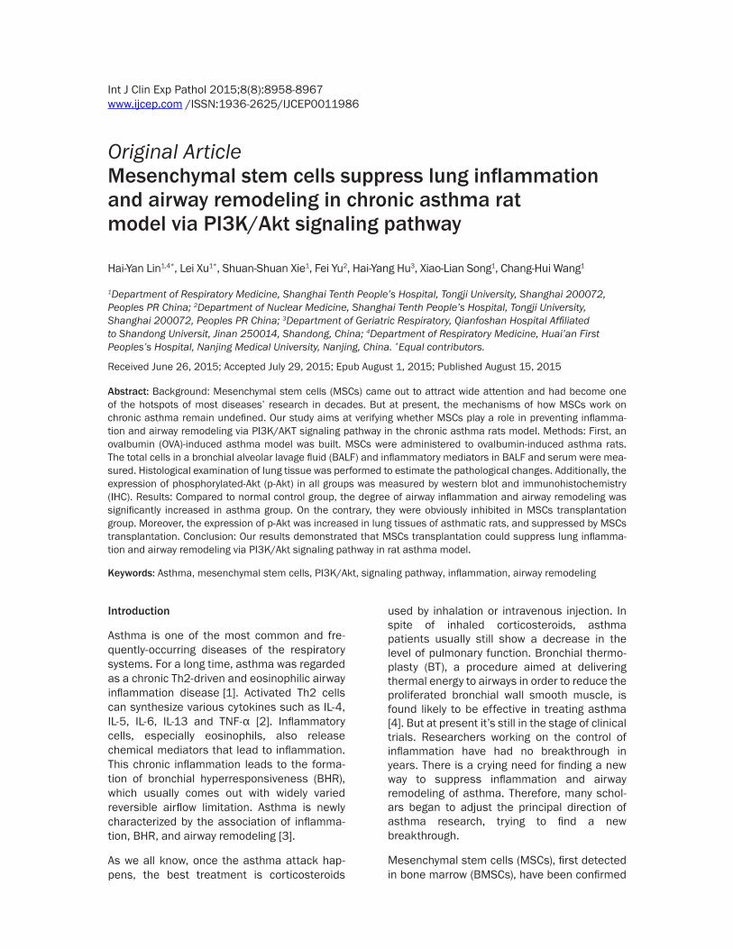
Figure 1. The scheme for building a chronic asthma rat model. Rats were sensitized on days 1 and 8. Then since day 15, rats were challenged for 6 weeks to establish a chronic asthma model. MSCs transplantation was carried out on day 22 and day 29.
MSCs suppress lung inflammation and airway remodeling in rat asthma model
8961 Int J Clin Exp Pathol 2015;8(8):8958-8967
MSCs transplantation improved asthma symp-toms
We consulted an established rat asthma model for studying the effects of MSCs transplanta-tion on asthma (Figure 1). After challenging with OVA, the symptoms of nose scratching, shortness of breath, sneezing, and coughing were observed in asthma group from the 2nd week. The symptoms were more and more prominent and heavier as the time increased. However, there was no abnormality seen in the control group rats challenged with PBS.
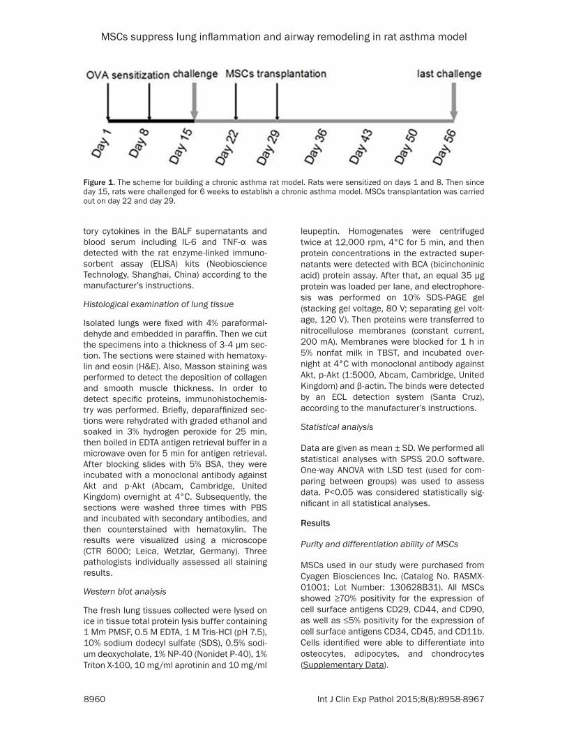
MSCs transplantation decreased BALF cell counts and cytokines in OVA-inhaled rats
In our experiments, we measured total cell counts and the percentage of eosinophils in BALF, and also the inflammatory mediators of TNF-α and IL-6 in both BALF and serum for the purpose of evaluating the anti-inflammatory effect of MSCs. As shown in Figure 2A and 2B, BALF total cell counts and the percentage of eosinophils in asthma group were increased more than normal control group (P<0.01) and MSCs control group. However, there was a sig-
Figure 2. MSCs transplantation decreased BALF cell counts and cytokines in OVA-inhaled rats. The total cell counts in BALF (A) were counted under a biological microscope, and the percentage of eosinophils in BALF (B) were fluores-cence-labeled for flow cytometry analysis. The levels of TNF-α and IL-6 in BALF (C) and serum (D) of normal control group (PBS), MSCs control group (PBS+MSC), asthma group (OVA), and MSCs transplantation group (OVA+MSC) were detected by means of ELISA. Data were expressed as the mean ± SD (n=6). *P<0.01 compared to normal control group. #P<0.01 compared to asthma group.
MSCs suppress lung inflammation and airway remodeling in rat asthma model
8962 Int J Clin Exp Pathol 2015;8(8):8958-8967
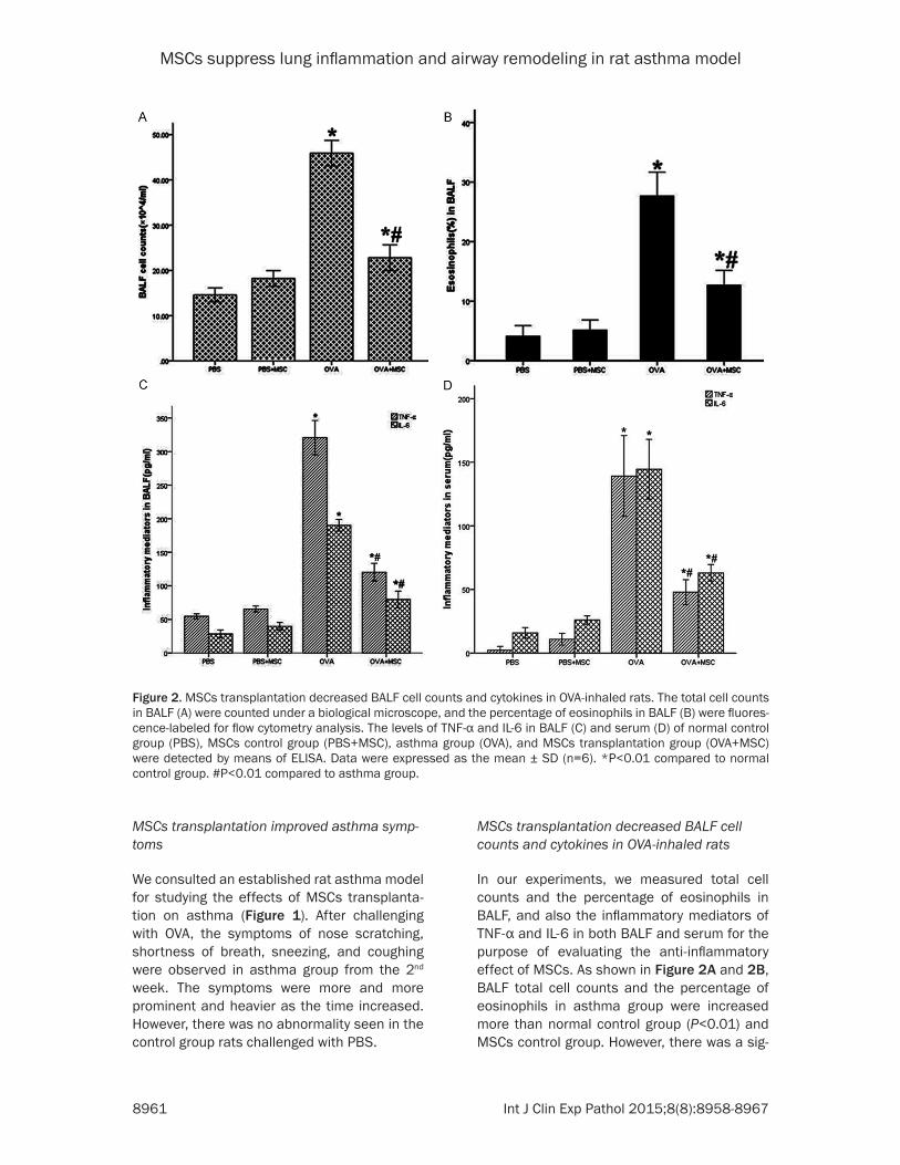
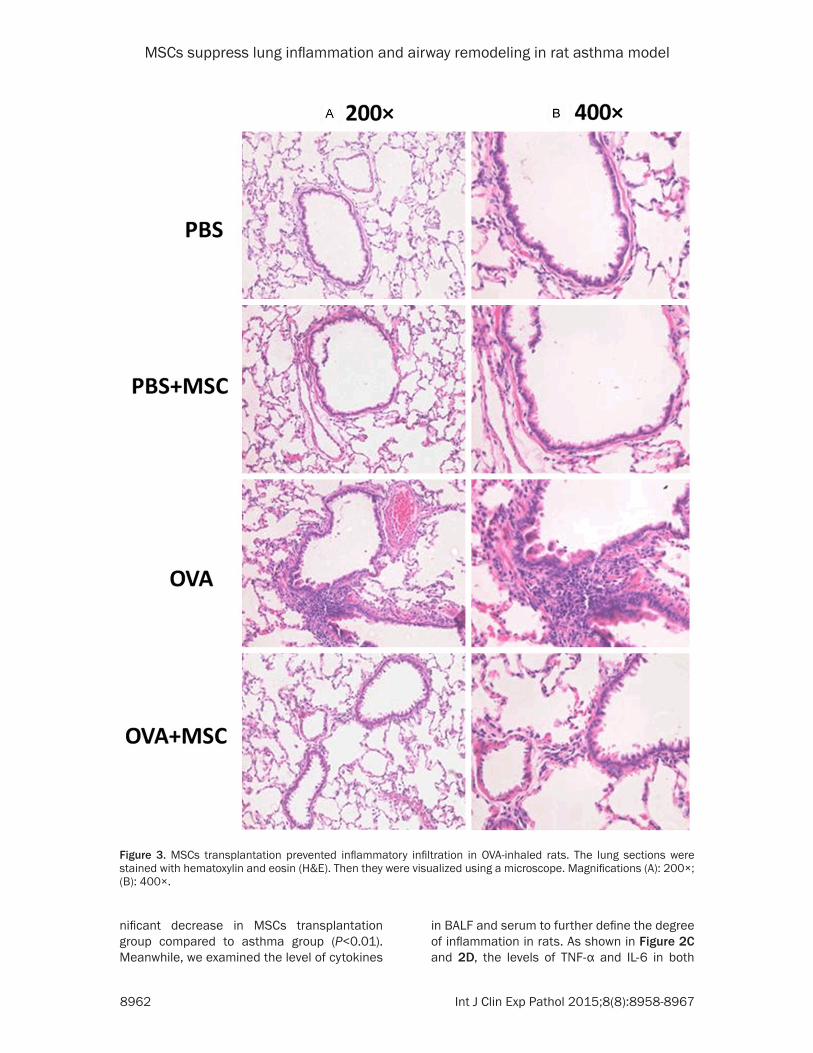
Figure 3. MSCs transplantation prevented inflammatory infiltration in OVA-inhaled rats. The lung sections were stained with hematoxylin and eosin (H&E). Then they were visualized using a microscope. Magnifications (A): 200×; (B): 400×.
nificant decrease in MSCs transplantation group compared to asthma group (P<0.01). Meanwhile, we examined the level of cytokines
in BALF and serum to further define the degree of inflammation in rats. As shown in Figure 2C and 2D, the levels of TNF-α and IL-6 in both
MSCs suppress lung inflammation and airway remodeling in rat asthma model
8963 Int J Clin Exp Pathol 2015;8(8):8958-8967
MSCs suppress lung inflammation and airway remodeling in rat asthma model
8964 Int J Clin Exp Pathol 2015;8(8):8958-8967
BALF and serum also presented obvious decline in MSCs transplantation group com-pared to asthma group (P<0.01), while the lev-els in asthma group performed a several-fold
increase than the control group (P<0.01). In addition, all these differences between normal control group and the MSCs control group had no statistical significance.
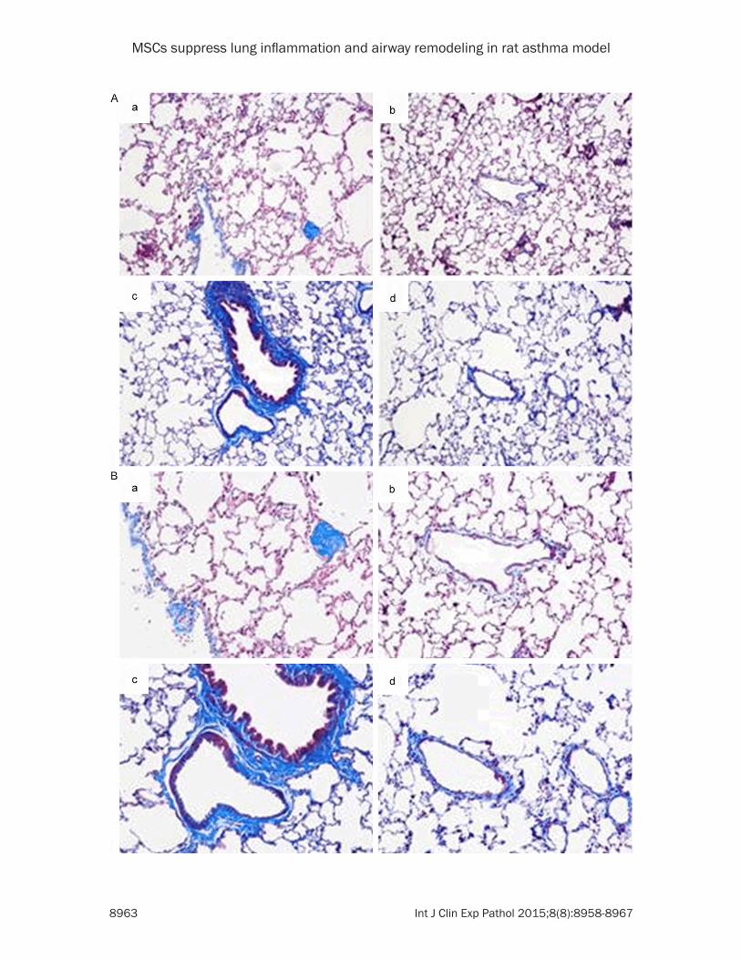
Figure 4. MSCs transplantation prevented airway remodeling in OVA-inhaled rats. Masson staining of lung tissues was performed to assess the deposition of collagen and the degree of muscular layer thickening of normal control group (a), MSCs control group (b), asthma group (c), and MSCs transplantation group (d). Magnifications (A): 100×; (B): 200×.
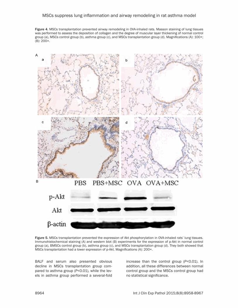
Figure 5. MSCs transplantation prevented the expression of Akt phosphorylation in OVA-inhaled rats’ lung tissues. Immunohistochemical staining (A) and western blot (B) experiments for the expression of p-Akt in normal control group (a), BMSCs control group (b), asthma group (c), and MSCs transplantation group (d). They both showed that MSCs transplantation had a lower expression of p-Akt. Magnifications (A): 200×.
MSCs suppress lung inflammation and airway remodeling in rat asthma model
8965 Int J Clin Exp Pathol 2015;8(8):8958-8967
MSCs transplantation prevented inflamma-tory infiltration and airway remodeling in OVA-inhaled rats
In order to observe the effect of MSCs on histo-pathological changes in lung tissues, hematox-ylin and eosin (H&E) staining were used in our study. As shown in Figure 3, the tissues of asth-ma group demonstrated that there were signifi-cant inflammatory cells infiltrating around the airway and vessels, alveolar structural damage, bronchial mucosal shedding and smooth mus-cle cell proliferation as compared to normal control group and MSCs control group. Nonetheless, the extent of pathological altera-tions was eased apparently after MSCs trans-plantation in OVA-inhaled rats. As shown in Figure 4, the mass of staining results showed that there persisted a large amount of collagen deposition and thickening of the muscular layer in asthma lungs while the remaining three groups did not see such obvious changes. The results suggested that MSCs transplantation was beneficial to airway remodeling.
MSCs transplantation prevented the expres-sion of Akt phosphorylation in OVA-inhaled rats’ lung tissues
To confirm the way that MSCs transplantation impact on asthma, we used a specific antibody to examine the phosphorylation of Akt in the OVA-inhaled rats’ lungs by immunohistochemis-try. In rat lung tissues, phosphorylated Akt (p-Akt) was mainly localized in the cytoplasm of smooth muscle cells, presenting with canary yellow to brown. As shown in Figure 5A, com-pared to the control group, the expression of p-Akt in lung tissues increased obviously; on the other side; there was a marked decrease in the expression of p-Akt due to the transplanta-tion of MSCs. To further confirming this, Western blot analysis was applied. As shown in Figure 5B, the results also revealed that MSCs transplantation could prevent the phosphoryla-tion of Akt in OVA-inhaled rats.
Discussion
Our results suggest that the transplantation of MSCs could prevent airway inflammation and also airway remodeling via PI3K/Akt pathway in chronic asthma rats.
To verify the effect of MSCs transplantation on asthma model, we established an OVA-induced
rat model to test their effects on inflammation and airway remodeling. OVA was the earliest and most widely used sensist Inogen in the home and overseas studies for establishing an animal model of bronchial asthma [15]. Rats were observed to have various symptoms, such as cough, sneeze, and nose scratch. The patho-logical examination of lung tissues demonstrat-ed that there were distinct inflammatory cells infiltrations around the airway of asthma mod-els rats; together they were helpful to prove the validity of the asthma model. With MSCs trans-planted to asthma rats, inflammatory cells and cytokines TNF-α, IL-6 in BALF and serum were reduced. The extent of inflammatory infiltration around the airway, deposition of collagen and airway remodeling were prevented by MSCs.
Airway remodeling can be distinctly sup-pressed. The inflammatory cells in BALF con-centration and the level of inflammatory media-tors, TNF-α and IL-6 in both BALF and serum were reduced by MSCs transplantation.
Airway inflammation is one major characteristic of asthma, giving priority to eosinophils and dominant Th2 cytokines. However, airway inflammation alone cannot fully explain the pathogenesis of asthma. Airway remodeling, also thought to be a feature of asthma, is involved in extracellular matrix (ECM) deposi-tion, epithelial-mesenchymal transition (EMT) [16], hyperplasia and hypertrophy of airway smooth muscle cells (ASMCs), and angiogene-sis [17]. Airway remodeling, together with chronic inflammation, will lead to irreversible airway obstruction and persistent airway hyper-responsiveness. As said above, MSCs have strong ability of proliferation, multiple differen-tiations potential, anti-inflammation and immu-nomodulatory effects [5, 6]. After infusing MSCs intravenously into rats, they were observed to bring about relocation in lungs, and simultaneously secrete anti-inflammatory molecules that have effects on the local and distal zones [18]. In line with our study, it proved that MSCs may be a potential therapy for asth-ma. A previous study also showed that MSCs transfer could obviously decrease inflammation and airway remodeling of Toluene Diisocyanate-induced murine asthma model [19].
PI3Ks are a family of proteins involved in cell proliferation, differentiation, apoptosis and glu-cose transportation [20]. Protein kinase B
MSCs suppress lung inflammation and airway remodeling in rat asthma model
8966 Int J Clin Exp Pathol 2015;8(8):8958-8967
(PKB, also called Akt), a downstream effector of PI3K, is a direct target gene of PI3K and the center link of this pathway. Usually, Akt phos-phorylation can be used as an index of PIK3 activity. Therefore, these observations spurred us to explore whether the effect of MSCs on asthma could be via a PI3K/Akt signaling path-way. In our study, we found that the expression of p-Akt was inhibited by MSCs transplantation while it was significantly increased in asthma model. Our results suggest that the effect of MSCs in the prevention of inflammation and air-way remodeling could be through a PI3K/Akt pathway. Masahide Takeda et al. [21] showed us that in PI3K γ-deficient mice, there was a prominent decline in the levels of inflammatory cell accumulation, airway remodeling and also BHR. PI3K p110δ was found necessary for treating airway remodeling and inflammation in asthma through induction of the production of IL-6 and calponin and α-smooth muscle actin in ASMCs [10]. Besides, PI3K inhibitors, LY294002, and wortmannin were both shown to play a useful role in asthma model [22, 23].
As far as we know, this is the first time a research was conducted to explore whether PI3K/Akt signaling pathway play a role in MSCs prevention of inflammation and airway remod-eling in chronic asthma. Although our results are remarkable, our limitations should be rec-ognized. Each group had a small sample size. It was not feasible to study on humans, so all our researches focused on animals.
In summary, our results demonstrated that MSCs transplantation can suppress lung inflammation and airway remodeling via PI3K/Akt signaling pathway in rat asthma model. There is a need for more detailed study in the future before MSCs can be applied to asthma patients.
Acknowledgements
This study was funded by the National Natural Science Foundation of China (No. 81100018, 81301993). The funders had no role in study design, data collection and analysis, decision to publish, or preparation of the manuscript.
Disclosure of conflict of interest
None.
Address correspondence to: Drs. Chang-Hui Wang and Xiao-Lian Song, Department of Respiratory
Medicine, Shanghai Tenth People’s Hospital, Tongji University, 301 Yanchang Rd (M), Shanghai 200072, Peoples PR China. E-mail: [email protected] (CHW); [email protected] (XLS)
References
[1] Parulekar AD, Atik MA, Hanania NA. Periostin, a novel biomarker of TH2-driven asthma. Curr Opin Pulm Med 2014; 20: 60-5.
[2] Kudo M, Ishigatsubo Y, Aoki I. Pathology of asthma. Front Microbiol 2013; 4: 263.
[3] Manuyakorn W, Howarth PH, Holgate ST. Airway remodelling in asthma and novel thera-py. Asian Pac J Allergy Immunol 2013; 31: 3-10.
[4] Dombret MC, Alagha K, Philippe Boulet L, Yves Brillet P, Joos G, Laviolette M, Louis R, Rochat T, Soccal P, Aubier M, Chanez P. Bronchial ther-moplasty: a new therapeutic option for the treatment of severe, uncontrolled asthma in adults. Eur Respir Rev 2014; 23: 510-8.
[5] Bruno S, Camussi G. Role of mesenchymal stem cell-derived microvesicles in tissue re-pair. Pediatr Nephrol 2013; 28: 2249-54.
[6] Inamdar AC, Inamdar AA. Mesenchymal stem cell therapy in lung disorders: pathogenesis of lung diseases and mechanism of action of mesenchymal stem cell. Exp Lung Res 2013; 39: 315-27.
[7] Lazebnik LB, Trubitsyna IE, Agafonov MA, Kniazev OV, Liundup AV. [Mesenchymal stro-mal cells transplantation in acute and chronic pancreatitis in rats]. Eksp Klin Gastroenterol 2011; 28-31.
[8] Park CH, Bae SH, Kim HY, Kim JK, Jung ES, Chun HJ, Song MJ, Lee SE, Cho SG, Lee JW, Choi JY, Yoon SK, Han NI, Lee YS. A pilot study of autologous CD34-depleted bone marrow mononuclear cell transplantation via the he-patic artery in five patients with liver failure. Cytotherapy 2013; 15: 1571-9.
[9] Lee JW, Fang X, Krasnodembskaya A, Howard JP, Matthay MA. Concise review: Mesenchymal stem cells for acute lung injury: role of para-crine soluble factors. Stem Cell 2011; 29: 913-9.
[10] Ge Q, Moir LM, Trian T, Niimi K, Poniris M, Shepherd PR, Black JL, Oliver BG, Burgess JK. The phosphoinositide 3’-kinase p110delta modulates contractile protein production and IL-6 release in human airway smooth muscle. J Cell Physiol 2012; 227: 3044-52.
[11] Song X, Xie S, Lu K, Wang C. Mesenchymal Stem Cells Alleviate Experimental Asthma by Inducing Polarization of Alveolar Macrophages. Inflammation 2015; 38: 485-92.
[12] Burgess JK, Lee JH, Ge Q, Ramsay EE, Poniris MH, Parmentier J, Roth M, Johnson PR, Hunt
MSCs suppress lung inflammation and airway remodeling in rat asthma model
8967 Int J Clin Exp Pathol 2015;8(8):8958-8967
NH, Black JL, Ammit AJ. Dual ERK and phos-phatidylinositol 3-kinase pathways control air-way smooth muscle proliferation: differences in asthma. J Cell Physiol 2008; 216: 673-9.
[13] Yang YG, Tian WM, Zhang H, Li M, Shang YX. Nerve growth factor exacerbates allergic lung inflammation and airway remodeling in a rat model of chronic asthma. Exp Ther Med 2013; 6: 1251-1258.
[14] Kabiri Rad M, Neamati A, Boskabady MH, Mahdavi-Shahri N, Mahmoudabady M. The preventive effect of Brassica napus L. oil on pathophysiological changes of respiratory sys-tem in experimental asthmatic rat. Avicenna J Phytomed 2013; 3: 56-63.
[15] Kucharewicz I, Bodzenta-Lukaszyk A, Buczko W. Experimental asthma in rats. Pharm Rep 2008; 60: 783-8.
[16] Hackett TL. Epithelial-mesenchymal transition in the pathophysiology of airway remodelling in asthma. Curr Opin Allergy Clin Immunol 2012; 12: 53-9.
[17] Bergeron C, Boulet LP. Structural changes in airway diseases: characteristics, mechanisms, consequences, and pharmacologic modula-tion. Chest 2006; 129: 1068-87.
[18] Fischer UM, Harting MT, Jimenez F, Monzon-Posadas WO, Xue H, Savitz SI, Laine GA, Cox CS Jr. Pulmonary passage is a major obstacle for intravenous stem cell delivery: the pulmo-nary first-pass effect. Stem Cells Dev 2009; 18: 683-92.
[19] Lee SH, Jang AS, Kwon JH, Park SK, Won JH, Park CS. Mesenchymal stem cell transfer sup-presses airway remodeling in a toluene diiso-cyanate-induced murine asthma model. Allergy Asthma Immunol Res 2011; 3: 205-11.
[20] Shepherd PR, Withers DJ, Siddle K. Phosphoinositide 3-kinase: the key switch mechanism in insulin signalling. Biochem J 1998; 333: 471-90.
[21] Takeda M, Ito W, Tanabe M, Ueki S, Kato H, Kihara J, Tanigai T, Chiba T, Yamaguchi K, Kayaba H, Imai Y, Okuyama K, Ohno I, Sasaki T, Chihara J. Allergic airway hyperresponsive-ness, inflammation, and remodeling do not develop in phosphoinositide 3-kinase gamma-deficient mice. J Allergy Clin Immunol 2009; 123: 805-12.
[22] Duan W, Aguinaldo Datiles AM, Leung BP, Vlahos CJ, Wong WS. An anti-inflammatory role for a phosphoinositide 3-kinase inhibitor LY294002 in a mouse asthma model. Int Immunopharm 2005; 5: 495-502.
[23] Dai Y, Li F, Wu L, Wang R, Li P, Yan S, Xu H, Xia M, Bai C. Roxithromycin treatment inhibits TGF-beta1-induced activation of ERK and AKT and down-regulation of caveolin-1 in rat airway smooth muscle cells. Respir Res 2014; 15: 96.