original article intervention of electro-acupuncture and ... · electro-acupuncture and acupuncture...
TRANSCRIPT
Int J Clin Exp Med 2016;9(5):7837-7845www.ijcem.com /ISSN:1940-5901/IJCEM0018965
Original ArticleIntervention of electro-acupuncture and acupuncture on expression of related genes on lacrimal gland cholinergic signaling pathway in treating dry eye rabbits
Wen Xu1, Lin Xie2, Yuliang Wang3, Weiping Gao1
1College of The First Clinical Medical, Nanjing University of Chinese Medicine, Nanjing 210000, China; 2Depart-ment of Pathology, Jiangsu Province Hospital of TCM, Nanjing 210000, China; 3Department of Ophthalmology, Jiangsu Province Hospital of TCM, Nanjing 210000, China
Received November 1, 2015; Accepted March 25, 2016; Epub May 15, 2016; Published May 30, 2016
Abstract: Purpose: To determine expression of related genes on lacrimal gland cholinergic signaling pathway at day 17 of electro-acupuncture and acupuncture interventions in dry eye rabbit models. Methods: 18 healthy 3-4 month old New Zealand white rabbits were randomly divided into control group (CON group), model group (MOD group) and electro-acupuncture and acupuncture treated group (EA group). Observing times of tear flows, tear break-up time and corneal staining scores in 3 groups. Assaying the contents of acetylcholine (Ach) and detecting the ef-fects to type 3 muscarinc acety-lcholine receptor (M3AChR), p44/p42 mitogen-activated protein kinase (MAPK) and 1,4,5-inositol trisphosphate receptor (IP3R). Results: Compared with control group, MOD and EA groups were significantly lower in tear flows and tear film break-time; and were significantly higher on Day 4. From Day 7 to Day 17, tears flows and tear film break-time of EA group were significantly higher than MOD group. About ach contents, MOD group was significantly lower than control group; EA group was significantly higher than MOD group in day 17. The acinar epithelial cells of the lacrimal gland were columnar, with small round cell nuclei in the basal part and abundant vesicular mucus in the glandular lumens in CON group. In day 17, M3AchR gene expression of EA was significantly lower than MOD group; Comparing with MOD group, MAPK gene expression of EA was significantly decreased. Conclusions: Repeated EA and acupuncture interventions have a time-dependent cumulative positive effect in dry eye rabbits, which is closely associated with its regulatory effects on M3AchR and MAPK levels.
Keywords: Electro-acupuncture, acupuncture, dry eye, M3AchR, MAPK, IP3
ncture had a significant neural regulation and protection effects [3-5], and could relieve dry eye symptoms [6]. Then, the mechanism of electro-acupuncture and acupuncture in the treatment of dry eye was not very clear. This study was through the observations of electro-acupuncture and acupuncture on dry eye model of rabbit lacrimal gland tissue M3AChR and the key gene expression effect during its mediated signal transduction to further explore the pos-sible mechanism of electro-acupuncture for the treatment of dry eye.
Materials and methods
Animals and grouping
A total of 18 3-4-month-old healthy New Zealand white rabbits (from the animal repro-duction base of Qinglong hill, Nanjing, SCXK-
Introduction
Dry eye is a common ocular surface disease, which can cause instability of the tear film and ocular surface damage. Its pathogenesis is complex and is mainly related to inflammation, cell apoptosis, abnormal neural regulation, the imbalance of sex hormones and other factors of related disorders at present [1]. In recent years, many studies had reported neural regu-lation played an integral role regulating lacrimal gland protein, electrolyte, and water secretion and hence tear volume and composition. The following two signal transduction pathways, MAPK signal transduction pathway and IP3 sig-nal transduction pathway, had gradually attract-ed people’s attention [2]. Those reports were rare about the specific intervention effect of electro-acupuncture and acupuncture. Acupu-

Intervention of electro-acupuncture and acupuncture in treating dry eye rabbits
7838 Int J Clin Exp Med 2016;9(5):7837-7845
su2012-0008), received regular feeding and body weight were 1.5~2.0 Kg. Anterior segment slit lamp microscope examination showed no abnormality. Schirmer I test ≥ 10 mm/5 min. Animals were randomly assigned (n = 6 in each group) to control (CON), dry eye model (MOD) and model plus electro-acupuncture and acu-puncture treatment (EA) groups. All experimen-tal procedures were approved by the Institute of Animal Center of Jiangsu provincial hospital of TCM, and performed according to the “Guidelines for Laboratory Animal Care and Use” of the Chinese Ministry of Science and Technology.
CON group: no treatment
MOD group: 1% atropine sulfate eye drops four times a day (8:00, 12:00, 16:00, 20:00) for a total of 17 days until the end of the experiment. Dropping three days later, Schirmer I test < 5 mm/5 min, the experimental dry eye rabbit model was made.
Electro-acupuncture and acupuncture group: The fourth days of model replication, rabbits were given electro-acupuncture and acupunc-ture treatment. And 1% atropine sulfate eye drops continued until the end of the trial. Acupoints: ching ming (BL1), cuan zu (BL2), sizukong temple (TE23), temporal (Extra-1), tongziliao (GB1), reference to “experimental
treatments one time a day. Specific points are shown in Figure 1.
Detection methods
Schirmer I test: Rabbits were kept immobile by intraperitoneal injection of 3 mg pentonbarbi-tal. The lower eyelid was pulled down slightly, then tear detection filter strips (Tianjin Jing Ming Pharmaceutical Co., Ltd.) were placed on the palpebral conjunctiva at a specified point approximately 1/3 of the distance from lateral canthus of the lower eyelid. Each eye in three groups was individually tested with the eyes open for 5 min. The length of moist folds is measured in centimetres. Each eye was tested 3 times, and the average length of moist folds was considered as the final length. After the test, eyes were turned closed to avoid exces-sive exposure and irritation of the ocular surface.
Tear film break-up time
One microliter of 0.1% liquid sodium fluorescein was dropped into the conjunctival sac. After 3 blinks, BUTs were recorded in seconds.
Corneal staining
One microliter of 0.1% liquid sodium fluorescein was dropped into the conjunctival sac. Ninety
Figure 1. Experimental acupuncture points image.
acupuncture of laboratory animals” [7]. Ching Ming hole needlepoint inward inclined to pierce the skin below 3 mm, cuan zu flat spines down 3 mm, tongzil-iao stab 3 mm, temporal stab 3 mm. The acupoints were no needle retaining needle for 15 min. The re- rearch chose WQ1002 as- aps cupping therapy app- aratus, using the density wave, the frequency of 2 Hz/15 Hz, pulse width 0.5 MS, intensity of 1 mA, retaining needle for 15 min. EA group was treated with 17-day continuous 1% atro-pine sulfate eye drops four times a day and 14 consec-utive days of electro-acu-puncture and acupuncture
Intervention of electro-acupuncture and acupuncture in treating dry eye rabbits
7839 Int J Clin Exp Med 2016;9(5):7837-7845
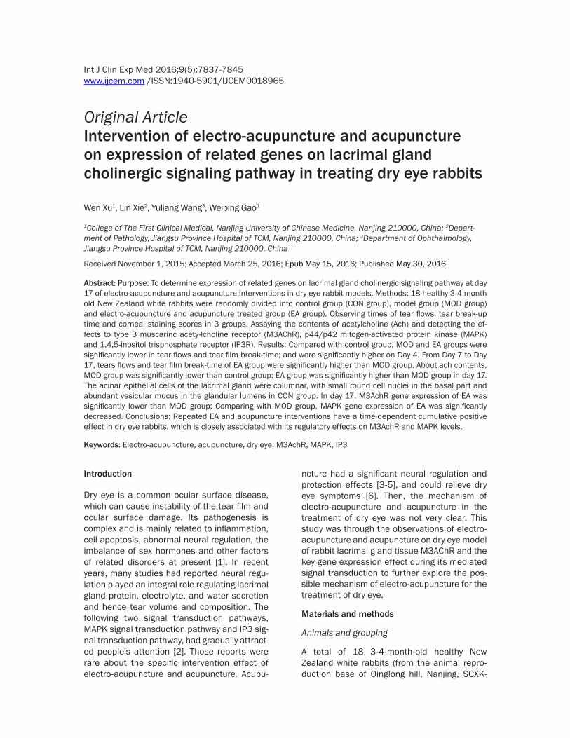
Figure 2. A. Tear flows of the rabbits in each group at different time-points. (*P < 0.001 vs. day 0, #P < 0.001 vs. Day 4 **P < 0.001 vs. MOD group). B. Tear film break-time of the rabbits in each group at different time-points. (*P < 0.001 vs. day 0, #P < 0.001 vs. day 4, **P < 0.001 vs. MOD group). C. Corneal staining scores of the rabbits in each group at different time-points. (*P < 0.001 vs. day 0, #P < 0.001 vs. day 4, **P < 0.001 vs. MOD group).
seconds later, corneal epithelial damage was graded with a co- balt blue filter under a slit-lamp microscope (Kanghua Science & Technology Co., Ltd, Chongqing, China). The cornea over the cor-nea center to do two vertical and horizontal lines was divided into 4 equal portions, which were scored respectively. The 4 scores were added to arrive at a final grade (total: 16 points). The fluo-rescein score was analyzed as previously described [8] with essential modification, briefly, as follows; each decile 0-3 degree, 0, no staining, 1; slightly punc-tate staining less than 5 spots; two dyed into five or more, divid-ed into three massive stained, and finally scored each decile of the sum out of 12 points.
Sampling
The normal group, model group, and EA group were given Schirmer I test; tear film break-up time, corneal staining scores on days 0, 4, 7, 10, 17, at a simi-lar time of the day (7AM) in the standard environment. Rabbits were killed humanely at 17 days after dry eye rabbit models were made, along with controls. Fo- llowing sacrifice, whole blood and sera were collected and stored at -80°C. Each lacrimal gland (right and left) was divided into three pieces: one fixed in 10% formalin solution for histo-logical analysis, one for ELISA analysis, and one stored at -80°C. All inspections were con-ducted by the same person in the same environment in a dou-ble-blind way.
Enzyme-linked immunosorbent assay (ELISA) in lacrimal gland
On day 17, lacrimal gland tissues were measured by enzyme-linked immunosorbent assay ACh content immediately after removing the right side of the sacrificed rabbit. Homogenizati-

Intervention of electro-acupuncture and acupuncture in treating dry eye rabbits
7840 Int J Clin Exp Med 2016;9(5):7837-7845
on was carried out on ice using a tissue homog-enizer and incubated for 1 min at 4°C with shaking. Homogenates were centrifuged and supernatants were collected. Protein concen-trations were estimated by the procedure of Taylor et al. [9] with BSA as the standard. The Ach concentration was measured by a competi-tive enzyme-linked immunoassay (ELISA) using a rabbit polyclonal Ach antibody (Nanjing sai yan Technology Development Co. Ltd., Nanjing, China) according to the manufacturer’s proto-col. Samples (or standard) and conjugate were added to each well, and the plate was incubat-ed for 1 h at room temperature without block-ing. After wells were washed several times with buffers and proper color developed, the optical density was measured at 450 nm using an ELISA reader (MutiRead 400; Authos Co., Vienna, Austria).
Histologic analysis
Lacrimal gland tissues for light microscopy were fixed in 10% neutral buffered formalin and embedded in paraffin using routine proce-dures. Four-micrometer thick sections were cut from the tissue blocks and stained with hema-toxylin-eosin (H&E). For H&E staining, the lacri-mal gland histologic evaluation revealed mor-phological change in lacrimal gland after acu- puncture and electro-acupuncture treatment.
Immunohistochemical analysis
The expression of anti-M3AchR, anti-MAPK, and anti-IP3 antigens (MaxVisionTM, Fuzhou
Figure 3. The contents of Ach in each group on day 17. (*P < 0.05 MOD vs. CON, #P < 0.05 EA vs. MOD).
Maixin Biotech. Co., Ltd) was examined in histological paraffin sections. For this, sections mea-suring 4 μm in thickness were collected on silanized slides and subsequently dewaxed in xylene and hydrated in decreasing con-centrations of ethanol. Tissues were then placed in citrate buffer (pH 6.0) and processed for heat-induced epitope retrieval for 10 minutes at 95°C. Slides were washed in deionized water, fol-lowed by buffer. A primary anti-body (1:50-100dilution, Fuzhou Maixin Biotech. Co., Ltd) was applied for 60 minutes followed by buffer washes (2 washes × 5 minutes each). A commercial sec-ondary kit (MaxVisionTM, Fuzhou
Maixin Biotech. Co., Ltd) was applied according to the manufacturer’s recommendations (30 minutes) and followed by buffer washes (2 washes × 5 minutes each). Chromogen (DAB) was applied to tissues for 5 minutes followed by buffer rinses. Tissues were then counter-stained with hematoxylin. As a negative control, sections incubated with non-specific immuno-globulin were used in place of the primary anti-body. Tissues were examined by a pathologist. For immunohistochemical evaluation of M3A- chR, MAPK, and IP3, we used semi-quantitative scoring analysis (H) as described by A. Scharl D [10]. 0 for negative reactivity and 1 to 3 accord-ing to the degree of intensity of reactivity to M3AchR, MAPK, and IP3. All indicators are membrane and (or) light yellow to brown appear-ance of fine particles in the cytoplasm, was higher than the background color colored posi-tive. Classification criteria: according to the depth of the lacrimal gland tissue staining and the percentage of positive cells one by one score for each slice, the mean was chosen for the staining intensity. Staining intensity scoring criteria: percentage of positive cells < 10% is negative (-), 10%-25% as a pale brown weakly positive (+), 25%-75%, dark brown positive (++), > 75% tan strong positive (+++). For statistical convenience, semi-quantitative scoring was given as 0, 1, 2, 3, respectively -, +, ++, +++. P < 0.05 was considered statistically significant.
Real-time quantitative RT-PCR of mRNA
Total RNA was extracted from the lacrimal gland samples using an RNAprep Pure Tissue

Intervention of electro-acupuncture and acupuncture in treating dry eye rabbits
7841 Int J Clin Exp Med 2016;9(5):7837-7845
kit (Tiangen Biotech Co. Ltd., Beijing, China). RNA quality and concentration were assessed and confirmed using the Eppendorf BioSpe- ctrometer (Eppendorf, Germany). Extracted RNA was converted to cDNA through reverse transcription using the QuantiTect Reverse Transcription Kit (Qiagen). RT-PCR was per-
formed using the QuantiTect SYBR Green PCR Kit (Qiagen). For the amplification of the desir- ed cDNA, the following gene-specific primers were used: GAPDH (housekeeping gene), M3- AchR, MAPK, and IP3. Primers were synthesiz- ed by Shanghai Sangon Biological Engineering (Shanghai, China). GAPDH as an internal refer-
Figure 4. HE staining results of the lacrimal gland acinar epithelial cells. In the pictures, the red arrow shows lym-phocyte cell, while the blue arrow shows acinar cell. A. CON group × 100; B. CON group × 400; C. MOD group × 100; D. MOD group × 400; E. EA group × 100; F. EA group × 400.
Intervention of electro-acupuncture and acupuncture in treating dry eye rabbits
7842 Int J Clin Exp Med 2016;9(5):7837-7845
ence, the upstream primer 5 ‘CAC GGT CAA GGC TGA GAA CG 3’, downstream primer 5 ‘GTA CTC GGC ACC AGC ATC AC 3’. CHRM3 upstream primer 5 ‘TTG ACA GGT ACT TTT CCA TC 3’, downstream primer: 5 ‘CAA GCT AGA CCA ATC ATC AC 3’; IP3R upstream primer: 5 ‘TAG CTG ACC GAA AGC AGA AT 3’, downstream primer: 5 ‘GCA AGC TCT TTG GGC TTC TC 3’; MAPK upstream primer: 5 ‘CCT CCA ACA TCC TGG TCA AC 3’, downstream primer: 5’ CCT GGT GCC CAC GAA GGA GT 3’. RT-PCR was conducted by bringing the temperature up to 95°C for 15 minutes, then 40 cycles of 15 seconds at 94°C, 30 seconds at 60°C, and 30 seconds at 72°C. The reactions for mRNA were automated by a 7900HT Real-Time PCR. For all RT-PCR experi-ments, sterile water was used as a negative control, and the murine housekeeping gene, GAP- DH, was used as a positive control. All quantita-tive PCR reactions, including no-template con-trols, were performed in triplicate. All calcula-tions and analyses were performed using SDS RQ Manager 1.1 software using the 2-ΔΔCt method [11].
Statistical analysis
Data from all groups were expressed as mean ± standard error of the mean. Statistical analy-
test, tear film break-time and corneal staining score. However, in day 4, MOD and AE groups were significantly lower in schirmer test and tear film break-time and higher in corneal stain-ing score compared with day0. Comparing with day 4, AE group was significantly increased in schirmer test and tear film break-time and decreased in corneal staining score. Moreover, in day 7, 10 and 17, AE group was also signifi-cantly improved schirmer test and tear film break-time, and significantly attenuated corne-al staining score compared with MOD group (data was shown in Figure 2A-C).
The contents of ach
About ach contents, MOD group was signifi-cantly lower than control group; EA group was significantly higher than MOD group in day 17 (P < 0.05, respectively) (Figure 3).
HE staining results
The acinar epithelial cells of the lacrimal gland were columnar, with small round cell nuclei in the basal part and abundant vesicular mucus in the glandular lumens in CON group (Figure 4A, 4B). In MOD group, we found the acinar was atrophied, lacking enough nuclei. We also found some inflammatory cells located in focal
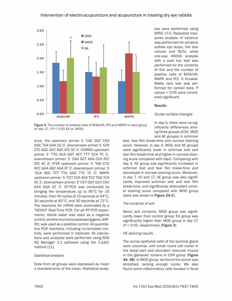
Figure 5. The number of positive cells of M3AchR, IP3 and MAPK in each group on day 17. (*P < 0.05 EA vs. MOD).
ses were performed using SPSS 17.0. Repeated mea-sures analysis of variance was performed for atropine sulfate eye drops, the tear volume and BUTs, while one-way ANOVA analysis with a post hoc test was performed for the contents of Ach and the number of positive cells of M3AchR, MAPK and IP3. A Kruskal-Wallis rank test was per-formed for ranked data. P values < 0.05 were consid-ered significant.
Results
Ocular surface changes
In day 0, there were no sig-nificantly differences amo- ng three groups (CON, MOD and AE groups) in schirmer

Intervention of electro-acupuncture and acupuncture in treating dry eye rabbits
7843 Int J Clin Exp Med 2016;9(5):7837-7845
areas (Figure 4C, 4D). In EA group, columnar epithelial cells expanded with abundant nuclei without inflammatory cells (Figure 4E, 4F).
The number of positive cells of M3AchR, MAPK and IP3
Immunohistochemiscal assay results showed that the number of positive cells in M3AchR was significantly increased in the electro-acu-puncture and acupuncture rabbits on day 17 (P < 0.05 EA vs. MOD). The number of positive cells in MAPK was significantly decreased in the electro-acupuncture and acupuncture rab-bits on day 17 (P < 0.05 EA vs.MOD). But the number of positive cells in IP3 had no obvious change in the electro-acupuncture and acu-puncture rabbits on day 17 (Figure 5). Cla- ssification criteria: 1) positive cells < 25% nega-tive (-); 2) positive cells in 25%-50% for weakly positive (+); 3) positive cells in 51%-75% posi-tive (++); 4) positive cells > 75% as strongly positive (+++). Its statistics, negative, weakly positive, positive, strong positive respectively denoted as 0, 1, 2, 3.
Gene expression of M3AchR, MAPK and IP3
In day 17, M3AchR gene expression of EA was significantly lower than MOD group; Comparing with MOD group, MAPK gene expression of EA was significantly decreased (Figure 6).
Discussion
Acupuncture is a traditional Chinese medicine and as an important part of treatment, it has been used to treat different types of diseases.
In many countries it is now being accepted as a complementary and alternative therapy. Animal and human experiments indicated that acu-puncture could be related to the release of opi-oids for pain relief, immune regulation, the release of neurotransmitters and hormones, as well as the dilation of blood vessels [11]. Many articles internationally had reported acupunc-ture had an undoubted efficacy in the treat-ment of dry eye [12-16]. EA is a new therapy, referring to the method of the pulse current applied to acupuncture needles and thus uni-form stimulation of acupuncture points, in order to ensure quantitative and qualitative unity [17]. EA at some acupoints can stimulate the cholinergic nerves [18, 19]. The experi-ments selected ching ming (BL1), cuan zu (BL2), sizukong temple (TE23), temporal (Extra-1), and tongziliao (GB1). Among them, ching ming (BL1), an acupoint of meridian of foot tai yang and the convergence of five meridians, can clear stagnated heat and is a key acupunc-ture point of treating ocular diseases. Cuan zu (BL2) an acupoint of the bladder meridian of foot tai yang, adjusts the qi and blood round the eye, and nourishing yin and clear away heat. Temporal (Extra-1) is an extraordinary point for attending eye diseases; tongziliao (GB1) is the first orifice of head and facial of gallbladder meridian of foot shao yang. It also can dispel wind, purge heat and improve eyesight. Sizu- kong temple (TE23) is an acupoint of the merid-ian of hand shao yang, attending diseases-con-gestion, swelling and pain of the eye. An orbital zygomatic branch of facial nerve and auriculo-temporal nerve branches are around sizukong temple (TE23), where the cloth zygomatic branch of the facial nerve and ear temporal nerve branches. The lacrimal gland secretion fibers are from the parasympathetic nerve of the facial nerve and sympathetic fibers of intra-cranial arterial plexus. So in the study, selecting these points stimulated the parasympathetic to release neurotransmitters, in order to promote lacrimal gland secretion. From the meridian and collateral circulation paths, the twelve meridians are related to the eye directly or indi-rectly, and surround the eye. So the theoretical base of acupuncture for the treatment of dry eye is to make better flow ‘qi’ and blood in the meridians around the eye, to balance Yin and Yang so as to open the lacrimal orifice [20].
Dry eye has long been considered a complicat-ed condition and its incidence rate has
Figure 6. M3AchR, IP3 and MAPK in each group on day 17 were assessed by Real-time PCR. 2-ΔΔCt was as the relative content. (*P < 0.05 EA vs. MOD).

Intervention of electro-acupuncture and acupuncture in treating dry eye rabbits
7844 Int J Clin Exp Med 2016;9(5):7837-7845
increased yearly. Abnormal neural regulation is one common cause, which can reduce tear secretion, eventually forming aqueous-deficien-cy dry eye. In this study, Comparing with Day 0, CON and EA groups were significantly decreased in tears flow and tear film break-up time, and significantly increased in corneal staining score (P < 0.05, respectively). Those results showed that the dry eye model was success. EA group was given electro-acupuncture and acupunc-ture treatment. From Day 7 to Day 17, Compared with MOD group, EA group was significantly higher in tears flow and tear film break-up time (P < 0.05, respectively), and was significantly lower in corneal staining score (P < 0.05, respectively). This result showed the electro-acupuncture and acupuncture treatment had effects to improve dry eye. However, the path-way was not clear.
The lacrimal gland is controlled by innervation, and the nerve terminal mainly contains neu-rotransmitter acetylcholine [21]. The choliner-gic receptors on lacrimal gland cells are the muscarinic M3 (glandular) subtype (M3AchR) [22]. M3AchR for neurotransmitter Ach and lac-rimal M3 cholinergic receptors are formed as the starting protein secretion pathway. The M3AchR is coupled to the Gq/11a subtype of G protein that is then connected to activate the enzyme phospholipase Cb, which breaks down the membrane phospholipids phosphatidylino-sitol bisphosphate into 1, 4, 5-inositol trisphos-phate (InsP3) and diacylglycerol (DAG) [23]. InsP3 is water-soluble and binds to its receptor on the endoplasmic reticulum. There are 3 dis-tinct subtypes of InsP3 receptors: types 1-3 that are expressed to varying degrees on in dif-ferent cell subtypes. InsP3 interaction with its receptor causes a rapid, immediate release of intracellular Ca2+ represented by a spike in Ca2+ response that quickly peaks [24]. Experiments confirmed IP3 may play a role in the connection gap that can be reduced to some extent in apoptosis [25]. Surprisingly, cholinergic ago-nists not only activate pathways that stimulate secretion, they also activate those that attenu-ate secretion. Cholinergic agonists activate p44-p42 mitogen-activated protein kinase (MAPK), also known as extracellular regulated kinase (ERK) 1/2, which decreases cholinergic agonist-activated protein secretion [26]. In this reach Day 17, Ach contents of EA group were significantly higher than MOD group (P < 0.05);
In EA group, M3AchR Protein and mRNA expres-sion were significantly increased and MAPK protein and mRNA expression were significant- ly decreased compared with MOD group (P < 0.05), However, the IP3 protein and mRNA expression were no significant between these two groups. Depend on these results; we inferred the Ach contents increase by inhibiting MAPK and stimulating M3AchR.
In this research, the rabbits were treated with 1% atropine sulfate eye drops to make dry eye models [4], the results showed that the model was success. From Day 7, the symptoms of EA group was significantly improved. The results of this study suggested electro-acupuncture and acupuncture treatment (EA group used treat-ment methods) improve dry eye by improving Ach contents, and Ach contents were increased by stimulating M3AchR and inhibiting MAPK.
Acknowledgements
Supported by the Natural Science Foundation of China (NSFC No. 81373746), the Natural Science Foundation of Jiangsu (No. BK2012- 856). The funders had no role in study design, data collection and analysis, decision to pub-lish, or preparation of the manuscript.
Disclosure of conflict of interest
None.
Address correspondence to: Weiping Gao, College of The First Clinical Medical, Nanjing University of Chinese Medicine, 282 Han Zhong Road, Nanjing 210000, China. E-mail: [email protected]
References
[1] Baudouin C. The pathology of dry eye. Surv Ophthalmol 2001; 45: 211-220.
[2] Dartt DA. Neural regulation of lacrimal gland secretory processes: relevance in dry eye dis-eases. Prog Retin Eye Res 2009; 28: 155-177.
[3] Fukuta H, Koshita M, Nakamura E, Nakamura H, Yamada A, Kawase Y, Ishigami T, Kurono Y, Iino S, Suzuki H. Acupuncture modulates me-chanical responses of smooth muscle pro-duced by transmural nerve stimulation in gas-tric antrum of genetically hyperglycemic rats. J Smooth Muscle Res 2009; 45: 167-85.
[4] Lee B, Sur B J, Kwon S, Jung E, Shim I, Lee H, Hahm DH. Acupuncture stimulation alleviates

Intervention of electro-acupuncture and acupuncture in treating dry eye rabbits
7845 Int J Clin Exp Med 2016;9(5):7837-7845
corticosterone-induced impairments of spatial memory and cholinergic neurons in rats. Evid Based Complement Alternat Med 2011; 2012: 670536-670536.
[5] Lee B, Sur B, Shim J, Hahm DH, Lee H. Acu-puncture stimulation improves scopolamine-induced cognitive impairment via activation of cholinergic system and regulation of BDNF and CREB expressions in rats. BMC Complement Altern Med 2014; 14: 338-338.
[6] Jang H, Lee S, Kim TH, Kim AR, Lee M, Lee JH. Acupuncture for dry eye syndrome after refrac-tive surgery: study protocol for a randomized controlled trial. Trials 2013; 14: 351-351.
[7] Li ZR. Experimental acupuncture. China Press of Traditional Chinese Medicine 2003; 314-319.
[8] Burgalassi S, Panichi L, Chetoni P, Saettone MF, Boldrini E. Development of a simple dry eye model in the albino rabbit and evaluation of some tear substitutes. Ophthalmic Res 1999; 31: 229-235.
[9] Taylor BM, Kolbasa KP, Chin JE, Richards IM, Fleming WE, Griffin RL, Fidler SF, Sun FF. Roles of adhesion molecules ICAM-1 and alpha4 in-tegrin in antigen-induced changes in microvas-cular permeability associated with lung inflam-mation in sensitized brown Norway rats. Am J Respir Cell Mol Biol 1997; 17: 757-766.
[10] Scharl A, Vierbuchen M, Conradt B, Moll W, Würz H, Bolte A. Immunohistochemical detec-tion of progesterone receptor in formalin-fixed and paraffin-embedded breast cancer tissue using a monoclonal antibody. Arch Gynecol Ob-stet 1990; 247: 63-71.
[11] Livak KJ, Schmittgen TD. Analysis of relative gene expression data using real-time quantita-tive PCR and the 2-ΔΔCt Method. Methods 2001; 25: 402-408.
[12] Leake R, Broderick JE. Current licensure for acupuncture in the United States. Altern Ther Health Med 1999; 5: 94-96.
[13] Kim TH, Kang JW, Kim KH, Kang KW, Shin MS, Jung SY, Kim AR, Jung HJ, Choi JB, Hong KE, Lee SD, Choi SM. Acu3puncture for the treat-ment of dry eye: a multicenter randomised controlled trial with active comparison inter-vention (artificial teardrops). PLoS One 2012; 7: 36638-36638.
[14] Gong L, Sun X. Treatment of intractable dry eyes: tear secretion increase and morphologi-cal changes of the lacrimal gland of rabbit af-ter acupuncture. Acupunct Electrother Res 2007; 32: 223-233.
[15] Lan WW, Tong L. Comments: Acupuncture for treating dry eye: a randomized placebo-con-trolled trial. Acta Ophthalmol 2011; 89: 371-372.
[16] Gong L, Sun X, Chapin WJ. Clinical curative ef-fect of acupuncture therapy on xerophthalmia. Am J Chin Med 2010; 38: 651-659.
[17] Humaidan P, Stener-Victorin E. Pain relief dur-ing oocyte retrieval with a new short duration electro-acupuncture technique-an alternative to conventional analgesic methods. Hum Rep 2004; 19: 1367-1372.
[18] Lee YC, Li TM, Tzeng CY, Chen YI, Ho WJ, Lin JG, Chang SL. Electroacupuncture at the zusanli (ST-36) acupoint induces a hypoglycemic ef-fect by stimulating the cholinergic nerve in a rat model of streptozotocine-induced insulin-dependent diabetes mellitus. Evid Based Com-plement Alternat Med 2011; 2011: 650263.
[19] Hsieh CL, Lin JG, Li TC. Changes of pulse rate and skin temperature evoked by electroacu-puncture stimulation with different frequency on both Zusanli acupoints in humans. Am J Chin Med 1999; 27: 11-18.
[20] Yang CS. Acupuncture Therapy. Shanghai: Shanghai Scientific & Technical Publish; 2002. pp. 163-165.
[21] Funaki C, Hodges RR, Dartt DA. Identification of the Raf-1 signaling pathway used by cAMP to inhibit p42/p44 MAPK in rat lacrimal gland acini: role in potentiation of protein secretion. Invest Ophthalmol Vis Sci 2010; 51: 6321-6328.
[22] Hootman SR, Picado-Leonard TM, Burnham DB. Muscarinic acetylcholine receptor struc-ture in acinar cells of mammalian exocrine glands. J Biol Chem 1985; 260: 4186-4194.
[23] Godfrey PP, Putney JW. Receptor-mediated me-tabolism of the phosphoinositides and phos-phatidic acid in rat lacrimal acinar cells. Bio-chem J 1984; 218: 187-195.
[24] Putney JW Jr. Type 3 inositol 1, 4, 5-trisphos-phate receptor and capacitative calcium entry. Cell Calcium 1997; 21: 257-261.
[25] Decrock E, Krysko DV, Vinken M, Kaczmarek A, Crispino G, Bol M, Wang N, De Bock M, De Vuyst E, Naus CC, Rogiers V, Vandenabeele P, Erneux C,Mammano F, Bultynck G, Leybaert L. Transfer of IP3 through gap junctions is critical, but not sufficient, for the spread of apoptosis. Cell Death Differ 2011; 19: 947-957.
[26] Ota I, Zoukhri D, Hodges RR, Rios JD, Te-pavcevic V, Raddassi I, Chen LL, Dartt DA. α1-Adrenergic and cholinergic agonists activate MAPK by separate mechanisms to inhibit se-cretion in lacrimal gland. Am J Physiol Cell Physiol 2003; 284: 168-178.