optokinetic nystagmus in harbor seals (phoca vitulina)
TRANSCRIPT
Available online at www.sciencedirect.com
www.elsevier.com/locate/visres
Vision Research 48 (2008) 304–315
Optokinetic nystagmus in harbor seals (Phoca vitulina)
Frederike D. Hanke a, Wolf Hanke b, Klaus-Peter Hoffmann a, Guido Dehnhardt a,*
a University of Bochum, General Zoology & Neurobiology, ND 7/31, D-44780 Bochum, Germanyb Organismic and Evolutionary Biology, Harvard University, Lauder Laboratory, 26 Oxford Street, Cambridge, MA 02138, USA
Received 12 June 2007; received in revised form 24 August 2007
Abstract
Harbor seals experience motion due to self-motion and to movement in the external world. However, motion vision has not beenstudied yet in marine mammals moving in the underwater world. To open up this research, optokinetic nystagmus (OKN) as a basicmotion sensing and retinal image stabilizing reflex was studied in four harbor seals during stimulation with moving black-and-whitestripe patterns. All seals responded with optokinetic eye movements. Detailed measurements obtained with one animal revealed a mod-erate gain for horizontal binocular OKN. Monocularly stimulated, the seal displayed a symmetrical OKN with slightly strongerresponses to leftward moving stimuli, and, surprisingly, a symmetrical OKN was found in the vertical domain.� 2007 Elsevier Ltd. All rights reserved.
Keywords: Harbor seal; Phoca vitulina; Optokinetic nystagmus; Eye movements; Motion vision
1. Introduction
The origin of perceived motion can lie in the subjectitself, in the external world or in a combination of both.If an object is in motion, the observer’s eyes usually movein pursuit of it in order to stabilize the object’s image on theretina and, therefore, to retain a high level of resolvingpower (Yarbus, 1967). When a large portion of the visualfield moves, the eyes move smoothly with the field (slowphase) interrupted by saccades (fast phase) in the oppositedirection. This rhythmic oscillation of the eye is calledoptokinetic nystagmus (OKN). In concert with othermechanisms like the smooth pursuit system or the vestibu-lar-ocular reflex (VOR), the OKN nullifies or at leastreduces the slip of the retinal image (Collewijn, 1985;Schor, 1993) caused by rotations of eye, head or body.Consequently, images on the retina are stabilized (Walls,1942, 1962) and conditions for clear and unblurred visionmaintained.
0042-6989/$ - see front matter � 2007 Elsevier Ltd. All rights reserved.doi:10.1016/j.visres.2007.11.012
* Corresponding author. Fax: +49 221 97750604.E-mail address: [email protected] (G. Dehnhardt).
Optokinetic nystagmus (OKN) has been studied indetail in a variety of organisms. But so far, no studies haveanalyzed motion vision including basic principles, e.g. theability to perform optokinetic eye movements, in marinemammals. This field of research is of great interest espe-cially in marine mammals, as, on the one hand, they movein the low structured, three-dimensional underwater world,and, on the other hand, their own locomotion involves,beside translation, rotations along all axis. Thus, visualperception might be different from that of terrestrial mam-mals (Schusterman & Thomas, 1966), and general featuresof the optokinetic nystagmus found in terrestrial speciescould also be different in marine mammals.
It appears highly probable that marine mammals ingeneral and harbor seals in particular possess the abilityof seeing motion. Harbor seals are amphibious mammals.Under water, they prey on diverse vertebrate and inverte-brate species which are normally moving. The ability tosee motion would imply that the predator can see themoving object from a significantly greater distance thanif it was static. Furthermore, motion provides the preda-tor with important information about direction anddistance, because near features move faster across the
F.D. Hanke et al. / Vision Research 48 (2008) 304–315 305
retina than distant ones (Miles, 1998). Motion vision alsohelps to separate figure from background, and motioninformation helps to detect and assess self-motion(Nakayama, 1985). The OKN stabilizes the eye withrespect to whole-field motion which enhances the sensitiv-ity to individual moving objects, e.g. prey, in the visualfield (Schor, 1993). This is of high importance concerningthe detection of prey and the onset of smooth pursuit tosuccessfully hunt it.
In line with these considerations, we show in this studythat a marine mammal, the harbor seal, shows optokineticresponses under water. On the one hand, we tested underwater because harbor seals are nearly emmetropic in water(Hanke, Dehnhardt, Schaeffel, & Hanke, 2006), and allessential activities requiring high level performance of thesensory organs are displayed in this medium. On the otherhand, we tested under water because we wanted to revealpossible adaptations to the underwater environment har-bor seals share with fully aquatic species, as e.g. fish. Theoptokinetic systems of fish have been studied already (seee.g. Dieringer, Reichenberger, & Graf, 1992; Easter,1972; Marsh & Baker, 1997). However, only results forhorizontal OKN are reported, and these are very variableamong (and within) species. Dieringer et al. (1992) proposethat the optokinetic responses may reflect species specificdifferences in the movement of the organism or in theenvironment.
We tried to assess the nature of optokinetic eye move-ments in the horizontal domain under binocular andmonocular viewing conditions to compare them to resultsobtained in fish and to theoretical predictions from thevisual consequences of forward locomotion and eyeplacement (Grasse & Cynader, 1988) which have beendiscussed for several terrestrial species. During forwardmovement, flow fields in frontally eyed terrestrial animalsare composed of flow lines with the predominant asym-metries in the vertical and no significant asymmetrybetween naso-temporal and temporal-nasal. Frontal eyeplacement therefore leads to symmetrical binocularOKN as a response to a leftward and rightward horizon-tal stimulus movement. As harbor seals are frontallyeyed, one could expect to find a symmetric horizontalOKN.
The vertical OKN in terrestrial species investigated sofar shows a remarkable asymmetry with higher gain forupward moving stimuli (Grasse & Cynader, 1988; Matsuo& Cohen, 1984; Takahashi & Igarashi, 1977). The reducedsensitivity to a downward moving stimulus has beenexplained in terms of preventing the eye from rotatingdownwards while walking over a highly-textured ground(Schor, 1993). In the swimming harbor seal, such a texturedground does not play a prominent role because the seal fre-quently experiences the ground, the water surface, both, ornone of them. The orientation to the ground and the sur-face can change when the seal rolls its body. With respectto its aquatic lifestyle, we considered it interesting to inves-tigate the vertical OKN in the harbor seal.
Under monocular viewing conditions, frontal-eyed ver-tebrates with an area of high resolution in the retina dis-play a symmetric response to horizontally movingstimuli. The question whether there is a high resolutionarea (area centralis) in the retina of the harbor seal is notyet answered. According to Jamieson and Fisher (1971),harbor seals do not possess any kind of area centralis. Incontrast to this finding, recent studies on retinal topogra-phy in other seals (Stellar sea lion, Mass and Supin,2005; Harp seal, Mass & Supin, 2003; Fur seal, Mass &Supin, 1992) have always assessed a definite area of highganglion cell density similar to the area centralis in terres-trial carnivores. Therefore, we would expect to find a highresolution area in the retina of harbor seals with moderntechniques as well, which, together with frontal eye place-ment, make a symmetric monocular OKN in harbor sealslikely.
2. Material and methods
2.1. Experimental animals
Harbor seals were studied in our Marine Mammal Laboratory in theZoo of Cologne, Germany. The optokinetic response (OKN) was testedin four male animals between 7 and 12 years of age (maximum age of har-bor seals in captivity and in the wild up to 35 yrs, cited after King (1983)and our own records). All animals had previous experience with visualtasks. The data of the velocity tuning of horizontal binocular OKN pre-sented in this study represent measurements of one animal (‘‘Henry’’, 9yrs old). Three additional seals (‘‘Sam’’, ‘‘Bill’’, ‘‘Nick’’) were tested forthe presence or absence of optokinetic responses under binocular viewingconditions but were not quantified further. Vertical binocular and monoc-ular OKN was examined in one seal (‘‘Henry’’).
The experiments were conducted according to the guidelines of theGerman animal protection law.
2.2. Optokinetic measurements
2.2.1. Experimental set-up
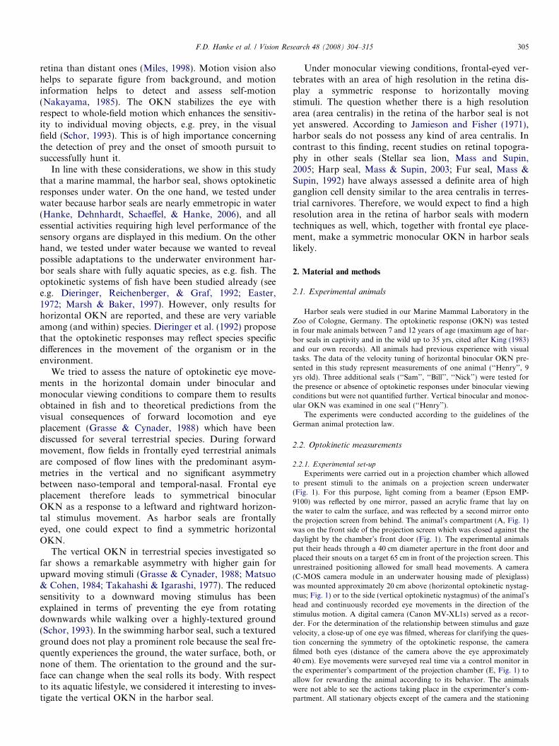
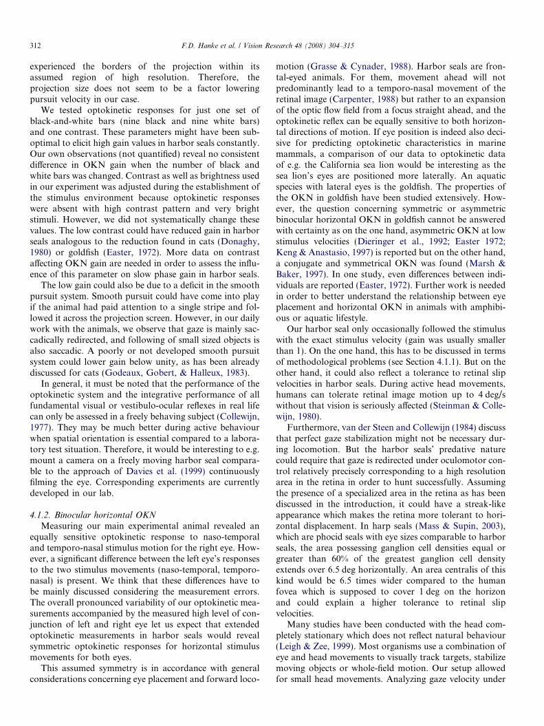
Experiments were carried out in a projection chamber which allowedto present stimuli to the animals on a projection screen underwater(Fig. 1). For this purpose, light coming from a beamer (Epson EMP-9100) was reflected by one mirror, passed an acrylic frame that lay onthe water to calm the surface, and was reflected by a second mirror ontothe projection screen from behind. The animal’s compartment (A, Fig. 1)was on the front side of the projection screen which was closed against thedaylight by the chamber’s front door (Fig. 1). The experimental animalsput their heads through a 40 cm diameter aperture in the front door andplaced their snouts on a target 65 cm in front of the projection screen. Thisunrestrained positioning allowed for small head movements. A camera(C-MOS camera module in an underwater housing made of plexiglass)was mounted approximately 20 cm above (horizontal optokinetic nystag-mus; Fig. 1) or to the side (vertical optokinetic nystagmus) of the animal’shead and continuously recorded eye movements in the direction of thestimulus motion. A digital camera (Canon MV-XL1s) served as a recor-der. For the determination of the relationship between stimulus and gazevelocity, a close-up of one eye was filmed, whereas for clarifying the ques-tion concerning the symmetry of the optokinetic response, the camerafilmed both eyes (distance of the camera above the eye approximately40 cm). Eye movements were surveyed real time via a control monitor inthe experimenter’s compartment of the projection chamber (E, Fig. 1) toallow for rewarding the animal according to its behavior. The animalswere not able to see the actions taking place in the experimenter’s com-partment. All stationary objects except of the camera and the stationing
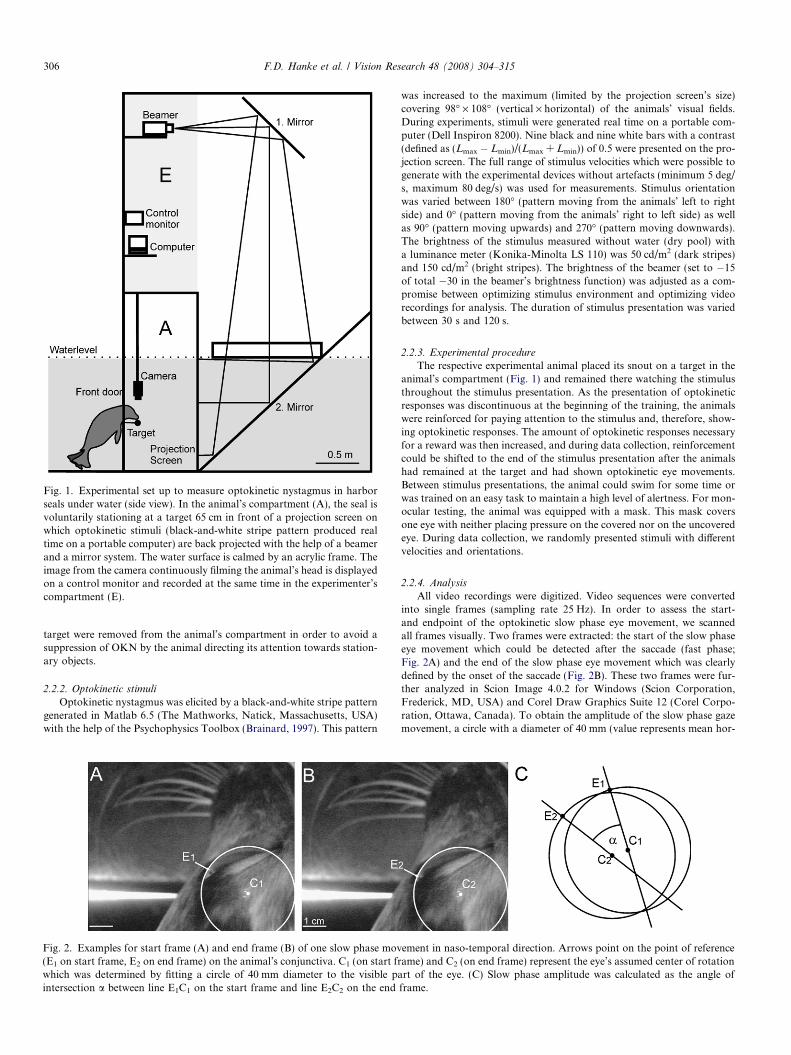
Fig. 1. Experimental set up to measure optokinetic nystagmus in harborseals under water (side view). In the animal’s compartment (A), the seal isvoluntarily stationing at a target 65 cm in front of a projection screen onwhich optokinetic stimuli (black-and-white stripe pattern produced realtime on a portable computer) are back projected with the help of a beamerand a mirror system. The water surface is calmed by an acrylic frame. Theimage from the camera continuously filming the animal’s head is displayedon a control monitor and recorded at the same time in the experimenter’scompartment (E).
306 F.D. Hanke et al. / Vision Research 48 (2008) 304–315
target were removed from the animal’s compartment in order to avoid asuppression of OKN by the animal directing its attention towards station-ary objects.
2.2.2. Optokinetic stimuli
Optokinetic nystagmus was elicited by a black-and-white stripe patterngenerated in Matlab 6.5 (The Mathworks, Natick, Massachusetts, USA)with the help of the Psychophysics Toolbox (Brainard, 1997). This pattern
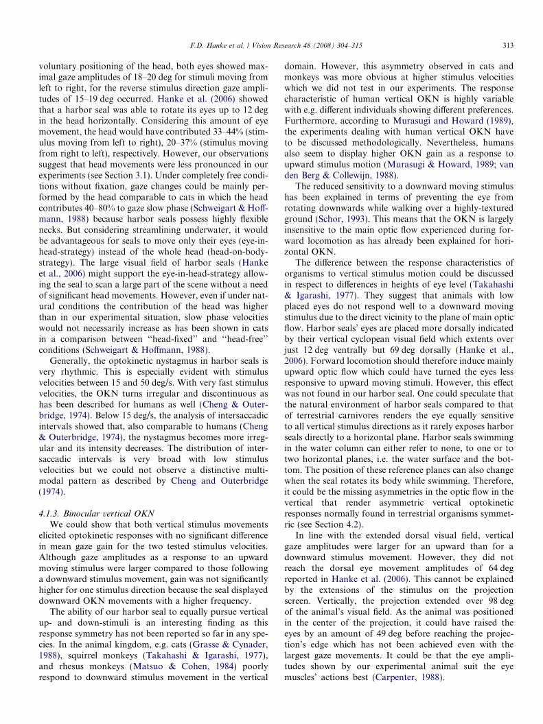
Fig. 2. Examples for start frame (A) and end frame (B) of one slow phase mov(E1 on start frame, E2 on end frame) on the animal’s conjunctiva. C1 (on start frwhich was determined by fitting a circle of 40 mm diameter to the visible paintersection a between line E1C1 on the start frame and line E2C2 on the end
was increased to the maximum (limited by the projection screen’s size)covering 98� · 108� (vertical · horizontal) of the animals’ visual fields.During experiments, stimuli were generated real time on a portable com-puter (Dell Inspiron 8200). Nine black and nine white bars with a contrast(defined as (Lmax � Lmin)/(Lmax + Lmin)) of 0.5 were presented on the pro-jection screen. The full range of stimulus velocities which were possible togenerate with the experimental devices without artefacts (minimum 5 deg/s, maximum 80 deg/s) was used for measurements. Stimulus orientationwas varied between 180� (pattern moving from the animals’ left to rightside) and 0� (pattern moving from the animals’ right to left side) as wellas 90� (pattern moving upwards) and 270� (pattern moving downwards).The brightness of the stimulus measured without water (dry pool) witha luminance meter (Konika-Minolta LS 110) was 50 cd/m2 (dark stripes)and 150 cd/m2 (bright stripes). The brightness of the beamer (set to �15of total �30 in the beamer’s brightness function) was adjusted as a com-promise between optimizing stimulus environment and optimizing videorecordings for analysis. The duration of stimulus presentation was variedbetween 30 s and 120 s.
2.2.3. Experimental procedure
The respective experimental animal placed its snout on a target in theanimal’s compartment (Fig. 1) and remained there watching the stimulusthroughout the stimulus presentation. As the presentation of optokineticresponses was discontinuous at the beginning of the training, the animalswere reinforced for paying attention to the stimulus and, therefore, show-ing optokinetic responses. The amount of optokinetic responses necessaryfor a reward was then increased, and during data collection, reinforcementcould be shifted to the end of the stimulus presentation after the animalshad remained at the target and had shown optokinetic eye movements.Between stimulus presentations, the animal could swim for some time orwas trained on an easy task to maintain a high level of alertness. For mon-ocular testing, the animal was equipped with a mask. This mask coversone eye with neither placing pressure on the covered nor on the uncoveredeye. During data collection, we randomly presented stimuli with differentvelocities and orientations.
2.2.4. Analysis
All video recordings were digitized. Video sequences were convertedinto single frames (sampling rate 25 Hz). In order to assess the start-and endpoint of the optokinetic slow phase eye movement, we scannedall frames visually. Two frames were extracted: the start of the slow phaseeye movement which could be detected after the saccade (fast phase;Fig. 2A) and the end of the slow phase eye movement which was clearlydefined by the onset of the saccade (Fig. 2B). These two frames were fur-ther analyzed in Scion Image 4.0.2 for Windows (Scion Corporation,Frederick, MD, USA) and Corel Draw Graphics Suite 12 (Corel Corpo-ration, Ottawa, Canada). To obtain the amplitude of the slow phase gazemovement, a circle with a diameter of 40 mm (value represents mean hor-
ement in naso-temporal direction. Arrows point on the point of referenceame) and C2 (on end frame) represent the eye’s assumed center of rotationrt of the eye. (C) Slow phase amplitude was calculated as the angle of
frame.
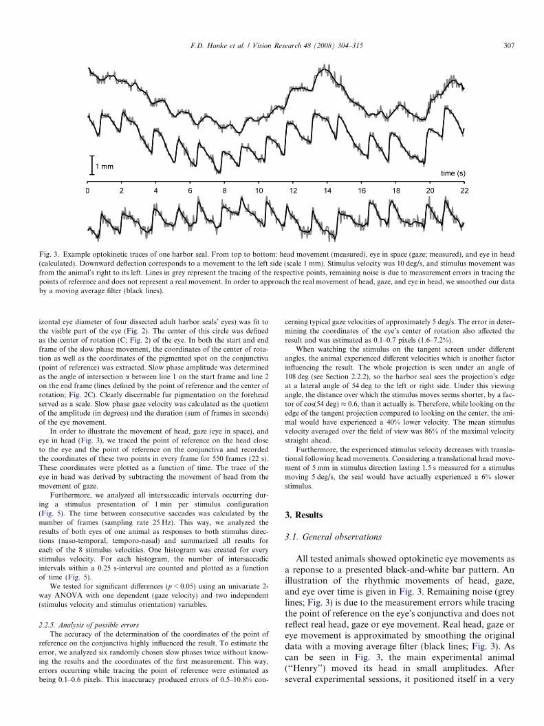
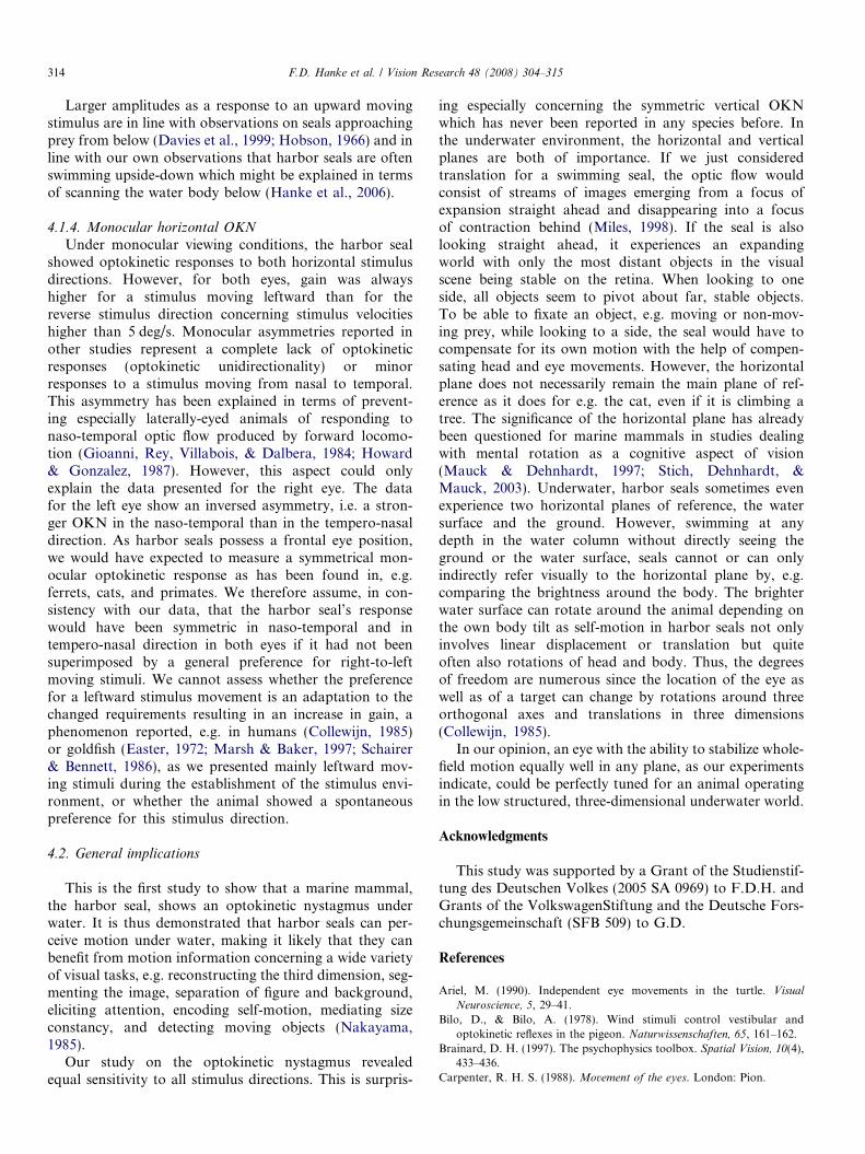
Fig. 3. Example optokinetic traces of one harbor seal. From top to bottom: head movement (measured), eye in space (gaze; measured), and eye in head(calculated). Downward deflection corresponds to a movement to the left side (scale 1 mm). Stimulus velocity was 10 deg/s, and stimulus movement wasfrom the animal’s right to its left. Lines in grey represent the tracing of the respective points, remaining noise is due to measurement errors in tracing thepoints of reference and does not represent a real movement. In order to approach the real movement of head, gaze, and eye in head, we smoothed our databy a moving average filter (black lines).
F.D. Hanke et al. / Vision Research 48 (2008) 304–315 307
izontal eye diameter of four dissected adult harbor seals’ eyes) was fit tothe visible part of the eye (Fig. 2). The center of this circle was definedas the center of rotation (C; Fig. 2) of the eye. In both the start and endframe of the slow phase movement, the coordinates of the center of rota-tion as well as the coordinates of the pigmented spot on the conjunctiva(point of reference) was extracted. Slow phase amplitude was determinedas the angle of intersection a between line 1 on the start frame and line 2on the end frame (lines defined by the point of reference and the center ofrotation; Fig. 2C). Clearly discernable fur pigmentation on the foreheadserved as a scale. Slow phase gaze velocity was calculated as the quotientof the amplitude (in degrees) and the duration (sum of frames in seconds)of the eye movement.
In order to illustrate the movement of head, gaze (eye in space), andeye in head (Fig. 3), we traced the point of reference on the head closeto the eye and the point of reference on the conjunctiva and recordedthe coordinates of these two points in every frame for 550 frames (22 s).These coordinates were plotted as a function of time. The trace of theeye in head was derived by subtracting the movement of head from themovement of gaze.
Furthermore, we analyzed all intersaccadic intervals occurring dur-ing a stimulus presentation of 1 min per stimulus configuration(Fig. 5). The time between consecutive saccades was calculated by thenumber of frames (sampling rate 25 Hz). This way, we analyzed theresults of both eyes of one animal as responses to both stimulus direc-tions (naso-temporal, temporo-nasal) and summarized all results foreach of the 8 stimulus velocities. One histogram was created for everystimulus velocity. For each histogram, the number of intersaccadicintervals within a 0.25 s-interval are counted and plotted as a functionof time (Fig. 5).
We tested for significant differences (p < 0.05) using an univariate 2-way ANOVA with one dependent (gaze velocity) and two independent(stimulus velocity and stimulus orientation) variables.
2.2.5. Analysis of possible errors
The accuracy of the determination of the coordinates of the point ofreference on the conjunctiva highly influenced the result. To estimate theerror, we analyzed six randomly chosen slow phases twice without know-ing the results and the coordinates of the first measurement. This way,errors occurring while tracing the point of reference were estimated asbeing 0.1–0.6 pixels. This inaccuracy produced errors of 0.5–10.8% con-
cerning typical gaze velocities of approximately 5 deg/s. The error in deter-mining the coordinates of the eye’s center of rotation also affected theresult and was estimated as 0.1–0.7 pixels (1.6–7.2%).
When watching the stimulus on the tangent screen under differentangles, the animal experienced different velocities which is another factorinfluencing the result. The whole projection is seen under an angle of108 deg (see Section 2.2.2), so the harbor seal sees the projection’s edgeat a lateral angle of 54 deg to the left or right side. Under this viewingangle, the distance over which the stimulus moves seems shorter, by a fac-tor of cos(54 deg) � 0.6, than it actually is. Therefore, while looking on theedge of the tangent projection compared to looking on the center, the ani-mal would have experienced a 40% lower velocity. The mean stimulusvelocity averaged over the field of view was 86% of the maximal velocitystraight ahead.
Furthermore, the experienced stimulus velocity decreases with transla-tional following head movements. Considering a translational head move-ment of 5 mm in stimulus direction lasting 1.5 s measured for a stimulusmoving 5 deg/s, the seal would have actually experienced a 6% slowerstimulus.
3. Results
3.1. General observations
All tested animals showed optokinetic eye movements asa reponse to a presented black-and-white bar pattern. Anillustration of the rhythmic movements of head, gaze,and eye over time is given in Fig. 3. Remaining noise (greylines; Fig. 3) is due to the measurement errors while tracingthe point of reference on the eye’s conjunctiva and does notreflect real head, gaze or eye movement. Real head, gaze oreye movement is approximated by smoothing the originaldata with a moving average filter (black lines; Fig. 3). Ascan be seen in Fig. 3, the main experimental animal(‘‘Henry’’) moved its head in small amplitudes. Afterseveral experimental sessions, it positioned itself in a very
308 F.D. Hanke et al. / Vision Research 48 (2008) 304–315
constant way in front of the screen with the body axisperpendicular to the projection showing pronounced eyemovements accompanied by small head movements.
The establishment of the stimulus condition proved tobe difficult. Our main experimental animal did neither reactwith optokinetic eye movements when presented with asmall sized (0.7 m · 1.5 m) random dot display nor withthe projection of a swarm of herring (comparable size asthe finally used black-and-white stripe pattern). Very fast,bright, and high contrast stripe patterns did not lead toan optokinetic nystagmus either. Under these circum-stances, the animal just showed saccades or no eye move-ments at all. The same observation was made when theanimal’s head was further fixed with a lower jaw station.Without any head fixation, head movements were per-formed against the stimulus direction coupled with eyemovements compensating head movements (vestibulo-ocu-lar reflex, VOR) or the animal was swimming vigorouslyagainst the front door of the projection chamber in direc-tion to the screen. Reducing the overall illumination inthe animal’s compartment and using a black-and-whitestripe pattern with reduced contrast (contrast = 0.5) finallyproved to be an appropriate optokinetic stimulusconfiguration.
After the establishment of the stimulus condition withour main experimental animal, the other animals showedoptokinetic responses when presented with the stimulusconfiguration for the first time. However, the rhythmic pat-tern was often interrupted by fixation phases.
During detailed measurements with our main experi-mental animal, all stimulus velocities resulted in the presen-tation of optokinetic responses. With the onset of thestimulus, the eyes always made a saccade against the stim-ulus orientation. The following eye movements rarely ledto the very temporal or nasal corner of the orbit, butinstead a saccade repositioned the eye more centrallybefore the following eye movement led to the maximal
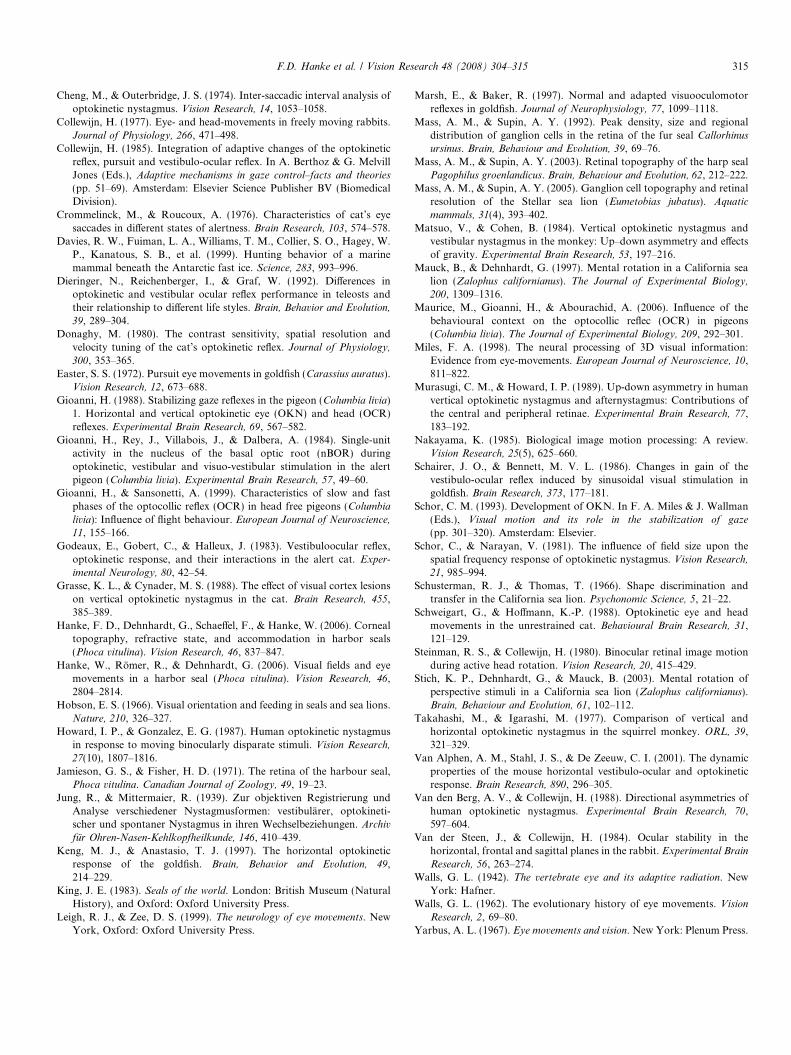
Fig. 4. Results of binocular horizontal optokinetic measurements of one harboplotted versus stimulus velocity for binocular viewing conditions for the lefillustrating the optokinetic response of the respective eye to temporo-nasally (
angle of rotation. Especially with stimuli moving temp-oro-nasally, the saccades did not reposition the eye in theoriginal starting position but the eyes continuously driftedto very temporal positions over the complete stimuluspresentation time. Thus, the average eye position (the‘‘Schlagfeld’’, Jung & Mittermaier, 1939) is shifted againstthe direction of stimulus motion. Velocities up to 20–25 deg/s were followed by an unbroken stream of optoki-netic nystagmus, but with higher velocities, the optokineticresponse was more and more discontinuous, and some-times saccades were elicited in direction of the stimulusand against it.
3.2. Binocular optokinetic nystagmus
3.2.1. Horizontal optokinetic nystagmus
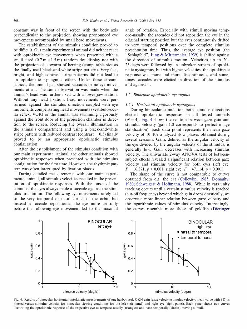
During binocular stimulation both stimulus directionselicited optokinetic responses in all tested animals(N = 4). Fig. 4 shows the relation between gaze gain andstimulus velocity (gain 1.0 corresponds to perfect imagestabilization). Each data point represents the mean gazevelocity of 10–109 analyzed slow phases obtained duringseveral sessions. Gain, defined as the angular velocity ofthe eye divided by the angular velocity of the stimulus, isgenerally low. Gain decreases with increasing stimulusvelocity. The univariate 2-way ANOVA tests of between-subject effects revealed a significant relation between gazevelocity and stimulus velocity for both eyes (left eye:F = 16.371, p < 0.001; right eye: F = 47.114, p < 0.001).
The shape of the curve is not comparable to curvesobtained from e.g. the cat (Collewijn, 1985; Donaghy,1980; Schweigart & Hoffmann, 1988). While in cats unitytracking occurs until a certain stimulus velocity is reached(cut-off frequency) beyond which gain drops drastically, weobserve a more linear relation between gaze velocity andthe logarithmic values of stimulus velocity. Interestingly,the curves resemble more those of goldfish (Dieringer
r seal. OKN gain (gaze velocity/stimulus velocity; mean value with SD) ist (left panel) and right eye (right panel). Each panel shows two curvestriangles) and naso-temporally (circles) moving stimuli.
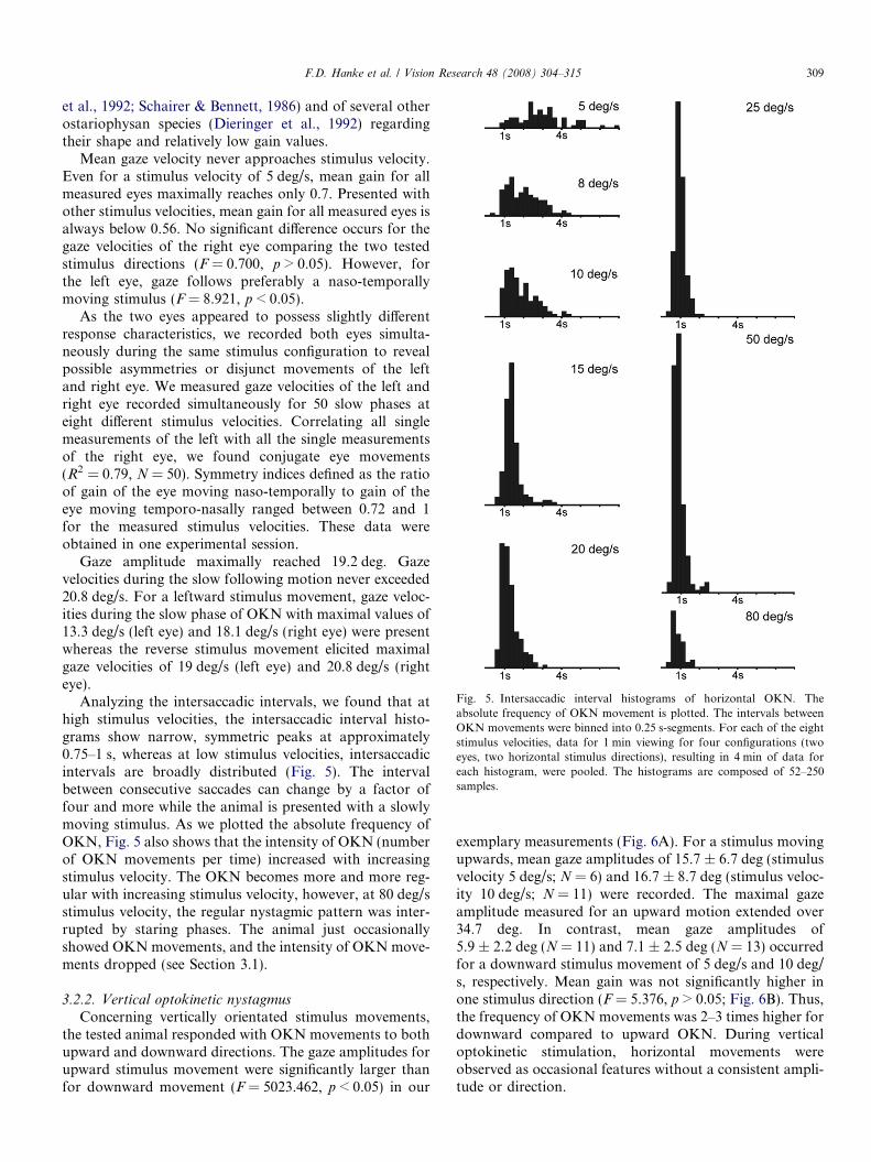
Fig. 5. Intersaccadic interval histograms of horizontal OKN. Theabsolute frequency of OKN movement is plotted. The intervals betweenOKN movements were binned into 0.25 s-segments. For each of the eightstimulus velocities, data for 1 min viewing for four configurations (twoeyes, two horizontal stimulus directions), resulting in 4 min of data foreach histogram, were pooled. The histograms are composed of 52–250samples.
F.D. Hanke et al. / Vision Research 48 (2008) 304–315 309
et al., 1992; Schairer & Bennett, 1986) and of several otherostariophysan species (Dieringer et al., 1992) regardingtheir shape and relatively low gain values.
Mean gaze velocity never approaches stimulus velocity.Even for a stimulus velocity of 5 deg/s, mean gain for allmeasured eyes maximally reaches only 0.7. Presented withother stimulus velocities, mean gain for all measured eyes isalways below 0.56. No significant difference occurs for thegaze velocities of the right eye comparing the two testedstimulus directions (F = 0.700, p > 0.05). However, forthe left eye, gaze follows preferably a naso-temporallymoving stimulus (F = 8.921, p < 0.05).
As the two eyes appeared to possess slightly differentresponse characteristics, we recorded both eyes simulta-neously during the same stimulus configuration to revealpossible asymmetries or disjunct movements of the leftand right eye. We measured gaze velocities of the left andright eye recorded simultaneously for 50 slow phases ateight different stimulus velocities. Correlating all singlemeasurements of the left with all the single measurementsof the right eye, we found conjugate eye movements(R2 = 0.79, N = 50). Symmetry indices defined as the ratioof gain of the eye moving naso-temporally to gain of theeye moving temporo-nasally ranged between 0.72 and 1for the measured stimulus velocities. These data wereobtained in one experimental session.
Gaze amplitude maximally reached 19.2 deg. Gazevelocities during the slow following motion never exceeded20.8 deg/s. For a leftward stimulus movement, gaze veloc-ities during the slow phase of OKN with maximal values of13.3 deg/s (left eye) and 18.1 deg/s (right eye) were presentwhereas the reverse stimulus movement elicited maximalgaze velocities of 19 deg/s (left eye) and 20.8 deg/s (righteye).
Analyzing the intersaccadic intervals, we found that athigh stimulus velocities, the intersaccadic interval histo-grams show narrow, symmetric peaks at approximately0.75–1 s, whereas at low stimulus velocities, intersaccadicintervals are broadly distributed (Fig. 5). The intervalbetween consecutive saccades can change by a factor offour and more while the animal is presented with a slowlymoving stimulus. As we plotted the absolute frequency ofOKN, Fig. 5 also shows that the intensity of OKN (numberof OKN movements per time) increased with increasingstimulus velocity. The OKN becomes more and more reg-ular with increasing stimulus velocity, however, at 80 deg/sstimulus velocity, the regular nystagmic pattern was inter-rupted by staring phases. The animal just occasionallyshowed OKN movements, and the intensity of OKN move-ments dropped (see Section 3.1).
3.2.2. Vertical optokinetic nystagmus
Concerning vertically orientated stimulus movements,the tested animal responded with OKN movements to bothupward and downward directions. The gaze amplitudes forupward stimulus movement were significantly larger thanfor downward movement (F = 5023.462, p < 0.05) in our
exemplary measurements (Fig. 6A). For a stimulus movingupwards, mean gaze amplitudes of 15.7 ± 6.7 deg (stimulusvelocity 5 deg/s; N = 6) and 16.7 ± 8.7 deg (stimulus veloc-ity 10 deg/s; N = 11) were recorded. The maximal gazeamplitude measured for an upward motion extended over34.7 deg. In contrast, mean gaze amplitudes of5.9 ± 2.2 deg (N = 11) and 7.1 ± 2.5 deg (N = 13) occurredfor a downward stimulus movement of 5 deg/s and 10 deg/s, respectively. Mean gain was not significantly higher inone stimulus direction (F = 5.376, p > 0.05; Fig. 6B). Thus,the frequency of OKN movements was 2–3 times higher fordownward compared to upward OKN. During verticaloptokinetic stimulation, horizontal movements wereobserved as occasional features without a consistent ampli-tude or direction.
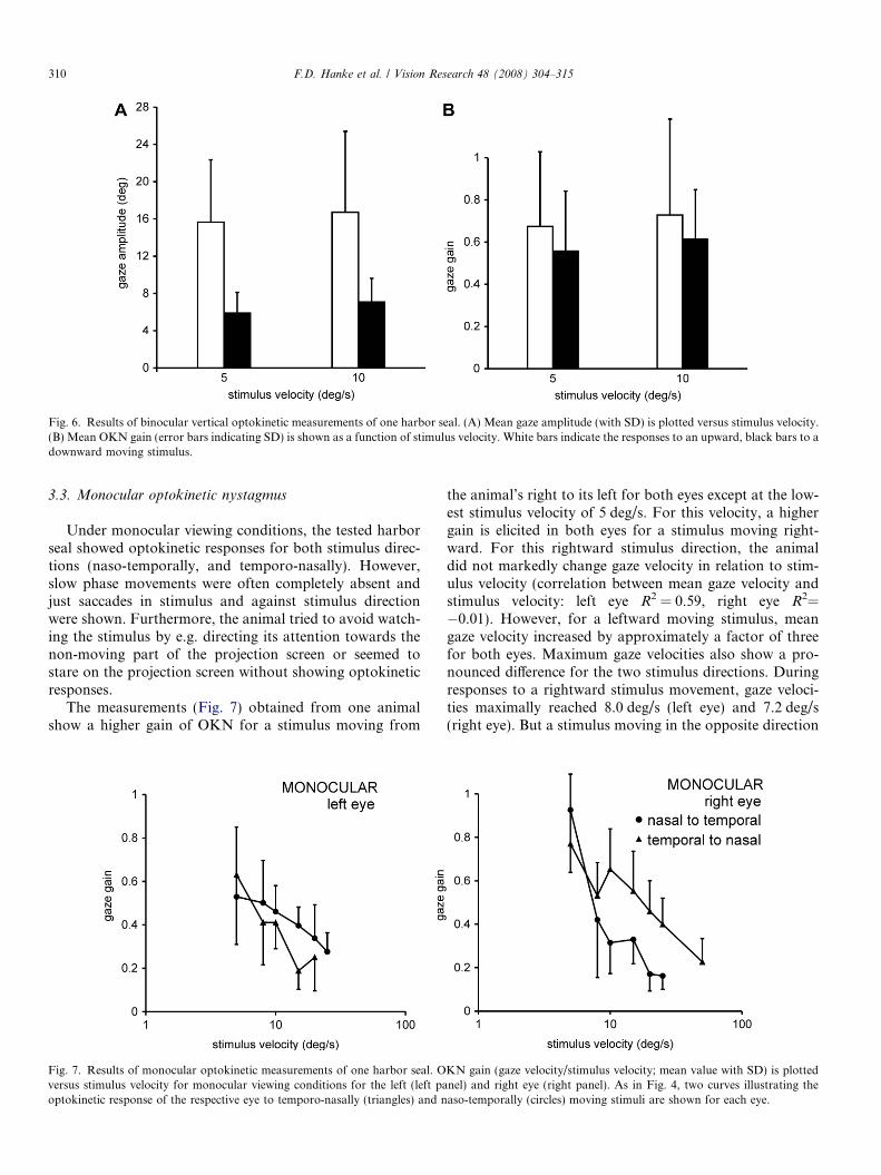
Fig. 6. Results of binocular vertical optokinetic measurements of one harbor seal. (A) Mean gaze amplitude (with SD) is plotted versus stimulus velocity.(B) Mean OKN gain (error bars indicating SD) is shown as a function of stimulus velocity. White bars indicate the responses to an upward, black bars to adownward moving stimulus.
310 F.D. Hanke et al. / Vision Research 48 (2008) 304–315
3.3. Monocular optokinetic nystagmus
Under monocular viewing conditions, the tested harborseal showed optokinetic responses for both stimulus direc-tions (naso-temporally, and temporo-nasally). However,slow phase movements were often completely absent andjust saccades in stimulus and against stimulus directionwere shown. Furthermore, the animal tried to avoid watch-ing the stimulus by e.g. directing its attention towards thenon-moving part of the projection screen or seemed tostare on the projection screen without showing optokineticresponses.
The measurements (Fig. 7) obtained from one animalshow a higher gain of OKN for a stimulus moving from
Fig. 7. Results of monocular optokinetic measurements of one harbor seal. Oversus stimulus velocity for monocular viewing conditions for the left (left paoptokinetic response of the respective eye to temporo-nasally (triangles) and n
the animal’s right to its left for both eyes except at the low-est stimulus velocity of 5 deg/s. For this velocity, a highergain is elicited in both eyes for a stimulus moving right-ward. For this rightward stimulus direction, the animaldid not markedly change gaze velocity in relation to stim-ulus velocity (correlation between mean gaze velocity andstimulus velocity: left eye R2 = 0.59, right eye R2=�0.01). However, for a leftward moving stimulus, meangaze velocity increased by approximately a factor of threefor both eyes. Maximum gaze velocities also show a pro-nounced difference for the two stimulus directions. Duringresponses to a rightward stimulus movement, gaze veloci-ties maximally reached 8.0 deg/s (left eye) and 7.2 deg/s(right eye). But a stimulus moving in the opposite direction
KN gain (gaze velocity/stimulus velocity; mean value with SD) is plottednel) and right eye (right panel). As in Fig. 4, two curves illustrating theaso-temporally (circles) moving stimuli are shown for each eye.
F.D. Hanke et al. / Vision Research 48 (2008) 304–315 311
(from the animal’s right to its left) elicited maximal gazevelocities of 11.6 deg/s (left eye) and 24.1 deg/s (righteye), respectively.
4. Discussion
4.1. Characteristics of optokinetic measurements in the
harbor seal
4.1.1. Methodological problems
One major limitation of this study is the fact that thepresented data are measurements of just one animal whichwas available for extended measurements. The other ani-mals could only be tested occasionally. Therefore, we can-not assess whether the detailed data obtained in one animalare representative for the species. However, the main exper-imental animal (‘‘Henry’’) behaves normally, is experimen-tally highly experienced and does not show any visualdeficits during daily tasks which make our data concerningthis individual reliable.
In our study, slow phase gain is generally low. The ques-tion arises whether unity-tracking would have occurredwith stimulus velocities below 5 deg/s. Unfortunately, wecould not present these slow velocities due to artefacts inprojection. However, in a variety of species, high gain val-ues are only found at low stimulus velocities. Mice e.g. dis-play gains of 0.7–0.8 constantly as a response to stimulusvelocities not exceeding 8 deg/s, whereas gain decreasedat higher stimulus velocities (Van Alphen, Stahl, & DeZeeuw, 2001).
If unity-tracking was found below 5 deg/s, our measure-ments would have been beyond the cut-off frequency atwhich gaze gain falls below one half its maximum (Ariel,1990; Donaghy, 1980). The low cut-off frequency couldbe due to an ineffective stimulus configuration using a pro-jected black-and-white bar pattern on a tangent screen.Collewijn (1985) reports that a real rotating drum is moreeffective than a projected pattern, and a random dot pat-tern is a better stimulus than a regular pattern in rabbitsand cats. The effect of the stimulus configuration is alsoeasily seen by comparing the results of optomotor studiesin the cat. Donaghy (1980) used sinusoidally modulatedgratings, Schweigart and Hoffmann (1988) used a randomdot pattern projected on a tangent screen, and Collewijn(1985) used an optokinetic drum lined with a random dotpattern. Despite the fact that all studies found high gainvalues at stimulus velocities lower than the cut-off fre-quency, the estimated cut-off frequencies varied. WhileDonaghy (1980) reports a cut-off frequency of 4–8 deg/s,the break of the gain curve seems to occur above 20 deg/s (Schweigart & Hoffmann, 1988) or at 60 deg/s accordingto Collewijn (1985). Testing harbor seals under variousstimulus conditions could clarify whether comparableeffects would occur.
Generally, various factors could have negatively influ-enced gain as well as the overall optokinetic reaction. These
factors need to be considered concerning their effect on notonly binocular but also on monocular OKN.
One of these factors influencing gain might be a changein alertness and attention of the animal during the courseof the experiment. A reduced level of motivation couldlower the overall optokinetic response (see Collewijn,1985 for review; for saccades: Crommelinck & Roucoux,1976). To maintain alertness in ‘‘head-fixed’’ situations,Schweigart and Hoffmann (1988) injected Met-amphet-amine-hydrochloride subcutaneously. In their study, thiswas not necessary in ‘‘head-free’’ conditions in which theanimal had to perform a special task. Our experimental sit-uation also involved a training situation which forced theanimal to maintain alertness and concentration to receivea fish reward. In addition, the soft training or pauses madeafter each stimulus presentation should have assured highresponsiveness of the animal. We could not observe anyobvious loss of motivation during an experimental sessionand during the whole time of data collection but it couldhave occurred unnoticed. Besides applying medication,another experimental approach to assure a high level ofalertness could be in line with experiments in pigeons (Bilo& Bilo, 1978; Gioanni, 1988; Gioanni & Sansonetti, 1999;Maurice, Gioanni, & Abourachid, 2006). Pigeons experi-encing frontal airflow in a flying posture displayed higheroptokinetic, optocollic and vestibular gain. This can bedue to the pigeon’s increased attention induced by the air-flow in flying condition or might indicate that the sensingof self-motion is crucial for a high level performance ofoptokinetic, optocollic and vestibular reflexes. Harbor sealscould show a comparable effect according to the behavioralcontext, e.g. as a response to water flow mimicking swim-ming behavior.
The tangent projection could also lead to a reduction ingain. Due to the tangent projection screen, the stimulusvelocity was 40% slower at the periphery compared to thecenter (see Section 2.2.5). The mean velocity of the wholeprojection amounted to 86% of the maximum velocity inthe center. This change in velocity can account for someof the variation observed concerning slow phase gain evenif it is unclear which velocity information—central, periph-eral or an average over the whole or central field of view—the animal used.
Gaze velocity is also affected by the projection size.Goldfish e.g. display slow tracking velocities if the targetsize is reduced (Easter, 1972). In humans, pursuit velocityonly reaches high values with full-field stimulation (Schor,1993). It was speculated that smaller stimulus fields restrictthe velocity range because of the proximity of stationaryedges to the fovea (Schor & Narayan, 1981). So far, it isnot known whether harbor seals possess an area centralis,but they probably do (see Section 1). However, the stimu-lus, covering 98� · 108�, is completely filling the binocularvisual field of harbor seals which extents over 67� measuredin air, leading to a binocular visual field of at least 42� cal-culated for underwater conditions (Hanke, Romer, &Dehnhardt, 2006). Thus, the animal should not have
312 F.D. Hanke et al. / Vision Research 48 (2008) 304–315
experienced the borders of the projection within itsassumed region of high resolution. Therefore, theprojection size does not seem to be a factor loweringpursuit velocity in our case.
We tested optokinetic responses for just one set ofblack-and-white bars (nine black and nine white bars)and one contrast. These parameters might have been sub-optimal to elicit high gain values in harbor seals constantly.Our own observations (not quantified) reveal no consistentdifference in OKN gain when the number of black andwhite bars was changed. Contrast as well as brightness usedin our experiment was adjusted during the establishment ofthe stimulus environment because optokinetic responseswere absent with high contrast pattern and very brightstimuli. However, we did not systematically change thesevalues. The low contrast could have reduced gain in harborseals analogous to the reduction found in cats (Donaghy,1980) or goldfish (Easter, 1972). More data on contrastaffecting OKN gain are needed in order to assess the influ-ence of this parameter on slow phase gain in harbor seals.
The low gain could also be due to a deficit in the smoothpursuit system. Smooth pursuit could have come into playif the animal had paid attention to a single stripe and fol-lowed it across the projection screen. However, in our dailywork with the animals, we observe that gaze is mainly sac-cadically redirected, and following of small sized objects isalso saccadic. A poorly or not developed smooth pursuitsystem could lower gain below unity, as has been alreadydiscussed for cats (Godeaux, Gobert, & Halleux, 1983).
In general, it must be noted that the performance of theoptokinetic system and the integrative performance of allfundamental visual or vestibulo-ocular reflexes in real lifecan only be assessed in a freely behaving subject (Collewijn,1977). They may be much better during active behaviourwhen spatial orientation is essential compared to a labora-tory test situation. Therefore, it would be interesting to e.g.mount a camera on a freely moving harbor seal compara-ble to the approach of Davies et al. (1999) continuouslyfilming the eye. Corresponding experiments are currentlydeveloped in our lab.
4.1.2. Binocular horizontal OKN
Measuring our main experimental animal revealed anequally sensitive optokinetic response to naso-temporaland temporo-nasal stimulus motion for the right eye. How-ever, a significant difference between the left eye’s responsesto the two stimulus movements (naso-temporal, temporo-nasal) is present. We think that these differences have tobe mainly discussed considering the measurement errors.The overall pronounced variability of our optokinetic mea-surements accompanied by the measured high level of con-junction of left and right eye let us expect that extendedoptokinetic measurements in harbor seals would revealsymmetric optokinetic responses for horizontal stimulusmovements for both eyes.
This assumed symmetry is in accordance with generalconsiderations concerning eye placement and forward loco-
motion (Grasse & Cynader, 1988). Harbor seals are fron-tal-eyed animals. For them, movement ahead will notpredominantly lead to a temporo-nasal movement of theretinal image (Carpenter, 1988) but rather to an expansionof the optic flow field from a focus straight ahead, and theoptokinetic reflex can be equally sensitive to both horizon-tal directions of motion. If eye position is indeed also deci-sive for predicting optokinetic characteristics in marinemammals, a comparison of our data to optokinetic dataof e.g. the California sea lion would be interesting as thesea lion’s eyes are positioned more laterally. An aquaticspecies with lateral eyes is the goldfish. The properties ofthe OKN in goldfish have been studied extensively. How-ever, the question concerning symmetric or asymmetricbinocular horizontal OKN in goldfish cannot be answeredwith certainty as on the one hand, asymmetric OKN at lowstimulus velocities (Dieringer et al., 1992; Easter 1972;Keng & Anastasio, 1997) is reported but on the other hand,a conjugate and symmetrical OKN was found (Marsh &Baker, 1997). In one study, even differences between indi-viduals are reported (Easter, 1972). Further work is neededin order to better understand the relationship between eyeplacement and horizontal OKN in animals with amphibi-ous or aquatic lifestyle.
Our harbor seal only occasionally followed the stimuluswith the exact stimulus velocity (gain was usually smallerthan 1). On the one hand, this has to be discussed in termsof methodological problems (see Section 4.1.1). But on theother hand, it could also reflect a tolerance to retinal slipvelocities in harbor seals. During active head movements,humans can tolerate retinal image motion up to 4 deg/swithout that vision is seriously affected (Steinman & Colle-wijn, 1980).
Furthermore, van der Steen and Collewijn (1984) discussthat perfect gaze stabilization might not be necessary dur-ing locomotion. But the harbor seals’ predative naturecould require that gaze is redirected under oculomotor con-trol relatively precisely corresponding to a high resolutionarea in the retina in order to hunt successfully. Assumingthe presence of a specialized area in the retina as has beendiscussed in the introduction, it could have a streak-likeappearance which makes the retina more tolerant to hori-zontal displacement. In harp seals (Mass & Supin, 2003),which are phocid seals with eye sizes comparable to harborseals, the area possessing ganglion cell densities equal orgreater than 60% of the greatest ganglion cell densityextends over 6.5 deg horizontally. An area centralis of thiskind would be 6.5 times wider compared to the humanfovea which is supposed to cover 1 deg on the horizonand could explain a higher tolerance to retinal slipvelocities.
Many studies have been conducted with the head com-pletely stationary which does not reflect natural behaviour(Leigh & Zee, 1999). Most organisms use a combination ofeye and head movements to visually track targets, stabilizemoving objects or whole-field motion. Our setup allowedfor small head movements. Analyzing gaze velocity under
F.D. Hanke et al. / Vision Research 48 (2008) 304–315 313
voluntary positioning of the head, both eyes showed max-imal gaze amplitudes of 18–20 deg for stimuli moving fromleft to right, for the reverse stimulus direction gaze ampli-tudes of 15–19 deg occurred. Hanke et al. (2006) showedthat a harbor seal was able to rotate its eyes up to 12 degin the head horizontally. Considering this amount of eyemovement, the head would have contributed 33–44% (stim-ulus moving from left to right), 20–37% (stimulus movingfrom right to left), respectively. However, our observationssuggest that head movements were less pronounced in ourexperiments (see Section 3.1). Under completely free condi-tions without fixation, gaze changes could be mainly per-formed by the head comparable to cats in which the headcontributes 40–80% to gaze slow phase (Schweigart & Hoff-mann, 1988) because harbor seals possess highly flexiblenecks. But considering streamlining underwater, it wouldbe advantageous for seals to move only their eyes (eye-in-head-strategy) instead of the whole head (head-on-body-strategy). The large visual field of harbor seals (Hankeet al., 2006) might support the eye-in-head-strategy allow-ing the seal to scan a large part of the scene without a needof significant head movements. However, even if under nat-ural conditions the contribution of the head was higherthan in our experimental situation, slow phase velocitieswould not necessarily increase as has been shown in catsin a comparison between ‘‘head-fixed’’ and ‘‘head-free’’conditions (Schweigart & Hoffmann, 1988).
Generally, the optokinetic nystagmus in harbor seals isvery rhythmic. This is especially evident with stimulusvelocities between 15 and 50 deg/s. With very fast stimulusvelocities, the OKN turns irregular and discontinuous ashas been described for humans as well (Cheng & Outer-bridge, 1974). Below 15 deg/s, the analysis of intersaccadicintervals showed that, also comparable to humans (Cheng& Outerbridge, 1974), the nystagmus becomes more irreg-ular and its intensity decreases. The distribution of inter-saccadic intervals is very broad with low stimulusvelocities but we could not observe a distinctive multi-modal pattern as described by Cheng and Outerbridge(1974).
4.1.3. Binocular vertical OKN
We could show that both vertical stimulus movementselicited optokinetic responses with no significant differencein mean gaze gain for the two tested stimulus velocities.Although gaze amplitudes as a response to an upwardmoving stimulus were larger compared to those followinga downward stimulus movement, gain was not significantlyhigher for one stimulus direction because the seal displayeddownward OKN movements with a higher frequency.
The ability of our harbor seal to equally pursue verticalup- and down-stimuli is an interesting finding as thisresponse symmetry has not been reported so far in any spe-cies. In the animal kingdom, e.g. cats (Grasse & Cynader,1988), squirrel monkeys (Takahashi & Igarashi, 1977),and rhesus monkeys (Matsuo & Cohen, 1984) poorlyrespond to downward stimulus movement in the vertical
domain. However, this asymmetry observed in cats andmonkeys was more obvious at higher stimulus velocitieswhich we did not test in our experiments. The responsecharacteristic of human vertical OKN is highly variablewith e.g. different individuals showing different preferences.Furthermore, according to Murasugi and Howard (1989),the experiments dealing with human vertical OKN haveto be discussed methodologically. Nevertheless, humansalso seem to display higher OKN gain as a response toupward stimulus motion (Murasugi & Howard, 1989; vanden Berg & Collewijn, 1988).
The reduced sensitivity to a downward moving stimulushas been explained in terms of preventing the eye fromrotating downwards while walking over a highly-texturedground (Schor, 1993). This means that the OKN is largelyinsensitive to the main optic flow experienced during for-ward locomotion as has already been explained for hori-zontal OKN.
The difference between the response characteristics oforganisms to vertical stimulus motion could be discussedin respect to differences in heights of eye level (Takahashi& Igarashi, 1977). They suggest that animals with lowplaced eyes do not respond well to a downward movingstimulus due to the direct vicinity to the plane of main opticflow. Harbor seals’ eyes are placed more dorsally indicatedby their vertical cyclopean visual field which extents overjust 12 deg ventrally but 69 deg dorsally (Hanke et al.,2006). Forward locomotion should therefore induce mainlyupward optic flow which could have turned the eyes lessresponsive to upward moving stimuli. However, this effectwas not found in our harbor seal. One could speculate thatthe natural environment of harbor seals compared to thatof terrestrial carnivores renders the eye equally sensitiveto all vertical stimulus directions as it rarely exposes harborseals directly to a horizontal plane. Harbor seals swimmingin the water column can either refer to none, to one or totwo horizontal planes, i.e. the water surface and the bot-tom. The position of these reference planes can also changewhen the seal rotates its body while swimming. Therefore,it could be the missing asymmetries in the optic flow in thevertical that render asymmetric vertical optokineticresponses normally found in terrestrial organisms symmet-ric (see Section 4.2).
In line with the extended dorsal visual field, verticalgaze amplitudes were larger for an upward than for adownward stimulus movement. However, they did notreach the dorsal eye movement amplitudes of 64 degreported in Hanke et al. (2006). This cannot be explainedby the extensions of the stimulus on the projectionscreen. Vertically, the projection extended over 98 degof the animal’s visual field. As the animal was positionedin the center of the projection, it could have raised theeyes by an amount of 49 deg before reaching the projec-tion’s edge which has not been achieved even with thelargest gaze movements. It could be that the eye ampli-tudes shown by our experimental animal suit the eyemuscles’ actions best (Carpenter, 1988).
314 F.D. Hanke et al. / Vision Research 48 (2008) 304–315
Larger amplitudes as a response to an upward movingstimulus are in line with observations on seals approachingprey from below (Davies et al., 1999; Hobson, 1966) and inline with our own observations that harbor seals are oftenswimming upside-down which might be explained in termsof scanning the water body below (Hanke et al., 2006).
4.1.4. Monocular horizontal OKN
Under monocular viewing conditions, the harbor sealshowed optokinetic responses to both horizontal stimulusdirections. However, for both eyes, gain was alwayshigher for a stimulus moving leftward than for thereverse stimulus direction concerning stimulus velocitieshigher than 5 deg/s. Monocular asymmetries reported inother studies represent a complete lack of optokineticresponses (optokinetic unidirectionality) or minorresponses to a stimulus moving from nasal to temporal.This asymmetry has been explained in terms of prevent-ing especially laterally-eyed animals of responding tonaso-temporal optic flow produced by forward locomo-tion (Gioanni, Rey, Villabois, & Dalbera, 1984; Howard& Gonzalez, 1987). However, this aspect could onlyexplain the data presented for the right eye. The datafor the left eye show an inversed asymmetry, i.e. a stron-ger OKN in the naso-temporal than in the tempero-nasaldirection. As harbor seals possess a frontal eye position,we would have expected to measure a symmetrical mon-ocular optokinetic response as has been found in, e.g.ferrets, cats, and primates. We therefore assume, in con-sistency with our data, that the harbor seal’s responsewould have been symmetric in naso-temporal and intempero-nasal direction in both eyes if it had not beensuperimposed by a general preference for right-to-leftmoving stimuli. We cannot assess whether the preferencefor a leftward stimulus movement is an adaptation to thechanged requirements resulting in an increase in gain, aphenomenon reported, e.g. in humans (Collewijn, 1985)or goldfish (Easter, 1972; Marsh & Baker, 1997; Schairer& Bennett, 1986), as we presented mainly leftward mov-ing stimuli during the establishment of the stimulus envi-ronment, or whether the animal showed a spontaneouspreference for this stimulus direction.
4.2. General implications
This is the first study to show that a marine mammal,the harbor seal, shows an optokinetic nystagmus underwater. It is thus demonstrated that harbor seals can per-ceive motion under water, making it likely that they canbenefit from motion information concerning a wide varietyof visual tasks, e.g. reconstructing the third dimension, seg-menting the image, separation of figure and background,eliciting attention, encoding self-motion, mediating sizeconstancy, and detecting moving objects (Nakayama,1985).
Our study on the optokinetic nystagmus revealedequal sensitivity to all stimulus directions. This is surpris-
ing especially concerning the symmetric vertical OKNwhich has never been reported in any species before. Inthe underwater environment, the horizontal and verticalplanes are both of importance. If we just consideredtranslation for a swimming seal, the optic flow wouldconsist of streams of images emerging from a focus ofexpansion straight ahead and disappearing into a focusof contraction behind (Miles, 1998). If the seal is alsolooking straight ahead, it experiences an expandingworld with only the most distant objects in the visualscene being stable on the retina. When looking to oneside, all objects seem to pivot about far, stable objects.To be able to fixate an object, e.g. moving or non-mov-ing prey, while looking to a side, the seal would have tocompensate for its own motion with the help of compen-sating head and eye movements. However, the horizontalplane does not necessarily remain the main plane of ref-erence as it does for e.g. the cat, even if it is climbing atree. The significance of the horizontal plane has alreadybeen questioned for marine mammals in studies dealingwith mental rotation as a cognitive aspect of vision(Mauck & Dehnhardt, 1997; Stich, Dehnhardt, &Mauck, 2003). Underwater, harbor seals sometimes evenexperience two horizontal planes of reference, the watersurface and the ground. However, swimming at anydepth in the water column without directly seeing theground or the water surface, seals cannot or can onlyindirectly refer visually to the horizontal plane by, e.g.comparing the brightness around the body. The brighterwater surface can rotate around the animal depending onthe own body tilt as self-motion in harbor seals not onlyinvolves linear displacement or translation but quiteoften also rotations of head and body. Thus, the degreesof freedom are numerous since the location of the eye aswell as of a target can change by rotations around threeorthogonal axes and translations in three dimensions(Collewijn, 1985).
In our opinion, an eye with the ability to stabilize whole-field motion equally well in any plane, as our experimentsindicate, could be perfectly tuned for an animal operatingin the low structured, three-dimensional underwater world.
Acknowledgments
This study was supported by a Grant of the Studienstif-tung des Deutschen Volkes (2005 SA 0969) to F.D.H. andGrants of the VolkswagenStiftung and the Deutsche Fors-chungsgemeinschaft (SFB 509) to G.D.
References
Ariel, M. (1990). Independent eye movements in the turtle. Visual
Neuroscience, 5, 29–41.Bilo, D., & Bilo, A. (1978). Wind stimuli control vestibular and
optokinetic reflexes in the pigeon. Naturwissenschaften, 65, 161–162.Brainard, D. H. (1997). The psychophysics toolbox. Spatial Vision, 10(4),
433–436.Carpenter, R. H. S. (1988). Movement of the eyes. London: Pion.
F.D. Hanke et al. / Vision Research 48 (2008) 304–315 315
Cheng, M., & Outerbridge, J. S. (1974). Inter-saccadic interval analysis ofoptokinetic nystagmus. Vision Research, 14, 1053–1058.
Collewijn, H. (1977). Eye- and head-movements in freely moving rabbits.Journal of Physiology, 266, 471–498.
Collewijn, H. (1985). Integration of adaptive changes of the optokineticreflex, pursuit and vestibulo-ocular reflex. In A. Berthoz & G. MelvillJones (Eds.), Adaptive mechanisms in gaze control–facts and theories
(pp. 51–69). Amsterdam: Elsevier Science Publisher BV (BiomedicalDivision).
Crommelinck, M., & Roucoux, A. (1976). Characteristics of cat’s eyesaccades in different states of alertness. Brain Research, 103, 574–578.
Davies, R. W., Fuiman, L. A., Williams, T. M., Collier, S. O., Hagey, W.P., Kanatous, S. B., et al. (1999). Hunting behavior of a marinemammal beneath the Antarctic fast ice. Science, 283, 993–996.
Dieringer, N., Reichenberger, I., & Graf, W. (1992). Differences inoptokinetic and vestibular ocular reflex performance in teleosts andtheir relationship to different life styles. Brain, Behavior and Evolution,
39, 289–304.Donaghy, M. (1980). The contrast sensitivity, spatial resolution and
velocity tuning of the cat’s optokinetic reflex. Journal of Physiology,
300, 353–365.Easter, S. S. (1972). Pursuit eye movements in goldfish (Carassius auratus).
Vision Research, 12, 673–688.Gioanni, H. (1988). Stabilizing gaze reflexes in the pigeon (Columbia livia)
1. Horizontal and vertical optokinetic eye (OKN) and head (OCR)reflexes. Experimental Brain Research, 69, 567–582.
Gioanni, H., Rey, J., Villabois, J., & Dalbera, A. (1984). Single-unitactivity in the nucleus of the basal optic root (nBOR) duringoptokinetic, vestibular and visuo-vestibular stimulation in the alertpigeon (Columbia livia). Experimental Brain Research, 57, 49–60.
Gioanni, H., & Sansonetti, A. (1999). Characteristics of slow and fastphases of the optocollic reflex (OCR) in head free pigeons (Columbia
livia): Influence of flight behaviour. European Journal of Neuroscience,
11, 155–166.Godeaux, E., Gobert, C., & Halleux, J. (1983). Vestibuloocular reflex,
optokinetic response, and their interactions in the alert cat. Exper-
imental Neurology, 80, 42–54.Grasse, K. L., & Cynader, M. S. (1988). The effect of visual cortex lesions
on vertical optokinetic nystagmus in the cat. Brain Research, 455,385–389.
Hanke, F. D., Dehnhardt, G., Schaeffel, F., & Hanke, W. (2006). Cornealtopography, refractive state, and accommodation in harbor seals(Phoca vitulina). Vision Research, 46, 837–847.
Hanke, W., Romer, R., & Dehnhardt, G. (2006). Visual fields and eyemovements in a harbor seal (Phoca vitulina). Vision Research, 46,2804–2814.
Hobson, E. S. (1966). Visual orientation and feeding in seals and sea lions.Nature, 210, 326–327.
Howard, I. P., & Gonzalez, E. G. (1987). Human optokinetic nystagmusin response to moving binocularly disparate stimuli. Vision Research,
27(10), 1807–1816.Jamieson, G. S., & Fisher, H. D. (1971). The retina of the harbour seal,
Phoca vitulina. Canadian Journal of Zoology, 49, 19–23.Jung, R., & Mittermaier, R. (1939). Zur objektiven Registrierung und
Analyse verschiedener Nystagmusformen: vestibularer, optokineti-scher und spontaner Nystagmus in ihren Wechselbeziehungen. Archiv
fur Ohren-Nasen-Kehlkopfheilkunde, 146, 410–439.Keng, M. J., & Anastasio, T. J. (1997). The horizontal optokinetic
response of the goldfish. Brain, Behavior and Evolution, 49,214–229.
King, J. E. (1983). Seals of the world. London: British Museum (NaturalHistory), and Oxford: Oxford University Press.
Leigh, R. J., & Zee, D. S. (1999). The neurology of eye movements. NewYork, Oxford: Oxford University Press.
Marsh, E., & Baker, R. (1997). Normal and adapted visuooculomotorreflexes in goldfish. Journal of Neurophysiology, 77, 1099–1118.
Mass, A. M., & Supin, A. Y. (1992). Peak density, size and regionaldistribution of ganglion cells in the retina of the fur seal Callorhinus
ursinus. Brain, Behaviour and Evolution, 39, 69–76.Mass, A. M., & Supin, A. Y. (2003). Retinal topography of the harp seal
Pagophilus groenlandicus. Brain, Behaviour and Evolution, 62, 212–222.Mass, A. M., & Supin, A. Y. (2005). Ganglion cell topography and retinal
resolution of the Stellar sea lion (Eumetobias jubatus). Aquatic
mammals, 31(4), 393–402.Matsuo, V., & Cohen, B. (1984). Vertical optokinetic nystagmus and
vestibular nystagmus in the monkey: Up–down asymmetry and effectsof gravity. Experimental Brain Research, 53, 197–216.
Mauck, B., & Dehnhardt, G. (1997). Mental rotation in a California sealion (Zalophus californianus). The Journal of Experimental Biology,
200, 1309–1316.Maurice, M., Gioanni, H., & Abourachid, A. (2006). Influence of the
behavioural context on the optocollic reflec (OCR) in pigeons(Columbia livia). The Journal of Experimental Biology, 209, 292–301.
Miles, F. A. (1998). The neural processing of 3D visual information:Evidence from eye-movements. European Journal of Neuroscience, 10,811–822.
Murasugi, C. M., & Howard, I. P. (1989). Up-down asymmetry in humanvertical optokinetic nystagmus and afternystagmus: Contributions ofthe central and peripheral retinae. Experimental Brain Research, 77,183–192.
Nakayama, K. (1985). Biological image motion processing: A review.Vision Research, 25(5), 625–660.
Schairer, J. O., & Bennett, M. V. L. (1986). Changes in gain of thevestibulo-ocular reflex induced by sinusoidal visual stimulation ingoldfish. Brain Research, 373, 177–181.
Schor, C. M. (1993). Development of OKN. In F. A. Miles & J. Wallman(Eds.), Visual motion and its role in the stabilization of gaze
(pp. 301–320). Amsterdam: Elsevier.Schor, C., & Narayan, V. (1981). The influence of field size upon the
spatial frequency response of optokinetic nystagmus. Vision Research,
21, 985–994.Schusterman, R. J., & Thomas, T. (1966). Shape discrimination and
transfer in the California sea lion. Psychonomic Science, 5, 21–22.Schweigart, G., & Hoffmann, K.-P. (1988). Optokinetic eye and head
movements in the unrestrained cat. Behavioural Brain Research, 31,121–129.
Steinman, R. S., & Collewijn, H. (1980). Binocular retinal image motionduring active head rotation. Vision Research, 20, 415–429.
Stich, K. P., Dehnhardt, G., & Mauck, B. (2003). Mental rotation ofperspective stimuli in a California sea lion (Zalophus californianus).Brain, Behaviour and Evolution, 61, 102–112.
Takahashi, M., & Igarashi, M. (1977). Comparison of vertical andhorizontal optokinetic nystagmus in the squirrel monkey. ORL, 39,321–329.
Van Alphen, A. M., Stahl, J. S., & De Zeeuw, C. I. (2001). The dynamicproperties of the mouse horizontal vestibulo-ocular and optokineticresponse. Brain Research, 890, 296–305.
Van den Berg, A. V., & Collewijn, H. (1988). Directional asymmetries ofhuman optokinetic nystagmus. Experimental Brain Research, 70,597–604.
Van der Steen, J., & Collewijn, H. (1984). Ocular stability in thehorizontal, frontal and sagittal planes in the rabbit. Experimental Brain
Research, 56, 263–274.Walls, G. L. (1942). The vertebrate eye and its adaptive radiation. New
York: Hafner.Walls, G. L. (1962). The evolutionary history of eye movements. Vision
Research, 2, 69–80.Yarbus, A. L. (1967). Eye movements and vision. New York: Plenum Press.