optimal clearing and mounting media for confocal...
TRANSCRIPT
Optimal clearing and mounting media for confocal microscopy of thick specimens Kurt Thorn, Chester Chamberlain, and Nan Tang
Sample preparation for confocal microscopy typically involves fixing and staining, followed by clearing and mounting. Clearing involves treatment with solvent to dissolve lipids and other structures that cause light scattering and reduce the ability to image through thick specimens. Mounting involves transfer of the specimen into a medium which matches the refractive index the objective is designed for (n = 1.515 in the case of oil immersion lenses). This higher refractive index also helps to match the refractive index of the tissue. Mounting media often include chemicals to retard photobleaching. A wide variety of clearing and mounting media for confocal microscopy have been described in the literature; in several cases the same media are used for both clearing and mounting. Careful attention to clearing and mounting has enabled confocal microscopy of samples greater than 500 μm thick at low magnification [2, 11-12].
To identify optimal clearing and mounting media for confocal microscopy, we tested ten commonly used mounting and clearing media (Table I). In all cases except for DMSO->TDE, we used the same media for clearing and mounting. Our test specimens were lungs dissected from E11.5 mouse embryos and stained for E-cadherin using fluorescein and a tyramide signal amplification protocol. The lungs are about 200 μm thick and have a characteristic epithelial cell morphology, so they provide a good test of the ability to image deep into the specimen and of the preservation of the cell morphology and other fine details of the specimen.
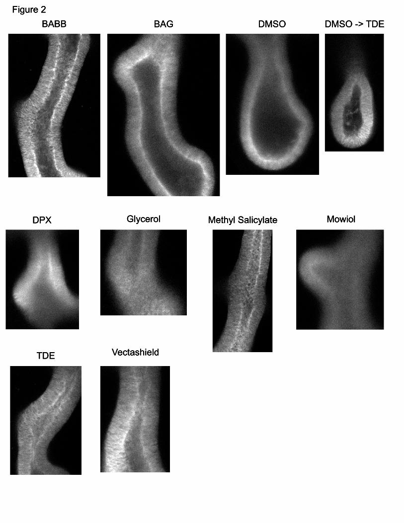
Each sample was first imaged with a Plan Apo 20x/0.75NA air lens. A z-stack was acquired at 5 μm steps through the entire thickness of the lung. The full field-of-view of the lens was imaged with 1024x1024 pixels, resulting in a pixel size of 621nm. These stacks are undersampled in all three dimensions, but provided sufficient resolution for initial evaluation of each mounting medium. X-Y and X-Z sections of these stacks are shown in figures 1 and 2. We found surprisingly large differences between the different mounting media tested, both in and in the ability to image through the full thickness of the specimen (seen in the X-Z sections) and in the preservation of cell morphology (seen in the X-Y sections).
BABB and methyl salicylate were the best mounting media for imaging through the full thickness of the sample, showing very little intensity decrease from the top of the lungs to the bottom of the lungs. Most other mounting media showed a rapid decrease in intensity in when imaging deeper into the sample, although TDE and Vectashield gave intermediate results. As this experiment was done with an air lens, and therefore none of the media were index-matched to the lens, this improvement in imaging depth likely results from improved clearing of the sample. Not surprisingly, the best mounting media were those that were most hydrophobic and the best clearing agents.
To evaluate the effectiveness of these mounting media for imaging with oil-immersion lenses, we re-imaged the best mounting media with a Plan Fluor 40x/1.3NA oil-immersion lens. These higher resolution images also enabled us to better evaluate the preservation of cell morphology. In general, the results at 40x were consistent with the results at 20x. Mounting in TDE results in by far the best preservation of cell

morphology of the mounting media tested; however, there is notable fall off in fluorescence intensity when imaging deep into the sample. BABB and methyl salicylate still gave the best penetration depth, with essentially no decrease in fluorescence intensity at the maximum thickness we were able to image (~150 μm). Mounting in BABB resulted in substantial perturbation of cell morphology, restricting its use to cases where maintenance of cell morphology is not critical. Mounting in methyl salicylate results in better preservation of cell morphology, though not as good as that seen when mounting in TDE. We also tested clearing with methyl salicylate followed by mounting in TDE to see if this would provide the large imaging depth of methyl salicylate, and the preservation of cell morphology of TDE. Unfortunately, this did not improve the imaging depth over mounting in TDE alone.
When imaging with oil lenses, a major limitation on the ability to image deeply in the sample is the short working distance of the lens. When working with mounting media well matched to the refractive index of the immersion oil and the coverslip, it should be possible to reduce the thickness of the coverslip (thereby increasing the effective working distance of the objective) without introducing spherical aberration. We tested this by mounting samples in methyl salicylate and TDE under #0 coverslips. These samples did not show any additional aberrations compared to those mounted under #1.5 coverslips. However, to obtain the maximum imaging depth available requires mounting the tissue as close as possible to the coverslip, which is difficult to do reproducibly. Probably the best way to do this is to use spacers between the coverslip and the slide which are just slightly thicker than the sample. This way, squashing the coverslip down will give the thinnest possible mount without squashing the sample.
In summary, methyl salicylate appears to be the preferred mounting medium when imaging depth is important. Methyl salicylate also has several advantages over BABB – its refractive index better matches that of immersion oil, it is less toxic (though not non- toxic), and the tissue can be seen when mounted in methyl salicylate, allowing mounting by brightfield (BABB requires mounting by fluorescence). Mounting in methyl salicylate has also been shown to reduce artifacts resulting from tissue shrinkage that may occur in other mounting media [7]. When mounting with either BABB or methyl salicylate, bear in mind that these compounds will dissolve vacuum grease and so this should not be used for making spacers or sealing slides. Nail polish can be used for sealing slides, and fragments of coverslips or silicone gaskets (available from Grace Bio-Labs) can be used as spacers.
When optimal preservation of cell morphology is important or when imaging depth is not as important (~<100 μm), TDE is the preferred mounting media. Of course, these media have only been tested for mounting embryonic mouse lungs; for other tissues the optimal mounting medium may be different.

Table I: Mounting media tested Name Composition R.I. Notes Source BABB 33% Benzyl Alcohol
67% Benzyl Benzoate 1.56 Requires
dehydration [1,2]
BAG 55% Benzyl Alcohol 45% Glycerol
1.515 Requires dehydration
[3]
DMSO 100% DMSO 1.48 [4] DMSO -> TDE Clear in DMSO
Mount in TDE 1.515 [4, 9]
DPX DPX 1.52-1.60
Requires dehydration
[5]
Glycerol 90% Glycerol in 20 mM Tris pH 8
1.46 [6]
Methyl Salicylate
100% Methyl Salicylate
1.536 Requires dehydration
[7]
Mowiol [8] TDE 97% 2,2’-
Thiodiethanol in water 1.515 [9]
Vectashield Vectashield 1.45 [10] R.I., refractive index. Figure 1: X-Z sections of embryonic mouse lungs acquired with a 20x/0.75NA lens. The X-axis runs horizontally; the Z-axis runs vertically. The top of the specimen is at the top of each image. The thickness of each image in Z is 150-200 μm. Good mounting media should give relatively constant intensities throughout the entire thickness of the specimen. Poor mounting media are those where the top of the specimen is significantly brighter than the bottom. Figure 2: X-Y sections of embryonic mouse lungs acquired with a 20x/0.75NA lens. The sections shown were in the middle of each Z-stack, typically 75-100 μm from the surface of the tissue. Good mounting media here are those which result in crisp, high-contrast images. Conversely, poor mounting media result in fuzzy, blurry images. Figure 3: X-Y sections of embryonic mouse lungs acquired with a 40x/1.3NA oil lens. The superior preservation of cell morphology provided by mounting in TDE can be clearly seen; samples mounted in BABB, BAG, or methyl salicylate show disorganization of the cell membranes. Vectashield also preserves cell morphology well; however the resulting image is noticeably blurrier than the TDE-mounted sample. References: 1. Klvmkowsky MW. Hanken J. Whole-Mount Staining Of Xenopus And Other
Vertebrates. In Kay BK, Peng HB, eds. Xenopus laevis: practical uses in cell and molecular biology. Methods Cell Biol 36:419, 1991.

2. Zucker RM. Hunter S. Rogers JM. Confocal Laser Scanning Microscopy of Apoptosis in Organogenesis-Stage Mouse Embryos. Cytometry 33:348, 1998.
3. Gustafsson MGL. Agard DA. Sedat JW. I5M: 3D Widefield Light Microscopy With Better Than 100nm Axial Resolution. J. Microsc. 195:10, 1999.
4. Grace AA. Llinas R. Morphological Artifacts Induced In Intracellularly Stained Neurons By Dehydration: Circumvention Using Rapid Dimethyl Sulfoxide Clearing. Neurosci. 16: 461, 1985.
5. http://www.sigmaaldrich.com/catalog/search/ProductDetail/ALDRICH/317616
6. http://nic.med.harvard.edu/Mounting%20Meda.pdf
7. Bucher D. Scholz M. Setter M. Obermayer K. Pfluger H-J. Correction methods for three-dimensional reconstructions from confocal images: I. tissue shrinking and axial scaling. J. Neurosci. Meth. 100: 135, 2000.
8. http://www.urmc.rochester.edu/research/pmic/Mowiol.html
9. Staudt T, Lang MC, Medda R, Engelhardt J, Hell SW. 2,2'-Thiodiethanol: A New Water Soluble Mounting Medium For High Resolution Optical Microscopy. Microsc. Res. Tech. 70:1, 2007.
10. http://www.vectorlabs.com/products.details.asp?prodID=427&locID=662374
11. Zucker RM. Whole Insect and Mammalian Embryo Imaging with Confocal Microscopy: Morphology and Apoptosis. Cytometry 69A: 1143, 2006
12. Miller CE. Thompson RP. Bigelow MR. Gittinger G. Trusk TC. Sedmera D. Confocal Imaging of the Embryonic Heart: How Deep? Microsc. Microanal. 11: 216, 2005.