optical properties of human colon tissues in the 350 – 2500 nm spectral range
TRANSCRIPT
This content has been downloaded from IOPscience. Please scroll down to see the full text.
Download details:
IP Address: 128.122.253.228
This content was downloaded on 17/10/2014 at 10:38
Please note that terms and conditions apply.
Optical properties of human colon tissues in the 350 – 2500 nm spectral range
View the table of contents for this issue, or go to the journal homepage for more
2014 Quantum Electron. 44 779
(http://iopscience.iop.org/1063-7818/44/8/779)
Home Search Collections Journals About Contact us My IOPscience
Quantum Electronics 44 (8) 779 – 784 (2014) © 2014 Kvantovaya Elektronika and Turpion Ltd
Abstract. We present the optical characteristics of the mucosa and submucosa of human colon tissue. The experiments are performed in vitro using a LAMBDA 950 spectrophotometer in the 350 – 2500 nm spectral range. The absorption and scattering coef-ficients and the scattering anisotropy factor are calculated based on the measured diffuse reflectance and total and collimated transmit-tance spectra using the inverse Monte Carlo method.
Keywords: integrating sphere spectrophotometry, inverse Monte Carlo method, mucous membrane, submucous membrane, absorp-tion coefficient, scattering coefficient, reduced scattering coeffi-cient, scattering anisotropy factor.
1. Introduction
Knowledge of the optical characteristics of tissues is one of the key issues in the development of mathematical models that adequately describe the propagation of light in biological tissues, which in turn is crucial for the development of new optical methods used in various fields of biology and medi-cine [1, 2]. At the same time, in spite of a considerable number of papers devoted to the determination of optical parameters of biological tissues [1 – 10], their optical properties in a wide wavelength range are currently poorly studied, even though the analysis of visible and near-IR radiation absorption by biological tissues is crucial to the development of the methods of optical diagnosis, endoscopic surgery, photodynamic and photothermal therapy of various diseases, including cancer.
A key issue in the prevention of colon cancer is the diag-nosis and treatment of pre-cancer [11 – 13]. Unfortunately, despite the undoubted social significance of the problem, laser scalpels in surgical endoscopy of the colon have not found wide application yet. In particular, this is due to the
lack of science-based criteria, which would allow endosco-pists and laser surgical instrument developers to use an opti-mal laser wavelength for surgery [13] and to choose the most suitable type of laser, respectively.
One of the criteria on which this selection can be done is an objective analysis of the optical characteristics of colon tis-sue. Previously, they were studied in papers [14 – 19]. However, the investigation of the optical characteristics was performed only in the visible wavelength range (360 – 685 nm) [14]; in the ranges of 400 – 1100 nm [15], 300 – 700 nm [16], 300 – 800 nm [17]; and only at particular wavelengths (850, 980 and 1060 nm [18], 476.5, 488, 496.5, 514.5, 532 nm [19]). At the same time, the rapid development of laser technology and the consequent emergence of new types of lasers require the determination of the optical characteristics of tissues in a much broader range of wavelengths. In addition, the authors of all the above papers investigated the optical properties of the entire bio-logical tissue on the whole rather than separate layers.
In light of the above, the purpose of this paper is to mea-sure the optical properties of mucous and submucous mem-branes of the human colon in the 350 – 2500 nm spectral range.
2. Materials and methods
In the study we used 20 samples of human colon tissue (10 samples of the mucosa and 10 samples of the submucosa) taken from different 40-to-60-year-old patients in the course of routine operations or histological sectioning. Immediately after surgery or autopsy the tissue samples were placed in a 0.9 % aqueous solution of NaCl and stored therein prior to the spectral measurements for 8 – 12 h at a temperature of 4 °C. The area of each sample was 500 – 600 mm2. To measure the thickness, the samples were placed between two glass slides; measurements were performed with a micrometer at several points. The average thickness of the samples was 0.56 ± 0.41 mm with a ±50 mm accuracy of each measurement.
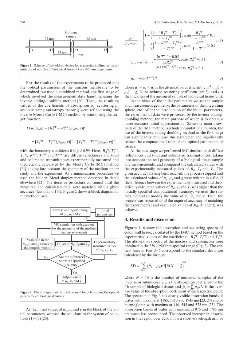
The optical properties of biological tissues were investi-gated in the 350 – 2500 nm spectral range using a LAMBDA 950 integrating sphere spectrophotometer (PerkinElmer, USA), which is a two-channel diffraction monochromator having an integrated control and signal registration system. The size of the light beam incident on the sample during the measurement of diffuse reflectance and total transmittance was 1 ́ 4 mm, and the scanning speed was 5 nm s–1. To mea-sure collimated transmittance we used a specially designed add-on device, which consisted of a holder to fix the sample of biological tissue and a system of four diaphragms (Fig. 1) with a diameter of 2 mm each. All measurements were per-formed at room temperature (~20 °C).
Optical properties of human colon tissues in the 350 – 2500 nm spectral range
A.N. Bashkatov, E.A. Genina, V.I. Kochubey, V.S. Rubtsov, E.A. Kolesnikova, V.V. Tuchin
A.N. Bashkatov, E.A. Genina, V.I. Kochubey, E.A. Kolesnikova N.G. Chernyshevsky Saratov State University, ul. Astrakhanskaya 83, 410012 Saratov, Russia; e-mail: [email protected], [email protected], [email protected], [email protected]; B.S. Rubtsov V.I. Razumovsky Saratov State Medical University, ul. Bol’shaya Kazach’ya 112, 410012 Saratov, Russia; e-mail: [email protected]; V.V. Tuchin N.G. Chernyshevsky Saratov State University, ul. Astrakhanskaya 83, 410012 Saratov, Russia; Institute of Precision Mechanics and Control, Russian Academy of Sciences, ul. Rabochaya 24, 410028 Saratov, Russia; University of Oulu, 90014, Oulu, P.O. Box 4500, Finland; e-mail: [email protected] Received 7 July 2014 Kvantovaya Elektronika 44 (8) 779 – 784 (2014) Translated by I.A. Ulitkin
PACS numbers: 87.19.xu; 87.50.wj; 87.50.wp; 87.64.Aa; 87.64.Cc; 87.80.Dj DOI: 10.1070/QE2014v044n08ABEH015613
A.N. Bashkatov, E.A. Genina, V.I. Kochubey, et. al.780
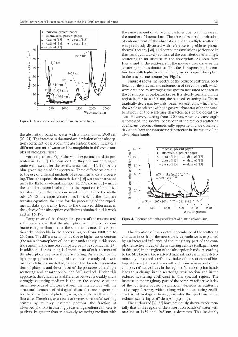
For the results of the experiments to be processed and the optical parameters of the mucous membrane to be determined, we used a combined method, the first stage of which involved the measurement data handling using the inverse adding-doubling method [20]. Then, the resulting values of the coefficients of absorption ma, scattering ms and scattering anisotropy factor g were refined using the inverse Monte Carlo (IMC) method by minimising the tar-get function
( , , ) [ ( , , )]F g R R gexpa s d d
calca s
2m m m m= -
+ [ ( , , )] [ ( , , )]T T g T T gexp expc c
calca s t t
calca s
2 2m m m m- + -
with the boundary conditions 0 G g G 0.99. Here, R expd , T
expt ,
T expc , Rd
calc, T tcalc and T c
calc are diffuse reflectances and total and collimated transmittances experimentally measured and theoretically calculated by the Monte Carlo (MC) method [21], taking into account the geometry of the medium under study and the experiment. As a minimisation procedure we used the Nelder – Mead simplex method described in detail elsewhere [22]. The iterative procedure continued until the measured and calculated data were matched with a given accuracy (less than 0.1 %). Figure 2 shows a block diagram of the method used.
As the initial values of ma, ms and g in the block of the ini-tial parameters, we used the solutions to the system of equa-tions (1) – (3) [20]:
, если .
, если .
T
R T
T
R
T
R T
T
R
11
1 4
10 1
194
1
1
10 1
<exp
exp exp
exp
exp
exp
exp exp
exp
expa s
s t
d t
t
d
t
d t
t
d
2
2
Hm m
m
+=
--
- -
-
--
- -
-
l
le
e
o
o
Z
[
\
]]]
]]
, (1)
( )
., если . ,
, если . ,
ln
ln ln
l R
TR
R
0 050 1
2 0 1>( )
exp
expexp
expa s d
td
dR T1 5exp expd t
Gm m+ =
-
+ +
l
Z
[
\
]]
]] (2)
( ) /ln T lexpt cm =- , (3)
where mt = ma + ms is the attenuation coefficient (cm–1); m's = ms(1 – g) is the reduced scattering coefficient (cm–1); and l is the thickness of the measured sample of biological tissue (cm).
In the block of the initial parameters we set the sample and measurement geometry, the parameters of the integrating sphere, etc. After the introduction of the initial parameters, the experimental data were processed by the inverse adding-doubling method, the main purpose of which is to obtain a more accurate initial approximation. Since the main draw-back of the IMC method is a high computational burden, the use of the inverse adding-doubling method at the first stage can significantly minimise this parameter and significantly reduce the computational time of the optical parameters of tissues.
At the next stage we performed MC simulation of diffuse reflectances and total and collimated transmittances, taking into account the real geometry of a biological tissue sample and measurements, and compared the calculated values with the experimentally measured values of Rd, Tt and Tс. The given accuracy having been reached, the process stopped and the calculated values of ma, ms and g were written in a file. If the difference between the experimentally measured and theo-retically calculated values of Rd, Tt and Tс was higher than the initially specified computational accuracy, we used the sim-plex method to modify the value of ma, ms and g. Then, the process was repeated until the required accuracy of matching the experimental and calculated values of Rd, Tt and Tc was achieved.
3. Results and discussion
Figures 3 – 6 show the absorption and scattering spectra of colon wall tissue, calculated by the IMC method based on the experimental values of the coefficients R exp
d , Texpt and T exp
c . The absorption spectra of the mucosa and submucosa were obtained in the 350 – 2500 nm spectral range (Fig. 3). The ver-tical lines in Figs 3 – 6 correspond to the standard deviation calculated by the formula
( ) /[ ( )]SD N N 1/
a aii
N2
1
1 2
m m= - -=
re o/ ,
where N = 10 is the number of measured samples of the mucosa or submucosa; mai is the absorption coefficient of the ith sample of biological tissue; and
i 1=/Na aim m=
Nr / is the aver-
age value of the absorption coefficient at each spectral point. The spectrum in Fig. 3 has clearly visible absorption bands of water with maxima at 1185, 1450 and 1945 nm [23, 24] and of haemoglobin with maxima at 410, 545 and 575 nm [25]. The absorption bands of water with maxima at 975 and 1785 nm are much less pronounced. The observed increase in absorp-tion in the region over 2200 nm is a short-wavelength arm of
75 mm
D D D D35 mm
95 mm
Biotissue sample
1 mm
Figure 1. Scheme of the add-on device for measuring collimated trans-mittance of samples of biological tissue; D is a Æ 2 mm diaphragm.
MC simulation with accountfor the geometry of the medium and measurements
Are the differencesbelow the specified
accuracy?
Initialparameters
Experimentallymeasured values of Rd, Tt, Tc
Values of Rd, Tt and Tc
Inverse adding-doublingof ma, ms and g
Output valuesof ma, ms and g
Variation in thema, ms and g values bythe simplex method
No
Yes
Figure 2. Block diagram of the method used for determining the optical parameters of biological tissues.
781Optical properties of human colon tissues in the 350 – 2500 nm spectral range
the absorption band of water with a maximum at 2950 nm [23, 24]. The increase in the standard deviation of the absorp-tion coefficient, observed in the absorption bands, indicates a different content of water and haemoglobin in different sam-ples of biological tissue.
For comparison, Fig. 3 shows the experimental data pre-sented in [15 – 19]. One can see that they and our data agree quite well, except for the results presented in [16, 17] for the blue-green region of the spectrum. These differences are due to the use of different methods of experimental data process-ing. Thus, the optical characteristics in [16] were reconstructed using the Kubelka – Munk method [26, 27], and in [17] – using the one-dimensional solution to the equation of radiative transfer in the diffusion approximation [28]. Since the meth-ods [26 – 28] are approximate ones for solving the radiative transfer equation, their use for the processing of the experi-mental data apparently leads to the observed differences in the values of the absorption coefficients obtained in this work and in [16, 17].
Comparison of the absorption spectra of the mucosa and submucosa shows that the absorption in the mucous mem-brane is higher than that in the submucous one. This is par-ticularly noticeable in the spectral region from 1000 nm to 2500 nm. The difference is mainly due to higher water content (the main chromophore of the tissue under study in this spec-tral region) in the mucosa compared with the submucosa [29]. In addition, there is an optical mechanism of enhancement of the absorption due to multiple scattering. As a rule, for the light propagation in biological tissues to be analysed, use is made of statistical modelling based on the discrete representa-tion of photons and description of the processes of multiple scattering and absorption by the MC method. Under this approach, the fundamental difference between a weakly and a strongly scattering medium is that in the second case, the mean free path of photons between the interactions with the structural elements of biological tissue that are responsible for the absorption of photons, is significantly less than in the first case. Therefore, as a result of overexposure of absorbing centres by multiply scattered photons, the fraction of absorbed photons in a strongly scattering medium can, ceteris paribus, be greater than in a weakly scattering medium with
the same amount of absorbing particles due to an increase in the number of interactions. The above-described mechanism of enhancement of the absorption due to multiple scattering was previously discussed with reference to problems photo-thermal therapy [30], and computer simulations performed in this work qualitatively confirmed the contribution of multiple scattering to an increase in the absorption. As seen from Figs 4 and 5, the scattering in the mucosa prevails over the scattering in the submucosa. This fact is responsible, in com-bination with higher water content, for a stronger absorption in the mucous membrane (see Fig. 3).
Figure 4 shows the spectra of the reduced scattering coef-ficient of the mucosa and submucosa of the colon wall, which were obtained by averaging the spectra measured for each of the 20 samples of biological tissue. It is clearly seen that in the region from 350 to 1300 nm, the reduced scattering coefficient gradually decreases towards longer wavelengths, which is on the whole consistent with the general character of the spectral behaviour of the scattering characteristics of biological tis-sues. However, starting from 1300 nm, when the wavelength is increased, the spectral behaviour of the reduced scattering coefficient becomes diametrically opposite and we observe a deviation from the monotonic dependence in the region of the absorption bands.
The deviation of the spectral dependence of the scattering characteristics from the monotonic dependence is explained by an increased influence of the imaginary part of the com-plex refractive index of the scattering centres (collagen fibres in this case) in the region of the absorption bands. According to the Mie theory, the scattered light intensity is mainly deter-mined by the complex refractive index of the scatterers of bio-logical tissue [31], and the growth of the imaginary part of the complex refractive index in the region of the absorption bands leads to a change in the scattering cross section and in the reduced scattering coefficient in this spectral region. The increase in the imaginary part of the complex refractive index of the scatterers causes a significant decrease in scattering anisotropy factor g, which, along with the scattering coeffi-cient ms of biological tissue, generates the spectrum of the reduced scattering coefficient ms’ = ms(1 – g).
The authors of [32, 33] have previously shown experimen-tally that in the region of the absorption bands of water with maxima at 1450 and 1945 nm, g decreases. This inevitably
0 500 1000 1500 2000 2500
0
10
20
30
40
50
60
1945 nm
1785 nm
1450 nm
1185 nm
975 nm
575 nm
545 nm
410 nm
Ab
sorp
tio
n c
oef
fici
ent /c
m–1
Wavelength/nm
– mucosa, present paper– submucosa, present paper– data of [15] – data of [18]– data of [16] – data of [19]– data of [17]
Figure 3. Absorption coefficient of human colon tissue.
0 500 1000 1500 2000 2500
0
20
40
60
80
100
120
m's(l) = 3.966´1010l–3.415
+ 538.083l– 0.611
m's(l) = 1.807´1010l–3.488 + 561.889l– 0.754
Red
uce
d s
catt
erin
g co
effi
cien
t /cm
–1
Wavelength/nm
– mucosa, present paper– submucosa, present paper– data of [14] – data of [17]– data of [15] – data of [18]– data of [16] – data of [19]
Figure 4. Reduced scattering coefficient of human colon tissue.
A.N. Bashkatov, E.A. Genina, V.I. Kochubey, et. al.782
leads to an increase in ms' and the appearance of bands in its spectrum, a decrease in scattering anisotropy factor in the absorption bands being proportional to the intensity of the absorption bands. The authors of [34, 35] developed a theory and built a computer model to explain this behaviour of the reduced scattering coefficient spectrum. The data presented in Fig. 4 are in good agreement with those mentioned above. In the 350 – 1300 nm region, the absorption of water is either insignificant or the absorption bands are characterised by low intensities (see Fig. 3 and the data of [23, 24]). Therefore, the formation of the ms' spectrum in this region is mainly deter-mined by the real part of the complex refractive index, and the ms' spectrum decreases rather monotonically towards longer wavelengths. In the 1300 – 2500 nm region, the absorption spectrum demonstrates sufficiently strong absorption bands of water, and so the formation of the spectrum is influenced not only by the real but also by the imaginary part of the com-plex refractive index of the scattering centres of biological tis-sue, which is manifested as an increase in light scattering in this spectral region with sufficiently strong peaks in the region of the absorption bands.
Comparison of our data with those presented in [14 – 19] shows a fairly good agreement between them (see Fig. 4). At the same time, one can clearly see that the data from [14, 19] lie in the range between the values of m s' for the mucous and submucous membranes, and the data from [15 – 18] are lower than the values of ms' for the submucous membrane, which is due to differences in the methods of storage and preparation of the samples for spectral measurements. Firstly, in all the previous studies [14 – 19], measurements were carried out without separating the mucosa and submucosa of colon tis-sues. In addition, when preparing samples [15, 16, 18], they were subjected to deep freezing at temperatures from 203 [15] to 77 K [18], and in the latter case, the samples were homogenised. It is obvious that deep freezing and homogeni-sation of tissue samples resulted in a change of the scattering characteristics. This is apparently the main reason for the observed differences between our data and the data presented in [15, 16, 18]. In the blue region of the spectrum, the differ-ences between our results and those of [17] are caused by the use of a one-dimensional solution to the equation of radiative transfer in the diffusion approximation [28] in the course of processing, which apparently led to a significant overestima-tion of the absorption coefficient and an underestimation of the reduced scattering coefficient. Our results of measure-ments and the data from [14, 19] (see Fig. 4) are in good agree-ment. Because the authors of [14, 19] did not separate the mucosa and submucosa, the averaging of the ms' values for the mucosa and submucosa give a better agreement with the data from [14, 19].
As was shown in papers [1 – 3, 5, 7, 36 – 40], in the visible and near-IR spectra the dependence of both the scattering coefficient and the reduced scattering coefficient is approxi-mated with good accuracy by a power function of the wave-length l, which has the form: ms'(l) = al–w, where the parame-ter a is determined by the concentration of the scattering cen-tres of biological tissue and the ratio of the refractive indices of the scatterers and their surrounding medium, and the parameter w (wavelength exponent) characterises the average size of the scatterers and determines the spectral behaviour of the scattering coefficient. Figure 4 shows the approximations of the spectra of the reduced scattering coefficients of the mucous and submucous membranes by the functions ms'(l) = 3.966 ́ 1010l–3.415 + 538.083l– 0.611 and ms'(l) = 1.807 ́ 1010l–
3.488 + 561.889l– 0.754, respectively, where l is taken in nano-metres. It is seen that these functions well approximate the experimental data in the 350 – 1300 nm spectral range, as opposed to the 1300 – 2500 nm spectral range demonstrating their discrepancy. The fact that the approximating functions are a combination of two power functions indicates the for-mation of the spectrum of the reduced scattering coefficient by at least two types of scatterers. The first term of the approximating function is responsible for the scattering of light caused by small enough (though not Rayleigh ones with w = 4) scatterers, such as mitochondria of cells, some collagen fibres, etc. The second term corresponds to sufficiently large scatterers (Mie scatterers), such as fibre bundles or their plexus as well as cell membranes or other sufficiently large components of epithelial cells.
Comparison of the functions approximating the ms' spectra of the mucous and submucous membranes indicates that the effective sizes of the scatterers of these types of tissues are vir-tually equal. At the same time it is clear that when compared to the submucosa, the mucous membrane comprises a larger number of small scatterers, which is in full accord with the structural features of biological tissues in question.
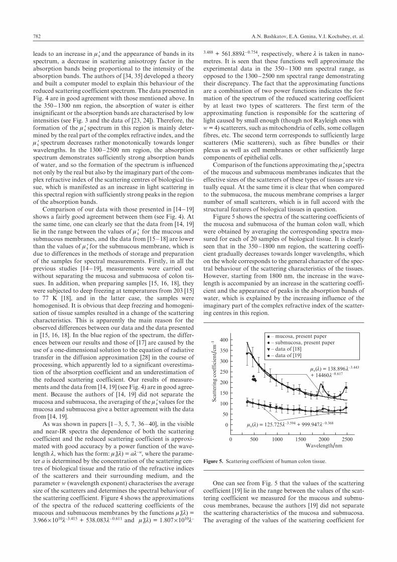
Figure 5 shows the spectra of the scattering coefficients of the mucosa and submucosa of the human colon wall, which were obtained by averaging the corresponding spectra mea-sured for each of 20 samples of biological tissue. It is clearly seen that in the 350 – 1800 nm region, the scattering coeffi-cient gradually decreases towards longer wavelengths, which on the whole corresponds to the general character of the spec-tral behaviour of the scattering characteristics of the tissues. However, starting from 1800 nm, the increase in the wave-length is accompanied by an increase in the scattering coeffi-cient and the appearance of peaks in the absorption bands of water, which is explained by the increasing influence of the imaginary part of the complex refractive index of the scatter-ing centres in this region.
One can see from Fig. 5 that the values of the scattering coefficient [19] lie in the range between the values of the scat-tering coefficient we measured for the mucous and submu-cous membranes, because the authors [19] did not separate the scattering characteristics of the mucosa and submucosa. The averaging of the values of the scattering coefficient for
0 500 1000 1500 2000 2500
0
50
100
150
200
250
300
350
400
Sca
tter
ing
coef
fici
ent /c
m–1
Wavelength/nm
ms(l) = 138.896 l–3.443
+ 14460l–0.617
ms(l) = 125.725l–3.594 + 999.947l– 0.368
– mucosa, present paper– submucosa, present paper– data of [18]– data of [19]
Figure 5. Scattering coefficient of human colon tissue.
783Optical properties of human colon tissues in the 350 – 2500 nm spectral range
the mucosa and submucosa will provide good agreement with the results of [19]. The data presented in [18] are almost identi-cal with our values of the scattering coefficient for the submu-cosa, which is apparently due to very deep freezing and homogenisation of tissue samples made in [18].
Figure 5 shows the approximations of the spectral depen-dences of the scattering coefficient of the mucosa and submu-cosa by the functions ms(l) = 138.896l–3.443 + 14460l– 0.617 и ms(l) = 125.725l–3.594 + 999.947l– 0.368, respectively. It can be seen that these functions well approximate the experimental data in the 350 – 1800 nm spectral range, and in the 1800 – 2500 nm range we observe a discrepancy between the experimental results and the approximating dependence. As in the case of ms', the approximating functions are a combina-tion of two power functions, indicating the formation of the scattering coefficient spectrum by at least two types of scat-terers.
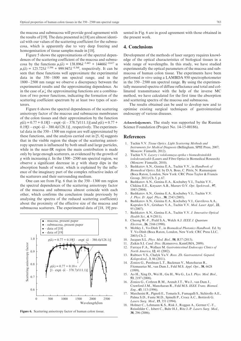
Figure 6 shows the spectral dependences of the scattering anisotropy factor of the mucous and submucous membranes of the colon tissues and their approximation by the function g(l) = 0.77 + 0.18[1 – exp(– (l – 378.7)/111.1)] and g(l) = 0.77 + 0.19[1 – exp(– (l – 380.4)/128.1)] , respectively. The experimen-tal data in the 350 – 1300 nm region are well approximated by these functions, and the analysis carried out in [5, 6] suggests that in the visible region the shape of the scattering anisot-ropy spectrum is influenced by both small and large particles, while in the near-IR region the main contribution is made only by large enough scatterers, as evidenced by the growth of g with increasing l. In the 1300 – 2500 nm spectral region, we observe a significant decrease in g with sharp dips in the absorption bands of water, which is explained by the influ-ence of the imaginary part of the complex refractive index of the scatterers and their surrounding medium.
One can see from Fig. 6 that in the 350 – 1300 nm region the spectral dependences of the scattering anisotropy factor of the mucosa and submucosa almost coincide with each other, which confirms the conclusion (made previously by analysing the spectra of the reduced scattering coefficient) about the proximity of the effective size of the mucosa and submucosa scatterers. The experimental data of [18, 19] pre-
sented in Fig. 6 are in good agreement with those obtained in the present work.
4. Conclusions
Development of the methods of laser surgery requires knowl-edge of the optical characteristics of biological tissues in a wide range of wavelengths. In this study, we have studied experimentally the optical parameters of the mucosa and sub-mucosa of human colon tissue. The experiments have been performed in vitro using a LAMBDA 950 spectrophotometer in the 350 – 2500 nm spectral range. By using the experimen-tally measured spectra of diffuse reflectance and total and col-limated transmittance with the help of the inverse MC method, we have calculated for the first time the absorption and scattering spectra of the mucosa and submucosa.
The results obtained can be used to develop new and to optimise existing surgical techniques of gastrointestinal endoscopy of various diseases.
Acknowledgements. The study was supported by the Russian Science Foundation (Project No. 14-15-00186).
References 1. Tuchin V.V. Tissue Optics. Light Scattering Methods and
Instruments for Medical Diagnosis (Bellingham, SPIE Press, 2007; Moscow: Fizmatlit, 2012).
2. Tuchin V.V. Lazery i volokonnaya optika v biomeditsinskikh issledovaniyakh (Lasers and Fibre Optics in Biomedical Research)(Moscow: Fizmatlit, 2010).
3. Bashkatov A.N., Genina E.A., Tuchin V.V., in Handbook of Biomedical Optics. Ed. by D.A. Boas, C. Pitris, N. Ramanujam (Boca Raton, London, New York: CRC Press Taylor & Francis Group, 2011) Ch. 5, p. 67.
4. Bashkatov A.N., Genina E.A., Kochubey V.I., Tuchin V.V., Chikina E.E., Knyazev A.B., Mareev O.V. Opt. Spektrosk., 97, 1043 (2004).
5. Bashkatov A.N., Genina E.A., Kochubey V.I., Tuchin V.V. J. Phys. D: Appl. Phys., 38, 2543 (2005).
6. Bashkatov A.N., Genina E.A., Kochubey V.I., Gavrilova A.A., Kapralov S.V., Grishaev V.A., Tuchin V.V. Med. Laser Appl., 22, 95 (2007).
7. Bashkatov A.N., Genina E.A., Tuchin V.V. J. Innovative Optical Health Sci., 4, 9 (2011).
8. Cheong W.-F., Prahl S.A., Welch A.J. IEEE J. Quantum Electron., 26, 2166 (1990).
9. Mobley J., Vo-Dinh T., in Biomedical Photonics Handbook. Ed. by T. Vo-Dinh (Boca Raton, London, New York: CRC Press LLC, 2003) Ch. 2.
10. Jacques S.L. Phys. Med. Biol., 58, R37 (2013).11. Zaikin S.I. Cand. Diss. (Kemerovo, KemGMA, 2009). 12. Farraye F.A., Wallace M. Gastrointestinal Endoscopy Clinics of
North America, 12, 41 (2002).13. Rubtsov V.S., Chalyk Yu.V. Ross. Zh. Gastroenterol. Gepatol.
Koloproktol., 3, 86 (2011). 14. Zonios G., Perelman L.T., Backman V., Manoharan R.,
Fitzmaurice M., van Dam J., Feld M.S. Appl. Opt., 38, 6628 (1999).
15. Ao H., Xing D., Wei H., Gu H., Wu G., Lu J. Phys. Med. Biol., 53, 2197 (2008).
16. Zonios G., Cothren R.M., Arendt J.T., Wu J., van Dam J., Crawford J.M., Manoharan R., Feld M.S. IEEE Trans. Biomed. Eng., 43, 113 (1996).
17. Marchesini R., Pignoli E., Tomatis S., Fumagalli S., Sichirollo A.E., Palma S.D., Fante M.D., Spinelli P., Croce A.C., Bottiroli G. Lasers Surg. Med., 15, 351 (1994).
18. Holmer C., Lehmann K.S., Risk J., Roggan A., Germer C.-T., Reissfelder C., Isbert C., Buhr H.J., Ritz J.-P. Lasers Surg. Med., 38, 296 (2006).
0 500 1000 1500 2000 2500
0.6
0.7
0.8
0.9
1.0
Sca
tter
ing
anis
otr
op
y fa
cto
r
Wavelength/nm
g(l) = 0.77 + 0.18´[1– exp(–(l – 378.7)/111.1)]
g(l) = 0.77 + 0.19[1 – exp(–(l – 380.4)/128.1)]
– mucosa, present paper– submucosa, present paper– data of [18]– data of [19]
Figure 6. Scattering anisotropy factor of human colon tissue.
A.N. Bashkatov, E.A. Genina, V.I. Kochubey, et. al.784
19. Wei H.-J., Xing D., Lu J.-J., Gu H.-M., Wu G.-Y., Jin Y. World J. Gastroenterology, 11, 2413 (2005).
20. Prahl S.A., van Gemert M.J.C., Welch A.J. Appl. Opt., 32, 559 (1993).
21. Wang L., Jacques S.L., Zheng L. Comput. Methods & Programs Biomed., 47, 131 (1995).
22. Bundy B. Basic Optimization Methods (Baltimore: Edward Arnold Publishers, 1984; Moscow: Radio i svyaz’, 1988).
23. Kou L., Labrie D., Chylek P. Appl. Opt., 32, 3531 (1993).24. Palmer K.F., Williams D. J. Opt. Soc. Am., 64, 1107 (1974).25. http://omlc.ogi.edu/spectra/hemoglobin/.26. Ishimaru A. Wave Propagation and Scattering in Random Media
(New York: Academic, 1978) Vol. 1.27. van Gemert M.J.C., Star W.M. Lasers Life Sci., 1, 287 (1987).28. Groenhuis R.A.J., Ferwerda H.A., Ten Bosch J.J. Appl. Opt., 22,
2456 (1983).29. Hidovic-Rowe D., Claridge E. Phys. Med. Biol., 50, 1071 (2005).30. Terentyuk G.S., Ivanov A.V., Polyanskaya N.I., Maksimova I.L.,
Skaptsov A.A., Chumakov D.S., Khlebtsov B.N., Khlebtsov N.G. Kvantovaya Elektron., 42 (5), 380 (2012) [ Quantum Electron., 42 (5), 380 (2012)].
31. Bohren C.F., Huffman D.R. Absorption and Scattering of Light by Small Particles (New York: Wiley, 1983; Moscow: Mir, 1986).
32. Du Y., Hu X.H., Cariveau M., Kalmus G.W., Lu J.Q. Phys. Med. Biol., 46, 167 (2001).
33. Ritz J.-P., Roggan A., Isbert C., Muller G., Buhr H., Germer C.-T. Lasers Surg. Med., 29, 205 (2001).
34. Fu Q., Sun W. Appl. Opt., 40, 1354 (2001).35. Sun W., Loeb N.G., Lin B. Appl. Opt., 44, 2338 (2005).36. Mourant J.R., Fuselier T., Boyer J., Johnson T.M., Bigio I.J.
Appl. Opt., 36, 949 (1997).37. Schmitt J.M., Kumar G. Appl. Opt., 37, 2788 (1998).38. Wang R.K. J. Modern Opt., 47, 103 (2000).39. Jacques S.L. J. Innovative Optical Health Sci., 4, 1 (2011).40. Shchyogolev S.Yu. J. Biomed. Opt., 4, 490 (1999).