optic nerve evaluation in glaucoma -covert to pdf nerve evaluation in glaucoma... · optic nerve...
TRANSCRIPT

Optic Nerve Evaluation in Glaucoma (TPG)
Introduction
Glaucoma is a progressive optic nerve disease characterized by retinal ganglion cell death and resultant axon loss, ultimately manifesting as excavation of the optic nerve head with consequent defects in retinal sensitivity that can be measured with visual field tests1,5,15,17. Glaucomatous damage may be due to elevated intraocular pressure, poor perfusion pressure to the optic nerve head, obstruction of axoplasmic flow within the ganglion cell axons, anatomic weakening of the lamina cribrosa, and programmed cell death of the ganglion cell axons2,9,14,16,20.
Knowing that 25‐35% of ganglion cells are lost before visual field defects are detected on standard automated perimetry and that careful evaluation of the optic nerve head has high specificity and good precision for glaucoma diagnosis1,13,14,15,19,21, accurate assessment of the optic nerve becomes essential for early diagnosis and management of glaucoma in order to prevent visual field defects before they occur. A sound knowledge of the subtle changes of the optic nerve and its surrounding tissues is thus critical in order for the astute clinician to become proficient in evaluating the optic nerve to be able to detect glaucoma as early as possible. Once characteristic cupping and visual field decline has occurred the diagnosis is easy – the challenge therefore lies in critically evaluating the optic nerve before significant cupping takes place. In the discussion that follows, we will first discuss the normal anatomy and non‐pathologic variations of the healthy optic nerve and peripapillary tissues, and will conclude by reviewing cardinal features to look for so that glaucomatous optic nerve damage can be detected as early as possible.
Naida Jakirlic, OD, FAAO
Dr. Jakirlic, graduated from SUNY College of Optometry and completed a residency in ocular disease at SUNY. She was a staff optometrist at two New York City hospitals for several years prior to joining the faculty at Western University of Health Sciences in 2014 where she is involved in both didactic and clinical education of first, second, and third year optometry students.
E‐mail address: [email protected]

Normal anatomy of the optic nerve
Ganglion cell axons make up the vast majority of the neuroretinal rim tissue of the optic disc. One to 1.5 million axons leave via the optic nerve head through the scleral canal, while the remainder of the neuroretinal rim is composed of capillaries and astrocytes2,8.
There are four distinct layers of the optic nerve head, including the surface, prelaminar, laminar, and retrolaminar layers8. The surface layer is the anterior limit of the optic nerve and is the point of contact with the vitreous8. Its peripheral edge is defined by the anterior limits of the scleral ring, and its posterior limit is where the axon bundles have completed a 90‐degree turn from the plane of the retina and reached the level of the choroid8. The prelaminar optic nerve is an indistinct segment of axons surrounded by outer retina, choriocapillaris, and choroid8. Within the laminar optic nerve, the ganglion cell axon bundles are wrapped in glial cells and confined in the rigid pores of the lamina cribrosa8. The retrolaminar optic nerve thickness is doubled by the presence of myelinating oligodendrocytes8.
The lamina cribrosa is a part of the scleral tissue through which ganglion cell axons exit as they leave the globe. It is composed of several sheets of connective tissue that is fenestrated to allow the passage of the nerve fiber bundles8,9,10. The superior and inferior poles have the largest pores, thus providing less structural support for the axon bundles leaving the nerve in these two areas8,9,10,13. This may explain why there is typically greater damage to the retinal ganglion cell axons in the superior and inferior poles of the optic nerve that we see clinically9. Furthermore, as ganglion cell axons are lost, the laminar dots become more exposed, which can also be visualized clinically. While laminar dots may certainly be present in healthy non‐glaucomatous eyes, their presence should alert the clinician to look for other signs of glaucoma. The lamina is also susceptible to thinning and bowing backward due to effects of the intraocular pressure, which results in a clinically visible deeper cup in glaucomatous eyes1,2,14.
The surface optic nerve receives its blood supply from small branches of the central retinal artery8,9. The pre‐laminar optic nerve is supplied by the short posterior ciliary arteries (SPCA), and their integrity is responsible for the reddish hue that is observed clinically in healthy optic nerves2,8,9. The laminar optic nerve is supplied by the Circle of Zinn‐Haller, which is composed of anastomoses from adjacent SPCA’s8,9. The retro‐laminar optic nerve head is supplied by axial vasculature from the central retinal artery, the pial vascular plexus, and the SPCA’s8,9. Knowing the vasculature of the optic nerve is critical when evaluating the health and integrity of the neuroretinal rim as it is necessary to remember that glaucoma is cupping without pallor1,11,14. If pallor of the optic nerve is observed, it indicates insult to the prelaminar vasculature and should always alert the clinician that a different or an additional optic neuropathy is present.
Venous drainage of the optic nerve occurs via the central retinal vein8,9. In chronic glaucoma, optociliary shunt vessels may appear due to disturbed retinal circulation. These vessels are pre‐existing venules that become more visible as they dilate to re‐route blood around an area of obstruction8. They can be differentiated from neovascular vessels as optociliary shunt vessels will not leak on fluorescein angiography, while neovascular vessels will always leak fluorescein dye.

Figure 5: Cvessels in optic neu
The optic head. It isthat formof the sclethereforewhere thedeviation assessmeLastly, it iscup, therepresent clnerve is pnecessaryindependglaucoma
A discussibecause nglaucomathe scleraextensionterminatetranspare
Color photoga patient witropathy.
cup, which iss devoid of axs its floor1,19eral canal10,1 viewing the e contour is loof vessels onnt as color cas critical to reefore large sclinically as smprobably normy for every C/ent finding bea in large nerv
on on normanon‐pathologa. Elschnig’s scal lip, is seen in of the scleraes just short oent nerve fibe
raph of optocth advanced g
s given the mxons and capi9. Its size is de11. The bordenerve stereosocated14. Then the surface an sometimesemember thacleral openingmall C/D ratiosmal, while an D ratio to be ecause it can ves, as well as
al optic nerve ic variations ocleral ring is ammediately aa between theof the optic dier bundles4,7.
ciliary shunt glaucomatous
ost attentionllaries and is ependent on er of the cup scopically unde best way toof the optic ns be very mislt the size of tgs will result is1,10,11,19. Taverage C/D considered dbe highly miss an under‐dia
anatomy is inof this area ma normal anatadjacent to the choroid andisc, thus allow. Choroidal cr
s Figurevesselsneurop
in glaucoma,pale due to vthe number and rim is deder high mago determine tnerve head14eading whenthe scleral opn large C/D raThe take‐homin a small nerdirectly in relasleading and agnosis of gla
ncomplete wmust be distintomic variatiohe optic disc d optic nerve;wing visualizarescents are a
e 6: Color phs in a patiepathy.
, is a central evisibility of theof nerve fibetermined by gnification is ethe contour is4. Clinicians mn determiningpening will deatios, whereame point hererve might be ation to the dcan lead to baucoma in sm
ithout addresnguished fromon whereby amargin11,12,; more simplyation of scleraalso a normal
hotograph ofent with gla
excavation ine collagenousrs leaving thecontour, not essential in acs by visualizinmust become g the cup‐to‐dtermine the sas small sclerae is that a largglaucomatoudisc size and nboth an over‐dmall nerves.
ssing the perim pathologic ca thin white ri,14. It is causey, the RPE andal tissue throu peripapillary
f optociliary aucomatous
the optic nes lamina cribre eye and theby color, ccurately asseng the point oproficient in tdisc (C/D) ratisize of the opal openings wge C/D in a larus – thereforenot as an diagnosis of
ipapillary arechanges seening, also knowed by an anted choroid tissugh the y variation, an
shunt optic
rve rosa e size
essing of this o. ptic will rge e it is
a in wn as erior sue
nd

are caused by non‐pathologic termination of the RPE prior to reaching the optic disc, thus causing the underlying choroid to be visible as a uniform slate‐gray crescent directly adjacent to the optic disc7. Choroidal crescents are typically present temporally, although they can span the optic disc 360 degrees7. These two normal variants should not be confused with zone alpha and zone beta peripapillary atrophy that will be discussed shortly.
Optic nerve evaluation in glaucoma
Slit lamp biomicroscopy with a fundus condensing lens is the ideal method to evaluate the optic nerve because it allows for a magnified stereoscopic view, thus enabling the observer to directly measure the vertical height of the optic disc6,18. While direct ophthalmoscopy provides good magnification, it unfortunately has the disadvantage of lacking stereopsis and is therefore not the ideal tool for optic nerve assessment. Indirect ophthalmoscopy is also sub‐optimal as it provides poor magnification and detail. Optic disc photography is an excellent tool for both documentation and monitoring for any progression; typically, it is performed at baseline and every year afterwards unless progression of disease is suspected6,10,18.
There are several key findings to look for when evaluating the optic nerve. First, the clinician should assess the optic disc size and shape. This allows for the determination of whether the cup size is normal or suspicious in relation to the size of the scleral opening6,11,21. Next, the size, color, and integrity of the neuroretinal rim tissue should be assessed6. Here it is critical to remember that glaucoma causes cupping without pallor, therefore any presence of pallor necessitates a different or additional workup for a non‐glaucomatous optic neuropathy. Next, the cup size and shape should be assessed in relation to the optic disc size. The clinician should then examine the retinal nerve fiber layer, the peripapillary region, and carefully look for the presence of optic disc hemorrhages, as well as other less obvious vascular changes that will be discussed below6.
Optic disc size
The scleral foramen through which the ganglion cell axons exit the eye ranges in diameter such that a large foramen will result in a large disc and a large cup, whereas a small foramen will result in the opposite6,10,11. Measurement of the vertical disc diameter is therefore essential in order for clinicians to assess whether an optic disc is small, average, or large6,10. Measurement of the optic disc size is best performed at the slit lamp using a narrow vertical beam of light and a fundus condensing lens6,10. Depending on the fundus lens that is used, the size measured at the slit lamp may need to be multiplied by a correction factor. A 60D condensing lens gives a 1:1 ratio, therefore it is the optimal lens for this purpose as the correction factor is simply 1.06. The correction factor for a 78D lens is 1.1, and for a 90D lens it is 1.36,21. Another way to document the vertical height is by simply recording the vertical height measured at the slit lamp and the condensing lens that was used without needing to multiply by the correction factor.
The average vertical height of the healthy optic nerve ranges from 1.8 to 2.0 millimeters18,19. A small vertical diameter is equal to or less than 1.5 millimeters; a large vertical diameter is greater than 2.2 millimeters6,18. This assessment becomes essential in order to avoid over‐testing and over‐diagnosis in large nerves due to the knee‐jerk reaction that any C/D greater than 0.4 is immediately suspicious for glaucoma. Perhaps even more importantly, it is necessary to carefully measure the vertical height of the optic nerve in order to prevent under‐diagnosing glaucoma in small optic nerves due to observing a C/D ratio that would not typically raise suspicion solely due to its low value. Thus it is imperative to keep in

mind that a C/D of 0.8 is probably not glaucomatous in a nerve with a 2.7 millimeter vertical height, whereas a C/D of 0.3 is probably glaucomatous in a nerve with a vertical height of 1.5 millimeters.
Neuroretinal Rim
The neuroretinal rim will reflect selective loss of ganglion cell axons and is the primary location of pathologic changes seen in glaucoma17. Because C/D ratio is often a poor indicator of early glaucoma,
clinicians should pay close attention to the width and health of the neuroretinal rim instead – we should be looking more carefully at the donut, not at the hole21. When assessing the neuroretinal rim tissue, its size, shape, and color must be taken into consideration6. The ISNT rule has been ingrained in us, and this is for a reason. When looking at the average nerve, the inferior rim tissue should always be the widest, followed by superior, nasal, and temporal, and this has to do with the anatomic distribution of the nerve fiber bundles as they leave through the scleral canal6,14. However, in normal anatomic variations whereby the nerve might be horizontally oval, the ISNT rule may not always apply, which is also important to keep in mind in order to avoid over‐diagnosing glaucoma in healthy eyes15.
As clinicians, we should look at the neuroretinal rim carefully and evaluate for any thinning and notching of the rim tissue, which would indicate glaucomatous damage. The typical sequence of neuroretinal rim loss in glaucoma is loss of rim tissue at the infero‐temporal and supero‐temporal poles, followed by the temporal rim, and lastly by the nasal rim17.
Peripapillary chorioretinal atrophy
Irregular pigmentation around the optic nerve is a non‐specific finding that can be seen in many healthy eyes3. However, its presence should raise suspicion for glaucoma, particularly if additional risk factors are identified in the patient. It differs from normal peripapillary variants discussed earlier in the respect that atrophy is typically irregular and patchy, whereas crescents are typically very uniform in color and shape. The clinical appearance of peripapillary atrophy is a moth‐eaten pattern of the RPE temporal to the optic nerve head; if truly associated with glaucoma, there is typically neuro‐retinal rim thinning adjacent to the area of atrophy1,3,4,5,6,11,22.
Zone alpha is a hypo and hyper‐pigmented area of RPE irregularity. Nasally it is always separated from the neuro‐retinal rim by either zone beta if present, or by the scleral ring if zone beta is absent, and temporally it is bounded by normal retina5,11,14. Zone alpha is a non‐specific finding that is present in both normal and glaucomatous eyes4,5.
Zone beta is an irregular area of complete RPE and choriocapillaris atrophy immediately adjacent to the optic disc, thus resulting in visualization of large choroidal vessels and underlying sclera5. It is thought to be caused by poor perfusion of the peripapillary area, and though it may also be present in normal eyes, it is much more common in glaucomatous eyes4,5,11,14.
The usefulness of zone alpha and zone beta is to help the clinician differentiate between glaucomatous and non‐glaucomatous optic nerves, particularly when additional anatomic changes suggest the presence of early glaucoma damage. Additionally, because progression of zone beta is associated with progression of glaucoma, it may be a subtle yet important indication to alert the clinician that more aggressive glaucoma therapy may be necessary in a patient already diagnosed with the disease4,5,14.

Figure 7: Color photograph demonstrating clear areas of zone alpha and beta peripapillary atrophy.
Vascular Changes
Several vascular changes can be observed in glaucomatous eyes, including optic disc hemorrhages, baring of circumlinear vessels, bayonetting of vessels, nasalization of vessels, optic nerve shunts, and retinal artery attenuation.
Optic disc hemorrhage, also known as drance hemorrhage, is a splinter or flame‐shaped hemorrhage located on the disc margin1,5,6,11,14,19. It is a hallmark of glaucomatous optic nerve damage and can be found in early and moderate stages of glaucoma, but is rare in advanced glaucomatous cupping likely due to absence of sufficient neuroretinal rim tissue1,5,6,11,14,19. It is usually located on the supero‐temporal and infero‐temporal disc margins and is more common in patients with normal tension glaucoma. It typically resolves within 6‐10 weeks of onset and is associated with localized RNFL defects and rim notching5. Though highly pathognomonic for glaucoma, it can also occur in patients with posterior vitreous detachment, BRVO, hypertensive retinopathy, and non‐arteritic anterior ischemic optic neuropathy, therefore its presence must always be considered in relation to the clinical picture at hand. In glaucoma patients, it is strongly indicative of disease progression and is a strong indicator that more aggressive therapy should be instituted.
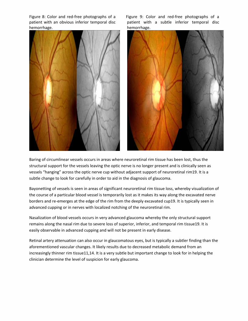
Figure 8: Color and red‐free photographs of a patient with an obvious inferior temporal disc hemorrhage.
Figure 9: Color and red‐free photographs of a patient with a subtle inferior temporal disc hemorrhage.
Baring of circumlinear vessels occurs in areas where neuroretinal rim tissue has been lost, thus the structural support for the vessels leaving the optic nerve is no longer present and is clinically seen as vessels “hanging” across the optic nerve cup without adjacent support of neuroretinal rim19. It is a subtle change to look for carefully in order to aid in the diagnosis of glaucoma.
Bayonetting of vessels is seen in areas of significant neuroretinal rim tissue loss, whereby visualization of the course of a particular blood vessel is temporarily lost as it makes its way along the excavated nerve borders and re‐emerges at the edge of the rim from the deeply excavated cup19. It is typically seen in advanced cupping or in nerves with localized notching of the neuroretinal rim.
Nasalization of blood vessels occurs in very advanced glaucoma whereby the only structural support remains along the nasal rim due to severe loss of superior, inferior, and temporal rim tissue19. It is easily observable in advanced cupping and will not be present in early disease.
Retinal artery attenuation can also occur in glaucomatous eyes, but is typically a subtler finding than the aforementioned vascular changes. It likely results due to decreased metabolic demand from an increasingly thinner rim tissue11,14. It is a very subtle but important change to look for in helping the clinician determine the level of suspicion for early glaucoma.

Retinal Nerve Fiber Layer (RNFL)
Discussion of the optic nerve in relation to glaucoma would be incomplete without mentioning the RNFL, which is composed of retinal ganglion cell axons which are covered by astrocytes and bundled by Muller cell processes14. The RNFL is seen as bright fine striations fanning off the optic disc and is most prominent infero‐temporally and supero‐temporally6,13,14. Healthy RNFL will obscure the details of underlying peripapillary retinal vascular walls, therefore RNFL loss will present as increased sharpness of vascular walls13. Clinical assessment of the RNFL requires a fundus condensing lens, bright light, and a red‐free filter at the slit lamp6,13. The green light produced by the filter is absorbed by the RPE and choroid, creating a dark background against which the light that is reflected by the RNFL is well contrasted against the dark background, which allows clinicians easy visualization of the RNFL.
RNFL defects in glaucoma are caused by selective damage to the superior and inferior arcuate bundles, with relative sparing of papillomacular and nasal bundles. Defects within the RNFL appear as darker zones in areas of expected brightness4. This will cause retinal vessels to appear redder and darker, and will allow small vessels to become more visible4.
Diffuse RNFL defects are the most common yet most difficult to detect. The clinician should compare the striations between the superior and inferior bundles of the same eye, as well as the striations between the right and left eyes and look for any loss of brightness, increased sharpness and prominence of peripapillary vessels, and clearer visualization of the underlying choroid.
Focal loss of the RNFL is easiest to identify, but is typically the least common and is usually associated with a notch at the disc or a current or prior disc hemorrhage6. It is seen as a dark slit or wedge in the bright superior or inferior striations traveling all the way back to the optic nerve head6,13.
Figure 1: Color photograph of superior temporal RNFL wedge defect.
Figure 2: Red‐free image of superior temporal and inferior temporal RNFL wedge defect.

Changes in the optic cup
Several characteristic changes of the optic cup can be observed clinically. They include increased cup size, increased depth, visualization and/or increase of laminar dots, vertical enlargement, and asymmetry between two eyes greater than 0.2 in the absence of disc size asymmetry1,2,16,17.
Therefore although the optic cup is not the only feature to consider when evaluating the optic nerve in glaucoma, it is still a very important one to take into consideration.
Conclusion
Evaluation of the optic nerve head is essential for the early diagnosis and management of glaucoma. Clinicians must keep in mind that the C/D ratio is not the only factor to consider when evaluating the optic nerve, and must therefore give due diligence to the neuroretinal rim and its surrounding peripapillary tissue and vasculature. Clinicians must also remember that glaucoma causes cupping without pallor, therefore the presence of pallor of the neuroretinal rim must prompt the investigation for a different or additional optic neuropathy. Once characteristic cupping has taken place, the diagnosis of glaucoma is easy; the challenge lies in diligently assessing the optic nerve and peripapillary tissues prior to development of significant structural and functional damage in order to make an accurate diagnosis as early as possible. By utilizing the steps outlined below, one should be able to become proficient in this evaluation over time in order to prevent under‐diagnosis, delayed diagnosis, as well as over‐diagnosis of a disease that is still in many ways very elusive even to the most seasoned clinician.
Figure 4: Color photograph of prominent laminar dot sign.

Optic Nerve Evaluation Checklist
o Measure the vertical size of the optic nerve at the slit lamp through dilated pupils o Determine the vertical and horizontal C/D ratio and consider it carefully in relation to the size of
the disc o Evaluate the integrity of the neuroretinal rim, specifically looking for the presence of diffuse
thinning, notching, and pallor – keep in mind that the ISNT rule should be observed for the average vertically‐oval optic nerve
o Evaluate for the presence of any vascular changes: disc hemorrhages, nasalization of vessels, baring of vessels, optociliary shunt vessels, and arteriole narrowing
o Evaluate for the presence and extent of peripapillary atrophy o Evaluate the integrity of the RNFL with a red‐free filter
Figure 3: Color photograph of the right eye demonstrating prominent laminar dot sign, inferior notch between 7‐8 o’clock, temporal zone beta and alpha peripapillary atrophy, and nasal zone beta peripapillary atrophy.

References:
1. Broadway DC, Nicolela MT, Drance SM. Optic Disc Appearances in Primary Open‐Angle Glaucoma. Survey of Ophthalmology. 1999;43(1):S223‐S243.
2. Burgoyne CF, Downs JC. Premise and prediction—how optic nerve head biomechanics underlies the susceptibility and clinical behavior of the aged optic nerve head. J Glaucoma. 2008; 17: 318‐328.
3. Chui TYP, Zhong Z, Burns SA. The relationship between peripapillary crescent and axial length: Implications for differential eye growth. Vision Research. 2011;51:2132‐38.
4. Curico et al. Peripapillary Chorioretinal Atrophy: Bruch's Membrane Changes and Photoreceptor Loss. Ophthalmology. 2000;107(2):334‐343.
5. De Moraes CG et al. Predictive factors within the optic nerve complex for glaucoma progression: disc hemorrhage and parapapillary atrophy. Asia‐Pacific J Ophthalmol. 2012; 1(2):105‐112.
6. Fingeret M et al. Five rules to evaluate the optic disc and retinal nerve fiber layer for glaucoma. Optometry. 2005;76(11):661‐668.
7. Grosvenor T. (2007). Primary Care Optometry. (pp. 153). St. Louis: Butterworth Heinmann Elsevier.
8. Hayreh SS. Blood supply of the optic nerve head and its role in optic atrophy, glaucoma, and oedema of the optic disc. Brit J Ophthal. 1969;53: 721‐748.
9. Hayreh, S.S. (2011). Ischemic Optic Neuropathies. (pp. 7‐35). New York: Springer. 10. Hoffmann EM et al. Optic Disk Size and Glaucoma. Survey of Ophthalmology. 2007;52(1):32‐49. 11. Jonas JB, Budde WM. Diagnosis and Pathogenesis of Glaucomatous Optic Neuropathy:
Morphological Aspects. Progress in Retinal and Eye Research. 2000;19(1):1‐40. 12. Jonas JB. Clinical implications of peripapillary atrophy in glaucoma. Current Opinion in
Ophthalmology. 2005;16:84‐88. 13. Jonas JB, Dichtl A. Evaluation of the Retinal Nerve Fiber Layer. Survey of Ophthalmology.
1996;40(5):369‐378. 14. Jonas JB, Budde WM, Panda‐Jonas S. Ophthalmoscopic Evaluation of the Optic Nerve Head.
Survey of Ophthalmology. 1999;43(4):293‐320. 15. Kampougeris G et al. Peripapillary retinal nerve fibre layer thickness measurement with SD‐OCT
in normal and glaucomatous eyes: distribution and correlation with age. International Journal of Ophthalmology. 2013;6(5):662‐665.
16. Lee EJ et al. Visualization of the Lamina Cribrosa Using Enhanced Depth Imaging Spectral‐Domain Optical Coherence Tomography. American Journal of Ophthalmology. 2011;152(1):87‐95.
17. Lee JLS, Nicolela MT, Chauhan BC. Rates of Neuroretinal Rim and Peripapillary Atrophy Area Change. Ophthalmology. 2009;116:840‐847.
18. Lim CS, O’Brien C, Bolton NM. A simple clinical method to measure the optic disk size in glaucoma. J Glaucoma. 1996;5:241–245.
19. Reis ASC, Toren A, Nicolela MT. Clinical Optic Disc Evaluation in Glaucoma. European Ophthalmic Review. 2012;6(2):92‐97
20. Sigal IA et al. Factors influencing optic nerve head biomechanics. Invest Ophthalmol Vis Sci. 2005; 46(11):4189‐99.
21. Spaeth GL et al. Systems for Staging the Amount of Optic Nerve Damage in Glaucoma: A Critical Review and New Material. Survey of Ophthalmology. 2006;51(4):293‐315.
22. Uhm KB et al. Peripapillary Atrophy in Normal and Primary Open‐Angle Glaucoma. Korean Journal of Ophthalmology. 1998;12:37‐50.

CE@HOME May/June Optic Nerve Evaluation in Glaucoma (TPG) Name_________________________________ License______________
6. Which of the following statements is TRUE about drance hemorrhages?
a. They are typically seen in very advanced glaucoma
b. They typically resolve in 1‐2 weeks c. They are indicative of disease stability
d. They may be caused by posterior vitreous detachment
7. Which of the following statements is TRUE about the
retinal nerve fiber layer? a. The nasal bundles are typically the brightest
b. The papillomacular bundles are typically the brightest
c. It is best visualized with a red‐free filter d. It is best visualized with a diffuse white light
8. Which of the following findings would arouse the MOST suspicion for possible glaucomatous optic nerve damage?
a. C/D ratio of 0.3 in a 1.5mm sized nerve b. C/D ratio of 0.3 in a 2.0mm sized nerve c. C/D ratio of 0.5 in a 2.2mm sized nerve d. C/D ratio of 0.8 in a 2.6mm sized nerve
9. Which of the following vascular changes is typically
seen in very advanced glaucomatous optic nerve damage?
a. Retinal artery attenuation b. Baring of circumlinear vessels c. Nasalization of blood vessels d. Increased sharpness of blood vessels
10. Approximately how many ganglion cells may be
lost before visual field defects are detected on standard automated perimetry?
a. 5‐15% b. 15‐25% c. 25‐35% d. 35‐45%
1. Approximately how many ganglion cell axons leave through the scleral canal in a healthy eye?
a. 0.5 million b. 1.5 million c. 2.5 million d. 3.0 million
2. Which of the following vascular channels
supplies arterial blood to the pre‐laminar optic nerve?
a. Central retinal artery b. Long posterior ciliary artery c. Short posterior ciliary artery d. Pial vascular plexus
3. Which of the following measurements of the
vertical height of the optic nerve would indicate a smaller than average disc size?
a. 1.5mm b. 1.8mm c. 2.0mm d. 2.2mm
4. When using a Volk 78D condensing fundus lens
to measure the vertical height of the optic nerve, which of the following is the correction factor that should be utilized in order to record the height of the optic nerve in millimeters?
5. a. 1.0x b. 1.1x c. 1.2x d. 1.3x
6. Which of the following is a TRUE statement
regarding zone alpha peripapillary atrophy?
a. It is caused by a complete loss of RPE and choriocapillaris
b. It is more commonly found in patients with glaucoma
c. Its temporal edge is bounded by normal retina
d. It is usually found in an area of neuroretinal rim thinning