optic disc size and retinal vein occlusions
TRANSCRIPT
Letter to the Editor
Optic disc size and retinal
vein occlusions
Jie Xu,1 Wei Bin Wei,1 Ya Xing Wang,1
Liang Xu1 and Jost B. Jonas1,2
1Beijing Institute of Ophthalmology,
Beijing Tongren Hospital, Capital Medi-cal University, Beijing, China2Department of Ophthalmology, Medi-cal Faculty Mannheim of the Ruprecht-
Karls-University Heidelberg, Heidelberg,Germany
doi: 10.1111/j.1755-3768.2011.02311.x
To the editor,
R etinal vein occlusions (RVOs)are potentially sight-threatening
retinal vascular diseases that usuallyoccur in middle-aged and elderly peo-ple. In recent population-based stud-ies, the age ⁄ gender-standardizedprevalence of any RVO was about fiveper 1000 (Rogers et al. 2010). Thesame studies showed that arterialhypertension and glaucomatous opticneuropathy were independent factorsassociated with RVOs (Liu et al. 2007;Rogers et al. 2010). As RVOs usuallyoccur at the site of the optic nervehead (except of extrapapillary branchRVOs), the optic nerve head has beenconsidered a ‘locus minoris resisten-ciae’. It appears obvious that a nar-row optic nerve scleral canal (i.e.small optic disc) should be a risk fac-tor for the development of a retinalvein occlusion. This may then be simi-lar to a small optic nerve head being arisk factor for the presence of discdrusen and nonarteritic anteriorischaemic optic neuropathy (Youet al. 2009a,b). Because the potentialrole of a small optic disc as risk factorfor a RVO was not systematicallyaddressed in a population-based studyyet, we performed this investigationon a population-based basis.
The Beijing Eye Study is a popula-tion-based prospective cohort study innorthern China. It included in total4439 subjects of 5324 subjects invitedto participate (response rate of83.4%) (Wang et al. 2006; Liu et al.2007; Jonas et al. 2009). The Medical
Ethics Committee of the Beijing Ton-gren Hospital had approved the studyprotocol, and all participants hadgiven informed consent, according tothe Declaration of Helsinki. The studyhas been described in detail recently(Wang et al. 2006; Liu et al. 2007).Using fundus photographs, the occur-rence of RVOs was assessed (Liu et al.2007). Colour optic disc slides servedfor the measurement of the optic discsize (Wang et al. 2006). Statisticalanalysis was performed using a com-mercially available statistical softwarepackage (spss for Windows, version19.0; SPSS, Chicago, IL, USA). Logis-tic regression was used to investigatethe associations of the binary depen-dent variable ‘presence of RVO’ withthe continuous or categorical indepen-dent variables, such as age, gender,education level, intraocular pressureand optic disc area.
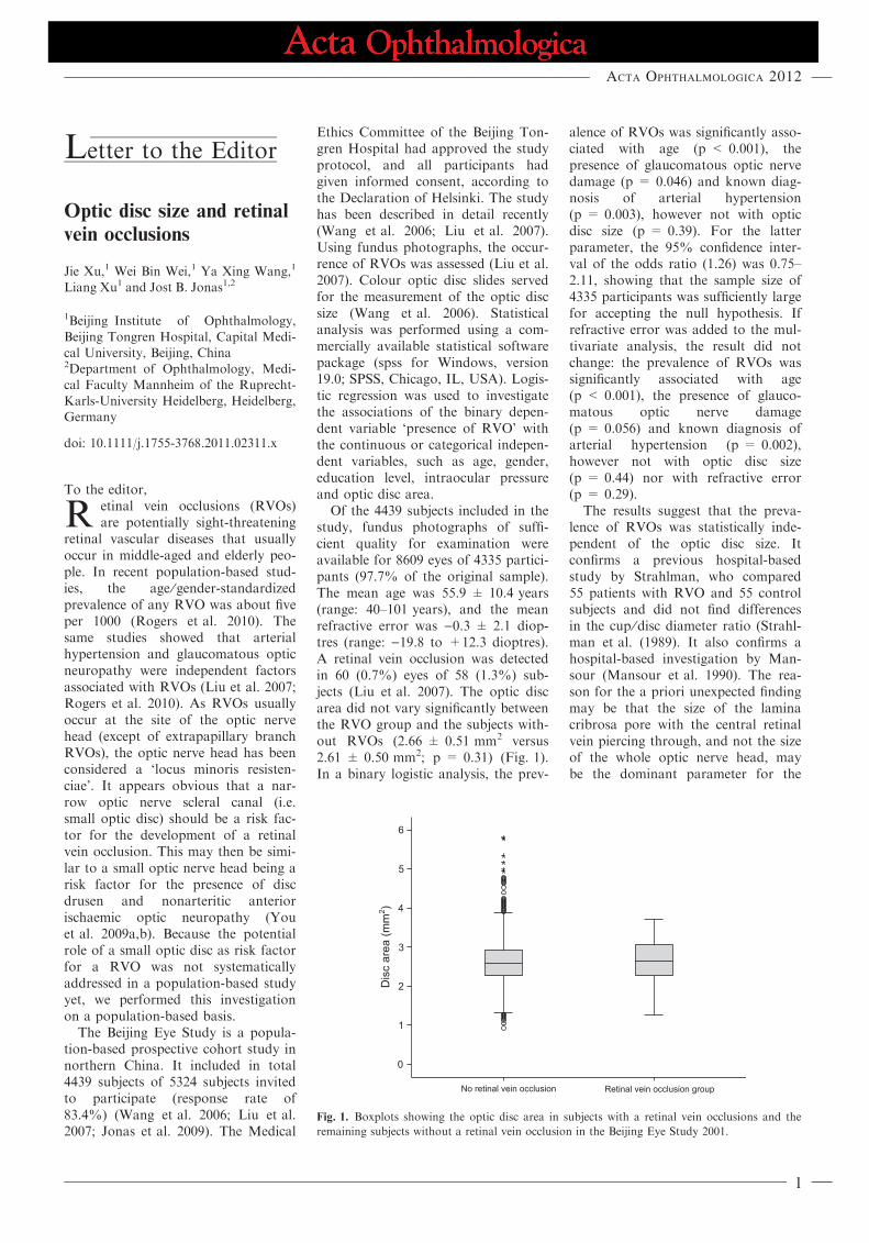
Of the 4439 subjects included in thestudy, fundus photographs of suffi-cient quality for examination wereavailable for 8609 eyes of 4335 partici-pants (97.7% of the original sample).The mean age was 55.9 ± 10.4 years(range: 40–101 years), and the meanrefractive error was )0.3 ± 2.1 diop-tres (range: )19.8 to +12.3 dioptres).A retinal vein occlusion was detectedin 60 (0.7%) eyes of 58 (1.3%) sub-jects (Liu et al. 2007). The optic discarea did not vary significantly betweenthe RVO group and the subjects with-out RVOs (2.66 ± 0.51 mm2 versus2.61 ± 0.50 mm2; p = 0.31) (Fig. 1).In a binary logistic analysis, the prev-
alence of RVOs was significantly asso-ciated with age (p < 0.001), thepresence of glaucomatous optic nervedamage (p = 0.046) and known diag-nosis of arterial hypertension(p = 0.003), however not with opticdisc size (p = 0.39). For the latterparameter, the 95% confidence inter-val of the odds ratio (1.26) was 0.75–2.11, showing that the sample size of4335 participants was sufficiently largefor accepting the null hypothesis. Ifrefractive error was added to the mul-tivariate analysis, the result did notchange: the prevalence of RVOs wassignificantly associated with age(p < 0.001), the presence of glauco-matous optic nerve damage(p = 0.056) and known diagnosis ofarterial hypertension (p = 0.002),however not with optic disc size(p = 0.44) nor with refractive error(p = 0.29).
The results suggest that the preva-lence of RVOs was statistically inde-pendent of the optic disc size. Itconfirms a previous hospital-basedstudy by Strahlman, who compared55 patients with RVO and 55 controlsubjects and did not find differencesin the cup ⁄disc diameter ratio (Strahl-man et al. (1989). It also confirms ahospital-based investigation by Man-sour (Mansour et al. 1990). The rea-son for the a priori unexpected findingmay be that the size of the laminacribrosa pore with the central retinalvein piercing through, and not the sizeof the whole optic nerve head, maybe the dominant parameter for the
Dis
c ar
ea (m
m2 )
No retinal vein occlusion Retinal vein occlusion group
Fig. 1. Boxplots showing the optic disc area in subjects with a retinal vein occlusions and the
remaining subjects without a retinal vein occlusion in the Beijing Eye Study 2001.
Acta Ophthalmologica 2012
1
development of an increased venousoutflow resistance at the level of theoptic nerve head. In a previous histo-morphometric study, the size of lam-ina cribrosa pore with the centralretinal vein passing through was sta-tistically independent of the optic discsize (Jonas et al. 1990). It may explainwhy, in agreement with the result ofour study, the disc size was notrelated with the prevalence of anRVO. From a clinical point of view, itindicates that a small or large opticdisc size is neither a risk factor nor aprotective factor for the developmentof an RVO.
ReferencesJonas JB, Muller-Bergh JA, Schlotzer-Schre-
hardt UM & Naumann GO (1990): Histo-
morphometry of the human optic nerve.
Invest Ophthalmol Vis Sci 31: 736–744.
Jonas JB, Xu L & Wang YX (2009): The
Beijing Eye Study. Acta Ophthalmol 87:
247–261.
Liu W, Xu L & Jonas JB (2007): Vein occlu-
sions in Chinese subjects. Ophthalmology
114: 1795–1796.
Mansour AM, Walsh JB & Henkind P
(1990): Optic disc size in central retinal
vein occlusion. Ophthalmology 97: 165–
166.
Rogers S, McIntosh RL, Cheung N et al.
(2010): The prevalence of retinal vein
occlusion: pooled data from population
studies from the United States, Europe,
Asia, and Australia. Ophthalmology 117:
313–319.
Strahlman ER, Quinlan PM, Enger C &
Elman MJ (1989): The cup-to-disc ratio
and central retinal vein occlusion. Arch
Ophthalmol 107: 524–525.
Wang Y, Xu L, Zhang L, Yang H, Ma Y &
Jonas JB (2006): Optic disc size in a popu-
lation-based study in Northern China. The
Beijing Eye Study. Br J Ophthalmol 90:
353–356.
You QS, Xu L, Wang YX & Jonas JB
(2009a): Prevalence of optic disc drusen in
an adult Chinese population: the Beijing
Eye Study. Acta Ophthalmol 87: 227–228.
You QS, Xu L, Wang YX & Jonas JB
(2009b): Frequency of non-arteritic anterior
ischaemic optic neuropathy in crowded
optic discs: the Beijing Eye Study. Acta
Ophthalmol 87: 354–355.
Correspondence:
Liang Xu, MD
Beijing Institute of Ophthalmology
17 Hougou Lane
Chong Wen Men
100005 Beijing
China
Tel: + 86 10 58265918
Fax: + 86 10 6512 5617
Email: [email protected]
and
Wei Bin Wei, MD
Beijing Tongren Hospital
Capital Medical University
Dong Jiao Min Xiang
Dong Chen District
100730 Beijing
China
Tel: + 86 10 58265916
Fax: + 86 10 6512 5617
Email: [email protected];
Acta Ophthalmologica 2012
2