open access bedside evaluation of dizzy patients · vertigo is the illusion of movements of oneself...
TRANSCRIPT

Copyright © 2013 Korean Neurological Association 203
Print ISSN 1738-6586 / On-line ISSN 2005-5013http://dx.doi.org/10.3988/jcn.2013.9.4.203
REVIEWJ Clin Neurol 2013;9:203-213
Introduction
Dizziness, which is one of the most common symptoms of patients referred for neurological evaluation, comprises a va-riety of symptoms. Even though each symptom may have a different pathophysiologic mechanism and significance, the description is often vague and intriguing. Accordingly, under-standing what is meant by “dizziness”, which can manifest as presyncope, disequilibrium, oscillopsia, and vertigo, should be the first step in evaluating dizzy patients.1
Lightheadedness or presyncope is a sensation of impending loss of consciousness due to a momentary decrease in the ce-rebral blood flow. Patients experience presyncopal dizziness when rapidly standing up from a relaxed supine or seated po-
sition. It may occur with multiple sensory disturbances, or-thostatic hypotension, and cardiac arrhythmias as well as hy-perventilation syndrome and panic attacks.
Disequilibrium is an imbalance or unsteadiness experi-enced while standing or walking. It is caused by various fac-tors including diminished vision, loss of vestibular function, defects in proprioception, and motor dysfunction from the central or peripheral nervous system. It also relates to nonves-tibular syndromes such as visual vertigo, presyncopal faint-ness, or somatoform phobic postural vertigo.
Oscillopsia is the subjective illusion of visual motion. While vertigo occurs with the eyes open or closed, oscillopsia only occurs when the eyes are open. Patients with acquired nystag-mus report spontaneous oscillopsia due to apparent motion of the visual scene from retinal slip. Patients with bilateral loss of the vestibulo-ocular reflex (VOR) frequently experience oscillopsia during head movements. Defects in eye move-ments due to inappropriate VOR cause retinal image motion during head motion.
Bedside Evaluation of Dizzy Patients
Young-Eun Huh, Ji-Soo KimDepartment of Neurology, Seoul National University College of Medicine, Seoul National University Bundang Hospital, Seongnam, Korea
Received June 19, 2013Revised July 31, 2013Accepted July 31, 2013
CorrespondenceJi-Soo Kim, MD, PhDDepartment of Neurology, College of Medicine, Seoul National University, Seoul National University Bundang Hospital, 82 Gumi-ro 173beon-gil, Bundang-gu, Seongnam 463-707, KoreaTel +82-31-787-7461Fax +82-31-719-6818E-mail [email protected]
In recent decades there has been marked progress in the imaging and laboratory evaluation of diz-zy patients. However, detailed history taking and comprehensive bedside neurotological evalua-tion remain crucial for a diagnosis of dizziness. Bedside neurotological evaluation should in-clude examinations for ocular alignment, spontaneous and gaze-evoked nystagmus, the vestibulo-ocular reflex, saccades, smooth pursuit, and balance. In patients with acute spontaneous vertigo, negative head impulse test, direction-changing nystagmus, and skew deviation mostly indicate central vestibular disorders. In contrast, patients with unilateral peripheral deafferentation invari-ably have a positive head impulse test and mixed horizontal-torsional nystagmus beating away from the lesion side. Since suppression by visual fixation is the rule in peripheral nystagmus and is frequent even in central nystagmus, removal of visual fixation using Frenzel glasses is required for the proper evaluation of central as well as peripheral nystagmus. Head-shaking, cranial vibra-tion, hyperventilation, pressure to the external auditory canal, and loud sounds may disclose un-derlying vestibular dysfunction by inducing nystagmus or modulating the spontaneous nystag-mus. In patients with positional vertigo, the diagnosis can be made by determining patterns of the nystagmus induced during various positional maneuvers that include straight head hanging, the Dix-Hallpike maneuver, supine head roll, and head turning and bending while sitting. Abnormal smooth pursuit and saccades, and severe imbalance also indicate central pathologies. Physicians should be familiar with bedside neurotological examinations and be aware of the clinical impli-cations of the findings when evaluating dizzy patients. J Clin Neurol 2013;9:203-213
Key Wordszz dizziness, bedside examination, nystagmus, head impulse test.
Open Access
cc This is an Open Access article distributed under the terms of the Cre-ative Commons Attribution Non-Commercial License (http://creative-commons.org/licenses/by-nc/3.0) which permits unrestricted non-com-mercial use, distribution, and reproduction in any medium, provided the ori-ginal work is properly cited.

Bedside Evaluation of Dizziness
204 J Clin Neurol 2013;9:203-213
Vertigo is the illusion of movements of oneself or the en-vironment due to an imbalance of tonic neural activity in the vestibular-cortical pathway. Although patients usually report rotational vertigo, occasionally they describe a sensation of linear displacement or tilt. Vertigo is commonly exacerbated by head movements and accompanied by nausea and vomit-ing. Vertigo may be due to unilateral injury to the peripheral vestibular organs such as the labyrinth or vestibular nerve. It also results from damage to the central vestibular structures including the vestibular nuclei, vestibular thalamic nuclei, vestibular cortex, and cerebellum. While peripheral vestibu-lar disorders are always characterized by a combination of perceptual, ocular motor, and postural symptoms and signs, central vestibular lesions may give rise to only some of them.
The primary aim of the bedside evaluation of a dizzy pa-tient is the detection of any vestibular deficits. While the ves-tibular impairments can be readily determined in patients with acute vertigo, meticulous examination is required in those with chronic dizziness. When the vertigo is induced in certain cir-cumstances, reproduction of those situations is important dur-ing the evaluation. One of the most important aims of the bed-side evaluation, especially in acute vertigo, is to differentiate the central from peripheral vestibular pathologies. Peripheral vestibular disorders resulting from damage to the labyrinth or vestibular nerve are usually benign, despite the possible pres-ence of severe dizziness. Conversely, central vestibular disor-ders may be fatal without prompt and proper management. The bedside neurotological examination should include eval-uation both for static and dynamic vestibular imbalances. Due to an anatomical proximity, patients with dizziness often also exhibit ocular motor impairments, and so a comprehensive neurotological examination should include an evaluation of the eye movements.2 Recent studies have indicated that simple bedside examinations such as tests for head impulse, gaze-evoked nystagmus (GEN), and skew deviation can reliably predict a central pathology in patients with isolated vertigo.3,4
Head Posture and Ocular Alignment
Bedside examination of dizzy patients should begin with ob-servation of the head posture (i.e., tilt or turn) and ocular alignment. Dysfunction of the pupil and eyelid may aid cen-tral localization of the vestibulopathy, as in lateral medullary infarction (Wallenberg syndrome) with Horner syndrome. Ocular misalignment is frequent in central vestibulopathy, and should be determined in nine cardinal positions of the gaze, along with the range of eye movements.
The ocular tilt reaction (OTR) refers to the triad of head tilt, ocular torsion, and skew deviation (Fig. 1).5 The OTR is known to reflect unilateral or asymmetric dysfunction of the
graviceptive pathways from the utricle. Skew deviation indi-cates a vertical misalignment of the eyes due to a supranucle-ar pathology.2 The presence of skew deviation may be in-ferred by vertical diplopia and can be confirmed by the cover test. This involves covering one eye and detecting a correc-tive vertical movement of the other eye to fixate the target, which indicates the presence of skew deviation. As a rule, the head is tilted toward the side of the lower eye and the ocular torsion occurs in the same direction with the upper poles of the eyes rotating to the lower eye.6 Since the graviceptive pathway responsible for the OTR is known to cross the mid-line just above the abducens nucleus level and ascend in the contralateral medial longitudinal fasciculus (MLF),7,8 lesions below the lower pons cause ipsiversive OTR, and more ros-tral lesions induce contraversive OTR. Cerebellar lesions may give rise to ipsi- or contraversive OTR depending upon the structures involved.9 Although it can occur in peripheral ves-tibular disorders, skew deviation observed in acute isolated vertigo more commonly indicates a central vestibular lesion.3
A tilt of the subjective visual vertical (SVV) is the most sensitive sign of vestibular tone imbalance in the roll plane.6 It may occur in lesions involving either the central or periph-
Fig. 1. The ocular tilt reaction (OTR). The OTR refers to the head tilt, ocular torsion, and skew deviation that are ascribed to asym-metry in the otolithic pathway from the utricle. The head tilt and ocular torsion occur toward the hypotropic eye.

Huh YE and Kim JS
www.thejcn.com 205
eral vestibular pathways. The bucket method is a simple and reliable bedside test for measuring a tilt of SVV,10 in which patients indicate the position of a bucket where the line on the bottom inside the bucket is estimated to be truly vertical while they sit upright and look into a plastic bucket. The ex-aminer then measures the SVV tilt using an external scale as the angle that the line is off the true vertical.
Spontaneous Nystagmus and Other Involuntary Eye Movements
The patterns of spontaneous nystagmus (when present) are most informative for evaluating patients with dizziness/verti-go.11 The proper evaluation of the spontaneous nystagmus re-quires observation of the direction and the effects of gaze on the intensity and direction of the nystagmus. In unilateral pe-
ripheral deafferentation, the spontaneous nystagmus is mixed horizontal-torsional beating away from the lesion side (Fig. 2).12 The nystagmus typically increases during the gaze in the direction of the spontaneous nystagmus, and decreases during the gaze in the opposite direction (Alexander’s law) (Fig. 2).13 Since the peripheral vestibular nystagmus is markedly sup-pressed by visual fixation, proper observation of the nystag-mus requires the removal of visual fixation using Frenzel glasses14 that have 20-diopter convex lenses to prevent fixa-tion and to magnify eye motion, allowing a better observation of the eyes (Fig. 3). When Frenzel glasses are unavailable, an ophthalmoscope can be used to observe nystagmus without fixation. By observing the optic disc with a direct ophthalmo-scope while the other eye is covered, the nystagmus can be examined without fixation.15 However, when determining the direction of the nystagmus, it should be borne in mind that the retina (optic disc) moves in the direction opposite to the cornea for horizontal and vertical nystagmus. Since the disc also moves vertically in torsional nystagmus, motion of the surrounding retinal vessels should be considered when deter-mining the direction of the nystagmus. In torsional nystag-mus the discs move in opposing directions vertically. The dis-tinguishing features of peripheral and central nystagmus are summarized in Table 1.
In contrast, the direction and fixation effects are variable in central vestibular nystagmus. Accordingly, when the charac-teristics of nystagmus do not conform to those of peripheral vestibular nystagmus, it should be considered central.16-19 However, even unidirectional horizontal-torsional nystagmus suppressed by visual fixation should not be simply regarded as peripheral unless other findings, such as a positive head im-pulse test or caloric paresis, are indicative of peripheral ves-
Fig. 2. Nystagmus in left peripheral vestibulopathy. In unilateral peripheral vestibular deafferentation, mixed torsional-horizontal nystagmus beating occurs toward the intact side. The nystagmus typically increases during the gaze in the direction of nystagmus and decreases during the gaze in the opposite direction (Alexan-der’s law), but never changes direction.
30
0
-30
Right
Left
2s
Eye
posit
ion
( deg
)
Fig. 3. Frenzel glasses remove visual fixation using 20-diopter convex lenses, and facilitate the detection of nystagmus by magnifying the eyes.

Bedside Evaluation of Dizziness
206 J Clin Neurol 2013;9:203-213
tibular lesions.
Induction or Modulation of Nystagmus
Various bedside maneuvers may induce nystagmus or modu-late the preexisting spontaneous nystagmus. Even in patients with spontaneous nystagmus, the modulation pattern may disclose an underlying pathology or aid in diagnosis. In pa-tients with compensated vestibulopathy, the induction of nys-tagmus by various maneuvers is crucial in disclosing the un-derlying vestibular imbalance.20
Gaze-evoked nystagmusGaze-evoked nystagmus refers to the nystagmus that devel-ops when patients take eccentric eye positions. Since GEN is caused by impaired gaze-holding in those positions, which causes centripetal drift of the eyes, GEN beats in the direc-tion of gaze.2,21 GEN is one of the most sensitive ocular mo-tor signs for central pathologies in patients with acute vestib-ular syndrome.3,22 GEN is attributed to dysfunction of the common neural integrator, which converts premotor eye ve-locity signals into an eye position signal and holds the eyes steady at an eccentric position in the orbit, and may occur in either the horizontal or vertical plane.2 The nucleus preposi-tus hypoglossi and medial vestibular nuclei are the main neu-ral integrators for horizontal eye movements,2 while the in-terstitial nucleus of Cajal is the main contributor to neural integration for vertical and torsional eye movements.23 The flocculus/paraflocculus also takes part in the integration of ocular motor signals; this role may depend on the feedback of eye movement signals by the cell groups of the paramedi-an tracts.24 The most common cause of GEN is medications, usually sedatives, tranquilizers, or anticonvulsants, or alco-hol. GEN due to medication usually occurs in both the hori-zontal and vertical planes.2
Gaze-evoked nystagmus should be differentiated from the end-point nystagmus that may be observed in extreme gazes even in normal subjects. End-point nystagmus is mostly tran-sient, with a low amplitude (slower drift) and frequency.25
Head-shaking nystagmusHead-shaking nystagmus (HSN) can be assessed using either a passive (by the examiner) or active (by the patient) head-shaking maneuver. The patient’s head is pitched forward by
approximately 20° to bring the horizontal semicircular canals (HCs) into the plane of stimulation, and then the head is shaken horizontally in a sinusoidal fashion at a rate of about 2-3 Hz with an amplitude of 20° for 15 seconds.26 In unilat-eral peripheral vestibulopathy, the typical pattern of HSN initially consists of contralesional nystagmus that decays over 20 seconds and then goes through a weak reversal.27 A combi-nation of Ewald’s second law and the velocity storage mech-anism provides an explanation for the mechanism of HSN. Since excitatory vestibular inputs are more effective than in-hibitory ones (Ewald’s second law), asymmetric vestibular inputs would be generated during horizontal head-shaking in peripheral vestibulopathies. These asymmetric vestibular in-puts are believed to accumulate in the central vestibular struc-tures (velocity storage) during head-shaking, and to discharge as contralesional nystagmus after the head-shaking. In con-trast, patterns of HSN in central vestibular disorders may vary. In general, central patterns of HSN include unusually strong HSN elicited by weak head-shaking, intense HSN in patients without caloric paresis, ipsilesional HSN, HSN in the direc-tion opposite to the spontaneous nystagmus, and perverted HSN (i.e., vertical or torsional nystagmus developing in re-sponse to horizontal head-shaking).26,28-30
Hyperventilation-induced nystagmusHyperventilation may elicit nystagmus [hyperventilation-in-duced nystagmus (HIN)] by revealing vestibular asymmetry in central as well as peripheral vestibular disorders, including compensated peripheral vestibulopathies, perilymph fistula, acoustic neuroma, lesions at the craniocervical junction, and cerebellar degeneration.31-33 To assess HIN, subjects are asked to hyperventilate for about 30 seconds while seated in the darkness, taking an average of one deep breath per second. HIN beating to the side of reduced caloric response or hear-ing loss may be a valuable sign for cerebellopontine-angle (CPA) tumors.34 Ipsilesional HIN, especially in patients with a small CPA tumor and less caloric asymmetry, may be as-cribed to an improvement of axonal conductance in partially demyelinated vestibular nerve fibers.
Vibration-induced nystagmusVibration applied to the forehead or mastoid induces nystag-mus [vibration-induced nystagmus (VIN)] in various vestib-ular disorders.35 In peripheral vestibulopathies, the direction
Table 1. Differentiation of peripheral and central nystagmus
Characteristic Central PeripheralPattern Variable Mixed horizontal-torsionalDirectionality Unidirectional or may change direction UnidirectionalFixation effect Variable Suppression

Huh YE and Kim JS
www.thejcn.com 207
of VIN is mostly toward the healthy side, with an exception in some patients with Ménière’s disease, in which VIN may beat toward the affected side.36 It has been proposed that VIN is the result of altered proprioceptive inputs from the neck muscles or direct stimulation of the vestibular receptors in the intact labyrinth after unilateral vestibular deafferentation.36
Positional testingPositional nystagmus refers to the nystagmus that develops in association with changes in the dependent position of the head in the direction of gravity. Positional testing is an essen-tial part of the vestibular examination for diagnosing position-al vertigo. Positional nystagmus may be either paroxysmal or persistent. Both peripheral and central vestibular disorders may produce positional nystagmus. The positional nystagmus is mostly paroxysmal in peripheral vestibular disorders, and is almost always observed in benign paroxysmal positional vertigo (BPPV), which is ascribed to otolithic debris that be-comes detached from the maculae of the otolithic organs and enters one of the semicircular canals.2 The patterns of posi-tional nystagmus in BPPV differ according to the canal in-
volved. In rare cases the positional nystagmus is observed in patients with lesions involving the inferior cerebellar ver-mis.37-39 This central positional nystagmus (CPN) may be ei-ther paroxysmal or persistent. The nystagmus is mostly verti-cal or apogeotropic, and may change direction depending on the positional maneuver. Prominent positional nystagmus in the absence of dizziness also suggests a central pathology.40
To stimulate each of the six semicircular canals with a max-imal intensity, patients usually undergo the Dix-Hallpike ma-neuver in either direction, with head turning to either side while supine. Various other maneuvers may be added to the routine positional test battery depending on the suspected disease and the positional changes that induces the vertigo.
The Dix-Hallpike maneuver is the gold-standard test for a diagnosis of BPPV involving the posterior semicircular canal (PC, PC-BPPV).41,42 While seated on the examination table, the patient’s head is turned 45° toward the side to be tested. The patient is then moved en bloc to a supine position, ending with the head hanging 20° below the examination table. This maneuver places the PC in the most dependent position (Fig. 4). In PC-BPPV the elicited nystagmus would be mixed up-
Fig. 4. The Dix-Hallpike maneuver for benign paroxysmal positional vertigo involving the right posterior semicircular canal (PC). After seat-ing the patient upright (A), the head is turned 45° in the direction of the involved ear (B: right ear in this figure). The patient is then moved from the sitting to the supine position, ending with the head hanging at 20° off the end of the examination table (C). The corresponding illus-trations demonstrate the orientation of the semicircular canals and location of the otolithic debris in the PC (viewed from the patient’s right side).
A B C

Bedside Evaluation of Dizziness
208 J Clin Neurol 2013;9:203-213
beat and torsional with the upper pole of the eyes beating to-ward the lower ear. When free-floating otolithic debris is pres-ent (canalolithiasis) in the PC being tested, nystagmus usually develops with a latency of several seconds (up to 30 seconds) and resolves within 1 minute (usually within 30 seconds). It may reverse direction upon sitting and tends to habituate with repeated testing (fatigue).41 In rare cases the Dix-Hallpike maneuver is contraindicated or not possible due to the pres-ence of a neck problem (e.g., cervical spine instability, cervi-cal disc herniation, prior cervical spine surgery, and vascular dissection), low back pain, or obesity.43 The side-lying test may be adopted as an alternative in such cases,44 wherein the patient is quickly laid en bloc toward the side being tested after the head is turned 45° away from the side to be tested (Fig. 5).
The HC can be maximally stimulated using the supine roll test, in which the patient’s head is first flexed forward about 30° to align the HC with the earth vertical, and then turned about 90° to each side. Two types of nystagmus may be ob-served in BPPV involving the HC (HC-BPPV): geotropic nystagmus beating toward the ground (lower ear) or apogeo-tropic nystagmus beating toward the ceiling (upper ear). Geotropic nystagmus is paroxysmal and is attributed to the
presence of free-floating otoconia (canalolithiasis) in the HC. The induced nystagmus is stronger when the affected ear is lower since the ampullopetal flow of the endolymph induces a stimulatory response in the HC (Fig. 6), and the stimulation is more effective in inducing the vestibular responses than the inhibition (according to the Ewald’s second law).2,45 Apo-geotropic nystagmus is usually persistent and is ascribed to otolith debris attached or near to the cupula (cupulolithiasis). In contrast to geotropic nystagmus, head turning to the healthy ear side elicits more intense nystagmus in apogeotropic HC-BPPV (Fig. 7).46
When determination of the involved side using the Ewald’s second law is difficult due to rather symmetrical responses during the supine roll test, the direction of lying-down nys-tagmus (LDN)47 or head-bending nystagmus (HBN)48 may aid lateralization of the involved ear. In geotropic HC-BPPV, LDN beats mostly toward the intact side, while HBN is di-rected to the affected side. By contrast, LDN is usually ipsile-sional and HBN is mostly contralesional in apogeotropic HC-BPPV. Patients with apogeotropic HC-BPPV may present a null head position in which the induced horizontal nystagmus disappears or becomes minimal. The null position is usually found when the head is turned to the affected side by 10-20°.49
Fig. 5. Side-lying test for diagnosis of right posterior canal benign paroxysmal positional vertigo. After seating the patient on the examination table (A), the head is turned 45° away from the involved ear (B). The patient then lies on the side of the involved ear (C). The corresponding il-lustrations demonstrate the orientation of the semicircular canals and location of the otolithic debris in the posterior canal (viewed from the front).
A B C

Huh YE and Kim JS
www.thejcn.com 209
At the null position, the heavy cupula is assumed to be aligned with the direction of the gravitational vector, resulting in no or minimal cupular deflection. The findings useful for lateraliz-
ing HC-BPPV are summarized in Table 2. However, CPN should be considered, especially when the repeated canalith-repositioning maneuvers fail to ameliorate the nystagmus and vertigo in apogeotropic positional nystagmus.
In the rare subtype of BPPV involving the anterior semicir-cular canal (AC, AC-BPPV), straight head hanging as well as the Dix-Hallpike maneuver on either side may elicit down-beat nystagmus with a small ipsitorsional component (i.e., with the upper poles of the eyes beating toward the affected side).23,24 AC-BPPV should be differentiated from CPN, es-pecially when the torsional component is minimal and the downbeat nystagmus is persistent.
Sound stimuli, the Valsalva maneuver, and tragal compressionVertigo and imbalance may be induced by sound stimuli to the ear. This so-called Tullio phenomenon is caused by a de-fect in the bone overlying the labyrinth, which is thought to create a third communication window in the wall of the laby-rinth that provides a low-resistance pathway for the transmis-sion of low-frequency sound energy through the labyrinth.50 Likewise, increased intracranial pressure during the Valsalva maneuver may produce similar symptoms and signs in pa-tients as those produced by the Tullio phenomenon.51 During a Valsalva maneuver against a closed glottis (in which the patient is asked to ‘‘bear down’’), the intracranial pressure appears to be transmitted to the labyrinth via the meninges and perilymph through the cochlear aqueduct to generate an endolymph flow in the semicircular canal.51 In contrast, the nasal Valsalva maneuver with an open glottis may directly increase the middle-ear pressure so as to induce endolymph flow.50 Pressure applied against the sealed external auditory canal (tragal compression) may also elicit vertigo in patients with a labyrinthine fistula (Hennebert’s sign).51 These ma-neuvers should be performed in patients with vertigo, oscil-lopsia, or imbalance induced by loud sounds, coughing, or nose blowing.
The Vestibulo-Ocular Reflex
Unilateral loss of vestibular function leads to static imbalance due to a difference in the tonic discharges between the ves-
Fig. 6. Supine roll test in geotropic benign paroxysmal positional vertigo involving the right horizontal canal. The head is turned about 90° to each side while supine. The corresponding illustra-tions demonstrate the migration of the otolithic debris in the hori-zontal canal in each position (arrows), and the direction of the in-duced nystagmus.
Left head turning
Left beating nystagmus Right beating nystagmus
Supine Right head turning
Fig. 7. Supine roll test in apogeotropic benign paroxysmal posi-tional vertigo involving the right horizontal canal. When the head is turned about 90° to each side while supine, deflection of the cupula due to attached otolithic debris induces apogeotropic nys-tagmus (arrows).
Left head turning
Right beating nystagmus Right beating nystagmus Left beating nystagmus
Supine Right head turning
Table 2. Lateralization of benign paroxysmal positional vertigo involving the horizontal canal
Lesion sideGeotropic nystagmus Apogeotropic nystagmus
Nystagmus intensity Stronger side Weaker sideLying-down nystagmus Contralesional IpsilesionalHead-bending nystagmus Ipsilesional ContralesionalNull point Uncommon Ipsilesional
Bedside Evaluation of Dizziness
210 J Clin Neurol 2013;9:203-213
tibular nuclei on the two sides, and decreased dynamic sensi-tivity during rotation due to deficits in the push-pull combina-tion of the responses. Loss of dynamic sensitivity results in reduced VOR gain, which can be identified using bedside tests.
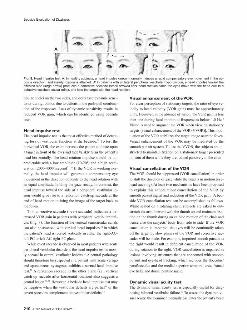
Head impulse testThe head impulse test is the most effective method of detect-ing loss of vestibular function at the bedside.52 To test the horizontal VOR, the examiner asks the patient to fixate upon a target in front of the eyes and then briskly turns the patient’s head horizontally. The head rotation impulse should be un-predictable with a low amplitude (10-20°) and a high accel-eration (2000-4000°/second2).53 If the VOR is working nor-mally, the head impulse will generate a compensatory eye movement in the direction opposite to the head rotation with an equal amplitude, holding the gaze steady. In contrast, the head impulse toward the side of a peripheral vestibular le-sion would give rise to a refixation catch-up saccade at the end of head motion to bring the image of the target back to the fovea.
This corrective saccade (overt saccade) indicates a de-creased VOR gain in patients with peripheral vestibular defi-cits (Fig. 8). The function of the vertical semicircular canals can also be assessed with vertical head impulses,54 in which the patient’s head is rotated vertically in either the right-AC-left-PC or left-AC-right-PC plane.
While overt saccade is observed in most patients with acute peripheral vestibular disorders, the head impulse test is most-ly normal in central vestibular lesions.55 A central pathology should therefore be suspected if a patient with acute vertigo and spontaneous nystagmus exhibits a normal head impulse test.22 A refixation saccade in the other plane (i.e., vertical catch-up saccade after horizontal rotation) also suggests a central lesion.56-58 However, a bedside head impulse test may be negative when the vestibular deficits are partial59 or the covert saccades complement the vestibular deficits.53
Visual enhancement of the VORFor clear perception of stationary targets, the ratio of eye ve-locity to head velocity (VOR gain) must be approximately unity. However, in the absence of vision, the VOR gain is less than one during head motion at frequencies below 1.0 Hz.2 Vision is used to augment the VOR when viewing stationary targets [visual enhancement of the VOR (VVOR)]. This mod-ulation of the VOR stabilizes the target image near the fovea. Visual enhancement of the VOR may be mediated by the smooth pursuit system. To test the VVOR, the subjects are in-structed to maintain fixation on a stationary target presented in front of them while they are rotated passively in the chair. Visual cancellation of the VORThe VOR should be suppressed (VOR cancellation) in order to shift the direction of gaze while the head is in motion (eye-head tracking). At least two mechanisms have been proposed to explain this cancellation: cancellation of the VOR by smooth pursuit signal and reduction of the VOR gain.2 A bed-side VOR cancellation test can be accomplished as follows. While seated on a rotating chair, subjects are asked to out-stretch the arm forward with the thumb up and maintain fixa-tion on the thumb during an en bloc rotation of the chair and hence also the subjects’ body from side to side. If the VOR cancellation is impaired, the eyes will be continually taken off the target by slow phases of the VOR and corrective sac-cades will be made. For example, impaired smooth pursuit to the right would result in deficient cancellation of the VOR during rotation to the right. VOR cancellation is impaired in lesions involving structures that are concerned with smooth pursuit and eye-head tracking, which includes the flocculus/paraflocculus and the medial superior temporal area, frontal eye field, and dorsal pontine nuclei.
Dynamic visual acuity testThe dynamic visual acuity test is especially useful for diag-nosing bilateral vestibular failure.60 To assess the dynamic vi-sual acuity, the examiner manually oscillates the patient’s head
Fig. 8. Head impulse test. A: In healthy subjects, a head impulse (arrow) normally induces a rapid compensatory eye movement in the op-posite direction, and steady fixation is attained. B: In patients with unilateral peripheral vestibular hypofunction, a head impulse toward the affected side (large arrow) produces a corrective saccade (small arrows) after head rotation since the eyes move with the head due to a defective vestibulo-ocular reflex, and lose the target with the head rotation.
A
B

Huh YE and Kim JS
www.thejcn.com 211
horizontally or vertically at about 2 Hz, while the patient reads a Snellen visual acuity chart. If the VOR gain is abnor-mal, visual acuity would deteriorate by more than two lines compared with the initial visual acuity measured when the head is still.
Head heave testThe head heave test can evaluate the translational VOR and the utricular function at the bedside.61 The examiner should move the head laterally with brief and rapid motions while the patient fixates on a target. The translational VOR would generate compensatory eye movements to keep the target sta-ble on the retina. A corrective saccade after the heave indicates vestibular hypofunction on the side toward which the head is moved.
Saccades
Saccades are rapid eye movements that shift the line of sight between successive points of fixation. The patient is instruct-ed to fixate alternatively on two targets, such as the tip of a pen and the examiner’s nose. Saccades can be examined in both the horizontal and vertical planes. The velocity, accura-cy, and conjugacy of the saccades should be determined at the bedside. Peripheral vestibular lesions do not impair sac-cades. Horizontal saccadic slowing is generally caused by dysfunction of the paramedian pontine reticular formation or the structures located more distally in the neural circuits for horizontal saccades,62 while vertical saccadic slowing is of-ten due to lesions involving the rostral interstitial nucleus of the MLF or more distal structures.63 In general, lesions in-volving the pons cause horizontal saccadic slowing, whereas lesions affecting the rostral midbrain cause vertical saccadic slowing. Internuclear ophthalmoplegia can also cause slow saccades and lagging of the adducting eye during attempted contraversive saccades. The slow adducting saccades can be easily detected by having the patient follow an optokinetic drum or tape. Rapid phases made by the eye on the lesion side are smaller and slower. Saccades may overshoot (hyper-metria) or undershoot (hypometria) the target, and corrective saccades would follow. These saccadic dysmetria are often caused by lesions involving the cerebellum (especially the dorsal vermis) or its connections.64,65 A unilateral lesion in the dorsal cerebellar vermis mostly induces contralateral saccadic hypermetria and ipsilateral hypometria, while lesions involv-ing the fastigial nucleus generate contralateral saccadic hy-pometria and ipsilateral hypermetria.64,65 Patients with Wal-lenberg syndrome make hypermetric saccades to the lesion side (ipsipulsion) due to lesions affecting the inferior cere-bellar peduncle.66
Smooth Pursuit
Smooth pursuit is a slow eye movement that holds the image of a small moving target on the fovea. The patient is asked to track a small target moving slowly in the horizontal or verti-cal direction with the head still. Corrective catch-up saccades occur if the pursuit movement does not match the target veloc-ity. Since many neural structures are involved generating a smooth pursuit, and various factors including age, medica-tions, and alertness can influence pursuit eye movements, im-paired pursuit generally does not allow either topographical or etiological classification.62 However, marked asymmetry of pursuit eye movements suggests a unilateral lesion along the pursuit pathway. Since patients with horizontal nystagmus may exhibit impaired smooth pursuit in the direction of the nystagmus, the evaluation of vertical smooth pursuit may be helpful in determining smooth pursuit deficits in acute ves-tibulopathy.67,68
Balance
The severity of imbalance and falling direction may provide important clues as to the underlying vestibular impairments.
Romberg testIn the Romberg test the patient is asked to stand with the feet close together with the eyes open, and then to close the eyes so as to eliminate visual cues. The test result is positive when the patient is stable with the eyes open but loses balance with the eyes closed. A positive test indicates defects in the pro-prioceptive function, but may be found in patients with acute unilateral vestibulopathy or severe bilateral vestibular defi-cits. Patients with cerebellar dysfunction exhibit postural im-balance both with the eyes open and closed. When the pres-ence of imbalance is unclear from the Romberg test, a tandem or sharpened Romberg test should be performed, in which the patient is asked to perform the same task as in Romberg test but while standing with the feet in a heel-to-toe position. In general, patients with vestibular neuritis, Wallenberg’s syn-drome, and cerebellar dysfunction tend to fall toward the le-sion side. To-and-fro sway can be evident in patients with BPPV, bilateral vestibulopathy, and downbeat or upbeat nys-tagmus.69
Stepping testThe stepping test is useful for detecting vestibular deficits by disrupting proprioceptive compensation. The examiner asks the patient to march in a fixed position with the arms extend-ed and eyes closed. A gradual turning toward the lesion side is observed in patients with a unilateral vestibular deficit.

Bedside Evaluation of Dizziness
212 J Clin Neurol 2013;9:203-213
Conclusion
A crucial aspect in the management of dizzy patients is dif-ferentiating central vestibular disorders from the more be-nign peripheral vestibular disorders. A thorough bedside ex-amination and careful history taking remain the most important diagnostic tools for dizziness/vertigo. Subtle ocular signs such as GEN, skew deviation, and a normal head impulse test are useful for determining the occurrence of acute stroke in pa-tients with acute dizziness/vertigo.
Conflicts of InterestThe authors have no financial conflicts of interest.
REFERENCES1. Baloh RW. Patient with dizziness. In: Baloh RW, Halmagyi GM. Dis-
orders of the Vestibular System. New York: Oxford University Press, 1996;157-170.
2. Leigh RJ, Zee DS. The Neurology of Eye Movements. 4th ed. New York: Oxford University Press, 2006.
3. Kattah JC, Talkad AV, Wang DZ, Hsieh YH, Newman-Toker DE. HINTS to diagnose stroke in the acute vestibular syndrome: three-step bedside oculomotor examination more sensitive than early MRI diffusion-weighted imaging. Stroke 2009;40:3504-3510.
4. Tarnutzer AA, Berkowitz AL, Robinson KA, Hsieh YH, Newman-Toker DE. Does my dizzy patient have a stroke? A systematic review of bedside diagnosis in acute vestibular syndrome. CMAJ 2011;183: E571-E592.
5. Westheimer G, Blair SM. The ocular tilt reaction--a brainstem oculo-motor routine. Invest Ophthalmol 1975;14:833-839.
6. Dieterich M, Brandt T. Ocular torsion and tilt of subjective visual vertical are sensitive brainstem signs. Ann Neurol 1993;33:292-299.
7. Diamond SG, Markham CH. Ocular counterrolling as an indicator of vestibular otolith function. Neurology 1983;33:1460-1469.
8. Halmagyi GM, Brandt T, Dieterich M, Curthoys IS, Stark RJ, Hoyt WF. Tonic contraversive ocular tilt reaction due to unilateral meso-di-encephalic lesion. Neurology 1990;40:1503-1509.
9. Baier B, Bense S, Dieterich M. Are signs of ocular tilt reaction in pa-tients with cerebellar lesions mediated by the dentate nucleus? Brain 2008;131(Pt 6):1445-1454.
10. Zwergal A, Rettinger N, Frenzel C, Dieterich M, Brandt T, Strupp M. A bucket of static vestibular function. Neurology 2009;72:1689-1692.
11. Serra A, Leigh RJ. Diagnostic value of nystagmus: spontaneous and induced ocular oscillations. J Neurol Neurosurg Psychiatry 2002;73: 615-618.
12. Baloh RW. Clinical practice. Vestibular neuritis. N Engl J Med 2003; 348:1027-1032.
13. Robinson DA, Zee DS, Hain TC, Holmes A, Rosenberg LF. Alexan-der’s law: its behavior and origin in the human vestibulo-ocular reflex. Ann Neurol 1984;16:714-722.
14. Hotson JR, Baloh RW. Acute vestibular syndrome. N Engl J Med 1998;339:680-685.
15. Zee DS. Ophthalmoscopy in examination of patients with vestibular disorders. Ann Neurol 1978;3:373-374.
16. Baloh RW, Yee RD. Spontaneous vertical nystagmus. Rev Neurol (Paris) 1989;145:527-532.
17. Böhmer A, Straumann D. Pathomechanism of mammalian downbeat nystagmus due to cerebellar lesion: a simple hypothesis. Neurosci Lett 1998;250:127-130.
18. Baloh RW, Spooner JW. Downbeat nystagmus: a type of central ves-tibular nystagmus. Neurology 1981;31:304-310.
19. Glasauer S, Hoshi M, Kempermann U, Eggert T, Büttner U. Three-dimensional eye position and slow phase velocity in humans with downbeat nystagmus. J Neurophysiol 2003;89:338-354.
20. Choi KD, Oh SY, Kim HJ, Koo JW, Cho BM, Kim JS. Recovery of vestibular imbalances after vestibular neuritis. Laryngoscope 2007; 117:1307-1312.
21. Büttner U, Grundei T. Gaze-evoked nystagmus and smooth pursuit deficits: their relationship studied in 52 patients. J Neurol 1995;242: 384-389.
22. Lee H, Sohn SI, Cho YW, Lee SR, Ahn BH, Park BR, et al. Cerebel-lar infarction presenting isolated vertigo: frequency and vascular top-ographical patterns. Neurology 2006;67:1178-1183.
23. Zapala DA. Down-beating nystagmus in anterior canal benign parox-ysmal positional vertigo. J Am Acad Audiol 2008;19:257-266.
24. Brantberg K, Bergenius J. Treatment of anterior benign paroxysmal positional vertigo by canal plugging: a case report. Acta Otolaryngol 2002;122:28-30.
25. Shallo-Hoffmann J, Schwarze H, Simonsz HJ, Mühlendyck H. A re-examination of end-point and rebound nystagmus in normals. Invest Ophthalmol Vis Sci 1990;31:388-392.
26. Choi KD, Oh SY, Park SH, Kim JH, Koo JW, Kim JS. Head-shaking nystagmus in lateral medullary infarction: patterns and possible mech-anisms. Neurology 2007;68:1337-1344.
27. Hain TC, Fetter M, Zee DS. Head-shaking nystagmus in patients with unilateral peripheral vestibular lesions. Am J Otolaryngol 1987;8:36-47.
28. Minagar A, Sheremata WA, Tusa RJ. Perverted head-shaking nystag-mus: a possible mechanism. Neurology 2001;57:887-889.
29. Kim JS, Ahn KW, Moon SY, Choi KD, Park SH, Koo JW. Isolated perverted head-shaking nystagmus in focal cerebellar infarction. Neurology 2005;64:575-576.
30. Huh YE, Kim JS. Patterns of spontaneous and head-shaking nystag-mus in cerebellar infarction: imaging correlations. Brain 2011;134(Pt 12):3662-3671.
31. Choi KD, Cho HJ, Koo JW, Park SH, Kim JS. Hyperventilation-in-duced nystagmus in vestibular schwannoma. Neurology 2005;64: 2062.
32. Robichaud J, DesRoches H, Bance M. Is hyperventilation-induced nystagmus more common in retrocochlear vestibular disease than in end-organ vestibular disease? J Otolaryngol 2002;31:140-143.
33. Walker MF, Zee DS. The effect of hyperventilation on downbeat nys-tagmus in cerebellar disorders. Neurology 1999;53:1576-1579.
34. Choi KD, Kim JS, Kim HJ, Koo JW, Kim JH, Kim CY, et al. Hyper-ventilation-induced nystagmus in peripheral vestibulopathy and cere-bellopontine angle tumor. Neurology 2007;69:1050-1059.
35. Karlberg M, Aw ST, Black RA, Todd MJ, MacDougall HG, Halma-gyi GM. Vibration-induced ocular torsion and nystagmus after uni-lateral vestibular deafferentation. Brain 2003;126(Pt 4):956-964.
36. Ohki M, Murofushi T, Nakahara H, Sugasawa K. Vibration-induced nystagmus in patients with vestibular disorders. Otolaryngol Head Neck Surg 2003;129:255-258.
37. Nam J, Kim S, Huh Y, Kim JS. Ageotropic central positional nystag-mus in nodular infarction. Neurology 2009;73:1163.
38. Arai M, Terakawa I. Central paroxysmal positional vertigo. Neurology 2005;64:1284.
39. Büttner U, Helmchen C, Brandt T. Diagnostic criteria for central ver-sus peripheral positioning nystagmus and vertigo: a review. Acta Oto-laryngol 1999;119:1-5.
40. Fernandez C, Alzate R, Lindsay JR. Experimental observations on postural nystagmus. II. Lesions of the nodulus. Ann Otol Rhinol Lar-yngol 1960;69:94-114.
41. Dix MR, Hallpike CS. The pathology symptomatology and diagnosis of certain common disorders of the vestibular system. Proc R Soc Med 1952;45:341-354.
42. Baloh RW, Honrubia V, Jacobson K. Benign positional vertigo: clinical and oculographic features in 240 cases. Neurology 1987;37:371-378.

Huh YE and Kim JS
www.thejcn.com 213
43. Humphriss RL, Baguley DM, Sparkes V, Peerman SE, Moffat DA. Contraindications to the Dix-Hallpike manoeuvre: a multidisciplinary review. Int J Audiol 2003;42:166-173.
44. Cohen HS. Side-lying as an alternative to the Dix-Hallpike test of the posterior canal. Otol Neurotol 2004;25:130-134.
45. McClure JA. Horizontal canal BPV. J Otolaryngol 1985;14:30-35.46. Baloh RW, Yue Q, Jacobson KM, Honrubia V. Persistent direction-
changing positional nystagmus: another variant of benign positional nystagmus? Neurology 1995;45:1297-1301.
47. Koo JW, Moon IJ, Shim WS, Moon SY, Kim JS. Value of lying-down nystagmus in the lateralization of horizontal semicircular canal be-nign paroxysmal positional vertigo. Otol Neurotol 2006;27:367-371.
48. Lee SH, Choi KD, Jeong SH, Oh YM, Koo JW, Kim JS. Nystagmus during neck flexion in the pitch plane in benign paroxysmal positional vertigo involving the horizontal canal. J Neurol Sci 2007;256:75-80.
49. Bisdorff AR, Debatisse D. Localizing signs in positional vertigo due to lateral canal cupulolithiasis. Neurology 2001;57:1085-1088.
50. Minor LB, Solomon D, Zinreich JS, Zee DS. Sound- and/or pressure-induced vertigo due to bone dehiscence of the superior semicircular canal. Arch Otolaryngol Head Neck Surg 1998;124:249-258.
51. Tilikete C, Krolak-Salmon P, Truy E, Vighetto A. Pulse-synchronous eye oscillations revealing bone superior canal dehiscence. Ann Neurol 2004;56:556-560.
52. Halmagyi GM, Curthoys IS. A clinical sign of canal paresis. Arch Neurol 1988;45:737-739.
53. Weber KP, Aw ST, Todd MJ, McGarvie LA, Curthoys IS, Halmagyi GM. Head impulse test in unilateral vestibular loss: vestibulo-ocular reflex and catch-up saccades. Neurology 2008;70:454-463.
54. Cremer PD, Halmagyi GM, Aw ST, Curthoys IS, McGarvie LA, Todd MJ, et al. Semicircular canal plane head impulses detect absent func-tion of individual semicircular canals. Brain 1998;121(Pt 4):699-716.
55. Newman-Toker DE, Kattah JC, Alvernia JE, Wang DZ. Normal head impulse test differentiates acute cerebellar strokes from vestibular neuritis. Neurology 2008;70(24 Pt 2):2378-2385.
56. Walker MF, Zee DS. Directional abnormalities of vestibular and op-tokinetic responses in cerebellar disease. Ann N Y Acad Sci 1999;871:
205-220.57. Walker MF, Zee DS. Cerebellar disease alters the axis of the high-ac-
celeration vestibuloocular reflex. J Neurophysiol 2005;94:3417-3429.58. Jeong SH, Kim JS, Baek IC, Shin JW, Jo H, Lee AY, et al. Perverted
head impulse test in cerebellar ataxia. Cerebellum 2013. [Epub ahead of print]
59. Perez N, Rama-Lopez J. Head-impulse and caloric tests in patients with dizziness. Otol Neurotol 2003;24:913-917.
60. Kim S, Oh YM, Koo JW, Kim JS. Bilateral vestibulopathy: clinical characteristics and diagnostic criteria. Otol Neurotol 2011;32:812-817.
61. Ramat S, Zee DS, Minor LB. Translational vestibulo-ocular reflex evoked by a “head heave” stimulus. Ann N Y Acad Sci 2001;942:95-113.
62. Gaymard B, Pierrot-Deseilligny C. Neurology of saccades and smooth pursuit. Curr Opin Neurol 1999;12:13-19.
63. Sharpe JA, Kim JS. Midbrain disorders of vertical gaze: a quantita-tive re-evaluation. Ann N Y Acad Sci 2002;956:143-154.
64. Robinson FR, Straube A, Fuchs AF. Role of the caudal fastigial nu-cleus in saccade generation. II. Effects of muscimol inactivation. J Neurophysiol 1993;70:1741-1758.
65. Takagi M, Zee DS, Tamargo RJ. Effects of lesions of the oculomotor vermis on eye movements in primate: saccades. J Neurophysiol 1998; 80:1911-1931.
66. Helmchen C, Straube A, Büttner U. Saccadic lateropulsion in Wallen-berg’s syndrome may be caused by a functional lesion of the fastigial nucleus. J Neurol 1994;241:421-426.
67. Chen L, Lee W, Chambers BR, Dewey HM. Diagnostic accuracy of acute vestibular syndrome at the bedside in a stroke unit. J Neurol 2011; 258:855-861.
68. Cnyrim CD, Newman-Toker D, Karch C, Brandt T, Strupp M. Bedside differentiation of vestibular neuritis from central “vestibular pseudo-neuritis”. J Neurol Neurosurg Psychiatry 2008;79:458-460.
69. Dieterich M, Grünbauer WM, Brandt T. Direction-specific impairment of motion perception and spatial orientation in downbeat and upbeat nystagmus in humans. Neurosci Lett 1998;245:29-32.