on learning and memory functions in
TRANSCRIPT

BEHAVIOURAL EFFECTS AND BRAIN
ACTIVITY OF MITRAGYNINE FROM
MITRAGYNA SPECIOSA KORTH (KRATOM) ON
LEARNING AND MEMORY FUNCTIONS IN RAT
FARAH WAHIDA BINTI SUHAIMI
UNIVERSITI SAINS MALAYSIA
2015

BEHAVIOURAL EFFECTS AND BRAIN
ACTIVITY OF MITRAGYNINE FROM
MITRAGYNA SPECIOSA KORTH (KRATOM) ON
LEARNING AND MEMORY FUNCTIONS IN RAT
by
FARAH WAHIDA BINTI SUHAIMI
Thesis submitted in fulfillment of the requirements
for the degree of
Doctor of Philosophy
December 2015

ii
ACKNOWLEDGEMENTS
First of all, I would like to express my gratitude to my supervisor, Dr Zurina
Hassan, who has been very helpful and supportive throughout this study. Her
constructive comments and valuable insights have guided me towards the completion
of the study. My utmost gratitude also goes to my co-supervisor, Professor Emeritus
Dato‟ Dr. Visweswaran Navaratnam, who has inspired me a lot with his brilliant
ideas.
Special thanks to our Director, Professor Sharif Mahsufi Mansor for his
guidance as well as for the facilities provided during the course of this study. Not
forgotten, all technical and non-technical staffs of the Centre for Drug Research,
Universiti Sains Malaysia especially Mr. Zulkeflee Ismail, Mrs. Zaiton Kader, Mrs.
Salmah Baba, Mr. Asokan A/L Muniandy and Miss Nur Aziah Hanapi. My
appreciation also goes to Mr. Chua Jen Keat and Mr. Zarif Mohamad Sofian for the
preparation of the mitragynine and Dr. V. Murugaiyah and Liew Wai Lam for their
assistance in enzyme assay.
I would like to extend my appreciation to my field supervisor, Professor
Christian P. Muller from Universitats Klinikum Erlangen, Germany for being very
helpful with the constructive ideas and comments. I am also grateful to Dr. Davide
Amato who had taught me the brain surgery technique and Dr. Fabio for his guidance
in animal behavioural technique.
I am truly indebted to my beloved father and mother, Suhaimi bin Zulkafli
and Zaiton binti Md. Jamil, my brothers and sisters and all my family members for

iii
their unconditional love, support, encouragement and endless patience throughout
my study in Universiti Sains Malaysia.
Many thanks to my friends, Nurul Hasnida Mohammad Yusoff and Norsyifa
Harun for being with me from the beginning of my study and to my other labmates,
Thenmoly, Fasehah and Nurul Aqmar, for the discussions of scientific, life and
personal affairs. I really enjoy the moment we spent together.
Last, but not least, I am grateful to the Ministry of Education for the financial
support through the Higher Institution Centre of Excellence grant and MyBrain15
scholarship which enable the completion of the research project.
iv
TABLE OF CONTENTS
Page
ACKNOWLEDGEMENTS ii
TABLE OF CONTENTS iv
LIST OF TABLES ix
LIST OF FIGURES x
LIST OF ABBREVIATIONS xv
LIST OF SYMBOLS xviii
LIST OF APPENDICES xix
ABSTRAK xx
ABSTRACT xxiii
CHAPTER ONE: INTRODUCTION 1
1.1 Learning and memory 1
1.1.1 Stages of learning and memory 1
1.1.2 Types of memory 2
1.1.3 Mechanisms of learning and memory 5
1.1.4 Memory modulatory mechanisms in the brain 7
1.1.4(a) GABA 8
1.1.4(b) Monoaminergic system 9
1.1.4(c) Cholinergic system 12
1.1.4(d) Opioidergic system 16
1.1.4(e) Endocannabinoid system 17
1.1.4(f) Histamine 18
1.1.4(g) Others 18

v
1.1.5 Memory circuitry in brain: interaction between neural
systems
19
1.2 Electroencephalogram (EEG) 23
1.3 Mitragyna speciosa 26
1.3.1 Botanical origin 26
1.3.2 Preparations and consumption 27
1.3.3 Epidemiology and legal status 28
1.3.4 Medicinal uses 30
1.3.5 Phytochemistry 30
1.3.6 Toxicology 33
1.3.7 Pharmacology 35
1.3.8 Pharmacological effects 36
1.3.9 Behavioural effects in human 40
1.3.10 Addictive liabilities 41
1.4 Cognitive effects of M. speciosa 44
1.5 Rationale of study 45
1.6 Scope of study 46
1.7 Objectives 49
CHAPTER TWO: MATERIALS AND METHODS 51
2.1 Animals 51
2.2 Chemicals and reagents 51
2.3 Experimental procedures 55
2.3.1 Acute effects of mitragynine on learning and memory
functions using passive avoidance task
55

vi
2.3.1(a) Acute effects of mitragynine on acquisition
(learning)
57
2.3.1(b) Acute effects of mitragynine on memory
consolidation
57
2.3.1(c) Acute effects of mitragynine on memory
retrieval
58
2.3.2 Effects of mitragynine on spatial learning and
reference memory of Morris water maze task
58
2.3.2(a) Experimental design 60
2.3.3 Chronic effects of mitragynine on learning and
memory functions using passive avoidance task
60
2.3.4 Chronic effects of mitragynine on learning and
memory functions during withdrawal using novel
object recognition task
60
2.3.5 Effects of 28 days treatment of mitragynine on brain
activity via wireless electroencephalogram (EEG)
recording
63
2.3.5(a) Surgical procedures 63
2.3.5(b) Experimental design 66
2.3.5(c) Brain histology 66
2.3.6 Role of opioid system in modulation of the
mitragynine effects on passive avoidance task
68
2.3.7 Role of GABAergic system in modulation of the
mitragynine effects on passive avoidance task
69

vii
2.3.8 Role of cholinergic system in modulation of the
mitragynine effects in Morris water maze task
70
2.3.9 Determination of cholinesterase activity in brain
samples
71
2.3.9(a) Tissue preparation 71
2.3.9(b) Determination of protein 72
2.3.9(c) Determination of cholinesterase activity 73
2.4 Statistical analysis 75
CHAPTER THREE: RESULTS 76
3.1 Acute effects of mitragynine on passive avoidance task 76
3.2 Effects of mitragynine on spatial learning task and reference
memory using Morris water maze task
79
3.3 Effects of chronic mitragynine treatment on memory retrieval
during withdrawal and abstinence
82
3.4 Effects of chronic mitragynine treatment on frontal cortex EEG 86
3.5 Effects of chronic mitragynine treatment on neocortex EEG 90
3.6 Effects of chronic mitragynine treatment on hippocampal EEG 94
3.7 Summary on the chronic effects of mitragynine on EEG from
frontal cortex, neocortex and hippocampus
99
3.8 Effects of naloxone on mitragynine-induced memory deficit 100
3.9 Effects of bicuculline on mitragynine-induced memory deficit 102
3.10 Effects of oxotremorine (0.1 mg/kg) and physostigmine (0.1
mg/kg) on spatial learning deficit induced by mitragynine
104
3.11 Determination of cholinesterase activity 110

viii
CHAPTER FOUR: DISCUSSION 115
4.1 Drugs of choice 115
4.2 Behavioural study 116
4.3 EEG study 123
4.4 Mechanisms underlying mitragynine-induced learning and
memory deficits in rats
129
4.5 Cholinesterase activity 136
4.6 Limitations 139
CHAPTER FIVE: CONCLUSION 140
5.1 Perspectives 142
REFERENCES 144
APPENDICES
LIST OF PUBLICATIONS

ix
LIST OF TABLES
Page
Table 2.1 List of chemicals and reagents 53
Table 2.2 Summary of reaction volumes and components for
cholinesterase assay
74

x
LIST OF FIGURES
Page
Figure 1.1 The traditional taxonomy of memory systems. 2
Figure 1.2 Molecular events underlie the early and late phases of
long-term potentiation.
7
Figure 1.3 Modulatory systems of learning and memory functions in
the brain.
8
Figure 1.4 Distribution of cholinergic neurons and their projections
in the rat brain.
13
Figure 1.5 The hippocampus and its connections. 22
Figure 1.6 The plant M. speciosa Korth. 27
Figure 1.7 Chemical structure of mitragynine and its major
analogues.
32
Figure 1.8 General outline of the study on the behavioural effects and
brain activity of mitragynine on learning and memory
functions in rats.
50
Figure 2.1 Schematic representation for passive avoidance task. 56
Figure 2.2 Schedule of drug‟s administration for acquisition during
passive avoidance task.
57
Figure 2.3 Schedule of drug‟s administration for memory
consolidation during passive avoidance task.
57
Figure 2.4 Schedule of drug‟s administration for memory retrieval
during passive avoidance task.
58

xi
Figure 2.5 Schematic representation of Morris water maze task
procedures.
59
Figure 2.6 Summary on study on chronic effects of mitragynine
using passive avoidance task and novel object recognition
task.
61
Figure 2.7 Schematic representation of novel object recognition task. 62
Figure 2.8
Figure 2.9
Figure 2.10
Illustration of the surface of the rat‟s skull.
Animal with wireless headstage plugged into the
implanted mating connector.
Groups of animals treated with naloxone and different
doses of mitragynine.
63
65
68
Figure 2.11 Groups of animals treated with bicuculline and different
doses of mitragynine.
69
Figure 2.12 Groups of animals treated with either oxotremorine or
physostigmine and different doses of mitragynine.
70
Figure 2.13 Principles of Ellman‟s assay. 73
Figure 3.1 Effects of mitragynine (1, 5, 10 mg/kg, i.p) on step-
through latencies during A. learning (administered pre-
training), B. consolidation (administered post-training)
and C. retrieval (administered pre-test) of passive
avoidance task in rats.
78
Figure 3.2 Effects of post-training mitragynine (1, 5, and 10 mg/kg,
i.p) on spatial acquisition of Morris water maze task.
80
Figure 3.3 Effects of post-training mitragynine on probe trial of
Morris water maze task (1, 5 and 10 mg/kg, i.p).
80

xii
Figure 3.4 Effects of post-training mitragynine on escape latency in a
presence of visible platform of Morris water maze task (1,
5 and 10 mg/kg, i.p).
81
Figure 3.5 Effects of chronic 28-days administration of mitragynine
(1, 5 and 10 mg/kg, i.p) on learning and memory in a
passive avoidance task during withdrawal.
83
Figure 3.6 Withdrawal effects from 28-days chronic administration
of mitragynine measured as discrimination ratio in novel
object recognition task.
85
Figure 3.7
Direct contact with objects in novel object recognition
task during retrieval testing.
85
Figure 3.8 Percentage changes in spectral power from baseline in the
frontal cortex. A. Day 1, B. Day 7, C. Day 14, D. Day 21,
E. Day 28.
89
Figure 3.9 Percentage changes in spectral power from baseline in the
neocortex. A. Day 1, B. Day 7, C. Day 14, D. Day 21, E.
Day 28.
93
Figure 3.10 Percentage changes in spectral power from baseline in the
hippocampus. A. Day 1, B. Day 7, C. Day 14, D. Day 21,
E. Day 28.
97
Figure 3.11 A. Photomicrograph of coronal section of the brain
showing the probe tract in hippocampus region, and B.
Schematic representation of the coronal section at the
coordinates, bregma: -4.3 mm.
98

xiii
Figure 3.12 Effects of post-training mitragynine + naloxone (0.4 or
1.0 mg/kg) on step-through latencies of passive avoidance
task.
101
Figure 3.13 Effects of post-training mitragynine + bicuculline (0.125
or 0.5 mg/kg) on step-through latencies of passive
avoidance task.
103
Figure 3.14 Effects of post-training vehicle in the presence of
oxotremorine (0.1 mg/kg) or physostigmine (0.1 mg/kg)
on spatial learning in Morris water maze task.
106
Figure 3.15 Effects of post-training vehicle in the presence of
oxotremorine (0.1 mg/kg) or physostigmine (0.1 mg/kg)
on reference memory in Morris water maze task.
106
Figure 3.16 Effects of post-training morphine in the presence of
oxotremorine (0.1 mg/kg) or physostigmine (0.1 mg/kg)
on spatial learning in Morris water maze task.
107
Figure 3.17 Effects of post-training morphine in the presence of
oxotremorine (0.1 mg/kg) or physostigmine (0.1 mg/kg)
on reference memory in Morris water maze task.
107
Figure 3.18 Effects of post-training mitragynine (5 mg/kg) in the
presence of oxotremorine (0.1 mg/kg) or physostigmine
(0.1 mg/kg) on spatial learning in Morris water maze task.
108
Figure 3.19 Effects of post-training mitragynine (5 mg/kg) in the
presence of oxotremorine (0.1 mg/kg) or physostigmine
(0.1 mg/kg) on reference memory in Morris water maze
task.
108

xiv
Figure 3.20 Effects of post-training mitragynine (10 mg/kg) in the
presence of oxotremorine (0.1 mg/kg) or physostigmine
(0.1 mg/kg) on spatial learning in Morris water maze task.
109
Figure 3.21 Effects of post-training mitragynine (10 mg/kg) in the
presence of oxotremorine (0.1 mg/kg) or physostigmine
(0.1 mg/kg) on reference memory in Morris water maze
task.
109
Figure 3.22 Graph absorbance (A595 nm) versus BSA standards
(mg/ml).
111
Figure 3.23 A. AChE and B. BuChE activity of frontal cortex. 112
Figure 3.24 A. AChE and B. BuChE activity of remaining cortex. 113
Figure 3.25 A. AChE and B. BuChE activity of hippocampus. 114

xv
LIST OF ABBREVIATIONS
5-HT 5-hydroxytriptamine
A595 Absorbance at 595 nm
Acetyl-CoA Acetyl coenzyme A
ACh Acetylcholine
AChE Acetylcholinesterase
ALT Alanine transaminase
AMPA α-amino-3-hydroxyl-5-methyl-4-isoxazole-propionate
ANOVA Analysis of variance
AP Anterior-posterior
AST Aspartate transaminase
ATPase Adenosinetriphosphatase
BDNF Brain derived neurotrophic factor
BSA Bovine serum albumin
BuChE Butyrylcholinesterase
Ca2+
Calcium ion
CaM Calcium sensor calmodulin
CaMKII calcium/calmodulin-dependent protein kinase II
cAMP Cyclic adenosine monophosphate
CB Cannabinoid receptors
ChAT Choline acetyltransferase
cm Centimetre
CNS Central nervous system
COOH Carboxyl group
COX Cyclooxygenase
CREB Cyclic AMP response element binding protein
DBB Diagonal band of Broca
DTNB 5,5′-Dithiobis(2-nitrobenzoic acid)

xvi
EEG Electroencephalogram
g Gram
G Group
GABA Gamma-aminobutyric acid
GTP Guanosine triphosphate
H2O2 Hydrogen peroxide
HDBB Horizontal limb of the diagonal band of Broca
HPLC High-performance liquid chromatography
h Hour
Hz Hertz
i.p Intraperitoneal
Iso-OMPA Tetraisopropyl pyrophosphoramide
JNK3 c-Jun N-terminal kinase 3
kg Kilogram
LTD Long-term depression
LTM Long-term memory
LTP Long-term potentiation
M Molarity
mA Miliampere
mAChR Muscarinic acetylcholine receptor
mg Miligram
Mg+
Magnesium ion
min Minutes
Mit 1 Mitragynine-treated group at 1 mg/kg
Mit 10 Mitragynine-treated group at 10 mg/kg
Mit 5 Mitragynine-treated group at 5 mg/kg
ML Mediolateral
ml Mililitre
mm Milimetre
xvii
Mor Morphine-treated group at 5 mg/kg
Na+
Sodium ion
nAChR Nicotinic acetylcholine receptor
NBM nucleus basalis magnocellularis
nm Nanometre
NMDA N-methyl-D-aspartate
OD Optical density
Oxo Oxotremorine
Phy Physostigmine
PKA Protein kinase A
PKC Protein kinase C
PKG Protein kinase G
s Second
SEM Standard error mean
STM Short-term memory
USM Universiti Sains Malaysia
V Ventral
Veh Vehicle-treated group
VDBB Vertical limb of the diagonal band of Broca
μl Microlitre
µm Micrometre
μV Microvolt

xviii
LIST OF SYMBOLS
β Beta
α Alpha
γ Gamma
δ Delta
ε Epsilon
μ Mu
κ Kappa
°C Degree celcius
% Percentage
θ Theta
< Less than
± Plus minus
~ Approximately

xix
LIST OF APPENDICES
Appendix A : Mating connector anchored with dental acrylic.
Appendix B : Guide cannula
Appendix C : Implantation of the mating connector with electrodes for
wireless EEG recording
Appendix D : Schematic representation of wireless EEG recording
Appendix E : Brain sectioning using tissue block
Appendix F(a): Animal ethics approval I
Appendix F(b): Animal ethics approval II
Appendix F(c): Animal ethics approval III
Appendix F(d): Animal ethics approval IV

xx
KESAN-KESAN TINGKAH LAKU DAN AKTIVITI OTAK OLEH
MITRAGININA DARIPADA MITRAGYNA SPECIOSA KORTH (KRATOM)
KE ATAS FUNGSI PEMBELAJARAN DAN MEMORI DALAM TIKUS
ABSTRAK
Mitraginina, sebatian alkaloid utama daripada Mitragyna speciosa Korth
telah dikaji secara meluas untuk kesan-kesan farmakologinya. Walau bagaimanapun,
sedikit maklumat yang diketahui tentang kesannya terhadap fungsi pembelajaran dan
memori. Dalam penyelidikan ini, kesan-kesan akut mitraginina terhadap peringkat-
peringkat yang berlainan dalam proses pembelajaran dan memori telah dikaji.
Pemberian pra-latihan, pasca-latihan dan pra-ujian mitraginina (1, 5 dan 10 mg/kg)
menjejaskan pemerolehan, pengukuhan dan dapatan semula memori dalam prosedur
penghindaran pasif, ke tahap yang sama seperti morfina. Penyelidikan ini seterusnya
mengkaji jika kesan mitraginina adalah bergantung kepada prosedur atau secara
umum ke atas prosedur tingkah laku yang lain. Hasil kajian menunjukkan bahawa
mitraginina (5 dan 10 mg/kg) menyebabkan defisit pembelajaran spasial dan
menjejaskan memori rujukan dalam prosedur berselirat air Morris. Kemudian, kesan-
kesan kronik mitraginina (28 hari) ke atas fungsi pembelajaran dan memori dinilai.
Hasil kajian menunjukkan bahawa kronik mitraginina menjejaskan memori dapatan
semula prosedur penghindaran pasif semasa penyisihan awal dan ketika abstinens.
Kesan-kesan mitraginina dinilai lagi menggunakan prosedur pengiktirafan objek
novel dalam haiwan yang sama. Hasil kajian menunjukkan bahawa haiwan mampu
mendiskriminasi antara objek novel dan biasa tetapi keupayaan untuk mengingati
objek yang telah dihadapi sebelum ini telah dilemahkan dalam haiwan yang dirawat
dengan dos mitraginina yang tinggi (10 mg/kg). Keputusan menunjukkan bahawa

xxi
dos mitraginina yang tinggi diperlukan untuk mengekalkan kesan-kesan kerosakan
memori oleh mitraginina dalam tugasan pembelajaran yang baru ketika abstinens.
Kesan-kesan mitraginina ke atas aktiviti otak dinilai lagi pada haiwan-haiwan yang
dirawat secara kronik untuk 28 hari. Mitraginina menyebabkan corak EEG yang
berbeza pada kedua-dua tisu kortikal (frontal dan neokorteks) dan hipokampus.
Pendedahan berulang kepada dos mitraginina yang sama pada 10 mg/kg
menyebabkan peningkatan dalam kuasa delta dan penurunan dalam kuasa alfa di
kedua-dua korteks frontal dan neokorteks. Sebaliknya, pendedahan berulang kepada
mitraginina menurunkan kuasa delta tanpa mengira dos tetapi hanya mitraginina
pada 5 dan 10 mg/kg menurunkan kuasa alfa di hipokampus. Perubahan dalam EEG
ini mencadangkan bahawa mitraginina mampu mencabar kestabilan rangkaian
kawasan setempat. Di samping itu, pemberian nalokson, antagonis opioid (1.0
mg/kg) dan bikukulina, antagonis GABAA (0.125 mg/kg) pasca-latihan memulihkan
memori yang terjejas disebabkan oleh mitraginina, mencadangkan penglibatan sistem
opioidergik dan GABAergik dalam modulasi kesan-kesan mitraginina ke atas fungsi
pembelajaran dan memori. Dalam prosedur berselirat air Morris, pemberian agonis
kolinergik, oksotrimorina (0.1 mg/kg) pasca-latihan, memulihkan defisit
pembelajaran spasial yang disebabkan oleh mitraginina (5 dan 10 mg/kg). Pemberian
fisostigmina (0.1 mg/kg), antikolinesterase hanya memulihkan kesan kerosakan oleh
mitraginina terhadap pembelajaran spasial pada 5 mg/kg tetapi tidak pada dos yang
lebih tinggi (10 mg/kg). Keputusan ini mungkin menunjukkan bahawa defisit
pembelajaran spasial yang disebabkan oleh mitraginina adalah berkolerasi dengan
pengurangan paras asetilkolina di kawasan-kawasan otak yang berkaitan dengan
memori. Pra-rawatan mitraginina selama 12 hari berturut-turut membawa kepada
perbezaan aktiviti kolinesterase di dalam otak. Tiada perbezaan dalam aktiviti

xxii
asetilkolinesterase (AChE) dilihat dalam korteks frontal. Lebihan korteks yang terdiri
daripada tisu kortikal utama menunjukkan penurunan manakala hipokampus
menunjukkan peningkatan aktiviti AChE dalam kumpulan yang telah dirawat dengan
mitraginina pada 10 mg/kg. Secara keseluruhannya, boleh disimpulkan bahawa
penggunaan mitraginina boleh memberi kesan kepada fungsi pembelajaran dan
memori seperti yang ditunjukkan oleh prestasi pencapaian tingkah laku yang terjejas
dan perubahan dalam aktiviti otak.

xxiii
BEHAVIOURAL EFFECTS AND BRAIN ACTIVITY OF MITRAGYNINE
FROM MITRAGYNA SPECIOSA KORTH (KRATOM) ON LEARNING AND
MEMORY FUNCTIONS IN RAT
ABSTRACT
Mitragynine, the major alkaloid from Mitragyna speciosa Korth has been
widely studied for its pharmacological effects. However, little is known about its
effects on learning and memory functions. In present study, the acute effects of
mitragynine on different stages of learning and memory processes were investigated.
Pre-training, post-training and pre-test administration of mitragynine (1, 5 and 10
mg/kg) impaired the acquisition, memory consolidation and retrieval of the passive
avoidance task, to a similar degree as morphine. The study further investigated if the
effects of mitragynine are either task-dependent or common to other behavioural
tasks. The results showed that mitragynine (5 and 10 mg/kg) caused spatial learning
deficits and impaired reference memory in Morris water maze task. Then, chronic
effects of mitragynine (28 days) on learning and memory functions were assessed.
Results demonstrated that chronic mitragynine impaired memory retrieval of the
passive avoidance task during early withdrawal and abstinence. Effects of
mitragynine were further evaluated using novel object recognition task in the same
treated-animals. The results indicated that animals were able to discriminate between
the novel and familiar objects but the ability to remember the previously encountered
object was attenuated in the animals treated with high dose of mitragynine (10
mg/kg). The results suggested that high dose of mitragynine is required to maintain
the memory-impairing effects of mitragynine in a new learning task during
abstinence. The effects of mitragynine on brain activity were further evaluated in

xxiv
chronically-treated animals for 28 days. Mitragynine caused dissociative patterns in
EEG of both cortical tissues (frontal and neocortex) and hippocampus. Repeated
exposure to equal dose of mitragynine at 10 mg/kg caused an increase in delta power
and a decrease in alpha power in both frontal cortex and neocortex. On the contrary,
repeated exposure to mitragynine regardless of the doses decreased the delta power
but only mitragynine at 5 and 10 mg/kg decreased the alpha power in the
hippocampus. These changes in EEG may suggest that mitragynine was able to
challenge the local network stability. In addition, post-training administration of
naloxone, an opioid antagonist (1.0 mg/kg) and bicuculline, GABAA antagonist
(0.125 mg/kg) improved memory impairment induced by mitragynine, suggesting the
participation of the opioidergic and GABAergic systems in modulating the effects of
mitragynine in learning and memory functions. In Morris water maze task, post-
training administration of cholinergic agonist, oxotremorine (0.1 mg/kg), improved
the spatial learning deficit induced by mitragynine (5 and 10 mg/kg). The
administration of physostigmine (0.1 mg/kg), an anticholinesterase only reversed the
impairing effects of mitragynine on spatial learning at 5 mg /kg but not at higher
dose (10 mg/kg). These results may indicate that mitragynine-induced spatial
learning deficit is correlated with the depletion of acetylcholine level in memory-
associated brain regions. Pre-treatment of mitragynine for 12 consecutive days led to
differential cholinesterase activities in brain. No difference in acetylcholinesterase
(AChE) activity was observed in frontal cortex. Remaining cortex which consists of
major cortical tissues showed a decrease whilst hippocampus showed enhanced
AChE activity in mitragynine-treated group at 10 mg/kg. Taken together, it can be
concluded that mitragynine consumption can affect learning and memory functions
as shown by the impaired behavioural performances and changes in brain activity.
1
CHAPTER ONE
INTRODUCTION
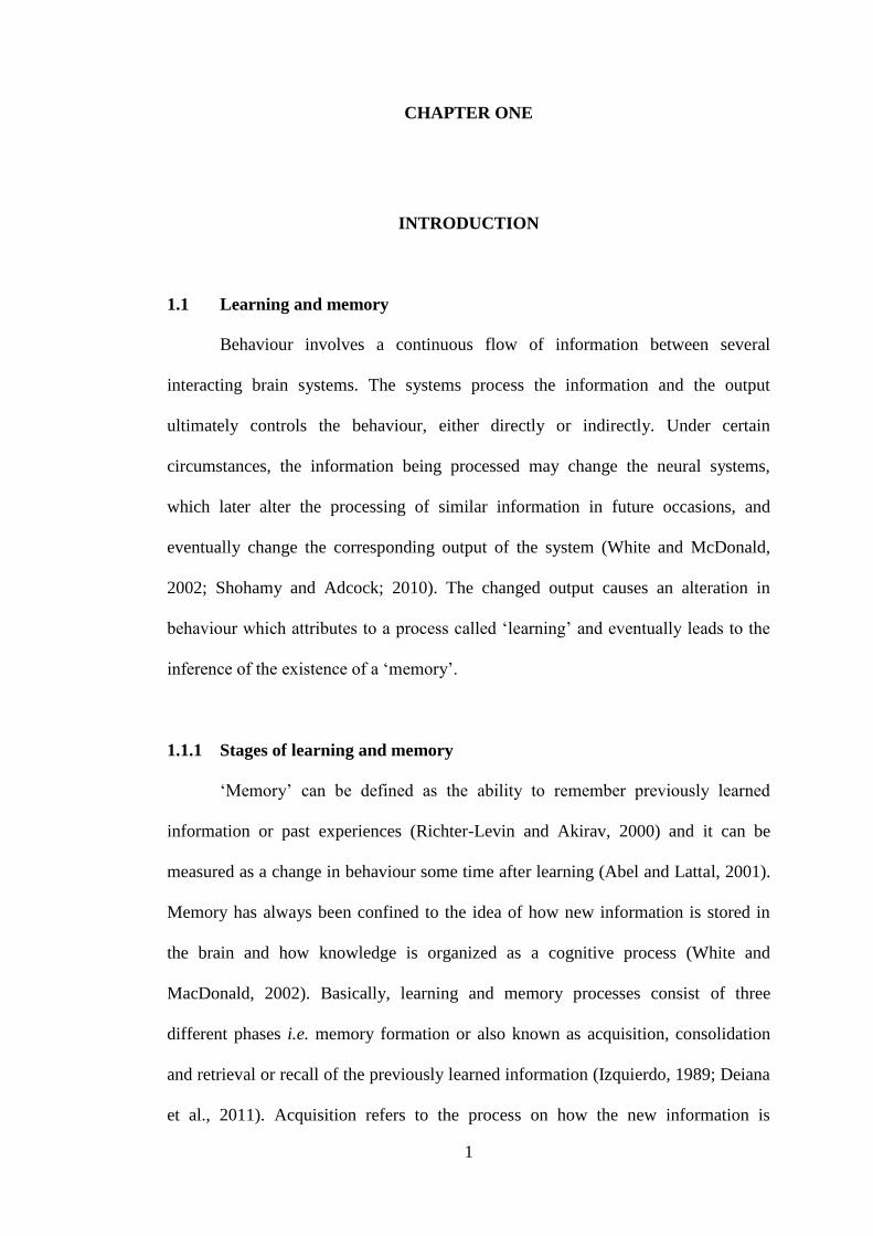
1.1 Learning and memory
Behaviour involves a continuous flow of information between several
interacting brain systems. The systems process the information and the output
ultimately controls the behaviour, either directly or indirectly. Under certain
circumstances, the information being processed may change the neural systems,
which later alter the processing of similar information in future occasions, and
eventually change the corresponding output of the system (White and McDonald,
2002; Shohamy and Adcock; 2010). The changed output causes an alteration in
behaviour which attributes to a process called „learning‟ and eventually leads to the
inference of the existence of a „memory‟.
1.1.1 Stages of learning and memory
„Memory‟ can be defined as the ability to remember previously learned
information or past experiences (Richter-Levin and Akirav, 2000) and it can be
measured as a change in behaviour some time after learning (Abel and Lattal, 2001).
Memory has always been confined to the idea of how new information is stored in
the brain and how knowledge is organized as a cognitive process (White and
MacDonald, 2002). Basically, learning and memory processes consist of three
different phases i.e. memory formation or also known as acquisition, consolidation
and retrieval or recall of the previously learned information (Izquierdo, 1989; Deiana
et al., 2011). Acquisition refers to the process on how the new information is

2
encoded whilst the consolidation refers to the state where the newly acquired
information that is labile and fragile undergoes a lingering process, becoming
stronger and resilient over time. Retrieval can be defined as a process of recall of the
stored information (Medina et al., 2008).
Figure 1.1: The traditional taxonomy of memory systems (Edited from Bird and
Burgess, 2008).
1.1.2 Types of memory
Classifications of memories are based on their duration (short- or long-term)
and their nature which could be archival (short- or long-term memory) or transient
(working memory) (Deiana et al., 2011; Izquierdo et al., 1999). In mid 1980s, the
term declarative and „non-declarative‟ were introduced with declarative memory
refers to one memory system and non-declarative memory refers to other additional
Memory
Short-term
memory
Long-term
memory
Visuospatial
sketchpad
Phonological
loop
Neocortical
sensory and
motor buffers
Declarative Non-
declarative
Contextual/
associative
Episodic
Recollection
Non-
contextual/
non-
associative
Semantic
Familiarity
Procedural
memory
Classical
conditioning
Priming

3
memory systems (Squire, 2004; Figure 1.1). Declarative memory refers to the
capacity of conscious recollection of facts or events that can be brought to mind and
described verbally. This kind of memory is often impaired in amnesia and is
dependent on structures in the medial temporal lobe and midline diencephalon.
Declarative memory allows the retained information to be compared and contrasted
and thus, supports the encoding of memories in terms of relationships among
multiple items and events. In contrast, non-declarative memory can be referred as
unconscious recollection of experiences such as habits and skills that are expressed
in performances. This kind of memory occurs due to modifications within
specialized performance systems and is revealed via reactivation of the systems
within which the process of learning occurred (Squire, 2004; Deiana et al., 2011).
Declarative memory can be subdivided into semantic and episodic memory.
The former constitutes the memory of meanings, facts, understanding and concept-
based knowledge, and the latter implies the ability to re-collect an event in the
environment in which it originally occurred and involves detailed elements (what),
location (where) and temporal occurrence (when). Spatial memory is usually
considered in the context of episodic memory and it implies the ability to attain
information about one‟s environment and its spatial orientation. Construction of
cognitive maps is based on the orientation in space as indicated by landmark cues.
Establishment of these cognitive maps follows either egocentric or allocentric
strategies. Egocentric involves self-to-object representational system whilst
allocentric involves relationship of oneself to three-dimensional arrangements of
objects in the environment (Deiana et al., 2011).
4
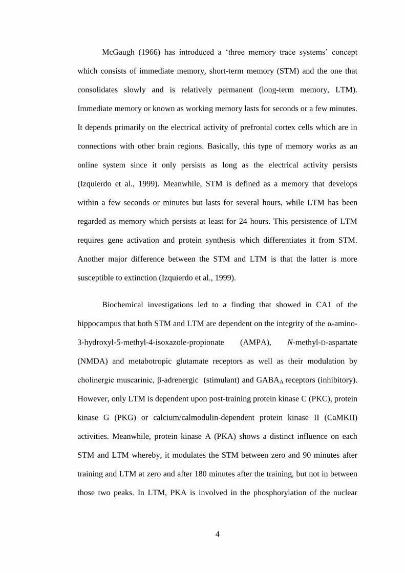
McGaugh (1966) has introduced a „three memory trace systems‟ concept
which consists of immediate memory, short-term memory (STM) and the one that
consolidates slowly and is relatively permanent (long-term memory, LTM).
Immediate memory or known as working memory lasts for seconds or a few minutes.
It depends primarily on the electrical activity of prefrontal cortex cells which are in
connections with other brain regions. Basically, this type of memory works as an
online system since it only persists as long as the electrical activity persists
(Izquierdo et al., 1999). Meanwhile, STM is defined as a memory that develops
within a few seconds or minutes but lasts for several hours, while LTM has been
regarded as memory which persists at least for 24 hours. This persistence of LTM
requires gene activation and protein synthesis which differentiates it from STM.
Another major difference between the STM and LTM is that the latter is more
susceptible to extinction (Izquierdo et al., 1999).
Biochemical investigations led to a finding that showed in CA1 of the
hippocampus that both STM and LTM are dependent on the integrity of the α-amino-
3-hydroxyl-5-methyl-4-isoxazole-propionate (AMPA), N-methyl-D-aspartate
(NMDA) and metabotropic glutamate receptors as well as their modulation by
cholinergic muscarinic, β-adrenergic (stimulant) and GABAA receptors (inhibitory).
However, only LTM is dependent upon post-training protein kinase C (PKC), protein
kinase G (PKG) or calcium/calmodulin-dependent protein kinase II (CaMKII)
activities. Meanwhile, protein kinase A (PKA) shows a distinct influence on each
STM and LTM whereby, it modulates the STM between zero and 90 minutes after
training and LTM at zero and after 180 minutes after the training, but not in between
those two peaks. In LTM, PKA is involved in the phosphorylation of the nuclear

5
transcription factor CREB1 (cAMP response element binding protein) (Izquierdo et
al, 1999; 2004).
On the contrary, it seems no involvement of NMDA receptors in the STM
formation in entorhinal cortex or elsewhere. In addition, the role of PKC and PKA
are both necessary for the formation of STM and LTM in entorhinal cortex
(Izquierdo et al., 1999).
1.1.3 Mechanisms of learning and memory
Learning and memory are basically supported by synaptic plasticity in the
brain. Synaptic plasticity can be referred as any lasting upregulation or
downregulation of synaptic strength including long-term potentiation (LTP), long-
term depression (LTD) or depotentiation, and the changes of the synaptic strength
can occur as a result of certain types of learning (Morgado-Bernal, 2011; Johnston et
al., 2003; Martin and Morris, 2002). It occurs at the dendritic spines, the major site of
excitatory synaptic transmission in the vertebrate brain. Studies on the initiation and
the maintenance of synaptic plasticity, particularly in CA1 pyramidal neurons of the
hippocampus, have shown that certain types of learning can induce an increase in the
number or morphological changes of the dendritic spines leading to a new or
strengthen existing synapses that may form the principle basis of memory (Morgado-
Bernal, 2011; Penzes and Jones, 2008).
Pharmacological manipulations have succeeded in establishing the major
molecular mechanisms of memory processes. Initially, activation of relevant
synapses causes a pre-synaptic release of glutamate which then activates the
ionotropic AMPA receptors (AMPAr). Activation of this AMPAr leads to an influx
of Ca2+
into the post-synaptic neuron thus depolarization occurs. Within a few

6
seconds, the local post-synaptic depolarization activates the NMDA receptors
causing the removal of Mg+
which blocks the channel of NMDA receptors and thus
giving rise to a massive influx of Ca2+
into the post-synaptic neuron. Consequently,
CaMKII, PKC and calcineurin are activated. Adenylyl cyclase is also activated by
the influx of Ca2+
or other modulatory inputs via G-protein-coupled receptors. As a
result, cyclic adenosine monophosphate (cAMP) level is increased and leads to
activation of PKA that eventually regulates the synthesis and availability of new
proteins via activation of CREB in the nucleus (Morgado-Bernal, 2011; Abel and
Lattal, 2001; Maren and Baudry, 1995; Figure 1.2).
In this complex process, Ca2+
binds to calcium sensor calmodulin (CaM) and
activates different CaMKs causing morphological changes of the cytoskeleton of the
neuron, for instance new dendritic spines or persistence enlargement of its head.
These morphological changes are strongly dependent on the protein synthesis, as
well as brain derived neurotrophic factor (BDNF) actions (Morgado-Bernal, 2011).
The formation and the maintenance of new dendritic spines provide a persistent
structural change upon which a long-term memory is established.

7
Figure 1.2: Molecular events underlie the early and late phases of long-term
potentiation (Adapted from Abel and Lattal, 2001).
1.1.4 Memory modulatory mechanisms in the brain
Extensive research has been carried out to investigate the neurobiological
processes and systems that contribute to the differences in memory strength.
Modulatory systems not only affect the neurobiological processes of newly acquired
information but also other mnemonic processes including working memory, memory
recall and memory extinction (Roozendaal and McGaugh, 2011; Cahill and
McGaugh, 1996; Figure 1.3).

8
+
Figure 1.3: Modulatory systems of learning and memory functions in the brain.
1.1.4(a) GABA
Gamma (γ)-aminobutyric acid (GABA) is the major inhibitory
neurotransmitter in brain that can act on three receptor subtypes: GABAA, GABAB
and GABAC. Both GABAA and GABAB are confined to the central nervous system
while GABAC is specific for retina (Lanctot et al., 2004). GABAA receptor is a
ligand-gated ion channel that can be found at both pre- and post-synaptic neurons.
Upon binding with GABAA agonist, flux of chloride ions increase leading to neurons
hyperpolarization and thus reduce the synaptic transmission of dopamine, serotonin
and acetylcholine (ACh). Other than its GABA-agonist binding site, GABAA
receptor has specific sites for benzodiazepines, barbiturates, steroids and ethanol
(Lanctot et al., 2004). Therefore, any interference in individual binding sites or at the
whole receptor unit may change the modulatory effect of GABA on other neuronal
+/-
-
+/-
+
+/-
+
-
+
-
- - +
-
- +
Memory formation/
Consolidation/
Retrieval
Glutamatergic
system
GABAergic
system
Opioidergic
system
Cholinergic
system
Noradrenergic
system
Dopaminergic
system
Serotonergic
system
Endocannabinoid
system
Histaminergic
system
Other modulators
e.g. Glucose, Bombesin

9
systems. Meanwhile, GABAB is linked to G-proteins and second-messenger system
that mediates calcium or potassium channels which in turn generating the slow
inhibitory post-synaptic potentials in several brain regions (Lanctot et al., 2004;
Staubli et al., 1999).
Bicuculline, GABAA antagonist has been proven pharmacologically to
facilitate the memory formation (Izquierdo et al., 1992; Izquierdo and McGaugh,
2000) while picrotoxin acts by blocking the chloride channel of GABAA receptors,
facilitates both memory formation and extinction (McGaugh et al., 1990; Izquierdo et
al., 2004). On the contrary, muscimol (GABAA agonist) that mimics the action of
GABA disrupts the memory performance and tiagabine, impairs the spatial learning
via inhibition of the transport of GABA away from the synaptic cleft (Izquierdo et
al., 2004).
The importance of GABAergic system is further illustrated in a study using
PWZ-029, an α5 GABAA selective inverse agonist. It was found that PWZ-029
administration was able to improve memory in object recognition task after 24-hours
delay in normal and scopolamine-treated rats. However, PWZ-029 seemed to be
ineffective to improve the performance in Morris water maze task in either normal or
scopolamine-treated rats (Milic et al., 2013).
1.1.4(b) Monoaminergic system
The role of dopaminergic, noradrenergic and serotonergic systems in
modulating the learning and memory processes in various memory-associated brain
regions including hippocampus, basolateral amygdala, entorhinal and parietal cortex
has been well established (Roozendaal and McGaugh, 2011; O‟Carroll et al., 2006;
Izquierdo and McGaugh; 2000). Pharmacological manipulation has shown that

10
dopamine uptake inhibition improves the acquisition of the inhibitory avoidance and
increases the hippocampal ACh release (Nail-Boucherie et al., 1998). However,
studies on dopaminergic modulation on learning and memory showed contrasting
results which may be due to the activation of different receptor subtypes and
different experimental design. For instance, activation of D1 receptor caused an
increase but activation of D2 receptor led to a decrease in the cAMP production.
Meanwhile, D3 receptor was not related to the adenylate cyclase and was not
affected by GTP which modulates the binding associated with D1 and D2 receptor
(Snyder, 1992; Sokoloff et al., 1990; Zarrindast, 2006). Administration of D1
receptor agonist resulted in enhanced cognitive performance in Morris water maze
task in rats (Hersi et al., 1995) or had no effects on radial-arm maze learning
(Wilkerson and Levin, 1999). Findings by Zarrindast et al. (1996) demonstrated that
administration of low and high doses of apomorphine, a mixed D1/D2 dopamine
receptor agonist in mice, improved or impaired memory retrieval in the active
avoidance task, respectively.
Noradrenergic neurons originated from locus coerulus innervate many brain
regions, ranging from the spinal cord and cerebellum to the forebrain (Sirvio and
MacDonald, 1999). The modulatory effects of noradrenergic receptors on learning
and memory may be mediated via α1-, α2-, β1-, and β2-adrenoceptors (Zarrindast,
2006). Previous study reported that activation of β-noradrenergic receptor, 3 to 4
hours after induction can help to maintain LTP in conscious rats (Straube and Frey,
2003) and thus enhancing the memory. Local infusion of norepinephrine into the
basolateral amygdala of rats also enhanced the consolidation of two different forms
of contextual fear conditioning (LaLumiere et al., 2003). In addition, previous study

11
stated the memory-enhancing effects were mediated by the β-adrenoceptors whilst
α1-adrenoceptors inhibited memory consolidation (Gibbs and Summers, 2001).
Serotonin (5-hydroxytriptamine, 5-HT) is a biogenic amine that is implicated
in various physiological functions such as sleep, pain perception, memory and mood
control (Polter and Li, 2010; Meneses and Perez-Garcia, 2007; Zarrindast, 2006;
Buhot et al., 2000; McEntee and Crook, 1991). The central nervous system (CNS)
effects of the serotonin are mediated by 5-HT receptor that can be classified into
seven main classes; 5-HT1, 5-HT2, 5-HT3, 5-HT4, 5-HT5, 5-HT6 and 5-HT7 which are
based on structural, transductional and operational features (Hannon and Hoyer,
2008; Hoyer et al., 2002). Some 5-HT receptors can be further subdivided into
several subtypes (Zarrindast, 2006). Activation of 5-HT receptors lead to diverse
effects on learning and memory processes and usually, activation of 5-HT1A
receptors promote an enhancement in memory consolidation and retrieval (Izquierdo
et al., 2004). In another study, impairment of short-term memory but not long-term
memory was observed due to serotonin depletion after intracerebroventricular
injection of serotonin neurotoxin and serotonin synthesis inhibitor in rats (Hritcu et
al., 2007).
A number of studies reported that 5-HT is potentially interacting with other
neural systems to contribute to a particular function (Lesch and Waider, 2012;
Ciranna, 2006). In entorhinal cortex, 5-HT activates 5-HT3 receptors on the
GABAergic neuron causing an inhibition of the cholinergic function (Ramirez et al.,
1996). 5-HT1A antagonist prevents the impairment of spatial learning induced by
intrahippocampal scopolamine, plausibly via favorable actions on other excitatory
neurotransmitters (Carli et al., 1995a). In another studies, activation of 5-HT1A
receptors in the hippocampal CA1 region causes impairment in spatial but not visual

12
discrimination in rats (Carli et al., 1995b) whilst blockade of 5-HT2A receptors in the
medial prefrontal cortex impairs the recognition memory in spontaneous novelty
preference task (Bekinschtein et al., 2013).
Taken together, dopaminergic, noradrenergic and serotonergic play a pivotal
role in modulating the learning and memory, and it seems possible that these neural
systems work with other neural systems to modulate the learning and memory
processes.
1.1.4(c) Cholinergic system
The involvement of cholinergic system in learning and memory processes has
gained a great interest among researchers (Blokland, 1996; Robinson et al., 2011;
Deiana et al., 2011; Miranda et al., 2003) and numerous studies have confirmed the
participation of cholinergic system in modulating learning and memory processes
(Easton et al., 2012; Pych et al., 2005; Farr et al., 2000). In rodent, there are at least
six cholinergic cell groups that project to different brain areas. Both medial septal
nucleus (Ch1) and vertical (VDBB) limb of the diagonal band of Broca (Ch2) from
the basal forebrain innervate the hippocampal formation. The horizontal (HDBB)
limb of the diagonal band of Broca (Ch3) projects to olfactory bulb and thalamic
reticular region whilst nucleus basalis magnocellularis (NBM, Ch4) issuing fibers to
the thalamus, cerebral cortex and amygdala. Finally, pedunculopontine (Ch5) and
laterodorsal tegmentum (Ch6) which are part of the midbrain regions provide
projections to cortical and various thalamic regions. In addition, there are numerous
cholinergic fibers that are intrinsic to specific regions, such as striatum (Deiana et al.,
2011; Zarrindast, 2006; Everitt and Robbins, 1997; Figure 1.4).

13
Figure 1.4: Distribution of cholinergic neurons and their projections in the rat brain.
Abbreviations: MS, medial septum (cell group Ch1); VDBB, vertical limb nucleus of
the diagonal band Broca (cell group Ch2); HDBB, horizontal limb nucleus of the
diagonal band Broca (cell group Ch3); NBM, nucleus basalis magnocellularis (cell
group Ch4); tpp, pedunculopontine tegmental nucleus (cell group Ch5); dltn,
laterodorsal tegmental nucleus (cell group Ch6); PFC, prefrontal cortex; ICj, islands
of Calleja; SN, substantia nigra; IP, interpeduncular nucleus; DR, dorsal raphe; LC,
locus coerulus (Adapted from Everitt and Robbins, 1997).
ACh is synthesized via the acetylation of choline with acetyl-CoA by enzyme
choline acetyltransferase (ChAT) in the nerve ending. Accumulation of ACh in the
synaptic vesicle is driven by proton-pumping ATPase. The quantal release of ACh
into the synaptic cleft is triggered by depolarization-induced Ca2+
flux. ACh will bind
to the both pre- and post-synaptic muscarinic (mAChR) and nicotinic (nAChR)
receptors (Deiana et al., 2011; Taylor and Brown, 1999). Muscarinic receptors are G-
proteins coupled receptors and can be classified into M1, M2, M3, M4 and M5. M1
receptors are widely distributed in CNS particularly in cerebral cortex, hippocampus
and amygdala. It is also a dominant mAChR subtype in sympathetic ganglia and
exocrine glands. M2 receptors are abundant in myocardium and M3 receptors are
mainly found in smooth muscle and exocrine glands as well as in cortex,

14
hippocampus and thalamus. Meanwhile, M4 receptors showed a scattered appearance
in cortex and striatum whilst M5 receptors are dense in hippocampus, habenula and
thalamus (Deiana et al., 2011).
nAChRs are ligand-gated ion channels with pentameric shape consisting of
α2-α10, β2-β4, γ, δ and ε (in the adult muscle) subunits that are arranged
symmetrically around the central pore. Activation of these receptors leads to a rapid
increase in cellular permeability to Na+ and Ca
2+, depolarization and excitation. α2 is
scattered in the hippocampus and cortex and α3 is abundant in layer four of cortex,
dorsal and ventral thalamus including lateral geniculate. α4 can be found mainly in
cortex, thalamus, amygdala and hippocampus whilst α7 is moderately distributed in
hippocampus, cortex, cerebellum and brainstem. β2 is dense in thalamus but less in
cortex and dorsal hippocampus (Deiana et al., 2011).
Lesions of the cholinergic system or systemic and direct brain injections of
cholinergic drugs have been used in many studies for its behavioural effects. Local
administration of muscarinic antagonist, scopolamine into the amygdala interfered
with the acquisition of the conditioned place preference task (McIntyre et al., 1998).
In general, many findings have demonstrated that local administration of cholinergic
agonists and antagonists into the striatum, hippocampus and amygdala, enhanced or
impaired learning and memory performance in task associated with the neural
system, respectively (Gold, 2003).
The availability of the new method called in vivo microdialysis/high
performance liquid chromatography (HPLC) has enabled the measurement of ACh
releases (Schlereth et al., 2006; Gold, 2003; McIntyre et al., 2002; Stancampiano et
al., 1999) from the hippocampus of behaving animals. It has been reported that the

15
increase and decrease in the release of ACh, particularly in hippocampus was
correlated with the enhancement and impairment of learning and memory (Gold,
2003). The ACh release may reflect the degree of activation and participation of the
cholinergic system in the hippocampus in contributing to the performance of the
learning and memory in a particular task.
Cholinesterases i.e. acetylcholinesterase and butyrylcholinesterase are pivotal
enzymes in cholinergic system. Acetylcholinesterase is present in both central
nervous system and peripheral nervous system such as at the parasympathetic and
sympathetic ganglia, parasympathetic end-organs, motor end-plates and sweat
glands. This enzyme primarily functions to hydrolyze the ACh at these junctions and
eventually terminates the cholinergic transmission (Padilla et al., 1999; Pepeu and
Giovannini, 2010; Miao et al., 2010). Meanwhile, the role of butyrylcholinesterase is
an enigma even though reports indicated that this enzyme plays a role during
developmental processes (Zugno et al., 2013). In contrast to acetylcholinesterase,
butyrylcholinesterase hydrolyzes both acetylcholine and other esters.
Acetylcholinesterase activity may be an indicator on how the brain is
functioning in relation to learning and memory. Isomae et al. (2003) reported that T-
82, a novel acetylcholinesterase inhibitor ameliorated the memory impairment
induced by acetylcholinergic dysfunction in inhibitory avoidance task. A study found
an increase in acetylcholinesterase activity in amnesic brain (Biradar et al., 2012),
but in the imaging study of Alzheimer patients, acetylcholinesterase appeared to be
reduced in the amygdala (-33%), hippocampus (-14%) and neocortex (-20%)
(Shinotoh et al., 2003). Therefore, it is possible that consumption of mitragynine may
also leads to the alteration of the cholinesterase activity in the brain.

16
1.1.4(d) Opioidergic system
Opioid peptides such as enkephalin and β-endorphin are synthesized and
released from both peripheral and CNS (Asai et al., 2007; Lin and Pan, 1995;
Carrasco et al., 1982). Extensive studies have shown that administration of opioid
peptide agonists and antagonists were able to modulate learning and memory
processes in a variety of tasks employing both appetitive and aversive motivation, via
binding to opioid receptors that are widely distributed in the memory-associated
brain regions. Opioid agonists act via G-protein coupled opioid receptors known as
μ-, δ- and κ-receptors (Feng et al., 2012; Al-Hasani and Bruchas, 2011; McDonald,
2005). Binding to the opioid receptors causes activation of potassium channels,
inhibition of calcium channels, inhibition of adenylyl cyclase which leads to reduced
cAMP production as well as inhibition of transmitter release (Dacher and Nugent,
2011; McDonald, 2005), thus affecting the learning and memory processes.
Opioid agonists impair memory, but opioid antagonists have memory-
enhancing effects in several paradigms including inhibitory and active avoidance,
aversively-motivated Y-maze discrimination, and appetitively-motivated spatial
learning (McGaugh, 1989; Zhu et al., 2011). A recent study reported that
administration of opioid agonists inhibited the acquisition of passive avoidance task
at all stages (Zarrindast et al., 2013). However, the inhibitory effects of opioid
agonist on memory could be reversed by naloxone, an opioid antagonist (Izquierdo,
1979). Repeated treatment of morphine impaired the acquisition of both simple
appetitive and cued operant learning but withdrawal for five weeks alleviated the
impairment. Single morphine exposure caused a disruption in the retrieval of the
operant memory, but no effect was observed in rats after five-week withdrawal
(Wang et al., 2006).

17
Opioid agonist may also interact with other neural systems in the brain. It has
been reported that opioid agonists inhibited noradrenaline release from the
hippocampus, thus suggesting how it regulates the learning and memory processes
(Matsumoto et al., 1994). Interaction with the cholinergic system occurs via binding
to the opioid receptors located on the cholinergic interneurons causing an inhibition
on the acetylcholine release in the brain, thus contributing to the memory impairment
(Xi-Geng et al., 2002). It has been reported that administration of opioid agonist,
morphine into the medial septum causes a reduction in the ACh release in the
hippocampus (Ragozzino and Gold, 1995).
1.1.4(e) Endocannabinoid system
Endocannabinoid receptors, particularly cannabinoid receptors type 1 (CB1)
which are widely distributed in the brain has been shown to be involved in learning
and memory functions (Moreno and Campolongo, 2014; Peterfi et al., 2012; Castillo
et al., 2012; Serrano and Parsons, 2011; De Oliveira Alvares et al., 2008).
Cannabinoids have disruptive effects on the learning and memory processes. This
was shown through impairment of spatial memory in 8-arm radial maze and
inhibition of the acquisition and retrieval of passive avoidance task upon
administration of delta 9-tetrahydrocannabinol, the major psychoactive compound of
marijuana (Mishima et al., 2001). In another study, spatial learning deficit induced
by delta 9-tetrahydrocannabinol was blocked by SR241716A, a specific CB1
antagonist in mice (Da and Takahashi, 2002). Anandamide, the main endogenous
ligand, impaired memory consolidation of one-trial inhibitory avoidance in mice but
did not affect the retrieval when given before testing (Costanzi et al., 2003). The
endocannabinoid system also controls the extinction of the aversive conditioned

18
responses in rat, presumably in the basolateral amygdala (Marsicano et al., 2002).
The amygdala has been characterized by a great presence of CB1 receptors, which
are responsible for the local GABAergic transmission (Katona et al., 2001). CB1
cannabinoid receptors may play a pivotal role in endogenous defense against
excitotoxicity (Marsicano et al., 2003), in modulation of retrograde signaling in the
hippocampus (Wilson and Nicoll, 2001) and in LTP induction (Carlson et al., 2002).
1.1.4(f) Histamine
Emerging evidences have shown that brain histamine plays a role in the
modulation of memory consolidation via histamine type 1 (H1), type 2 (H2) and type
3 (H3) receptors (Serafim et al., 2012; Kohler et al., 2011; De Almeida and
Izquierdo, 1986; 1988). Recent study showed that post-training histamine and
histamine H3 receptor antagonist thioperamide in the basolateral amygdala enhanced
and impaired memory consolidation of the step-down inhibitory avoidance task,
respectively (Benetti and Izquierdo, 2013). On the contrary, another study reported
that systemic administration of H3 receptor antagonist, thioperamide facilitated
memory consolidation with no influence on retrieval (Orsetti et al., 2001). In
addition, a study by Cangioli et al. (2002) has shown that activation of H3 receptors
by R-α-methyl-histamine in the basolateral amygdala improved the consolidation of
the fear memory and enhanced the release of ACh.
1.1.4(g) Others
Glucose has been known to influence memory modulation (Izquierdo et al.,
2004; Smith et al., 2011). Extracellular fluid of glucose in the brain was found to be
low and even fluctuated across brain areas during spontaneous alternation testing

19
(McNay et al., 2001). In another study, glucose enhanced, and glucose-uptake
inhibitor 2-deoxyglucose impaired memory consolidation in a day-old chick
inhibitory avoidance model (Gibbs and Summers, 2002). Systemic or intra-amygdala
administration of glucose led to the facilitation of the onset of extinction of drug-
induced conditioned reward (Schroeder and Packard, 2003).
Another factor that may modulate learning and memory is bombesin. It was
found that intrahippocampal infusion of RC-3095, the bombesin/gastrin-releasing
peptide antagonist caused an impairment in the inhibitory avoidance in rats, after
given immediately or two hours after training. This finding may suggest that the
receptors for bombesin are present in the hippocampus, thus being able to modulate
the memory consolidation (Roesler et al., 2003).
1.1.5 Memory circuitry in brain: interaction between neural systems
Based on the lesion of different brain areas, several neural systems have been
shown to play a significant role in the processing information of different types of
learning and memory (Gold, 2004; White and McDonald, 2002). Critical interactions
between these neural systems will determine the time it takes to learn a task, and to
the extent of the content and the strength of particular memories. Lesion in a
particular area of the brain leads to a decreased contribution of a neural system to a
memory processing from its normal proportional participation to a value of zero.
However, if the brain damage led to enhancement of learning, it could be suggested
that the neural system that contributes to the efficient learning is „released‟ from its
competition by a system that contributes to a less efficient learning (Gold, 2004).
However, the degree of efficiency of the system varies between tasks.

20
As the memory research keeps developing, most of them have been
concerned with the role of hippocampal formation in the formation and consolidation
of the explicit memory (Wang and Morris, 2010; Richter-Levin and Akirav, 2000;
Richter-Levin, 2004; Thierry et al., 2000). The hippocampal formation consists of
the entorhinal cortex, dentate gyrus, the individual CA fields of the hippocampus
proper and the subicular complex. Hippocampal formation works together with the
subcortical and cortical networks where it is believed to store the long-term memory
(Bird and Burgess, 2008; Wang and Morris, 2010; White and McDonald, 2002;
Figure 1.5).
Prefrontal cortex is mainly implicated in the working memory as well as in
temporal ordering of spatial and non-spatial events and in organization and planning
of response. Both hippocampal formation and prefrontal cortex are anatomically and
functionally associated and due to their reciprocal connections with the temporal and
parietal association areas of the cerebral cortex, prefrontal cortex may serve as an
intermediate link between these neocortical areas and the hippocampus (Thierry et
al., 2000). Direct projection from the hippocampal formation to the prefrontal cortex
was first described in 1977, in which projections from subiculum to the medial
frontal cortex were found (Rosene and Van Hoesen, 1977). There were also
reciprocal connections between caudal portion of the presubiculum and the
dorsolateral prefrontal cortex (Goldman-Rakic et al., 1984). Direct projections from
the hippocampal CA1 field to the medial and orbito prefrontal cortex were also
observed (Barbas and Blatt, 1995).
Both prefrontal cortex and hippocampus further innervate the ventral
striatum, particularly the nucleus accumbens which consists of two main regions, the
„core‟ and the „shell‟. The „core‟ is innervated preferentially by pre-limbic/medial

21
orbito areas of the prefrontal cortex while the „shell‟ is innervated by the
CA1/subiculum regions of the hippocampus. The nucleus accumbens plays a pivotal
role in integrating information from the limbic system and cortical regions into goal-
directed behaviours. Nucleus accumbens sends direct projections into several regions
including pallidum, substantia nigra, hypothalamus and several mesencephalic areas
(Thierry et al., 2000; Mogenson et al., 1982).
The amygdala is one of the brain regions that has a significant contribution to
learning and memory processes. Neurons from the basal nucleus project to the
entorhinal cortex, CA3 and CA1 fields of the hippocampus, subiculum and
parasubiculum. These anatomical interactions are believed to be involved in the
encoding and consolidation (Pare, 2003; McGaugh et al., 1996; Richter-Levin and
Akirav, 2000), but not in the retrieval or expression of the emotionally based
experiences, suggesting that amygdala only facilitates memory storage in other brain
areas (Pare, 2003). It has been shown that amygdala activity modulated the induction
of the hippocampal LTP (Akirav and Richter-Levin, 1999; 2002). Other than the
hippocampus, the amygdala also receives extensive projections from cortical areas of
the temporal lobe, from the substantia nigra, ventral tegmental area, nucleus
accumbens and from brain stem areas (White and McDonald, 2002). In general, it
can be concluded that one or more brain systems can be involved in acquisition,
consolidation or retrieval processes of one particular types of memory.

22
Figure 1.5: The hippocampus and its connections. a) Hippocampus in the medial
temporal lobes b) Subcortical connections: red lines; cortical connections: black
lines; thickness of the black lines approximates to the strength of the connections.
Abbreviations: MDTN, medial dorsal thalamic nuclei; ATN, anterior thalamic
nuclei; TE & TEO, inferior temporal areas TE and TEO; LIP, lateral intraparietal
area; RSp, retrosplenial; Cing gyrus, cingulate gyrus (Adapted from Bird and
Burgess, 2008).

23
1.2 Electroencephalogram (EEG)
EEG can be referred as amplified electrical activity generated by connecting
neurons in the brain (Liu et al., 2005; Idris et al., 2014), thus is a reflection of the
functional connectivity in the brain network. It has been proven to be a very sensitive
and reliable tool for characterizing drug effects in the CNS (Palenicek et al., 2013;
Dimpfel, 2005). It can be classified into delta (0.1-4 Hz), theta (4-7 Hz), alpha (7-13
Hz), beta (13-30 Hz) and gamma (>30 Hz) bands based on range of frequencies
(Idris et al., 2014). Amplitude of brain waves is measured using microvolt unit (μV).
Amplitude with less than 20 μV, in between 20-50 μV and more than 50 μV are
considered as low, medium and high amplitude, respectively (Idris et al., 2014).
The slow wave delta band (0.1-4 Hz, 20-200 μV) is associated with deep
sleep and unconsciousness while theta band (4-7 Hz, 20-100 μV) which originated
from thalamus appears as consciousness slips towards drowsiness. The most
prominent rhythm in the brain activity is alpha band (7-13 Hz, 20-60 μV), with
round, sinusoidal or sometimes as sharp wave appearance. Alpha band appears
mainly in the posterior half of the head, over the parieto-occipital region of the brain.
This band indicates a relaxed awareness without any attention or concentration. Beta
band (13-30 Hz, 2-20 μV) which usually found at frontal and central region
represents the waking rhythm of the brain associated with active thinking or active
attention. Lastly, the gamma band (> 30 Hz, ~5-10 μV) occurs at certain occasion
when the sensory stimuli are given. EEG activity originated mainly from pyramidal
postsynaptic potentials which are in oscillations with several brain regions. The first
network is with thalamus, thus is referred as thalamocortical networks which is
modulated by the reticulo-thalamo-cortical circuits. The other network is the extra-
thalamic-cortical circuits which involve the reticular system, hypothalamus,

24
hippocampus, amygdala and basal forebrain nuclei. Finally, the network from the
other cortex is referred as cortical-cortical networks (Idris et al., 2014).
EEG characteristics across species are relatively similar (Moretti et al., 2013;
Sambeth et al., 2007). Changes in EEG characteristics induced by drugs were found
to be similar in human and rodents (Dimpfel, 2005; van Luijtelaar et al., 2002;
Dimpfel et al., 1992). These changes are useful in understanding the central activity
of drugs in human (Sambeth et al., 2007). The electrical power of each frequency
range changes in parallel with changes in particular behaviour or drug condition
without affecting the neighbouring frequencies. This notion has been proven true by
Stahl et al. (1997). Repeated exposure to d-amphetamine produced an increase in the
alpha activity via activation of the dopamine D2-like receptors, without affecting
other frequency bands. Finding by Dimpfel and Schober (2001) further supported the
notion. Administration of α2-adrenoceptor agonist that acts presynaptically led to a
great increase in electrical theta activity but not other frequencies. However, the
degree of increment was not similar between brain areas suggesting uneven
distribution of α2-adrenoceptor in different brain areas.
Considerable evidences support the role of cholinergic system in modulating
the brain activity (Dringenberg et al., 2002; Metherate et al., 1992). Blockade of
stimulation-induced EEG activation by intracortical application of anti-muscarinic
drugs suggested that acetylcholine promotes activation by a direct effect on cortical
neurons (Metherate et al., 1992). In another study, cholinergic transmitter was found
to modulate the changes in electrical delta activity which was intimately linked to
dopaminergic transmission as shown by the concomitant changes in alpha2 electrical
power (Dimpfel, 2005). The influence of the dopaminergic system on EEG activity
was further corroborated via administration of dopamine agonist and antagonist,