on a dynamic reaction-diffusion mechanism: the spatial...
TRANSCRIPT
On a dynamic reaction-diffusion mechanism: The spatial patterning of teeth prirnordia in the alligator
J. D. Murraya and P. M. Kulesab* Department of Applied Mathematics, University of Washington, Box 352420, Seattle,
Division of Biology, Beckman Institute (139-741, California Institute of Technology, Pasadena, W A 91985-4240, U S A
C A 91125, USA
It is now well established both theoretically and, more recently, experimentally, that steady-state spatial chemical concentration patterns can be formed by a number of specific reaction-diffusion systems. Reaction-diffusion models have been widely applied to biological pattern formation problems. Here we propose a model mechanism for the initiation and spatial positioning of teeth primordia in the alligator, Alligator mississippiensis, which, from 'a reaction-diffusion theory, introduces, among other things, a new element, namely the effect of domain growth on dynamic spatial pattern formation. Detailed embryological studies by Westergaard and Ferguson (B. Westergaard and M. W. J. Ferguson, J . Zool. Lond., 1986, 210, 575; 1987, 212, 191; Am. J . Anatomy, 1990, 187, 393) show that jaw growth plays a crucial role in the developmental patterning of the tooth initiation process. Based on biological data we develop a reaction-diffusion mechanism, which crucially includes domain growth. The model can reproduce the spatial pattern development of the first seven teeth primordia in the lower half jaw of A. mississippiensis. The results for the precise spatio temporal sequence compare well with detailed developmental experiments.
A fundamental theme in embryology is the development of pattern and form. Through a sophisticated orchestration of signals, a homogeneous mass of cells differentiate into specific cell types in a way which leads to functional tissue and organs. Pattern formation is just one of the crucial components of this morphogenesis.
We present a biological system which possesses an elegant mechanism for the formation of anatomical structure. The morphogenesis of this structure takes place in a developing embryo and is first observed in a precise spatial and temporal pattern. Experimental evidence has revealed that the spatial pattern forms dynamically: the timescale on which the pattern forms is comparable to the growth of the embryonic pat- terning domain. Although many of the biological details of the patterning process remain unknown, there is evidence that chemical components play a direct role in the formation of pattern. We will propose a mechanism by which the chemical dynamics interplay with the physical morphogenesis to repro- duce the essence of the observed biological pattern.
One of the proposed mechanisms of pattern formation is chemical gradients : the concentration of a soluble substance (morphogen) may help to instruct a homogeneous population of cells to act. The basis chemical theory that two homoge- neously distributed solutions could interact to produce stable spatial patterns was first put forward by Turing.' These pat- terns would represent areas of different chemical concentra- tion whose interactions would produce an ordered stable spatial structure. The basic mechanism of this pa t tep forma- tion, local autocatalysis and long-range inhibition, couples non-linear chemical kinetics with diffusion to produce stable spatial structure. Non-linear chemical kinetics are rich in theory and have a wide application in experiment (see for example, ref. 2 and 3). This work formed the basis for the theory of reaction-diffusion in morphogenesis, Since then, reaction-diffusion theory has been a mechanism by which scientists have described spatial patterns in nature (see ref. 4 for an extensive review) and demonstrated in a chemical context in theory and laboratory experiment^.^-^
The development of teeth primordia in the vertebrate jaw is an example of one of the elegent processes by which nature creates spatial pattern from otherwise shapeless tissue. Cells
collectively work together to build each tooth primordium which fit into a precise spatial and temporal sequence of teeth primordia. This all takes place as the jaw is dynamically growing. In the end, the pattern of teeth primordia form the foundation for the functioning dentition.
Experimental studies which detail the initiation and spatial pattern of teeth primordia provide a database from which observations and hypotheses can be incorporated into a theo- retical modelling framework. In this paper, we address two fundamental questions on the initiation and patterning of teeth primordia. First, what are the mechanisms by which an individual tooth primordium is initiated? Secondly, how is the precise spatial distribution of these teeth primordia deter- mined? Using the available biological data, we construct a dynamic reaction-diffusion mechanisms for teeth primordia initiation which we simulate numerically and compare with experimental data. We then use the model to predict possible experimental outcomes which may help to guide further experiment. Knowledge of dentition development and palate formation and their interaction are crucial to the understand- ing of the human birth defects of cleft lip and palate.
The model mechanism gives rise to a system of three highly non-linear coupled reaction-diffusion equations. Our analysis differs to previous studies since we include domain growth, the role of which is crucially important. We believe this is the first extensive study of the effect of domain growth on the spatio temporal solutions.
Biology Vertebrate teeth may vary in size and shape yet most pass through similar stages of development. The first sign of developing structure of the tooth organ is the tooth primor- dium. The tooth primordium first becomes evident in the for- mation of a placode, which is a localised thickening of the oral epithelium. Through a series of complex tissue interactions, which occur while the jaw is growing, these clumps of epithe- lial cells invaginate into the underlying mesenchyme and cause a local aggregation of mesenchymal cells, forming a tooth bud. In some vertebrates, early primordia degenerate into the mesenchyme and are resorbed or shed, while in others even
J . Chem. Soc., Faraday Trans., 1996,92(16), 2927-2932 2927
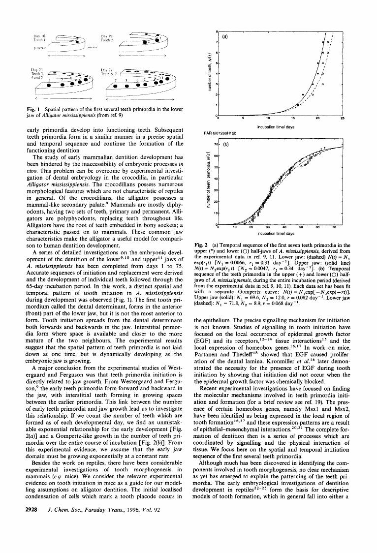
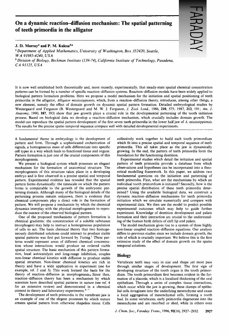
Fig. 1 jaw of Alligator mississippiensis (from ref. 9)
Spatial pattern of the first several teeth primordia in the lower
6 - v
2 g 5 - 0
c 4 - c
z 3- 5 2 -
1 -
early primordia develop into functioning teeth. Subsequent teeth primordia form in a similar manner in a precise spatial and temporal sequence and continue the formation of the functioning dentition.
The study of early mammalian dentition development has been hindered by the inaccessibility of embryonic processes in viuo. This problem can be overcome by experimental investi- gation of dental embryology in the crocodilia, in particular Alligator mississippiensis. The crocodilians possess numerous morphological features which are not characteristic of reptiles in general. Of the crocodilians, the alligator possesses a mammal-like secondary palate.* Mammals are mostly diphy- odonts, having two sets of teeth, primary and permanent. Alli- gators are polyphyodonts, replacing teeth throughout life. Alligators have the root of teeth embedded in bony sockets; a characteristic passed on to mammals. These common jaw characteristics make the alligator a useful model for compari- son to human dentition development.
A series of detailed investigations on the embryonic devel- opment of the dentition of the 1 0 w e r ~ ~ ' ~ and upper" jaws of A. mississippiensis has been completed from days 1 to 75. Accurate sequences of initiation and replacement were derived and the development of individual teeth followed through the 65-day incubation period. In this work, a distinct spatial and temporal pattern of tooth intiation in A . mississippiensis during development was observed (Fig. 1). The first tooth pri- mordium called the dental determinant, forms in the anterior (front) part of the lower jaw, but it is not the most anterior to form. Tooth initiation spreads from the dental determinant both forwards and backwards in the jaw. Interstitial primor- dia form where space is available and closer to the more mature of the two neighbours. The experimental results suggest that the spatial pattern of teeth primordia is not laid down at one time, but is dynamically developing as the embryonic jaw is growing.
A major conclusion from the experimental studies of West- ergaard and Ferguson was that teeth primordia initiation is directly related to jaw growth. From Westergaard and Fergu- son,g the early teeth primordia form forward and backward in the jaw, with interstitial teeth forming in growing spaces between the earlier primordia. This link between the number of early teeth primordia and jaw growth lead us to investigate this relationship. If we count the number of teeth which are formed as of each developmental day, we find an unmistak- able exponential relationship for the early development [Fig. 2(a)] and a Gompertz-like growth in the number of teeth pri- mordia over the entire course of incubation [Fig. 2(b)]. From this experimental evidence, we assume that the early jaw domain must be growing exponentially at a constant rate.
Besides the work on reptiles, there have been considerable experimental investigations of tooth morphogenesis in mammals (e.g. mice). We consider the relevant experimental evidence on tooth initiation in mice as a guide for our model- ling assumptions on alligator dentition. The initial localised condensation of cells which mark a tooth placode occurs in
0 5 10 15 20 25
incubation time/ days FAR 6/01286H/ 2b
incubation time/ days
Fig. 2 (a) Temporal sequence of the first seven teeth primordia in the upper (*) and lower (0) half-jaws of A. mississippiensis, derived from the experimental data in ref. 9, 11. Lower jaw: (dashed) N(t) = N , exp(r,t) [ N , = 0.0066, rl = 0.31 day-']. Upper jaw: (solid line) N(t ) = N,exp(r, t) [N, = 0.0047, r2 = 0.34 day-']. (b) Temporal sequence of the teeth primordia in the upper (+) and lower (0) half- jaws of A. mississippiensis, during the entire incubation period (derived from the experimental data in ref. 9, 10, 11). Each data set has been fit with a separate Gompertz curve: N ( t ) = Nlexp[ -N,exp(-rt)]. Upper jaw (solid): N , = 69.6, N , = 12.0, r = 0.082 day-'. Lower jaw (dashed): N , = 71.8, N , = 8.9, r = 0.068 day-'.
the epithelium. The precise signalling mechanism for initiation is not known. Studies of signalling in tooth initiation have focused on the local occurrence of epidermal growth factor (EGF) and its receptors,' 2-14 tissue interaction^'^ and the local expression of homeobox genes.I6.l7 In work on mice, Partanen and Thesleff l8 showed that EGF caused prolifer- ation of the dental lamina. Kronmiller et al.I4 later demon- strated the necessity for the presence of EGF during tooth initiation by showing that initiation did not occur when the the epidermal growth factor was chemically blocked.
Recent experimental investigations have focused on finding the molecular mechanisms involved in teeth primordia initi- ation and formation (for a brief review see ref. 19). The pres- ence of certain homeobox genes, namely Msxl and Msx2, have been identified as being expressed in the local region of tooth and these expression patterns are a result of epithelial-mesenchymal interactions.20v21 The complete for- mation of dentition then is a series of processes which are coordinated by signalling and the physical interaction of tissue. We focus here on the spatial and temporal intitiation sequence of the first several teeth primordia.
Although much has been discovered in identifying the com- ponents involved in tooth morphogenesis, no clear mechanism as yet has emerged to explain the patterning of the teeth pri- mordia. The early embryological investigations of dentition development in form the basis for descriptive models of tooth formation, which in general fall into either a
2928 J . Chem. SOC., Faraday Trans., 1996, Vol. 92
prepattern or dynamic model category. Prepattern models rely on the pattern being imposed from an external source, while in dynamic models the pattern arises as a result of growth of the system. E d m ~ n d ~ ~ ? ~ ' proposed the 'Zahnreihe' theory of tooth initiation where a chemical stimulus passes through the jaw, initiating prepatterned tooth sites. Osborn2* contradicted this theory and presented his own descriptive clone m0de1~'*~' in which teeth primordia are initiated by the dynamics of the growing clone. The experimental results of Westergaard and Fergusong-' ' confirm the inadequacies of the Zahnreihe theory and have also led to a rejection"." of the clone model based on the criticism that new teeth do not develop in the sequence suggested by the growing clone. The first quantitative model for tooth initiation was developed by Sneyd et aL31 in the form of a mechanochemical model which describes how the mechanical movements of cells and related tissues could create the structure and form of the tooth pri- mordium. This model, although it only produces the first tooth primordium, is useful in showing the necessity to incorporate the growth of the jaw domain. Clearly, any pro- posed model mechanism for tooth initiation must be capable of reproducing the spatial and temporal sequence of teeth pri- mordia in the alligator from the experimental data.
Model mechanism
Here, we propose a new dynamic mechanism for the initiation and spatial patterning of the teeth primordia using a mathe- matical model which is a quantitative realisation of the experi- mental work of Westergaard and Ferguson.'-' ' We investigate whether certain components are sufficient to gener- ate the observed spatio temporal sequence of teeth primordia. We show how this linking of a patterning mechanism and the physical jaw growth is essential to produce the observed spatio temporal sequence for the first seven teeth primordia in the alligator. Subsequent primordia development is compli- cated by tissue interaction and placode resorption and is not addressed here.
Our aim is to show that the proposed mechanism for the initiation and spatial positioning of the teeth primordia, which we now construct, is sufficient to explain the pattern of tooth sites in A . mississippiensis. The interplay of chemical com- ponents leads us to suggest a chemical mechanism for the ini- tiation of the teeth primordia, where certain chemicals react and diffuse so that gradients in their concentrations develop. Turing' showed that diffusion can drive a stable equilibrium chemical system unstable to form a steady-state pattern and inspired the development of reactiondiffusion models to explain the chemical basis of morphogenesis (see for example, Murray4 for a review). Experimental evidence requires that the pattern of teeth primordia arises dynamically as a result of jaw growth and not as a result of a prepattern of tooth initi- ation sites. Thus, we consider a dynamic reaction-diffusion system, capable of forming pattern, which is mediated by an inhibitor related to the concentration of epidermal growth factor, EGF. Based on the experiments of Kronmiller et ~ 1 . ' ~ we assume the existence of an inhibitory substance whose con- centration decreases as the concentration of EGF increases, and vice-versa. Novel about our approach is the incorporation of the physical growth of the domain, which is dictated by the experimental evidence of jaw growth.'-' '
Based on the biological data,32 we consider the patterning of the teeth primordia as an epithelial process in the half jaw of A. mississippiensis. We consider the half jaw as one- dimensional along the posterior-anterior axis from the back to the front of the jaw. Let x represent the length along this axis. A major conclusion from the experimental work by Westergaard and Fergu~on,~- ' ' is that teeth primordia forma- tion is directly related to jaw growth. If we count the number of teeth primordia which are formed as of each developmental
day, we find an unmistakable exponential relationship for the early development (Fig. 2), and a Gompertz-like growth in the number of teeth primordia over the entire course of develop- ment. This forms an integral part of our model mechanism. So, based on the experimental data which reflects jaw growth, we reasonably consider that the length of the jaw, L(t), grows at a constant strain rate r, according to
L(t) = Lo exp(rt) (1) The effect of this growth is to dilute the chemical concentra- tions. Estimates for Lo and r are obtained from the experi- mental data in Fig. 2.
For the basis of the patterning mechanism, we take by way of example a simple reactiondiffusion system, namely the Schnakenberg mechanism33 with a substrate chemical and an activator chemical, where u and u represent their respective concentrations in space and time. This is similar to the system presented by Gierer and M e i ~ ~ h a r d t . ~ ~ Incorporating jaw growth (1) into the model, the mathematical equations of the reaction-diffusion system expressed on a non-dimensionalised domain 0 d x d 1, in a reference frame fixed to the growing domain, are
rate of change = reaction kinetics in substrate and bifurcation
concentration 'parameter', c(x, t )
dilution diffusion due to jaw
growth (2)
a v at - = Y[b - U ~ V ]
rate of change = reaction kinetics in activator
concentration
dilution diffuse due to jaw
growth (3) where d is the diffusion coefficient ratio, h and b are non- dimensional parameters involved in the reaction kinetics and the exp( - 2rt) factor follows from the transformation to the fixed frame. For certain ranges of parameter values and a large enough domain size, the reaction4iffusion system (2)- (3) is capable of producing spatial patterns in u and v. We introduce a bifurcation parameter to spatial pattern, the inhibitor c(x, t) (a function of space and time). The conserva- tion equation for the inhibitor is
ac
at = -6c -
rate of change = degradation in inhibitor
a Z c
ax2 - rc + p exp(-2rt) -
dilution diffusion due to jaw
growth (4)
where p is the diffusion coefficient and 6 is the first-order kinetics degradation constant. We choose the boundary con-
J . Chem. SOC., Faraday Trans., 1996, Vol. 92 2929
ditions for u and 0 to be no-flux, while for c there is no flux only at the anterior end, x = 1. We assume there is a decaying source of c at the posterior end, whose primary purpose as bifurcation parameter is simply to initiate the patterning process.
The patterning mechanism for placode initiation is pri- marily controlled by the inhibitor and the length of the jaw domain. When the inhibitor, c, is above a certain threshold, pattern formation is inhibited. For c below this threshold, the pattern formation mechanism is switched on and spatial pattern is laid down in u and u, but only when the subthresh- old portion of the domain is large enough. Eventually, the substrate u crosses an upper threshold and stimulates the spe- cific area of the epithelium to initiate a placode, setting the spatial position of tooth primordium 1; the so-called dental determinant. Experimental evidenceg*' suggests that the dental determinant (and each subsequent tooth primordium) then becomes a source of inhibitor factor (simulating an inhi- bition zone). So, we assume that when u rises above a certain threshold, at the location of the peak in u, a new source of the inhibitor is initiated. Subsequent teeth primordia are laid down in a likewise manner.
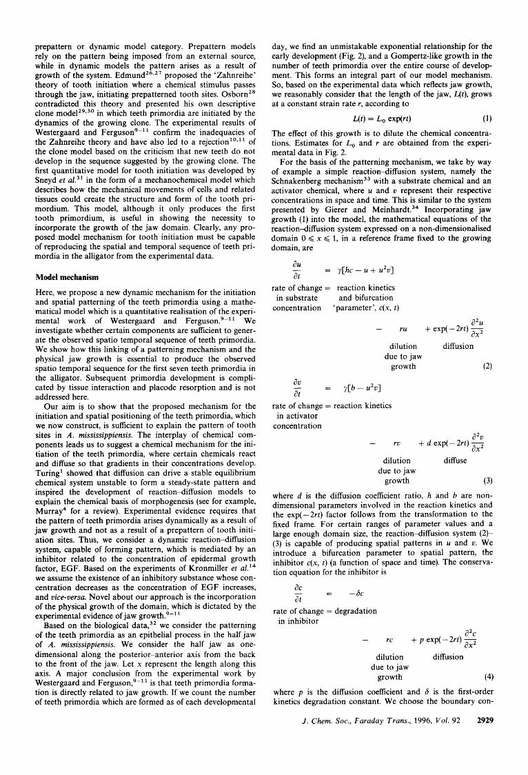
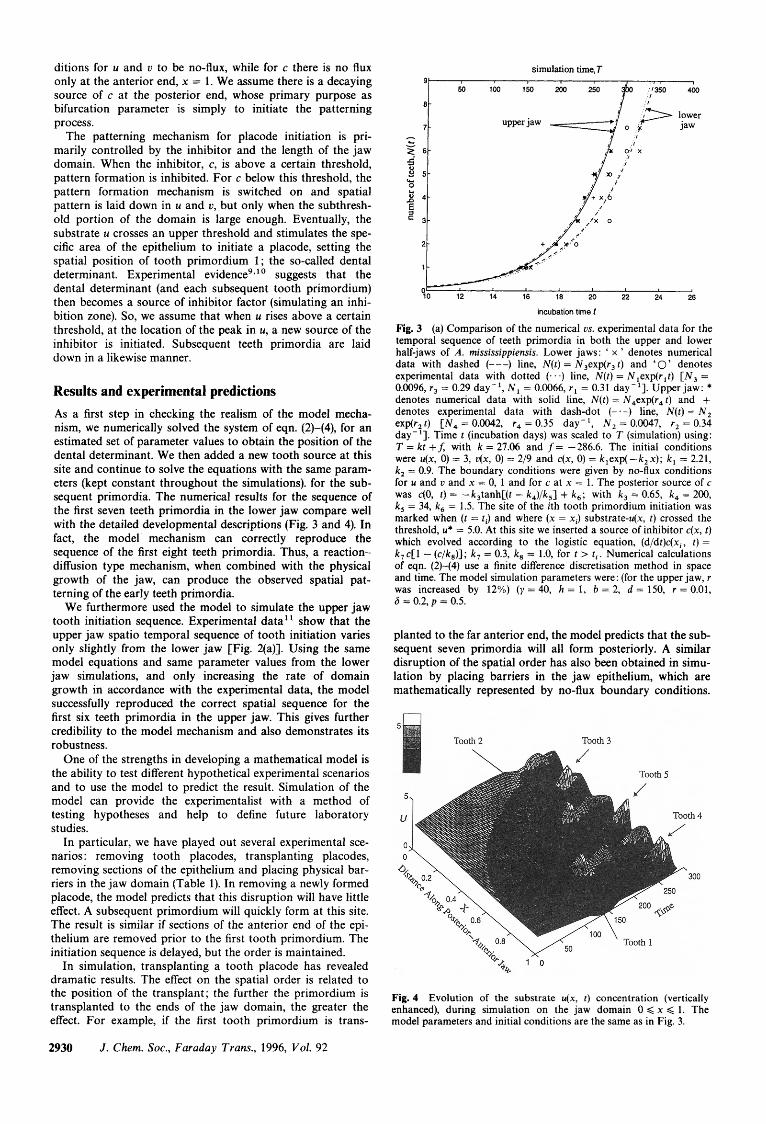
Results and experimental predictions As a first step in checking the realism of the model mecha- nism, we numerically solved the system of eqn. (2)-(4), for an estimated set of parameter values to obtain the position of the dental determinant. We then added a new tooth source at this site and continue to solve the equations with the same param- eters (kept constant throughout the simulations). for the sub- sequent primordia. The numerical results for the sequence of the first seven teeth primordia in the lower jaw compare well with the detailed developmental descriptions (Fig. 3 and 4). In fact, the model mechanism can correctly reproduce the sequence of the first eight teeth primordia. Thus, a reaction- diffusion type mechanism, when combined with the physical growth of the jaw, can produce the observed spatial pat- terning of the early teeth primordia.
We furthermore used the model to simulate the upper jaw tooth initiation sequence. Experimental data' ' show that the upper jaw spatio temporal sequence of tooth initiation varies only slightly from the lower jaw [Fig. 2(a)]. Using the same model equations and same parameter values from the lower jaw simulations, and only increasing the rate of domain growth in accordance with the experimental data, the model successfully reproduced the correct spatial sequence for the first six teeth primordia in the upper jaw. This gives further credibility to the model mechanism and also demonstrates its robustness.
One of the strengths in developing a mathematical model is the ability to test different hypothetical experimental scenarios and to use the model to predict the result. Simulation of the model can provide the experimentalist with a method of testing hypotheses and help to define future laboratory studies.
In particular, we have played out several experimental sce- narios : removing tooth placodes, transplanting placodes, removing sections of the epithelium and placing physical bar- riers in the jaw domain (Table 1). In removing a newly formed placode, the model predicts that this disruption will have little effect. A subsequent primordium will quickly form at this site. The result is similar if sections of the anterior end of the epi- thelium are removed prior to the first tooth primordium. The initiation sequence is delayed, but the order is maintained.
In simulation, transplanting a tooth placode has revealed dramatic results. The effect on the spatial order is related to the position of the transplant; the further the primordium is transplanted to the ends of the jaw domain, the greater the effect. For example, if the first tooth primordium is trans-
simulation time, T 9 1
50 100 150 200 250 '350 400
A - I
ri
2/
incubation time 1
Fig. 3 (a) Comparison of the numerical us. experimental data for the temporal sequence of teeth primordia in both the upper and lower half-jaws of A. mississippiensis. Lower jaws: ' x ' denotes numerical data with dashed (---) line, N(t) = N3exp(r3 t ) and '0' denotes experimental data with dotted (. . .) line, N(t) = Nlexp(rlt) [ N , = 0.0096, r3 = 0.29 day-', N , = 0.0066, r l = 0.31 day-']. Upper jaw: * denotes numerical data with solid line, N ( t ) = N4exp(r4t) and + denotes experimental data with dash-dot (-.-) line, N ( t ) = N , exp(r, t ) [ N , = 0.0042, r4 = 0.35 day-', N , = 0.0047, r, = 0.34 day-']. Time t (incubation days) was scaled to T (simulation) using: T = kt +f, with k = 27.06 and f= -286.6. The initial conditions were u(x, 0) = 3, u(x, 0) = 2/9 and c(x, 0) = k,exp( - k , x); k , = 2.21, k, = 0.9. The boundary conditions were given by no-flux conditions for u and u and x = 0, 1 and for c at x = 1. The posterior source of c was c(0, t ) = -k,tanh[(t - k4)/k5] + k , ; with k , = 0.65, k , = 200, k , = 34, k , = 1.5. The site of the ith tooth primordium initiation was marked when (t = ti) and where (x = x i ) substrate-u(x, t ) crossed the threshold, u* = 5.0. At this site we inserted a source of inhibitor c(x, t ) which evolved according to the logistic equation, (d/dt)c(xi, t ) = k , c[1 - (c/k8)]; k, = 0.3, k, = 1.0, for t > t i . Numerical calculations of eqn. (2)-(4) use a finite difference discretisation method in space and time. The model simulation parameters were: (for the upper jaw, r was increased by 12%) ( y = 40, h = 1, b = 2, d = 150, r = 0.01, 6 = 0.2, p = 0.5.
planted to the far anterior end, the model predicts that the sub- sequent seven primordia will all form posteriorly. A similar disruption of the spatial order has also been obtained in simu- lation by placing barriers in the jaw epithelium, which are mathematically represented by no-flux boundary conditions.
Fig. 4 Evolution of the substrate u(x, t ) concentration (vertically enhanced), during simulation on the jaw domain 0 < x < 1. The model parameters and initial conditions are the same as in Fig. 3.
2930 J . Chem. SOC., Faraday Trans., 1996, Vol. 92
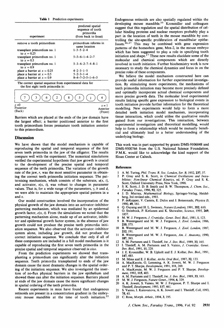
Table 1 Prediction experiments
experiment
predicted spatial sequence of tooth
primordia (from back to front)
remove a tooth primordium
transplant primordium no. 1
transplant primordium no. 1
transplant primordium no. 1
place a barrier at x = 0.25 place a barrier at x = 0.5 place a barrier at x = 0.9
to x = 0.25
to x = 0.5
to x = 0.9
primordium reforms in same location
1-3-5-2-4
5-3-6-1-4-2-7
5-3-6-2-7-4-8-1
4-2-3- 1-5 5-2-3- 1-4 8-4-7-2-5-1-6-3
The correct spatial sequence from experimental data for the first eight teeth primordia is:
x =o Posterior (Back)
x = I Anterior
(Front)
Barriers which are placed at the ends of the jaw domain have the largest effect; a barrier positioned anterior to the first tooth primordium forces premature tooth initiation anterior to this primordium.
Discussion We have shown that the model mechanism is capable of reproducing the spatial and temporal sequence of the first seven teeth primordia in the jaw of the alligator. The results compare well with the experiment. The numerical simulations verified the experimental hypothesis that jaw growth is crucial to the development of the precise spatial and temporal sequence of the teeth primordia. The variation of the growth rate of the jaw, r, was the most sensitive parameter in obtain- ing the correct teeth primordia initiation sequence. The pat- ternising mechanism, which consists of the substrate, u(x, t), and activator, v(x, t), was robust to changes in parameter values. That is, for a wide range of the parameters, y, b and d, we were able to maintain the appropriate spatial pattern in u and u.
Our model construction involved the incorporation of the physical growth of the jaw domain into an activator-inhibitor patterning mechanism, which was mediated by an epidermal growth factor, c(x, t). From the simulations we noted that the patterning mechanism alone, made up of an activator, inhibi- tor and epidermal growth factor system, in the absence of jaw growth could not produce the precise teeth primordia initi- ation sequence. We also observed that the activator-inhibitor system alone, including jaw growth, did not produce the correct initiation sequence. We conclude that only if all of these components are included in a full model mechanism is it capable of reproducing the first seven teeth primordia in the precise spatial and temporal sequence observed in viuo.
From the prediction experiments we showed that trans- planting a primordium can significantly alter the initiation sequence. Teeth primordia transplanted to ends of the jaw domain cause the most dramatic changes in the spatial order- ing of the initiation sequence. We also investigated the inser- tion of no-flux physical barriers in the jaw epithelium and showed these can alter the initiation sequence. Barriers placed at ends of the jaw domain cause the more significant changes in spatial ordering of the teeth primordia.
Recent experiments in mice have found that endogenous retinoids are present in a concentration gradient in the embry- onic mouse mandible at the time of tooth init iati~n.~’
Endogenous retinoids are also spatially regulated within the developing mouse mandible.35 Kronmiller and colleagues suggest that this regulation and the spatial distribution of cel- lular binding proteins and nuclear receptors probably play a part in the location of teeth in the mouse mandible by con- trolling the site-specific proliferation of mandibular epithe- li~um.’~*’’ This may be combined with gene expression patterns of the homeobox gene, Msx-2, in the mouse embryo which has been suggested to play a role in specifying tooth initiation and shape.I7 These new results elucidate some of the molecular and chemical components which are directly involved in tooth initiation. Further biochemistry work is now necessary to study the chemical dynamics and focus in on the precise roles of these components.
We believe the model mechanism constructed here can provide useful information for further experimental investiga- tion. By stimulating more experiments, the mechanisms of teeth primordia initiation may become more precisely defined and optimally incorporate actual chemical components and more precise growth data. The molecular level experimental results linking specific gene expression to biological events in tooth initiation provide further information for the theoretical modelling. New experiments would help to form a more detailed teeth initiation model mechanism, incorporating tissue interaction, which could utilise the qualitative results gained from our investigations. This interaction, between experimental investigation and theoretical modelling, would help to form a relationship which would be mutually benefi- cial and ultimately lead to a better understanding of the underlying biology.
This work was in part supported by grants DMS-9106848 and DMS-9500766 from the U.S. National Science Foundation. P.M.K. would like to acknowledge the kind support of the Sloan Center at Caltech.
References 1 2
3
4
5
6 7
8 9
10
11
12 13
14
15 16
17
18 19 20
21
22
A. M. Turing, Phil. Trans. R . SOC. London, Ser. B, 1952,237,37. P. Gray and S. K. Scott, in Chemical Oscillations and Insta- bilities: Non-linear Chemical Kinetics, ed. P. Gray and S. K. Scott, Oxford University Press, New York, 1990. S. K. Scott, J. D. B. Smith and B. W. Thompson, J . Chem. SOC., Faraday Trans., 1996,92,325. J. D. Murray, Mathematical Biology, Springer-Verlag, Heidel- berg, 3rd revised edn., 1993. P. deKepper, V. Castets, E. Dulos and J. Boissonade, Physica D, 1990,49, 16 1. Q. Ouyang and H. L. Swinney, Nature (London), 1991,352,610. 0. Steinbock, P. Ketlunen and K. Showalter, Science, 1995, 269, 1857. M. W. J. Ferguson, J . Craniofac. Genet. Devl. Biol., 1981, 1, 123. B. Westergaard and M. W. J. Ferguson, J . 2001. London, 1986, 210, 575. B. Westergaard and M. W. J. Ferguson, J . Zool. London, 1987, 212, 191. B. Westergaard and M. W. J. Ferguson, Am. J . Anatomy, 1990, 187, 393. A. M. Partanen and I. Thesleff, Int. J . Dev. Biol., 1989,33, 165. I. Thesleff, A. M. Partanen and S. Vainio, J . Craniofac. Genet. Dev. Biol., 1991, 11, 229. J. E. Kronmiller, W. B. Upholt and E. J. Kollar, Dev. Bzol., 1991, 147,485. M. Mina and E. J. Kollar, Archs. Oral Biol., 1987,32, 123. A. MacKenzie, G. Lemming, A. K. Jowett, M. W. J. Ferguson and P. T. Sharpe, Development, 1991,111,269. A. MacKenzie, M. W. J. Ferguson and P. T. Sharpe, Develop- ment, 1992, 115, 403. A. M. Partanen and I. Thesleff, Znt. J . Dev. Biol., 1989,33, 165. M. W. J. Ferguson, Nature Genet., 1994,6, 329. A. K. Jowett, S. Vainio, M. W. J. Ferguson, P. T. Sharpe and I. Thesleff, Development, 1993, 117,461. S . Vainio, I. Karavanova, A. K. Jowett and I. Thesleff, Cell, 1993, 75,45. C. Rose, Morph. Arbeit., 1894, 3, 195.
J . Chem. SOC., Faraday Trans., 1996, Vol. 92 2931
23 24 25 26 27
28 29
30 31
M. W. Woerdeman, Arch. Mikrosk. Anat., 1919,92, 104. M. W. Woerdeman, Arch. Mikrosk. Anat., 1921,95,265. J. W. Osborn, Proc. R. SOC. London, Ser. B., 1971,179,261. A. G. Edmund, R. SOC. Can. Studia Varia., 1960,445. A. G. Edmund, in Biology of the Reptilia. I . Morphology A., ed. C. Gans, A. d’A. Bellairs and T. S. Parsons, Academic Press, London, 1969, p. 115. J. W. Osborn, Nature (London), 1970,225,343. J. W. Osborn, in Development, Function and Evolution of Teeth, ed. P. M. Butler and K. A. Joysey, Academic Press, New York, 1978, p. 171. J. W. Osborn, J. Theor. Biol., 1993, 165,429. J. Sneyd, A. Atri, M. W. J. Ferguson, M. A. Lewis, W. Seward and J. D. Murray, J. Theor. Biol., 1993, 165, 633.
32 E. J. Kollar and M. Mina, J . Craniofac. Genet. Dev. Biol., 1991, 11,223.
33 J. Schnakenberg, J. Theor. Biol., 1979,81,389. 34 A. Gierer and H. Meinhardt, Kybernetik, 1972,12,30. 35 J. E. Kronmiller and C. S. Beeman, Arch. Oral Biol., 1994, 39,
1071. 36 J. E. Kronmiller, W. B. Upholt and E. J. Kollar, Arch. Oral Biol.,
1992,37, 129. 37 J. E. Kronmiller, C. S. Beeman, K. Kweicien and T. Rollins, Arch.
Oral Biol., 1994, A39,839.
Paper 6/01286H ; Received 22nd February, 1996
2932 J . Chem. SOC., Faraday Trans., 1996, Vol. 92