omalizumab for severe asthma: efficacy beyond the atopic patient?
TRANSCRIPT
CURRENT OPINION
Omalizumab for Severe Asthma: Efficacy Beyond the AtopicPatient?
Christian Domingo
Published online: 2 April 2014
� Springer International Publishing Switzerland 2014
Abstract Several years ago, omalizumab became com-
mercially available for the treatment of severe asthma. It
remains the only monoclonal antibody to be marketed for
this purpose. Since then, many studies have been published
endorsing its efficacy and effectiveness. Concomitantly,
evidence of an overlap between atopic and non-atopic
severe asthma has emerged. However, there also appears to
be some disagreement regarding the value of omalizumab
in the management of non-atopic disease, as some studies
have failed to show any benefit in these patients. The recent
literature has also sought to identify appropriate prognostic
biomarkers for the use of omalizumab, other than immu-
noglobulin (IgE) levels. This article briefly summarizes the
evolution of asthma treatment, the pathophysiology of the
condition, and the method of action of omalizumab. The
author describes the controlled and uncontrolled studies
(also named ‘‘real-life studies’’) published in adult and
pediatric populations in different countries and expresses
his view on the current place of the drug in the manage-
ment of severe allergic asthma. He offers a personal per-
spective on the recent evidence for the use of omalizumab
in non-atopic patients, highlighting the implications for
current clinical practice and the gaps in our knowledge.
The author justifies his belief that omalizumab is not only
an IgE-blocking drug and should be considered as a dis-
ease-modifying therapy because of its multiple effects on
different biologic pathways. Finally, some areas for future
research are indicated.
1 Brief History of Asthma Treatment
Asthma is a common, chronic, inflammatory disorder of
the airways. It is clinically characterized by bronchial
hyper-responsiveness, reversible airflow limitation, and
recurrent episodes of wheezing, shortness of breath, chest
tightness, and cough. Asthma is in fact a complex syn-
drome with many clinical and inflammatory phenotypes
[1–6]. However, it took a long time to reach this conclu-
sion, and the lack of definition regarding the disease has
slowed the development of appropriate therapeutic
approaches. Two major breakthroughs have significantly
improved the clinical management of asthma. The first one
dates from 1992, when the first international guidelines for
asthma treatment recommended the systematic prescription
of inhaled corticosteroids as standard therapy for patients
with persistent disease [7]. During the next 20 years,
repeated attempts were made to find the most appropriate
dose of inhaled corticosteroids for use at the different
asthma severity steps. Some other drugs that are helpful in
the management of inflammation such as leukotriene
modifiers and for persistent bronchial smooth cell fiber
constriction such as long-acting b-agonists were marketed,
but none made an essential contribution to conceptually
improving asthma treatment, which seemed to have
advanced as far as it could [8]. These drugs attempt to treat
the last step of the pathophysiologic pathway of asthma,
that is, inflammation. At the same time, conventional,
subcutaneous, allergen-specific immunotherapy continued
to market new products and tried to find alternative
administration routes [9–14].
C. Domingo (&)
Pulmonary Service, Hospital de Sabadell (Corporacio Sanitaria i
Universitaria Parc Taulı), Parc Taulı 1, 08208 Sabadell
(Barcelona), Spain
e-mail: [email protected]
C. Domingo
Departament of Medicine, Universitat Autonoma de Barcelona
(UAB), Barcelona, Spain
Drugs (2014) 74:521–533
DOI 10.1007/s40265-014-0203-y
The second breakthrough dates from the early 2000s,
with the advent of what were known as ‘‘biologic treat-
ments,’’ that is, treatments that try to block the proteins or
molecules originating in cells, which in turn trigger or
modulate the asthma cascade. When the aim is to block the
production of intracellular proteins we call it anti-sense
therapy, because the procedure comprises the administra-
tion of molecules that are short, single-stranded, nucleic
acids complementary to target messenger RNA (mRNA),
which bind to receptor mRNA with levels of affinity and
avidity that can far surpass those shown by traditional
drugs targeting protein receptors [15]. These molecules are
thus a chain of oligonucleotides arranged in the opposite
sense (anti-sense) of the RNA strand to be blocked. This
approach is still at a very early stage of development.
Monoclonal antibodies (mAbs) represent a form of
immunotherapy using passive immunity in which pre-
formed antibodies against a target antigen are injected into
the body. Because of their specificity, mAbs can efficiently
target an antigen on a cell of interest or in the serum and
block the binding of cytokines, immunoglobulins (Igs),
hormones, or proteins that promote certain unwanted
functions including inflammatory and immune responses.
Ultimately, both methods can attenuate the expression of
disease-associated genes [16].
2 Physiopathology of Allergic Asthma
The induction, maintenance, and progression of the
inflammatory and remodeling responses of asthma are
driven by complex interactions of adaptive (i.e., dendritic
cells, B cells, and activated T cells) and innate immune
responses (i.e., macrophages and neutrophils) with struc-
tural cells of the airways (i.e., epithelial cells, airway
smooth muscle cells, and myofibroblasts). These interac-
tions lead to the secretion of preformed and/or newly
synthesized mediators, IgE, cytokines, growth factors, and
chemokines, which results in distinct asthma phenotypes
[17]. Classically, allergic diseases are generally defined as
significant pathologic changes caused by excessive reac-
tions of the immune system to innocuous substances. When
an atopic individual is exposed to an antigen, the dendritic
cells (specialized macrophages located in the organism’s
epithelium) internalize it, process it, and present it to a T
lymphocyte, via the major histocompatibility complex. In
this process, the T lymphocyte develops a T-helper 2 (Th2)
or a T-helper 1 (Th1) profile. The relative amount of each
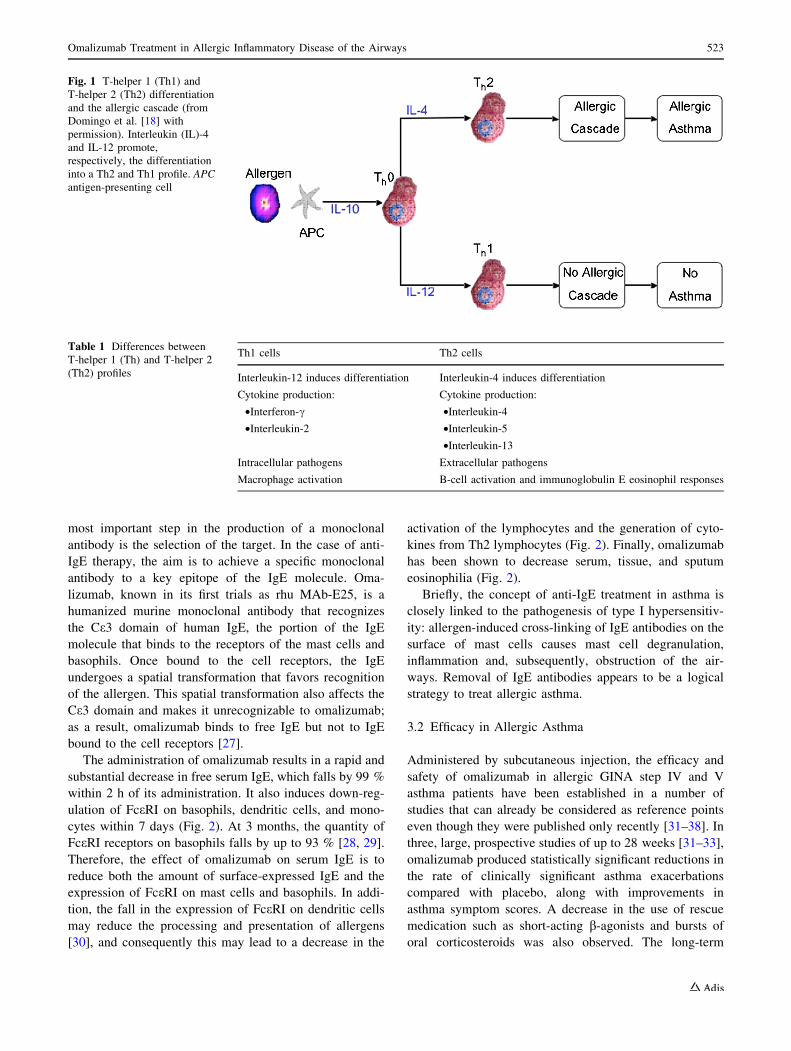
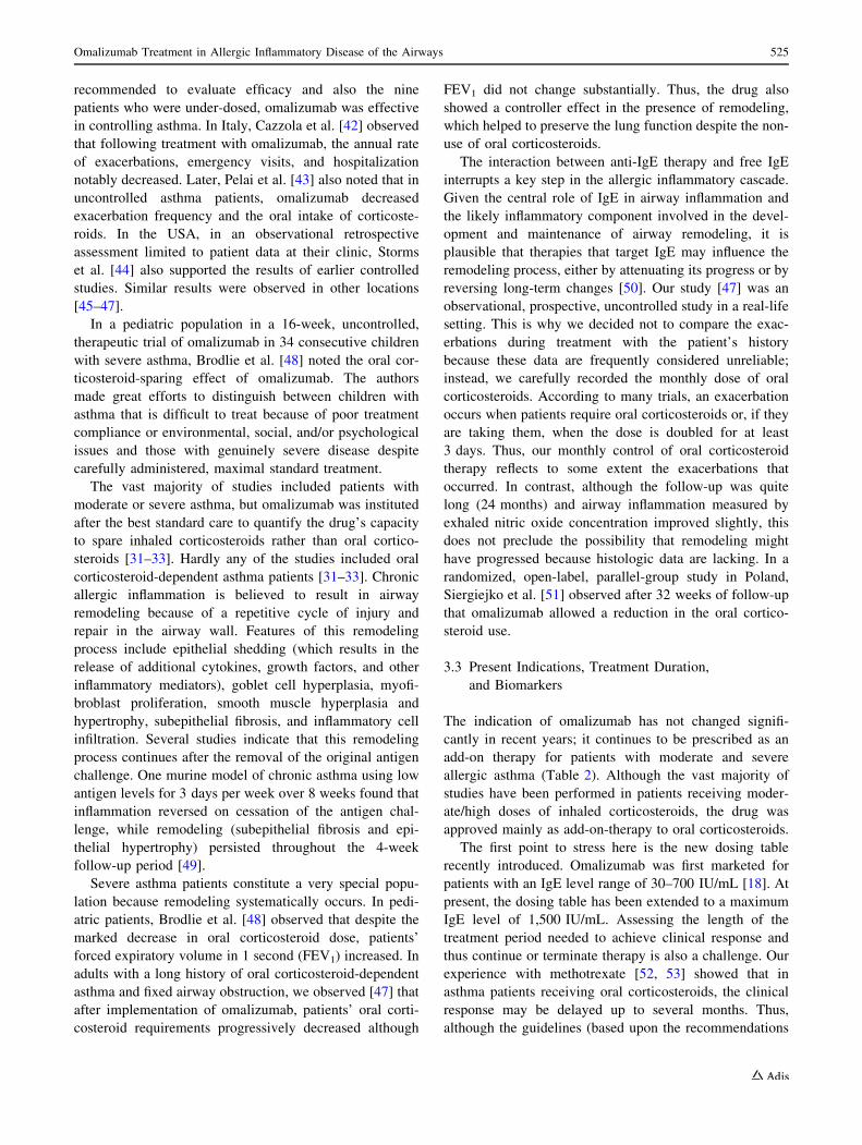
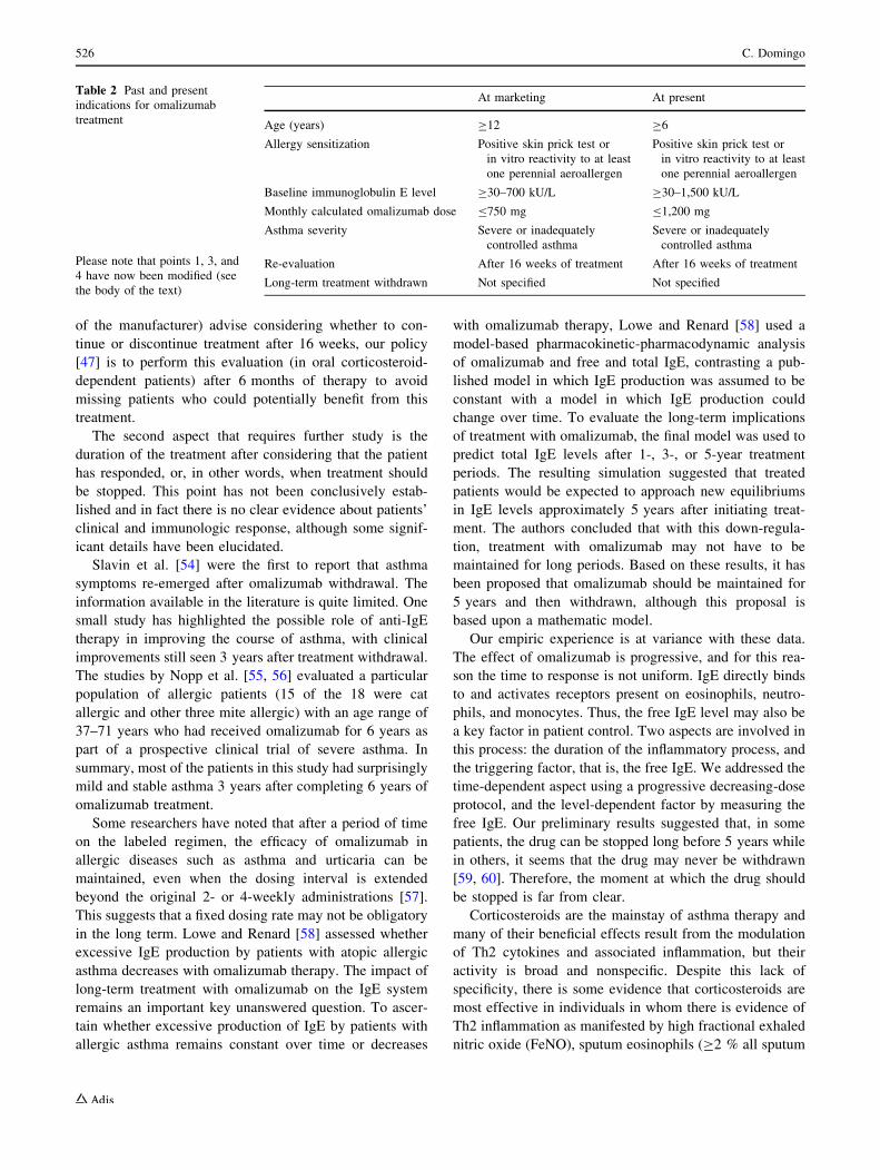
is both antigen and host dependent (Fig. 1) [18]. When the
antigen is an allergen, the lymphocyte differentiates into a
Th2 cell able to produce interleukin (IL)-4, which in turn
promotes the synthesis of IgE by the B lymphocytes
(Table 1). As well as regulating IgE, IL-4 stimulates the
migration and activation of the mast cells. Mast cells
contain in their membrane the high-affinity receptor for IgE
(FceRI). When this receptor binds to IgE, the molecule is
able to recognize the antigen. When two or more IgE
molecules bound to their receptor recognize the same
antigen molecule, they cause cross linking of the receptors,
a phenomenon that triggers a series of biochemical sig-
naling reactions that culminate in mast cell degranulation
[18] and the release of the immediate mediators of the
inflammation—histamine, leukotrienes, prostaglandins,
ILs, and cell growth factors. This phase lasts approximately
an hour. Later, a second phase may occur after 4–8 h of
exposure to the allergen. The chemotactic factors, IL-5, IL-
3, IL-13, and the cell growth factors released in the
immediate inflammation phase enhance eosinophil
recruitment. The eosinophils release IL-5, which in turn
enhances the recruitment of new eosinophils and perpetu-
ates the inflammation. This may lead to chronic allergic
inflammation in the case of continued exposure to the
allergen. On occasion, if the number of eosinophils and
mediators is very high, this process may persist without the
need for an allergenic stimulus [18, 19].
IgE plays a key role in the physiopathology of asthma
[20]. IgE binds to its high-affinity receptors, FceRI, on the
surface of mast cells and basophils via the Ce3 domain of
its Fc fragment. The correlation between the expression of
FceRI on the basophils and serum levels of IgE is well
established [21]. IgE itself [22] seems to up-regulate the
expression of FceRI on human basophils probably by
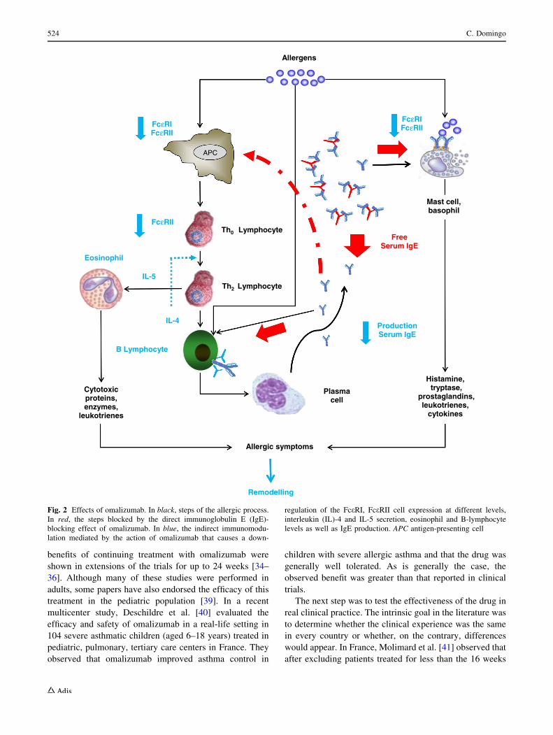
interacting with FceRI (Fig. 2).
Recent data have suggested that IgE has some additional
immunobiologic effects. This molecule may promote mast
cell survival through autocrine production of IL-6 [23]. Sur-
vival assays performed on cultures of human lung mast cells
provided further evidence that IgE and IL-6 contribute to the
pathogenesis of asthma, and it is speculated that anti-IgE
therapy may achieve its therapeutic effect through this
mechanism. IgE binds to dendritic cells and enhances allergen
uptake and presentation to T cells [24]. It has been reported
that dendritic cells in patients with mild atopic asthma bind
significantly more IgE than cells taken from healthy individ-
uals [25], and FceRI receptors are known to be up-regulated on
dendritic cells (as well as eosinophils, mast cells, and mac-
rophages) in patients with seasonal allergic rhinitis [26].
3 Omalizumab: The First Monoclonal Antibodies
Marketed for Asthma Treatment
3.1 Mechanism of Action
The monoclonal antibody omalizumab is an IgG of clonal
origin and is therefore specific for a single antigen. The
522 C. Domingo
most important step in the production of a monoclonal
antibody is the selection of the target. In the case of anti-
IgE therapy, the aim is to achieve a specific monoclonal
antibody to a key epitope of the IgE molecule. Oma-
lizumab, known in its first trials as rhu MAb-E25, is a
humanized murine monoclonal antibody that recognizes
the Ce3 domain of human IgE, the portion of the IgE
molecule that binds to the receptors of the mast cells and
basophils. Once bound to the cell receptors, the IgE
undergoes a spatial transformation that favors recognition
of the allergen. This spatial transformation also affects the
Ce3 domain and makes it unrecognizable to omalizumab;
as a result, omalizumab binds to free IgE but not to IgE
bound to the cell receptors [27].
The administration of omalizumab results in a rapid and
substantial decrease in free serum IgE, which falls by 99 %
within 2 h of its administration. It also induces down-reg-
ulation of FceRI on basophils, dendritic cells, and mono-
cytes within 7 days (Fig. 2). At 3 months, the quantity of
FceRI receptors on basophils falls by up to 93 % [28, 29].
Therefore, the effect of omalizumab on serum IgE is to
reduce both the amount of surface-expressed IgE and the
expression of FceRI on mast cells and basophils. In addi-
tion, the fall in the expression of FceRI on dendritic cells
may reduce the processing and presentation of allergens
[30], and consequently this may lead to a decrease in the
activation of the lymphocytes and the generation of cyto-
kines from Th2 lymphocytes (Fig. 2). Finally, omalizumab
has been shown to decrease serum, tissue, and sputum
eosinophilia (Fig. 2).
Briefly, the concept of anti-IgE treatment in asthma is
closely linked to the pathogenesis of type I hypersensitiv-
ity: allergen-induced cross-linking of IgE antibodies on the
surface of mast cells causes mast cell degranulation,
inflammation and, subsequently, obstruction of the air-
ways. Removal of IgE antibodies appears to be a logical
strategy to treat allergic asthma.
3.2 Efficacy in Allergic Asthma
Administered by subcutaneous injection, the efficacy and
safety of omalizumab in allergic GINA step IV and V
asthma patients have been established in a number of
studies that can already be considered as reference points
even though they were published only recently [31–38]. In
three, large, prospective studies of up to 28 weeks [31–33],
omalizumab produced statistically significant reductions in
the rate of clinically significant asthma exacerbations
compared with placebo, along with improvements in
asthma symptom scores. A decrease in the use of rescue
medication such as short-acting b-agonists and bursts of
oral corticosteroids was also observed. The long-term
Fig. 1 T-helper 1 (Th1) and
T-helper 2 (Th2) differentiation
and the allergic cascade (from
Domingo et al. [18] with
permission). Interleukin (IL)-4
and IL-12 promote,
respectively, the differentiation
into a Th2 and Th1 profile. APC
antigen-presenting cell
Table 1 Differences between
T-helper 1 (Th) and T-helper 2
(Th2) profiles
Th1 cells Th2 cells
Interleukin-12 induces differentiation Interleukin-4 induces differentiation
Cytokine production: Cytokine production:
•Interferon-c •Interleukin-4
•Interleukin-2 •Interleukin-5
•Interleukin-13
Intracellular pathogens Extracellular pathogens
Macrophage activation B-cell activation and immunoglobulin E eosinophil responses
Omalizumab Treatment in Allergic Inflammatory Disease of the Airways 523
benefits of continuing treatment with omalizumab were
shown in extensions of the trials for up to 24 weeks [34–
36]. Although many of these studies were performed in
adults, some papers have also endorsed the efficacy of this
treatment in the pediatric population [39]. In a recent
multicenter study, Deschildre et al. [40] evaluated the
efficacy and safety of omalizumab in a real-life setting in
104 severe asthmatic children (aged 6–18 years) treated in
pediatric, pulmonary, tertiary care centers in France. They
observed that omalizumab improved asthma control in
children with severe allergic asthma and that the drug was
generally well tolerated. As is generally the case, the
observed benefit was greater than that reported in clinical
trials.
The next step was to test the effectiveness of the drug in
real clinical practice. The intrinsic goal in the literature was
to determine whether the clinical experience was the same
in every country or whether, on the contrary, differences
would appear. In France, Molimard et al. [41] observed that
after excluding patients treated for less than the 16 weeks
Histamine, tryptase,
prostaglandins,leukotrienes,
cytokines
Allergic symptoms
FreeSerum IgE
Plasma cell
APC
Th0 Lymphocyte
FcεεRIFcεRII
IL-4
IL-5
Cytotoxicproteins,enzymes,
leukotrienes
Allergens
Th2 Lymphocyte
ProductionSerum IgE
FcεRIFcεRII
FcεRII
Eosinophil
B Lymphocyte
Remodelling
Mast cell, basophil
Fig. 2 Effects of omalizumab. In black, steps of the allergic process.
In red, the steps blocked by the direct immunoglobulin E (IgE)-
blocking effect of omalizumab. In blue, the indirect immunomodu-
lation mediated by the action of omalizumab that causes a down-
regulation of the FceRI, FceRII cell expression at different levels,
interleukin (IL)-4 and IL-5 secretion, eosinophil and B-lymphocyte
levels as well as IgE production. APC antigen-presenting cell
524 C. Domingo
recommended to evaluate efficacy and also the nine
patients who were under-dosed, omalizumab was effective
in controlling asthma. In Italy, Cazzola et al. [42] observed
that following treatment with omalizumab, the annual rate
of exacerbations, emergency visits, and hospitalization
notably decreased. Later, Pelai et al. [43] also noted that in
uncontrolled asthma patients, omalizumab decreased
exacerbation frequency and the oral intake of corticoste-
roids. In the USA, in an observational retrospective
assessment limited to patient data at their clinic, Storms
et al. [44] also supported the results of earlier controlled
studies. Similar results were observed in other locations
[45–47].
In a pediatric population in a 16-week, uncontrolled,
therapeutic trial of omalizumab in 34 consecutive children
with severe asthma, Brodlie et al. [48] noted the oral cor-
ticosteroid-sparing effect of omalizumab. The authors
made great efforts to distinguish between children with
asthma that is difficult to treat because of poor treatment
compliance or environmental, social, and/or psychological
issues and those with genuinely severe disease despite
carefully administered, maximal standard treatment.
The vast majority of studies included patients with
moderate or severe asthma, but omalizumab was instituted
after the best standard care to quantify the drug’s capacity
to spare inhaled corticosteroids rather than oral cortico-
steroids [31–33]. Hardly any of the studies included oral
corticosteroid-dependent asthma patients [31–33]. Chronic
allergic inflammation is believed to result in airway
remodeling because of a repetitive cycle of injury and
repair in the airway wall. Features of this remodeling
process include epithelial shedding (which results in the
release of additional cytokines, growth factors, and other
inflammatory mediators), goblet cell hyperplasia, myofi-
broblast proliferation, smooth muscle hyperplasia and
hypertrophy, subepithelial fibrosis, and inflammatory cell
infiltration. Several studies indicate that this remodeling
process continues after the removal of the original antigen
challenge. One murine model of chronic asthma using low
antigen levels for 3 days per week over 8 weeks found that
inflammation reversed on cessation of the antigen chal-
lenge, while remodeling (subepithelial fibrosis and epi-
thelial hypertrophy) persisted throughout the 4-week
follow-up period [49].
Severe asthma patients constitute a very special popu-
lation because remodeling systematically occurs. In pedi-
atric patients, Brodlie et al. [48] observed that despite the
marked decrease in oral corticosteroid dose, patients’
forced expiratory volume in 1 second (FEV1) increased. In
adults with a long history of oral corticosteroid-dependent
asthma and fixed airway obstruction, we observed [47] that
after implementation of omalizumab, patients’ oral corti-
costeroid requirements progressively decreased although
FEV1 did not change substantially. Thus, the drug also
showed a controller effect in the presence of remodeling,
which helped to preserve the lung function despite the non-
use of oral corticosteroids.
The interaction between anti-IgE therapy and free IgE
interrupts a key step in the allergic inflammatory cascade.
Given the central role of IgE in airway inflammation and
the likely inflammatory component involved in the devel-
opment and maintenance of airway remodeling, it is
plausible that therapies that target IgE may influence the
remodeling process, either by attenuating its progress or by
reversing long-term changes [50]. Our study [47] was an
observational, prospective, uncontrolled study in a real-life
setting. This is why we decided not to compare the exac-
erbations during treatment with the patient’s history
because these data are frequently considered unreliable;
instead, we carefully recorded the monthly dose of oral
corticosteroids. According to many trials, an exacerbation
occurs when patients require oral corticosteroids or, if they
are taking them, when the dose is doubled for at least
3 days. Thus, our monthly control of oral corticosteroid
therapy reflects to some extent the exacerbations that
occurred. In contrast, although the follow-up was quite
long (24 months) and airway inflammation measured by
exhaled nitric oxide concentration improved slightly, this
does not preclude the possibility that remodeling might
have progressed because histologic data are lacking. In a
randomized, open-label, parallel-group study in Poland,
Siergiejko et al. [51] observed after 32 weeks of follow-up
that omalizumab allowed a reduction in the oral cortico-
steroid use.
3.3 Present Indications, Treatment Duration,
and Biomarkers
The indication of omalizumab has not changed signifi-
cantly in recent years; it continues to be prescribed as an
add-on therapy for patients with moderate and severe
allergic asthma (Table 2). Although the vast majority of
studies have been performed in patients receiving moder-
ate/high doses of inhaled corticosteroids, the drug was
approved mainly as add-on-therapy to oral corticosteroids.
The first point to stress here is the new dosing table
recently introduced. Omalizumab was first marketed for
patients with an IgE level range of 30–700 IU/mL [18]. At
present, the dosing table has been extended to a maximum
IgE level of 1,500 IU/mL. Assessing the length of the
treatment period needed to achieve clinical response and
thus continue or terminate therapy is also a challenge. Our
experience with methotrexate [52, 53] showed that in
asthma patients receiving oral corticosteroids, the clinical
response may be delayed up to several months. Thus,
although the guidelines (based upon the recommendations
Omalizumab Treatment in Allergic Inflammatory Disease of the Airways 525
of the manufacturer) advise considering whether to con-
tinue or discontinue treatment after 16 weeks, our policy
[47] is to perform this evaluation (in oral corticosteroid-
dependent patients) after 6 months of therapy to avoid
missing patients who could potentially benefit from this
treatment.
The second aspect that requires further study is the
duration of the treatment after considering that the patient
has responded, or, in other words, when treatment should
be stopped. This point has not been conclusively estab-
lished and in fact there is no clear evidence about patients’
clinical and immunologic response, although some signif-
icant details have been elucidated.
Slavin et al. [54] were the first to report that asthma
symptoms re-emerged after omalizumab withdrawal. The
information available in the literature is quite limited. One
small study has highlighted the possible role of anti-IgE
therapy in improving the course of asthma, with clinical
improvements still seen 3 years after treatment withdrawal.
The studies by Nopp et al. [55, 56] evaluated a particular
population of allergic patients (15 of the 18 were cat
allergic and other three mite allergic) with an age range of
37–71 years who had received omalizumab for 6 years as
part of a prospective clinical trial of severe asthma. In
summary, most of the patients in this study had surprisingly
mild and stable asthma 3 years after completing 6 years of
omalizumab treatment.
Some researchers have noted that after a period of time
on the labeled regimen, the efficacy of omalizumab in
allergic diseases such as asthma and urticaria can be
maintained, even when the dosing interval is extended
beyond the original 2- or 4-weekly administrations [57].
This suggests that a fixed dosing rate may not be obligatory
in the long term. Lowe and Renard [58] assessed whether
excessive IgE production by patients with atopic allergic
asthma decreases with omalizumab therapy. The impact of
long-term treatment with omalizumab on the IgE system
remains an important key unanswered question. To ascer-
tain whether excessive production of IgE by patients with
allergic asthma remains constant over time or decreases
with omalizumab therapy, Lowe and Renard [58] used a
model-based pharmacokinetic-pharmacodynamic analysis
of omalizumab and free and total IgE, contrasting a pub-
lished model in which IgE production was assumed to be
constant with a model in which IgE production could
change over time. To evaluate the long-term implications
of treatment with omalizumab, the final model was used to
predict total IgE levels after 1-, 3-, or 5-year treatment
periods. The resulting simulation suggested that treated
patients would be expected to approach new equilibriums
in IgE levels approximately 5 years after initiating treat-
ment. The authors concluded that with this down-regula-
tion, treatment with omalizumab may not have to be
maintained for long periods. Based on these results, it has
been proposed that omalizumab should be maintained for
5 years and then withdrawn, although this proposal is
based upon a mathematic model.
Our empiric experience is at variance with these data.
The effect of omalizumab is progressive, and for this rea-
son the time to response is not uniform. IgE directly binds
to and activates receptors present on eosinophils, neutro-
phils, and monocytes. Thus, the free IgE level may also be
a key factor in patient control. Two aspects are involved in
this process: the duration of the inflammatory process, and
the triggering factor, that is, the free IgE. We addressed the
time-dependent aspect using a progressive decreasing-dose
protocol, and the level-dependent factor by measuring the
free IgE. Our preliminary results suggested that, in some
patients, the drug can be stopped long before 5 years while
in others, it seems that the drug may never be withdrawn
[59, 60]. Therefore, the moment at which the drug should
be stopped is far from clear.
Corticosteroids are the mainstay of asthma therapy and
many of their beneficial effects result from the modulation
of Th2 cytokines and associated inflammation, but their
activity is broad and nonspecific. Despite this lack of
specificity, there is some evidence that corticosteroids are
most effective in individuals in whom there is evidence of
Th2 inflammation as manifested by high fractional exhaled
nitric oxide (FeNO), sputum eosinophils (C2 % all sputum
Table 2 Past and present
indications for omalizumab
treatment
Please note that points 1, 3, and
4 have now been modified (see
the body of the text)
At marketing At present
Age (years) C12 C6
Allergy sensitization Positive skin prick test or
in vitro reactivity to at least
one perennial aeroallergen
Positive skin prick test or
in vitro reactivity to at least
one perennial aeroallergen
Baseline immunoglobulin E level C30–700 kU/L C30–1,500 kU/L
Monthly calculated omalizumab dose B750 mg B1,200 mg
Asthma severity Severe or inadequately
controlled asthma
Severe or inadequately
controlled asthma
Re-evaluation After 16 weeks of treatment After 16 weeks of treatment
Long-term treatment withdrawn Not specified Not specified
526 C. Domingo
inflammatory cells), and increased airway periostin. All of
these factors have been proposed as biomarkers for Th2
asthma [61].
The evolving data surrounding the heterogeneity of
asthma and its associated phenotypes have made the
development and use of biomarkers of utmost importance.
Periostin (encoded by POSTN) is a secreted, 90-kDa,
extracellular protein. Microarray studies of gene expression
in the airway epithelium of asthmatic patients have shown
a greater than four-fold increase in POSTN compared with
healthy controls [62], making periostin among the most
highly expressed genes in asthma.
The importance of periostin as a biomarker for Th2
inflammation was initially highlighted in a study by
Woodruff et al. [62] The authors used gene-expression
microarrays to demonstrate that POSTN, along with
SERPINB2 and CLCA1, was upregulated in the airway
epithelial cells of asthmatic patients. Baseline expression
of POSTN, SERPINB2, and CLCA1 was associated with
good clinical response to corticosteroids, and treatment
with corticosteroids led to the down-regulation of these
three genes [62]. The same group of investigators subse-
quently used this three-gene signature to identify Th2-high
and Th2-low phenotypes in asthmatic patients and vali-
dated their findings with bronchial biopsy specimens [63].
The three-gene mean obtained from the airway epithelial
brushings of asthmatic patients identified Th2-high and
Th2-low populations, correlated with other Th2 biomarkers
[FENO, blood eosinophils, and provocative concentration
of methacholine causing a 20 % drop in FEV1 (PC20)], and
predicted FEV1 improvement with inhaled corticosteroids
[64]. Other convincing data have contributed to the interest
in using periostin as a biomarker in asthma. A study by Jia
et al. [65] identified serum periostin as a systemic bio-
marker of airway eosinophilia in severe, uncontrolled
asthmatic patients. Peripheral blood and induced sputum
were collected and bronchoscopy was performed on severe,
uncontrolled asthmatic patients treated with high doses of
inhaled corticosteroids. Serum periostin was the single best
predictor of sputum and tissue eosinophilia, showing
superiority to blood eosinophils, IgE, and FENO. Impor-
tantly, serum periostin did not correlate with sputum or
tissue neutrophilia [65]. These findings further support the
role of periostin as a biomarker in Th2 or eosinophilic
inflammation. Supporting the previous work by Jia et al.
[65], Kanemitsu et al. [66] observed that serum periostin
levels were significantly higher in patients taking high-dose
inhaled corticosteroids, suggesting that serum periostin
may be a biomarker of eosinophilic airway inflammation
that is at least partly refractory to inhaled corticosteroids.
The ability of periostin to predict treatment response in
Th2-driven asthma has mainly been considered but not
limited to inhaled corticosteroids and lebrikizumab. In a
post hoc analysis of patients enrolled in the EXTRA study,
Hanania et al. [67] explored the potential of Th2 inflam-
matory biomarkers (FeNO, blood eosinophils, and serum
periostin) to serve as baseline predictors of the therapeutic
benefit of omalizumab treatment in patients with inade-
quately controlled, severe, allergic asthma. In this study of
uncontrolled, severe, persistent, allergic asthmatic patients,
exacerbation rate decreased by 30 % in the high serum
periostin group (50 ng/mL) compared with 3 % in the low
serum periostin group (\50 ng/mL) following treatment
with omalizumab. In any case the investigators were very
cautious, and concluded that their observations provided
preliminary information suggesting the presence of bio-
markers able to identify patients most likely to benefit from
omalizumab in relation to preventing exacerbations.
Finally, the role of free IgE levels as a possible biomarker
still needs to be established.
4 Omalizumab in Non-atopic Patients: Implications
for Current Clinical Practice
4.1 Gray Areas between Allergic and Non-allergic
Asthma: Atopy vs. Entopy
Atopic patients show a predisposition for allergic disease.
By definition, patients with an allergy have an IgE-medi-
ated allergic response involving a Th2 inflammatory
pathway. Allergic patients characteristically show affir-
mative responses to clinical tests for atopy, including
allergen skin prick tests and serum allergen-specific and
increased total IgE levels. Allergen-specific IgE was
detected many years ago in nasal secretions of nonatopic
patients with rhinitis [68]. Thus, the concept of localized
mucosal allergic disease in the absence of systemic atopy is
not new. Furthermore, when subjected to allergen nasal
provocation, a sub-group of non-allergic idiopathic patients
show nasal airway responses similar to those seen in
allergic rhinitis [69].
Powe et al. [70] reported for the first time mucosal
allergen capture in non-atopic rhinitis patients. They pro-
posed the term ‘‘entopy’’ (Greek ‘entopos’ meaning local
resident) to describe the phenomenon of a localized
mucosal response independent of systemic atopic respon-
ses. We believe that this concept has a wider implication
and may occur in allergic diseases of the skin, gastroin-
testinal tract, eye, and upper respiratory airways [70–73].
Interestingly, the study of Powe et al. [70] also suggests
that differential IgE longevity may occur, depending on the
nature of the allergen causing the immune response; thus
atopic status may change over time.
The distinction between atopic and non-atopic asthma
has also come under scrutiny and may not be as clear cut as
Omalizumab Treatment in Allergic Inflammatory Disease of the Airways 527
originally thought. Recent publications show that IgE may
be produced by T lymphocytes in non-allergic subjects
owing to the presentation of the allergen through the
mucosa. The observation of local IgE production (entopy)
not detectable by skin-prick testing) also suggests a pos-
sible role for IgE in non-atopic asthma. This IgE may bind
to the high-affinity receptors on the mast cells or basophils
and produce the same type of inflammation as in patients
considered allergic [74, 75]. As in atopic asthma, bronchial
biopsies from non-atopic asthmatic patients showed
enhanced expression of Th2-type cytokines (IL-4, IL-5,
and IL-13) and FceRI compared with controls [76]. The
finding that the immunopathology of atopic and non-atopic
asthma shares more similarities than differences [77]
somewhat blurs the distinction between the two forms.
4.2 Experience in Nasal Polyposis
Nasal polyps (NP) are benign edematous masses in the
nasal cavities, paranasal cavities, or both, with a probable
overall prevalence of approximately 2–4 %. The patho-
physiology of NP is characterized by a prominent, local
eosinophilic inflammation with high production of eosin-
ophil cationic protein, IL-5, and tissue IgE; mediators also
observed in asthma inflammation. Thus, in addition to
classic treatment options (local or systemic corticosteroids
and/or endoscopic sinus surgery), strategies to antagonize
IgE antibodies have been considered as possible alterna-
tives. Some reports have suggested the beneficial effect of
omalizumab in patients with NP and allergic asthma [78].
It has also been shown that the level of tissue inflammation
and local IgE formation in patients with NP is independent
of the presence of allergy. The next challenge was to test
whether omalizumab might be effective in allergic and
non-allergic patients with NP and asthma. Gevaert et al.
[79] conducted a randomized, double-blind, placebo-con-
trolled study of 24 allergic and non-allergic patients with
NP and co-morbid asthma. Omalizumab demonstrated
clinical efficacy in the treatment of NP with co-morbid
asthma, supporting the importance and functionality of
local IgE formation in the airways. The omalizumab group
comprised seven allergic and eight non-allergic patients;
both groups experienced similar benefits.
4.3 Hypothetical Mechanisms of Action
To date, many similarities have been found between
allergic and non-allergic asthma, including eosinophilic
inflammation and increased levels of IL-5, IL-4, and IL-13
[80, 81]. As stated previously [74–76] the presence of
asthma associated with increased local IgE levels is called
intrinsic asthma. Some publications have shown that IgE
may be produced by T lymphocytes in non-allergic patients
because of the presentation of the allergen through the
mucosa. This IgE may bind to the high-affinity receptors on
the mast cells or basophils and produce the same type of
inflammation as in the patients considered allergic [74, 75].
Recent evidence suggests that Staphylococcus aureus en-
terotoxins act as superantigens and induce local polyclonal
IgE formation combined with severe eosinophilic inflam-
mation [82, 83]. The marked local production of IgE
antibodies appears to be functional and involved in the
regulation of chronic inflammation [84].
The rationale for the use of omalizumab in non-allergic
patients is based on the relatively recent finding that IgE
can be produced locally in mucosal tissue, without any
increase in IgE levels in the blood. In allergic patients, a
significant decrease in surface FceRI expression caused by
omalizumab is also seen in dendritic cells, which are potent
antigen-presenting cells. In both basophils and dendritic
cells, the decrease in FceRI expression is proportional to
the reduction in serum free IgE [30]. In a very recent
randomized study, Garcıa et al. [76] evaluated the changes
in the expression of the FceRI in non-atopic patients. The
results clearly showed that, compared with placebo, oma-
lizumab decreased the level of FceRI on basophils and
plasmacytoid dendritic cells as much as it does in severe
atopic asthma.
At the present moment, three hypotheses have been
proposed to explain the possible mechanisms of anti-IgE
treatment in intrinsic asthma [85–87] The first one assumes
that patients with intrinsic asthma have a localized allergy
with high levels of allergen-specific IgE antibodies in the
airways. In this scenario, anti-IgE treatment would reduce
local allergic inflammation in the airways, a mechanism
similar to that found in allergic patients, with the difference
that the phenomenon occurs locally rather than generally.
Interestingly, an alternative or additional explanation may
be provided by Kalesnikoff et al. [87] who observed that by
binding to its high-affinity receptor (FceRI) IgE was able to
induce intracellular signaling pathways, resulting in the
production of cytokines (e.g., IL-4, IL-6, IL-13, tumor
necrosis factor-a) and the enhancement of mast cell sur-
vival without cross linking by allergens. In addition, IgE
can directly bind and activate receptors present on eosin-
ophils, neutrophils, and monocytes [85]. The third
hypothesis deals with the possibility that omalizumab
modulates innate immunity. It is known that plasmacytoid
dendritic cells (pDCs), involved in the allergic immune
response, play a crucial role in innate immune defenses
against (predominantly viral) infections. Dendritic cells of
patients with asthma display increased expression of the
high-affinity IgE receptor. Thus, pDCs play a role in both
innate and adaptive immunity; both pathways seem to
influence each other. By reducing the FceRI, omalizumab
seems to favor the anti-viral immune responses of the
528 C. Domingo
pDCs, and thus helps to prevent exacerbations of airway
diseases triggered by viral infections [86].
4.4 Clinical Evidence
The clinical evidence is still scarce. Some case reports
[88, 89] were followed by real-life observations suggest-
ing that omalizumab could be clinically effective in
patients with intrinsic asthma [85, 90]. In a 2-year study,
Perez de Llano et al. [90] analyzed 29 patients with
intrinsic asthma treated with omalizumab, finding that
omalizumab helped to increase asthma control, was
associated with a trend towards reduced exacerbation
rates, and improved lung function. As the authors noted,
the study had several limitations; the results were obtained
from a registry of patients with severe asthma in whom
participation was optional (and were therefore unlikely to
be representative of the country’s severe asthma popula-
tion), and the diagnosis of asthma was based solely on the
judgment of the chest physician caring for the patient. In a
study by our group, six non-atopic, oral corticosteroid-
dependent asthma patients were followed for 1 year. We
did not observe changes in eosinophil count, spirometry,
or FeNO values, but oral corticosteroid requirement
decreased: three patients considered responders did not
need prednisolone during the follow-up, and the mean
daily dose of prednisolone and the mean monthly accu-
mulated dose fell notably but did not reach statistical
significance probably because of the small number of
patients. We concluded that in some non-allergic asthma
patients, omalizumab might have a certain oral cortico-
steroid-sparing effect [85]. More recently, Garcıa et al.
[76] performed the first randomized study designed to
show immunobiologic changes in non-allergic patients
randomly treated with omalizumab and placebo but the
study lacked power to detect differences in secondary end-
points. In addition to the well-documented, primary out-
come benefits already outlined above, the authors
observed a statistically significant overall increase in
FEV1 compared with baseline of 250 mL (9.9 %), as well
as a trend toward improvement in the global evaluation of
treatment effectiveness in the omalizumab group. A
meticulous analysis of the manuscript shows that the
group of patients receiving omalizumab had an FEV1
lower than the placebo group (400 mL, 10 %) and there-
fore may have presented greater room for improvement.
To summarize, it seems that at least some non-allergic
patients can benefit from omalizumab, although to date
there is no clinically irrefutable evidence (as there is in the
case of allergic patients) to systematically recommend the
use of omalizumab in this population. The preliminary
findings highlight the need for further investigation to
better assess the drug’s clinical efficacy.
5 Anti-IgE: A Disease-Modifying Therapy?
Initially presented as an IgE-blocking drug, omalizumab
seems to offer many more benefits to patients. Two types of
receptor mediate the biologic activities of IgE: a high-
affinity receptor (FceRI) and a low-affinity receptor
(FceRII or CD23). After blocking free IgE, omalizumab
causes a down-regulation of FceRI on basophils owing to
the fact that the very low level of free IgE molecules
becomes too sparse to bind the receptors, and unoccupied
receptors on basophils are endocytosed and not replenished
(Fig. 2). Basophils have a life span of about 2 weeks; thus,
in longer periods, the old pool of basophils in the blood
will be replaced by a new pool which, because of the low
free IgE levels, has not undergone up-regulation of FceRI
[27].
Acting mostly through FceRII, IgE appears to play a
role in the induction of a Th2-type response and forms part
of a positive feedback loop leading to further increases in
IgE in the airway. By down-regulating FceRII expression,
anti-IgE may inhibit allergen presentation to T cells, pos-
sibly resulting in decreased allergen-specific T-cell acti-
vation and thus being able to block both the sensitization
and effector phases of allergen-specific immune responses
(Fig. 2).
The observation that omalizumab also downregulates
FceRII expression and the relationship established between
FceRII and IgE production suggests one possible way in
which treatment with omalizumab might reduce IgE pro-
duction (Fig. 2). Moreover, the binding of anti-IgE anti-
bodies to cell surface membrane IgE on B cells can alter
B-cell function, leading to changes in IgE production
in vitro [91]. In vivo, down-regulation was demonstrated
using the chimeric monoclonal anti-IgE CGP 51901, which
reduced both circulating IgE and IgE-expressing cells in a
mouse model system [92]. In turn, IgE acts as a trigger
factor in the releasing process of ILs. Anti-IgE has shown
to selectively decrease IL-5 production from mononuclear
cells in response to both mite allergen and ionomycin/PMA
(interferon-c production was unchanged) [93]. Finally, the
concentration of eosinophils, proinflammatory cells that
make a major contribution to the inflammation seen in
allergic diseases such as asthma, is also modulated by
omalizumab [94, 95].
6 Perspectives for Anti-IgE Blockers
Future research on omalizumab should focus on several
different levels. The drug’s effect on the down-regulation
of low-affinity receptors (FceRII or CD23) and the role of
free IgE and periostin [96] levels in patients treated with
the drug will have to be determined. A retrospective
Omalizumab Treatment in Allergic Inflammatory Disease of the Airways 529
analysis of the omalizumab database corroborates the
observation that deeper IgE suppression may be linked to
better outcomes in asthma [97]. Several markers have been
documented in addition to the absolute level of IgE, such as
a threefold variation in receptor expression for the same
level of IgE [98] and significant changes in cellular sen-
sitivity because of differences in receptor occupancy by
allergen-specific IgE, antigen valency, and IgE affinity for
allergen [99]. Taken together, the above data provide
additional justification for the development of a more
potent (i.e., higher affinity) anti-IgE monoclonal antibody
for the treatment of a variety of IgE-mediated allergic
diseases, including asthma [100]. Clinical studies will have
to investigate the criteria for starting a step-down process
in omalizumab dose in stabilized patients. The company
marketing the drug will be particularly interested in
showing that many patients eligible for omalizumab
treatment are still to be identified [101] and will try to
establish the association between regular omalizumab
treatment and asthma control. They will need to demon-
strate the reductions in severe exacerbations in oral corti-
costeroid-dependent patients and in emergency department
visits and hospitalizations [102] to persuade health insur-
ance companies and National Health Services to cover the
costs.
Acknowledgment The authors thank Michael Maudsley of the
University of Barcelona’s Language Service for revising the English.
Declaration of funding This study was partially funded by grants
obtained from the FIS (PI11/02303), FUCAP (Fundacio Catalana de
Pneumologia 2008) and SEPAR (2009).
Declaration of financial/other relationships The author declares
having received financial support for travel and speakers bureaus from
Novartis, Boehringer-Ingelheim, Esteve, Almirall, Chiesi, Menarini,
GSK, Takeda, Pfizer, Ferrer, and Astra-Zeneca.
The author also reports that he has no specific conflicts of interest
to report regarding this paper.
References
1. Banh HL. Unconventional treatment options in severe asthma:
an overivew. J Pharm Pharm Sci. 2011;14(3):387–99.
2. Bush A, Menzies-Gow A. Phenotypic differences between
pediatric and adult asthma. Proc Am Thorac Soc. 2009;6(8):
712–9.
3. Holgate ST, Polosa R. The mechanisms, diagnosis, and man-
agement of severe asthma in adults. Lancet. 2006;368(9537):
780–93.
4. Moore WC, Peters SP. Severe asthma: an overview. J Allergy
Clin Immunol. 2006 Mar;117(3):487–94; quiz 495.
5. de Carvalho-Pinto RM, Cukier A, Angelini L, Antonangelo L,
Mauad T, Dolhnikoff M, et al. Clinical characteristics and
possible phenotypes of an adult severe asthma population.
Respir Med. 2012;106(1):47–56.
6. National Heart, Lung, and Blood Institute, National Institutes of
Health. International consensus report on diagnosis and treat-
ment of asthma. Publication no. 92-3091, March 1992. Eur
Respir J. 1992;5(5):601–41.
7. Morjaria JB, Polosa R. Recommendation for optimal manage-
ment of severe refractory asthma. J Asthma Allergy. 2010;3:
43–56.
8. Domingo C. Ultra-LAMA, ultra-LABA, ultra-inhaled steroids?
The future has landed. Arch Bronconeumol. 2013;49(4):131–4.
9. Federal Register, Food and Drug Administration. Biological
products: allergenic extracts: implementation of efficacy review.
Docket no. 81 N-0096 ed; 1985. p. CRF Parts 600, 10 and 80.
10. American Academy of Allergy, Asthma and Immunology
(AAAAI). The use of standardized allergen extracts. J Allergy
Clin Immunol. 1997;99(5):583–6.
11. Bousquet J, Demoly P. Specific immunotherapy: an optimistic
future. Allergy. 2006;61(10):1155–8.
12. Frew AJ. Sublingual immunotherapy. N Engl J Med. 2008;
358(21):2259–64.
13. Ott H, Sieber J, Brehler R, Folster-Holst R, Kapp A, Klimek L,
et al. Efficacy of grass pollen sublingual immunotherapy for
three consecutive seasons and after cessation of treatment: the
ECRIT study. Allergy. 2009;64(1):179–86.
14. Pfaar O, Leitzbach S, Hormann K, Klimek L. Cluster protocols
in SCIT: enough evidence for practical use? Curr Opin Allergy
Clin Immunol. 2010;10(3):188–93.
15. Metzger WJ, Nyce JW. Respirable antisense oligonucleotide
(RASON) therapy for allergic asthma. BioDrugs. 1999;12(4):
237–43.
16. Polosa R, Casale T. Monoclonal antibodies for chronic refrac-
tory asthma and pipeline developments. Drug Discov Today.
2012;17(11–12):591–9.
17. Holgate ST, Polosa R. Treatment strategies for allergy and
asthma. Nat Rev Immunol. 2008;8(3):218–30.
18. Domingo C, Pacheco A, Hinojosa M, Bosque M. The relevance
of IgE in the pathogenesis of allergy: the effect of an anti-IgE
drug in asthma and other diseases. Recent Pat Inflamm Allergy
Drug Discov. 2007;1(2):151–64.
19. Abou Taam R, Scheinmann P, de Blic J. Perspectives pediatri-
ques des anti-IgE. Rev Fr Allergol Immunol Clin. 2005;45(7):
550–4.
20. Burrows B, Martinez FD, Halonen M, Barbee RA, Cline MG.
Association of asthma with serum IgE levels and skin-test
reactivity to allergens. N Engl J Med. 1989;320(5):271–7.
21. Malveaux FJ, Conroy MC, Adkinson NF Jr, Lichtenstein NF.
IgE receptors on human basophils: relationship to serum IgE
concentration. J Clin Invest. 1978;62(1):176–81.
22. MacGlashan D Jr, McKenzie-White J, Chichester K, Bochner
BS, Davis FM, Schroeder JT, et al. In vitro regulation of FceRIaexpression on human basophils by IgE antibody. Blood.
1998;91(5):1633–43.
23. Cruse G, Cockerill S, Bradding P. IgE alone promotes human
lung mast cell survival through the autocrine production of IL-6.
BMC Immunol. 2008;9:2.
24. Maurer D, Ebner C, Reininger B, Fiebiger E, Kraft D, Kinet JP,
et al. The high affinity IgE receptor (FceRI) mediates IgE-
dependent allergen presentation. J Immunol. 1995;154(12):
6285–90.
25. Holloway JA, Holgate ST, Semper AE. Expression of the high-
affinity IgE receptor on peripheral blood dendritic cells: differ-
ential binding of IgE in atopic asthma. J Allergy Clin Immunol.
2001;107(6):1009–18.
26. Rajakulasingam K, Durham SR, O’Brien F, Humbert M, Barata
LT, Reece L, et al. Enhanced expression of high-affinity IgE
receptor (FceRI) alpha chain in human allergen-induced rhinitis
530 C. Domingo
with co-localization to mast cells, macrophages, eosinophils,
and dendritic cells. J Allergy Clin Immunol. 1997;100(1):78–86.
27. Chang TW. The pharmacological basis of anti-IgE therapy. Nat
Biotechnol. 2000;18(2):157–62.
28. MacGlashan DW Jr, Bochner BS, Adelman DC, Jardieu PM,
Togias A, McKenzie-White J, et al. Down-regulation of FceRI
expression on human basophils during in vivo treatment of
atopic patients with anti-IgE antibody. J Immunol. 1997;158(3):
1438–45.
29. Djukanovic R, Wilson SJ, Kraft M, Jarjour NN, Steel M, Chung
KF, et al. Effects of treatment with anti-immunoglobulin E
antibody omalizumab on airway inflammation in allergic
asthma. Am J Respir Crit Care Med. 2004;170(6):583–93.
30. Prussin C, Griffith DT, Boesel KM, Lin H, Foster B, Casale TB.
Omalizumab treatment downregulates dendritic cell FceRI
expression. J Allergy Clin Immunol. 2003;112(6):1147–54.
31. Humbert M, Beasley R, Ayres J, Slavin R, Hebert J, Bousquet J,
et al. Benefits of omalizumab as add-on therapy in patients with
severe persistent asthma who are inadequately controlled despite
best available therapy (GINA 2002 step 4 treatment): INNO-
VATE. Allergy. 2005;60(3):309–16.
32. Soler M, Matz J, Townley R, Buhl R, O’Brien J, Fox H, et al.
The anti-IgE antibody omalizumab reduces exacerbations and
steroid requirement in allergic asthmatics. Eur Respir J.
2001;18(2):254–61.
33. Busse W, Corren J, Lanier BQ, McAlary M, Fowler-Taylor A,
Cioppa GD, et al. Omalizumab, anti-IgE recombinant human-
ized monoclonal antibody, for the treatment of severe allergic
asthma. J Allergy Clin Immunol. 2001;108(2):184–90.
34. Buhl R, Soler M, Matz J, Townley R, O’Brien J, Noga O, et al.
Omalizumab provides long-term control in patients with mod-
erate-to-severe allergic asthma. Eur Respir J. 2002;20(1):73–8.
35. Lanier BQ, Corren J, Lumry W, Liu J, Fowler-Taylor A, Gupta
N. Omalizumab is effective in the long-term control of severe
allergic asthma. Ann Allergy Asthma Immunol. 2003;91(2):
154–9.
36. Niven R, Chung KF, Panahloo Z, Blogg M, Ayre G. Effec-
tiveness of omalizumab in patients with inadequately controlled
severe persistent allergic asthma: an open-label study. Respir
Med. 2008;102(10):1371–8.
37. Holgate ST, Chuchalin AG, Hebert J, Lotvall J, Persson GB,
Chung KF, et al. Efficacy and safety of a recombinant anti-
immunoglobulin E antibody (omalizumab) in severe allergic
asthma. Clin Exp Allergy. 2004;34(4):632–8.
38. Ayres JG, Higgins B, Chilvers ER, Ayre G, Blogg M, Fox H.
Efficacy and tolerability of anti-immunoglobulin E therapy with
omalizumab in patients with poorly controlled (moderate-to-
severe) allergic asthma. Allergy. 2004;59(7):701–8.
39. Lanier B, Bridges T, Kulus M, Taylor AF, Berhane I, Vidaurre
CF. Omalizumab for the treatment of exacerbations in children
with inadequately controlled allergic (IgE-mediated) asthma.
J Allergy Clin Immunol. 2009;124(6):1210–6.
40. Deschildre A, Marguet C, Salleron J, Pin I, Rittie JL, Derelle J,
et al. Add-on omalizumab in children with severe allergic
asthma: a 1-year real life survey. Eur Respir J. 2013;42(5):
1224–33.
41. Molimard M, Buhl R, Niven R, Le Gros V, Thielen A, Thirlwell
J, et al. Omalizumab reduces oral corticosteroid use in patients
with severe allergic asthma: real-life data. Respir Med.
2010;104(9):1381–5.
42. Cazzola M, Camiciottoli G, Bonavia M, Gulotta C, Ravazzi A,
Alessandrini A, et al. Italian real-life experience of omalizumab.
Respir Med. 2010;104(10):1410–6.
43. Pelaia G, Gallelli L, Romeo P, Renda T, Busceti MT, Proietto A,
et al. Omalizumab decreases exacerbation frequency, oral intake
of corticosteroids and peripheral blood eosinophils in atopic
patients with uncontrolled asthma. Int J Clin Pharmacol Ther.
2011;49(12):713–21.
44. Storms W, Bowdish MS, Farrar JR. Omalizumab and asthma
control in patients with moderate-to-severe allergic asthma: a
6-year pragmatic data review. Allergy Asthma Proc. 2012;
33(2):172–7.
45. Tzortzaki EG, Georgiou A, Kampas D, Lemessios M, Markatos
M, Adamidi T, et al. Long-term omalizumab treatment in severe
allergic asthma: the South-Eastern Mediterranean ‘‘real-life’’
experience. Pulm Pharmacol Ther. 2012;25(1):77–82.
46. Ozgur ES, Ozge C, Ilvan A, Nayci SA. Assessment of long-term
omalizumab treatment in patients with severe allergic asthma
long-term omalizumab treatment in severe asthma. J Asthma.
2013;50(6):687–94.
47. Domingo C, Moreno A, Jose Amengual M, Monton C, Suarez
D, Pomares X. Omalizumab in the management of oral corti-
costeroid-dependent IGE-mediated asthma patients. Curr Med
Res Opin. 2011;27(1):45–53.
48. Brodlie M, McKean MC, Moss S, Spencer DA. The oral corti-
costeroid-sparing effect of omalizumab in children with severe
asthma. Arch Dis Child. 2012;97(7):604–9.
49. Kumar RK, Herbert C, Kasper M. Reversibility of airway
inflammation and remodelling following cessation of antigenic
challenge in a model of chronic asthma. Clin Exp Allergy.
2004;34(11):1796–802.
50. Rabe KF, Calhoun WJ, Smith N, Jimenez P. Can anti-IgE
therapy prevent airway remodeling in allergic asthma? Allergy.
2011;66(9):1142–51.
51. Siergiejko Z, Swiebocka E, Smith N, Peckitt C, Leo J, Peachey
G, et al. Oral corticosteroid sparing with omalizumab in severe
allergic (IgE-mediated) asthma patients. Curr Med Res Opin.
2011;27(11):2223–8.
52. Comet R, Domingo C, Larrosa M, Moron A, Rue M, Amengual
MJ, et al. Benefits of low weekly doses of methotrexate in ste-
roid-dependent asthmatic patients: a double-blind, randomized,
placebo-controlled study. Respir Med. 2006;100(3):411–9.
53. Domingo C, Moreno A, Amengual MJ, Comet R, Lujan M.
Twelve years’ experience with methotrexate for GINA treatment
step 5 asthma patients. Curr Med Res Opin. 2009;25(2):367–74.
54. Slavin RG, Ferioli C, Tannenbaum SJ, Martin C, Blogg M,
Lowe PJ. Asthma symptom re-emergence after omalizumab
withdrawal correlates well with increasing IgE and decreasing
pharmacokinetic concentrations. J Allergy Clin Immunol.
2009;123(1):107–13.e3.
55. Nopp A, Johansson SG, Ankerst J, Palmqvist M, Oman H. CD-
sens and clinical changes during withdrawal of Xolair after
6 years of treatment. Allergy. 2007;62(10):1175–81.
56. Nopp A, Johansson SG, Adedoyin J, Ankerst J, Palmqvist M,
Oman H. After 6 years with Xolair; a 3-year withdrawal follow-
up. Allergy. 2010;65(1):56–60.
57. Katz R, Rafi A, Do L, Lin M, Mangat R, Azad N, et al. Efficacy
of omalizumab using extended dose intervals. J Allergy Clin
Immunol. 2007;119(1 (Suppl.):s212.
58. Lowe PJ, Renard D. Omalizumab decreases IgE production in
patients with allergic (IgE-mediated) asthma; PKPD analysis of
a biomarker, total IgE. Br J Clin Pharmacol. 2011;72(2):306–20.
59. Domingo C, Pomares X. Can omalizumab be effective in
chronic eosinophilic pneumonia? Chest. 2013;143(1):274.
60. Domingo C, Pomares X, Casabon J, Garcia A, Veigas C,
Monton C. Decreasing dose protocol for omalizumab treatment
in oral corticosteroid allergic asthma patients. Amsterdam:
European Respiratory Society Annual Congress; 2011.
61. Wenzel SE. Asthma phenotypes: the evolution from clinical to
molecular approaches. Nat Med. 2012;18(5):716–25.
62. Woodruff PG, Boushey HA, Dolganov GM, Barker CS, Yang
YH, Donnelly S, et al. Genome-wide profiling identifies
Omalizumab Treatment in Allergic Inflammatory Disease of the Airways 531
epithelial cell genes associated with asthma and with treatment
response to corticosteroids. Proc Natl Acad Sci USA.
2007;104(40):15858–63.
63. Woodruff PG, Modrek B, Choy DF, Jia G, Abbas AR, Ellw-
anger A, et al. T-helper type 2-driven inflammation defines
major subphenotypes of asthma. Am J Respir Crit Care Med.
2009;180(5):388–95.
64. Bhakta NR, Solberg OD, Nguyen CP, Nguyen CN, Arron JR,
Fahy JV, et al. A qPCR-based metric of Th2 airway inflam-
mation in asthma. Clin Transl Allergy. 2013; 3(1):24.
65. Jia G, Erickson RW, Choy DF, Mosesova S, Wu LC, Solberg
OD, et al. Periostin is a systemic biomarker of eosinophilic
airway inflammation in asthmatic patients. J Allergy Clin
Immunol. 2012;130(3):647–54.e10.
66. Kanemitsu Y, Matsumoto H, Izuhara K, Tohda Y, Kita H,
Horiguchi T, et al. Increased periostin associates with greater
airflow limitation in patients receiving inhaled corticosteroids.
J Allergy Clin Immunol. 2013;132(2):305–12.
67. Hanania NA, Wenzel S, Rosen K, Hsieh HJ, Mosesova S, Choy
DF, et al. Exploring the effects of omalizumab in allergic
asthma: an analysis of biomarkers in the EXTRA study. Am J
Respir Crit Care Med. 2013;187(8):804–11.
68. Huggins K, Brostoff J. Local production of specific IgE anti-
bodies in allergic rhinitis patients with negative skin tests.
Lancet. 1975;2(7926):148–50.
69. Carney AS, Powe DG, Huskisson RS, Jones NS. Atypical nasal
challenges in patients with idiopathic rhinitis: more evidence for
the existence of allergy in the absence of atopy? Clin Exp
Allergy. 2002;32(10):1436–40.
70. Powe DG, Jagger C, Kleinjan A, Carney AS, Jenkins D, Jones
NS. ’Entopy’: localized mucosal allergic disease in the absence
of systemic responses for atopy. Clin Exp Allergy.
2003;33(10):1374–9.
71. Rondon C, Romero JJ, Lopez S, Antunez C, Martın-Casanez E,
Torres MJ, et al. Local IgE production and positive nasal
provocation test in patients with persistent nonallergic rhinitis.
J Allergy Clin Immunol. 2007;119(4):899–905.
72. Wise SK, Ahn CN, Schlosser RJ. Localized immunoglobulin E
expression in allergic rhinitis and nasal polyposis. Curr Opin
Otolaryngol Head Neck Surg. 2009;17(3):216–22.
73. Vicario M, Blanchard C, Stringer KF, Collins MH, Mingler MK,
Ahrens A, et al. Local B cells and IgE production in the
oesophageal: mucosa in eosinophilic oesophagitis. Gut.
2010;59(1):12–20.
74. Humbert M, Menz G, Ying S, Corrigan CJ, Robinson DS,
Durham SR, et al. The immunopathology of extrinsic (atopic)
and intrinsic (non-atopic) asthma: more similarities than dif-
ferences. Immunol Today. 1999;20(11):528–33.
75. Mehlhop PD, Blake K. Impact of inadequately controlled
asthma: a need for targeted therapy? J Clin Pharm Ther.
2004;29(3):189–94.
76. Garcia G, Magnan A, Chiron R, Contin-Bordes C, Berger P,
Taille C, et al. A proof-of-concept, randomized, controlled trial
of omalizumab in patients with severe, difficult-to-control, no-
natopic asthma. Chest. 2013;144(2):411–9.
77. Owen CE. Immunoglobulin E: role in asthma and allergic dis-
ease: lessons from the clinic. Pharmacol Ther. 2007;113(1):
121–33.
78. Vennera M del C, Picado C, Mullol J, Alobid I, Bernal-Spre-
kelsen M. Efficacy of omalizumab in the treatment of nasal
polyps. Thorax. 2011;66(9):824–5.
79. Gevaert P, Calus L, Van Zele T, Blomme K, De Ruyck N,
Bauters W, et al. Omalizumab is effective in allergic and non-
allergic patients with nasal polyps and asthma. J Allergy Clin
Immunol. 2013;131(1):110–6.e1.
80. Ying S, Humbert M, Barkans J, Corrigan CJ, Pfister R, Menz G,
et al. Expression of IL-4 and IL-5 mRNA and protein product by
CD4? and CD8? T cells, eosinophils, and mast cells in bron-
chial biopsies obtained from atopic and nonatopic (intrinsic)
asthmatics. J Immunol. 1997;158(7):3539–44.
81. Humbert M, Durham SR, Kimmitt P, Powell N, Assoufi B,
Pfister R, et al. Elevated expression of messenger ribonucleic
acid encoding IL-13 in the bronchial mucosa of atopic and no-
natopic subjects with asthma. J Allergy Clin Immunol.
1997;99(5):657–65.
82. Van Zele T, Gevaert P, Watelet JB, Claeys G, Holtappels G,
Claeys C, et al. Staphylococcus aureus colonization and IgE
antibody formation to enterotoxins is increased in nasal polyp-
osis. J Allergy Clin Immunol. 2004;114(4):981–3.
83. Zhang N, Gevaert P, van Zele T, Perez-Novo C, Patou J, Hol-
tappels G, et al. An update on the impact of Staphylococcus
aureus enterotoxins in chronic sinusitis with nasal polyposis.
Rhinology. 2005;43(3):162–8.
84. Zhang N, Holtappels G, Gevaert P, Patou J, Dhaliwal B, Gould
H, et al. Mucosal tissue polyclonal IgE is functional in response
to allergen and SEB. Allergy. 2011;66(1):141–8.
85. Domingo C, Pomares X, Angrill N, Rudi N, Amengual M,
Mirapeix RM. Effectiveness of omalizumab in non-allergic
severe asthma. J Biol Regul Homeost Agents. 2013;27(1):45–53.
86. Lommatzsch M, Korn S, Buhl R, Virchow JC. Against all odds:
anti-IgE for intrinsic asthma? Thorax. 2014;63(1):94–6.
87. Kalesnikoff J, Huber M, Lam V, Damen JE, Zhang J, Siraganian
RP, et al. Monomeric IgE stimulates signaling pathways in mast
cells that lead to cytokine production and cell survival. Immu-
nity. 2001;14(6):801–11.
88. van den Berge M, Pauw RG, de Monchy JG, van Minnen CA,
Postma DS, Kerstjens HA. Beneficial effects of treatment with
anti-IgE antibodies (omalizumab) in a patient with severe
asthma and negative skin-prick test results. Chest. 2011;139(1):
190–3.
89. Menzella F, Piro R, Facciolongo N, Castagnetti C, Simonazzi A,
Zucchi L. Long-term benefits of omalizumab in a patient with
severe non-allergic asthma. Allergy Asthma Clin Immunol.
2011;7(1):9.
90. Perez de Llano L, Vennera Mdel C, Alvarez FJ, Medina JF,
Borderias L, Pellicer C, et al. Effects of omalizumab in non-
atopic asthma: results from a Spanish multicenter registry.
J Asthma. 2013;50(3):296–301.
91. Stampfli MR, Miescher S, Aebischer I, Zurcher AW, Stadler BM.
Inhibition of human IgE synthesis by anti-IgE antibodies requires
divalent recognition. Eur J Immunol. 1994;24(9):2161–7.
92. Davis FM, Gossett LA, Pinkston KL, Liou RS, Sun LK, Kim
YW, et al. Can anti-IgE be used to treat allergy? Springer Semin
Immunopathol. 1993;15(1):51–73.
93. Takaku Y, Soma T, Nishihara F, Nakagome K, Kobayashi T,
Hagiwara K, et al. Omalizumab attenuates airway inflammation
and interleukin-5 production by mononuclear cells in patients
with severe allergic asthma. Int Arch Allergy Immunol.
2013;161(Suppl 2):107–17.
94. Walsh GM. Profile of reslizumab in eosinophilic disease and its
potential in the treatment of poorly controlled eosinophilic
asthma. Biologics. 2013;7:7–11.
95. Domingo X, Bosque M, Valdesoiro L, Larramona H, Vigil L,
Maestro A, et al. Induced sputum versus exhaled nitric oxide for
the evaluation of airway inflammation in allergic pediatric
asthma patients treated with omalizumab. Chest. 2013;144(4_
Meeting abstracts):761A.
96. Parulekar AD, Atik MA, Hanania NA. Periostin, a novel bio-
marker of Th2-driven asthma. Curr Opin Pulm Med. 2014;20(1):
60–5.
532 C. Domingo
97. Lowe PJ, Tannenbaum S, Gautier A, Jimenez P. Relationship
between omalizumab pharmacokinetics, IgE pharmacodynamics
and symptoms in patients with severe persistent allergic (IgE-
mediated) asthma. Br J Clin Pharmacol. 2009;68(1):61–76.
98. Saini SS, Klion AD, Holland SM, Hamilton RG, Bochner BS,
Macglashan DW Jr. The relationship between serum IgE and
surface levels of FceRI on human leukocytes in various diseases:
correlation of expression with FceRI on basophils but not on
monocytes or eosinophils. J Allergy Clin Immunol.
2000;106(3):514–20.
99. Kleine-Tebbe J, Erdmann S, Knol EF, MacGlashan DW Jr,
Poulsen LK, Gibbs BF. Diagnostic tests based on human baso-
phils: potentials, pitfalls and perspectives. Int Arch Allergy
Immunol. 2006;141(1):79–90.
100. Arm J, Bottoli I, Skerjanec A, Groenewegen A, Lowe P, Maahs
S. QGE031 high affinity anti-IgE: tolerability, safety, pharma-
cokinetics and pharmacodynamics in atopic subjects. European
Respiratory Society Annual Congress, Barcelona; 2013 (abstract
no. 850518).
101. Buhl R, Marco AG, Cohen D, Canonica GW. Eligibility for
treatment with omalizumab in Italy and Germany. Respir Med.
2014;108(1):50–6.
102. Lafeuille MH, Gravel J, Zhang J, Gorsh B, Figliomeni M, Le-
febvre P. Association between consistent omalizumab treatment
and asthma control. J Allergy Clin Immunol Pract. 2013;1(1):
51–7.
Omalizumab Treatment in Allergic Inflammatory Disease of the Airways 533