oliver-sharpen lectures on the nature of flutter and fibrillation of the auricle
TRANSCRIPT

No. 5094.
APRIL 16, 1921.
Oliver-Sharpen LecturesON THE
NATURE OF FLUTTER AND FIBRILLATIONOF THE AURICLE.
Delivered before the Royal College of Physicians ofLondon on April 12th, 1921,
BY THOMAS LEWIS, M.D., F.R.C.P. LOND., F.R.S.,PHYSICIAN TO THE STAFF OF THE MEDICAL RESEARCH COUNCIL;
PHYSICIAN TO UNIVERSITY COLLEGE HOSPITAL, LONDON;HONORARY CONSULTING PHYSICIAN TO THE
MINISTRY OF PENSIONS.
LECTURE I.-AURICULAR FLUTTER.
RAFTER some introductory remarks, in which hementioned as his co-workers Dr. Thomas Cotton andDr. A. N. Drury, of this country; Dr. H. S. Feil, Dr.W. D. Stroud, and Dr. H. A. Bulger, of the UnitedStates; and Dr. C. C. Iliescu, of B acharest, Dr. Lewisproceeded:-] ]
SOME NORMAL FEATURES OF THE CONTRACTIONPROCESS.
Let me first briefly describe the relevant events whennormal heart muscle contracts in response to a naturalor artificial stimulus. The first event of which we
possess knowledge is that it passes into what is termedthe state of excitaticn. By this we mean that themuscle develops a charge, just as a battery develops acharge, and that currents flow through it and thesurrounding tissues. The charge is such that theactive muscle becomes relatively negative to thatwhich is still inactive, in the sense that the zincterminal of a copper-zinc battery is negative to thecopper. These electrical changes, associated withmuscular activity, are of sufficient magnitude to berecorded by means of sensitive instruments, and theyyield the pictures now spoken of as electrocardiograms.The electrical change in the muscle is the immediateprecursor of contraction 1; first the muscle becomesexcited (or charged), secondly it contracts. The twoevents are separated in the mammalian auricle by asmall time interval of three-hundredths of a second orless.Now the process of excitation is not simultaneous in
all parts of the muscle; it begins at the point firststimulated and spreads from this point. Stimulate apoint in the centre of a sheet of muscle and the wavespreads concentrically, as does the ripple on the surfaceof a pond into which a pebble is thrown. It is con-ducted from one muscle element to the next as a wave,which is termed the zvave of excitation. Similarlywith the contraction process itself ; this also spreadsas a wave which accurately follows the course travelledby the wave of excitation. It may be likened to thesecond ripple on the pond. For our present purposesand to simplify the conception we may regard thesewaves as one, signalling itself as it proceeds, firstelectrically (first ripple), then mechanically (secondripple). When, therefore, I speak of the wave ofexcitation in the auricle and of the course it takes, youmay picture in your minds the wave of contractionwithout fear of serious misconception. Nevertheless,I should, perhaps, be guilty of inaccuracy if I spoke ofthe wave of contraction, because our studies have beenmainly, though not exclusively, directed to the waveof excitation ; for movements of the latter are to berecorded with far greater accuracy than movements ofthe former.Movement of the excitation wave is recorded by
connecting a sensitive galvanometer directly to thesurface of the muscle. As the wave passes the chosenpoint the galvanometer yields a sharp deflection, andthis may be photographed and accurately timed againstthe events in some other region of the muscle. I donot propose in the present lectures to enter upon a
1 That the electrical change precedes the contraction is at themoment the accepted view, though the interval is acknowledged tobe very short.
detailed description of this delicate method, but amcontent to declare that we can time the arrival of thisexcitation wave at any point of the muscle surface withsuch accuracy that the error is no more, and usuallyless, than one-thousandth of a second; that by usingtwo or more recording instruments simultaneously wecan trace out the paths taken by waves of excitation asthey travel through the muscle ; and that we canestimate very accurately the time lost in the passagefrom point to point in different circumstances.The wave of excitation, the first of the contraction
process, travels until it involves the whole surface ofthe muscle ; it travels in every available direction untilit has exhausted all tracts of responsive muscle. Start-ing in the centre, it travels until it comes to theboundaries ; there it ends, for at the boundary it findsno further tracts of responsive muscle through which topass. Appropriately you may liken the spread of theexcitation wave to the flame of a prairie fire. It spreadsconcentrically wherever it finds inflammable material;it cannot return over its scorched wake, neither can itproceed when it reaches the bounds of the dried acres.The wave of excitation leaves the muscle over whichit passes in a non-inflammable, or, as it is termed,refractory state, and that muscle is incapable of carry-ing a second wave of excitation until its originalinflammable state is restored. The period during whichthis restoration is effected is called the refractory periodof the muscle ; in the natural beating auricle of the dogit lasts from a fifth to a tenth of a second ; its durationis approximately the same as the duration of thecontracted state of the muscle.From these preliminary observations I pass to what
may be termed the " ring experiment."THE RING EXPERIMENT.
The ring experiment was first performed by Mayer in1908; he used the contractile bell of the jelly-fish andsubsequently he utilised rings of muscle cut from theventricles of turtles. His experiments were repeatedand amplied by Mines in 1913 upon rings of muscle cutfrom the auricles of teleostean fishes. In 1914 bothMines and Garrey reported that they had obtainedsimilar effects in rings of muscle cut from the dog’sventricle.The experiment from our present standpoint is
fundamental; I shall describe it therefore in somedetail.
Suppose that we stimulate a ring of muscle, such asis depicted in Fig. 1, at a, the centre point of its lowestquadrant, and start in it by means of a single shock awave of excitation. This wave, as soon as it hasinvolved the whole cross section of the ring. developstwo crests, which travel rapidly along the twolimbs of the circle until, as shown in the serial
diagrams (Fig 1, 1-4), they meet at point 9, thecentre point of the highest quadrant. At theinstant the crests meet the whole ring has becomeinvolved by the wave; the whole has passed into astate of excitation; it has all become "refractory."When, therefore, the two crests meet at point b move-ment is brought to an end; like two waves of flame,two waves of excitation meeting do not over-ride, eachcrest forms an impassable barrier to the other. Thering of muscle remains in this state of excitation for aperiod and then recovers. It recovers in the order inwhich it has become involved, it begins to be" responsive"in its lowest quadrant (at a) first of all. The wave of"responsiveness" travels similarly as two crests alongthe two limbs of the ring (Fig. 1, 5-8) until they alsomeet and coalesce at b, the centre of the highest.quadrant. The ring of muscle has now returned to itsoriginal condition; it is once again wholly in theresponsive state.Such is the series of events when the muscle of the
ring responds to a single stimulus. If the ring is stimu-lated by means of slow rhythmic shocks each shockawakens a like series of events, providing that theintervals between the shocks are amply sufficient to
permit recovery. Each stimulus yields a double-crestedwave of response, which ultimately involves the wholering. It will be evident that if in rhythmic stimulationthe second shock falls upon the ring while it is in the
Q

786
FiG. l.-A diagram illustrating the progress of a single wave passingthrough a ring of muscle as a result of stimulating it at a. Theblack portion of the ring represents the refractory state, and thefigure shows its progress through the ring till it involves thewhole (4); later the figure shows its subsidence.
condition represented by Fig. 1, 6, a fresh wave may bepropagated at a before the response to the first shockhas subsided at b. In such circumstances two waveswill be moving through the ring at one time, as depictedin Fig. 2, 6-8.We, come now to a different form of reaction; it is
that which especially concerns us. It happens whenthe rate of rhythmic stimulation is raised to a criticalpoint-namely, when the intervals between the shocksare such that each falls on the muscle at a time whenresponsiveness in this muscle has not wholly recovered.In such circumstances the wave propagated by stimula-tion may be unable to travel with its usual freedom inall directions. From time to time it is found that awave propagated from the point of stimulation can
pass in one direction only. It starts well enough,perhaps, and reaches two points of the ring A and B(Fig. 3) ; but at A it finds the muscle still insufficiently
FiG. 4.-Examples of auricular flutter, clinical and experimental.The upper or clinical curve was taken from a patient in whomflutter was continuous. The auricle (P) is beating at a rate of228 per minute and regularly; the ventricle (R) at half that rate.The ventricle responds to alternate beats of the auricle ; the beatof the auricle to which there is no response falls with the beatof the ventricle. The lower or experimental curve shows theend of a period of flutter and the return of the normal rhythm.The first half of this curve is to be compared with the uppercurve. During the period of flutter the auricle (P) is beatingrapidly and regularly; the ventricle (R) at half the rate. Theventricle responds to alternate beats of the auricle; the beat ofthe auricle to which there is no response falls with the beat of theventricle.
responsive, while at B recoveryis more advanced. At A progresscomes to an end, while at B thecrest continues to move. Sup-pose that at this instant stimuliare withdrawn ; the conditionsare peculiar. Instead of twocrests advancing to meet andinterfere, there is now but one,and it is free to circulate, for asit comes to each new segment(C, D, E) of the ring it finds thissegment excitable (Fig. 3,2 to 4).The single-crested wave con-
sequently progresses around thewhole ring, and, coming back toits starting-point, finds that thismuscle also has recovered(Fig. 3, 5); it proceeds so longas the conditions remain un-
altered ; it continues its course
as a circulating wave which has no ending (Fig. 3, 6 to 10).Thus it has been shown that, the conditions beingnicely controlled, the last stimulus of a series mayoriginate, not a singleresponse of the ring,but an unlimitedseries of responses.Mines saw the wavecontinue to circulatefor minutes, Mayersaw it continue for
very many hours ;this is what is meant
by circus movement;
FiG. 2.-A second stimulus enters thering while it is in the condition shownin Fig. 1, 6. While the first wave issubsiding the second wave spreads.
it is constituted by a wave of response, which travelscontinuously along a re-entrant path of muscle.
AURICULAR FLUTTER.
We may now examine a condition of the mammalianauricle, to-day called auricular flutter. Flutter, as we
FIG. 3.-A diagram to illustrate the establishment of a circus move-ment in a ring of muscle. The ring is stimulated in its lowerquadrant and the wave spreads to A and to B. At A it is blocked,but from B it continues around the ring. When it arrives at E(4) the refractory state at A is passing, and so the wave continuesto travel around the circle (5-10).
now understand it, was first clearly described in a
patient by Hertz and Goodhart in 1909 ; they studiedtheir case by means of venous curves, showing theauricle to be beating continuously and regularly at arate of 234 per minute. Their description was quicklyfollowed by the more ample records of Jolly andRitchie, and of other writers. The rate of theauricular beating in flutter is from 230 to 350 perminute; the action of this chamber is remarkable,not only for its high rate, but for its regularity. Itis to be recognised with the greatest certainty inelectrocardiograms, of which examples are shown inFigs. 4 and 5. The condition is also remarkablebecause it continues uninterruptedly for long periodsof time. Sometimes it may occur in short paroxysms,more usually it comes to stay for months or years.Its nature until quite recently has remained altogethermysterious, though very numerous observations haveindicated its close affinity to the condition calledfibrillation. Thus flutter is often converted intofibrillation by the administration of digitalis. Therehave been few satisfactory records of a similar experi-
FIG. 5.-Examples of auricular flutter, clinical and experimental. The upper or experi-mental curve was taken from a long period of experimental flutter. The auricle (P) isbeating regularly and continuously at a rate of about 350 per minute ; the ventricle (R S)is beating much more slowly. The lower curve is from a patient in whom flutter hadpersisted for many months. The auricle (P) is beating regularly and continuously at arate of about 270 per minute; the ventricle (R S) is beating much more slowly.
787
mental condition. The term "nutter" was used byMcWilliam as early as 1887; that was in the dayswhen the two conditions, flutter and fibrillation, werenot definitely distinguished. The first electrocardiogramsof experimental flutter-and these curves are alonereally distinctive-have been obtained during the pastfew years. We are now certain that a conditionidentical with that found clinically may be producedexperimentally in the dog’s auricle ; this we have beentaught by clinical and experimental electrocardio-
graphy.
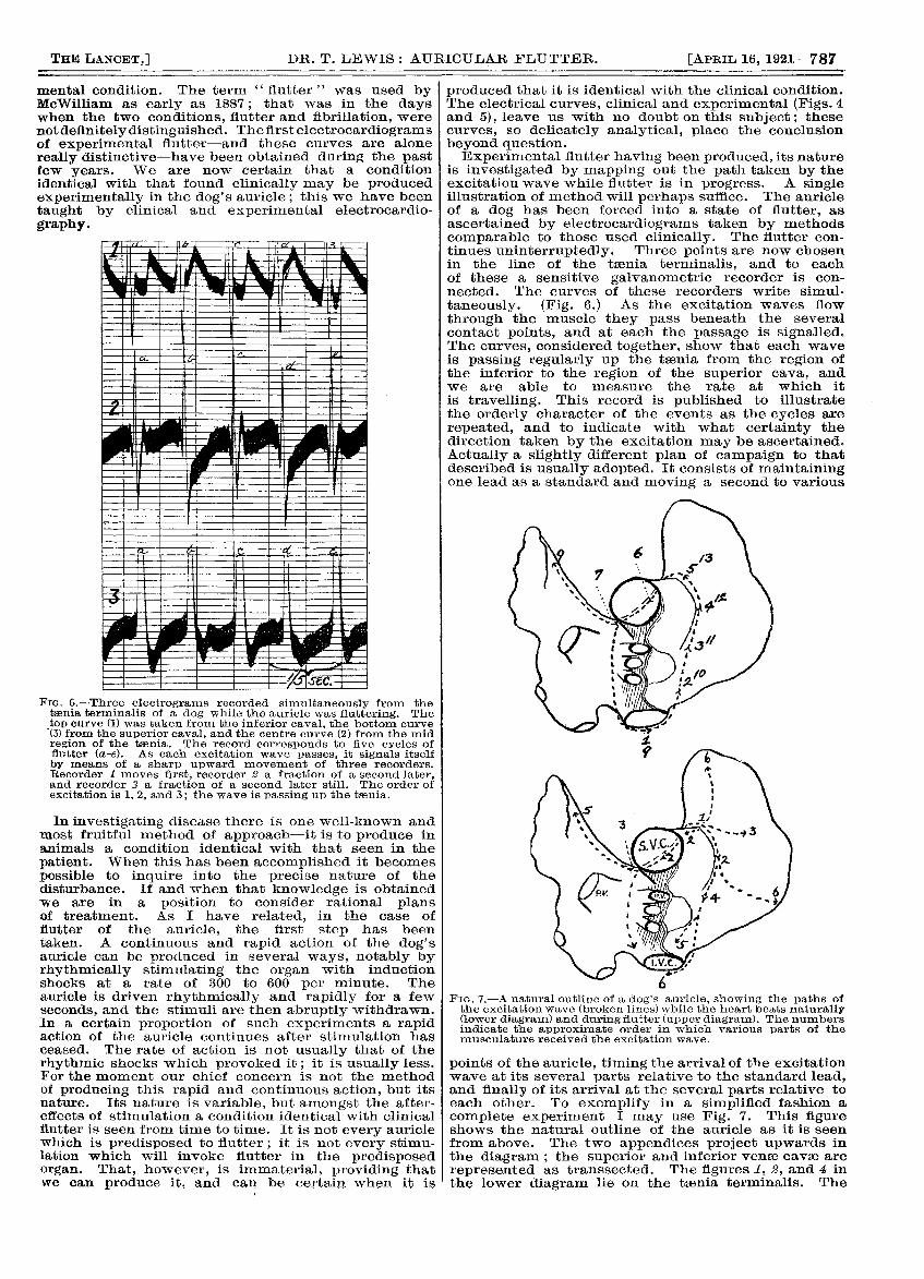
Fie. 6.-Three electrograms recorded simultaneously from thetsenia, terminalis of a dog while the auricle was fluttering. Thetop curve (1) was taken from the inferior caval, the bottom curve(3) from the superior caval, and the centre curve (2) from the midregion of the taenia. The record corresponds to five cycles offlutter (a-e). As each excitation wave passes, it signals itselfby means of a sharp upward movement of three recorders.Recorder moves first, recorder 2 a fraction of a second later,and recorder 3 a fraction of a second later still. The order ofexcitation is 1, 2, and 3; the wave is passing up the taania.
In investigating disease there is one well-known andmost fruitful method of approach-it is to produce inanimals a condition identical with that seen in thepatient. When this has been accomplished it becomespossible to inquire into the precise nature of thedisturbance. If and when that knowledge is obtainedwe are in a position to consider rational plansof treatment. As I have related, in the case offlutter of the auricle, the first step has beentaken. A continuous and rapid action of the dog’sauricle can be produced in several ways, notably byrhythmically stimulating the organ with inductionshocks at a rate of 300 to 600 per minute. Theauricle is driven rhythmically and rapidly for a fewseconds, and the stimuli are then abruptly withdrawn.In a certain proportion of such experiments a rapidaction of the auricle continues after stimulation hasceased. The rate of action is not usually that of therhythmic shocks which provoked it; it is usually less.For the moment our chief concern is not the methodof producing this rapid and continuous action, but itsnature. Its nature is variable, but amongst the after-effects of stimulation a condition identical with clinicalflutter is seen from time to time. It is not every auriclewhich is predisposed to flutter ; it is not every stimu-lation which will invoke flutter in the predisposedorgan. That, however, is immaterial, providing thatwe can produce it, and can be certain when it is
produced that it is identical with the clinical condition.The electrical curves, clinical and experimental (Figs. 4and 5), leave us with no doubt on this subject; thesecurves, so delicately analytical, place the conclusionbeyond question.Experimental flutter having been produced, its nature
is investigated by mapping out the path taken by theexcitation wave while flutter is in progress. A singleillustration of method will perhaps suffice. The auricleof a dog has been forced into a state of flutter, asascertained by electrocardiograms taken by methodscomparable to those used clinically. The flutter con-tinues uninterruptedly. Three points are now chosenin the line of the toania terminalis, and to eachof these a sensitive galvanometric recorder is con-
nected. The curves of these recorders write simul-
taneously. (Fig. 6.) As the excitation waves flow
through the muscle they pass beneath the severalcontact points, and at each the passage is signalled.The curves, considered together, show that each waveis passing regularly up the taenia from the region ofthe inferior to the region of the superior cava, andwe are able to measure the rate at which itis travelling. This record is published to illustratethe orderly character of the events as the cycles arerepeated, and to indicate with what certainty thedirection taken by the excitation may be ascertained.Actually a slightly different plan of campaign to thatdescribed is usually adopted. It consists of maintainingone lead as a standard and moving a second to various
0
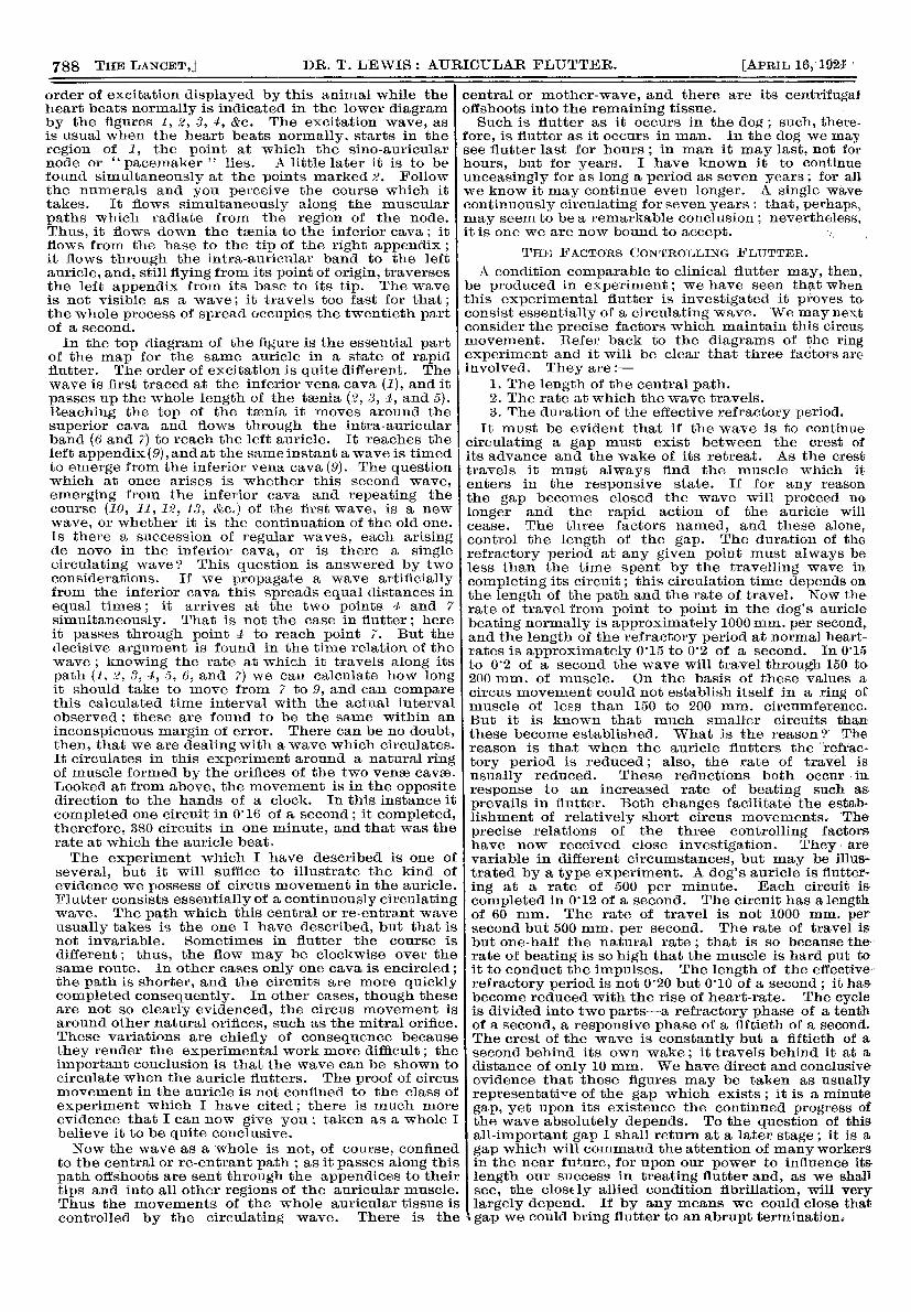
FiG. 7.-A natural outline of a dog’s auricle, showing the paths ofthe excitation wave (broken lines) while the heart beats naturally(lower diagram) and during flutter (upper diagram). The numbersindicate the approximate order in which various parts of themusculature received the excitation wave.
points of the auricle, timing the arrival of the excitationwave at its several parts relative to the standard lead,and finally of its arrival at the several parts relative toeach other. To exemplify in a simplified fashion acomplete experiment I may use Fig. 7. This figureshows the natural outline of the auricle as it is seenfrom above. The two appendices project upwards inthe diagram ; the superior and inferior venae cavae arerepresented as transsected. The figures 1, 2, and 4 inthe lower diagram lie on the taenia terminalis. The
788
order of excitation displayed by this animal while theheart beats normally is indicated in the lower diagramby the figures 1, 2, 5, , &c. The excitation wave, asis usual when the heart beats normally, starts in theregion of 1, the point at which the sino-auricularnode or "pacemaker lies. A little later it is to befound simultaneously at the points marked ,’!. Followthe numerals and you perceive the course which ittakes. It flows simultaneously along the muscularpaths which radiate from the region of the node.Thus, it flows down the tsenia to the inferior cava ; itflows from the base to the tip of the right appendix ;it flows through the intra-auricular band to the leftauricle, and, still flying from its point of origin, traversesthe left appendix from its base to its tip. The waveis not visible as a wave; it travels too fast for that;the whole process of spread occupies the twentieth partof a second.In the top diagram of the figure is the essential part
of the map for the same auricle in a state of rapidflutter. The order of excitation is quite different. Thewave is first traced at the inferior vena cava (1), and itpasses up the whole length of the taenia (.?, 3, 4, and 5).Reaching the top of the taenia it moves around the
superior cava and flows through the intra-auricularband (6 and 7) to reach the left auricle. It reaches theleft appendix (9), and at the same instant a wave is timedto emerge from the inferior vena cava (9). The questionwhich at once arises is whether this second wave,emerging from the inferior cava and repeating thecourse (10, 11, 12, 13, &c.) of the first wave, is a newwave, or whether it is the continuation of the old one.Is there a succession of regular waves, each arisingde novo in the inferior cava, or is there a singlecirculating wave? This question is answered by twoconsiderations. If we propagate a wave artificiallyfrom the inferior cava this spreads equal distances inequal times; it arrives at the two points 4 and 7 I
simultaneously. That is not the case in flutter; hereit passes through point to reach point. But thedecisive argument is found in the time relation of thewave ; knowing the rate at which it travels along itspath (1, ., 3, dt, o, 6, and 7) we can calculate how longit should take to move from 7 to 9, and can comparethis calculated time interval with the actual intervalobserved; these are found to be the same within aninconspicuous margin of error. There can be no doubt,then, that we are dealing with a wave which circulates.It circulates in this experiment around a natural ringof muscle formed by the orifices of the two venae cavse.Looked at from above, the movement is in the oppositedirection to the hands of a clock. In this instance it
completed one circuit in 0"16 of a second; it completed,therefore, 380 circuits in one minute, and that was therate at which the auricle beat.The experiment which I have described is one of
several, but it will suffice to illustrate the kind ofevidence we possess of circus movement in the auricle.Flutter consists essentially of a continuously circulatingwave. The path which this central or re-entrant waveusually takes is the one I have described, but that isnot invariable. Sometimes in flutter the course isdifferent ; thus, the flow may be clockwise over thesame route. In other cases only one cava is encircled;the path is shorter, and the circuits are more quicklycompleted consequently. In other cases, though theseare not so clearly evidenced, the circus movement isaround other natural orifices, such as the mitral orifice.These variations are chiefly of consequence becausethey render the experimental work more difficult; theimportant conclusion is that the wave can be shown tocirculate when the auricle flutters. The proof of circusmovement in the auricle is not confined to the class of
experiment which I have cited; there is much moreevidence that I can now give you ; taken as a whole Ibelieve it to be quite conclusive.Now the wave as a whole is not, of course, confined
to the central or re-entrant path ; as it passes along thispath offshoots are sent through the appendices to theirtips and into all other regions of the auricular muscle.Thus the movements of the whole auricular tissue iscontrolled by the circulating wave. There is the
central or mother-wave, and there are its centrifugaloffshoots into the remaining tissue. ’
Such is flutter as it occurs in the dog ; such, there-fore, is flutter as it occurs in man. In the dog we maysee flutter last for hours ; in man it may last, not forhours, but for years. I have known it to continueunceasingly for as long a period as seven years ; for allwe know it may continue even longer. A single wavecontinuously circulating for seven years : that, perhaps,may seem to be a remarkable conclusion ; nevertheless,it is one we are now bound to accept. ’i
,
THE FACTORS CONTROLLING FLUTTER.
A condition comparable to clinical flutter may, then,be produced in experiment; we have seen that whenthis experimental flutter is investigated it proves toconsist essentially of a circulating wave. We may nextconsider the precise factors which maintain this circusmovement. Refer back to the diagrams of the ringexperiment and it will be clear that three factors areinvolved. They are :-
1. The length of the central path.2. The rate at which the wave travels.3. The duration of the effective refractory period.
It must be evident that if the wave is to continuecirculating a gap must exist between the crest ofits advance and the wake of its retreat. As the cresttravels it must always find the muscle which itenters in the responsive state. If for any reasonthe gap becomes closed the wave will proceed nolonger and the rapid action of the auricle willcease. The three factors named, and these alone,control the length of the gap. The duration of the
refractory period at any given point must always beless than the time spent by the travelling wave incompleting its circuit; this circulation time depends onthe length of the path and the rate of travel. Now therate of travel from point to point in the dog’s auriclebeating normally is approximately 1000 mm. per second,and the length of the refractory period at normal heart-rates is approximately 0’15 to 0’2 of a second. In 0’15to 0’2 of a second the wave will travel through 150 to200 mm. of muscle. On the basis of these values acircus movement could not establish itself in a ring ofmuscle of less than 150 to 200 mm. circumference.But it is known that much smaller circuits thanthese become established. What is the reason?’ Thereason is that when the auricle flutters the "refrac-tory period is reduced; also, the rate of travel is
usually reduced. These reductions both occur inresponse to an increased rate of beating such asprevails in flutter. Both changes facilitate the estab-lishment of relatively short circus movements. Theprecise relations of the three controlling factorshave now received close investigation. They arevariable in different circumstances, but may be illus*trated by a type experiment. A dog’s auricle is flutter-ing at a rate of 500 per minute. Each circuit is
completed in 0’12 of a second. The circuit has a lengthof 60 mm. The rate of travel is not 1000 mm. persecond but 500 mm. per second. The rate of travel isbut one-half the natural rate; that is so because therate of beating is so high that the muscle is hard put toit to conduct the impulses. The length of the effectiverefractory period is not 0’20 but 0’10 of a second ; it hasbecome reduced with the rise of heart-rate. The cycleis divided into two parts-a refractory phase of a tenthof a second, a responsive phase of a fiftieth of a second.The crest of the wave is constantly but a fiftieth of asecond behind its own wake; it travels behind it at adistance of only 10 mm. We have direct and conclusiveevidence that these figures may be taken as usuallyrepresentative of the gap which exists ; it is a minute
gap, yet upon its existence the continued progress ofthe wave absolutely depends. To the question of thisall-important gap I shall return at a later stage ; it is agap which will command the attention of many workersin the near future, for upon our power to influence itslength our success in treating flutter and, as we shallsee, the closely allied condition fibrillation, will verylargely depend. If by any means we could close thatgap we could bring flutter to an abrupt termination.