oculomotor nerve schwannoma: a case report · pdf filethe magnetic reso-oculomotor nerve...
TRANSCRIPT
Copyright © 2014 The Korean Brain Tumor Society and The Korean Society for Neuro-Oncology 43
INTRODUCTION
Schwannomas are slowly growing peripheral nerve tumors that account for 6% to 8% of all intracranial tumors. They usually arise from the schwann cell layer of the vestibular branch of the eighth nerve or less commonly from the fifth nerve, the seventh nerve, and lower cranial nerve [1]. Oculo-motor schwannomas without neurofibromatosis is very rare [2]. The first report of an isolated oculomotor nerve schwan-noma was described by Kovacs in an autopsy in 1927 [3]. Af-ter then, approximately 40 cases of oculomotor nerve schwan-nomas have been described in the literature. The author reports a case of oculomotor schwannoma mimicking an optic nerve origin tumor that was removed surgically.
CASE REPORT
A 41-year-old female presented with one week history of blurred vision. A physical examination revealed relative af-ferent pupillary defect and decreased visual acuity of the left side. Visual acuity of the left eye was 20/200 with the Snellen’s chart (usual visual acuity was 200/200), and inferior hemi vi-sual field defect of left eye was observed. The magnetic reso-
Oculomotor Nerve Schwannoma: A Case ReportYong-Hwan Cho1,2, Kyung-Su Sung1,2, Young-Jin Song1,2, Dae-Cheol Kim1,3, Sunseob Choi1,4, Ki-Uk Kim1,2
1Brain Tumor Institute Medical Science Research Center, Departments of 2Neurosurgery, 3Pathology, 4Radiology, College of Medicine, Dong-A University, Busan, Korea
Received March 7, 2014Revised April 1, 2014Accepted April 3, 2014
CorrespondenceKi-Uk KimDepartment of Neurosurgery, College of Medicine, Dong-A University, 26 Daesingongwon-ro, Seo-gu, Busan 602-715, KoreaTel: +82-51-240-5241Fax: +82-51-242-6714E-mail: [email protected]
Schwannomas account for about 8% of intracranial tumors and 90% are vestibular schwannomas. Oculomotor schwannoma without neurofibromatosis is extremely rare. A 41-year-old female presented with complaints of blurred vision, and the neurologic examination revealed afferent pupillary defect and decreased visual acuity of the left side. Brain magnetic resonance image showed an extra axial mass in the left superior orbital fissure. The patient underwent major surgery via the fronto-temporal ap-proach. The tumor originated from the oculomotor nerve and was subtotally removed under micro-scopic surgery. The pathological findings confirmed the tumor as a schwannoma. After surgery, ptosis and medial gaze limitation of the left eye was detected, but the symptoms improved gradually.
Key Words Schwannoma; Oculomotor nerve; Surgery.
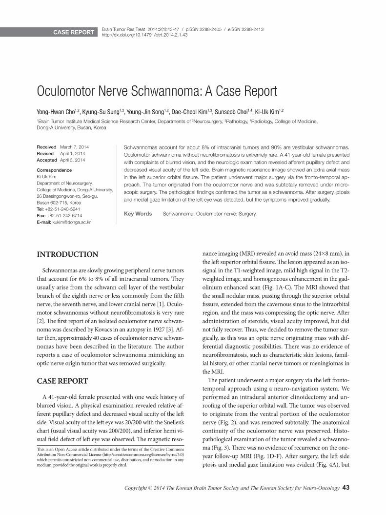
nance imaging (MRI) revealed an avoid mass (24×8 mm), in the left superior orbital fissure. The lesion appeared as an iso-signal in the T1-weighted image, mild high signal in the T2-weighted image, and homogeneous enhancement in the gad-olinium enhanced scan (Fig. 1A-C). The MRI showed that the small nodular mass, passing through the superior orbital fissure, extended from the cavernous sinus to the intraorbital region, and the mass was compressing the optic nerve. After administration of steroids, visual acuity improved, but did not fully recover. Thus, we decided to remove the tumor sur-gically, as this was an optic nerve originating mass with dif-ferential diagnostic possibilities. There was no evidence of neurofibromatosis, such as characteristic skin lesions, famil-ial history, or other cranial nerve tumors or meningiomas in the MRI.
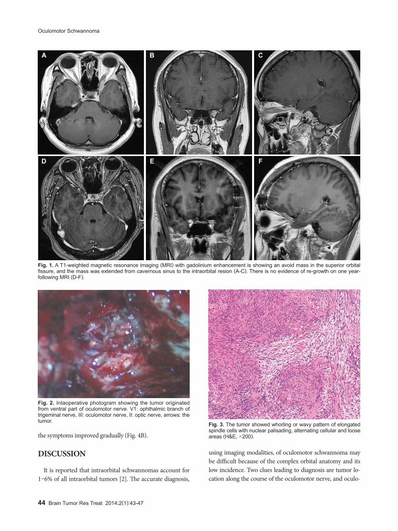
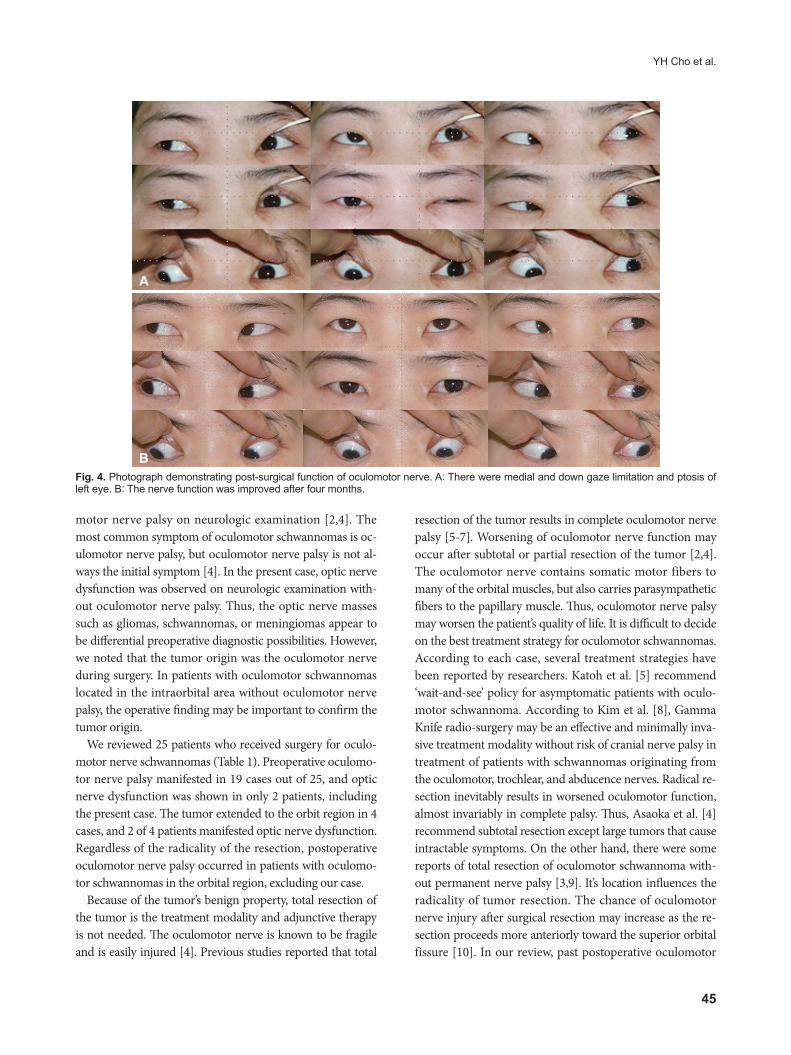
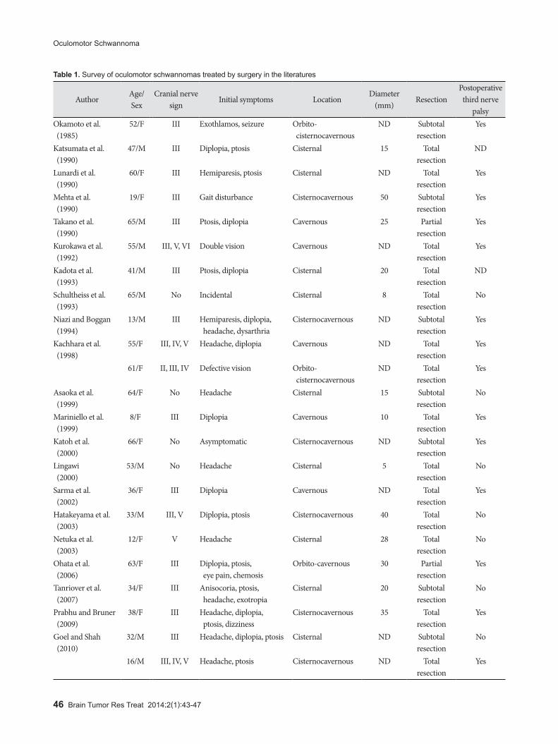
The patient underwent a major surgery via the left fronto-temporal approach using a neuro-navigation system. We performed an intradural anterior clinoidectomy and un-roofing of the superior orbital wall. The tumor was observed to originate from the ventral portion of the oculomotor nerve (Fig. 2), and was removed subtotally. The anatomical continuity of the oculomotor nerve was preserved. Histo-pathological examination of the tumor revealed a schwanno-ma (Fig. 3). There was no evidence of recurrence on the one-year follow-up MRI (Fig. 1D-F). After surgery, the left side ptosis and medial gaze limitation was evident (Fig. 4A), but
CASE REPORT Brain Tumor Res Treat 2014;2(1):43-47 / pISSN 2288-2405 / eISSN 2288-2413http://dx.doi.org/10.14791/btrt.2014.2.1.43
This is an Open Access article distributed under the terms of the Creative Commons Attribution Non-Commercial License (http://creativecommons.org/licenses/by-nc/3.0) which permits unrestricted non-commercial use, distribution, and reproduction in any medium, provided the original work is properly cited.
44 Brain Tumor Res Treat 2014;2(1):43-47
Oculomotor Schwannoma
the symptoms improved gradually (Fig. 4B).
DISCUSSION
It is reported that intraorbital schwannomas account for 1–6% of all intraorbital tumors [2]. The accurate diagnosis,
using imaging modalities, of oculomotor schwannoma may be difficult because of the complex orbital anatomy and its low incidence. Two clues leading to diagnosis are tumor lo-cation along the course of the oculomotor nerve, and oculo-
A
D
B
E
C
F
Fig. 1. A T1-weighted magnetic resonance imaging (MRI) with gadolinium enhancement is showing an avoid mass in the superior orbital fissure, and the mass was extended from cavernous sinus to the intraorbital resion (A-C). There is no evidence of re-growth on one year-following MRI (D-F).
Fig. 2. Intaoperative photogram showing the tumor originated from ventral part of oculomotor nerve. V1: ophthalmic branch of trigeminal nerve, III: oculomotor nerve, II: optic nerve, arrows: the tumor. Fig. 3. The tumor showed whorling or wavy pattern of elongated
spindle cells with nuclear palisading, alternating cellular and loose areas (H&E, ×200).
YH Cho et al.
45
motor nerve palsy on neurologic examination [2,4]. The most common symptom of oculomotor schwannomas is oc-ulomotor nerve palsy, but oculomotor nerve palsy is not al-ways the initial symptom [4]. In the present case, optic nerve dysfunction was observed on neurologic examination with-out oculomotor nerve palsy. Thus, the optic nerve masses such as gliomas, schwannomas, or meningiomas appear to be differential preoperative diagnostic possibilities. However, we noted that the tumor origin was the oculomotor nerve during surgery. In patients with oculomotor schwannomas located in the intraorbital area without oculomotor nerve palsy, the operative finding may be important to confirm the tumor origin.
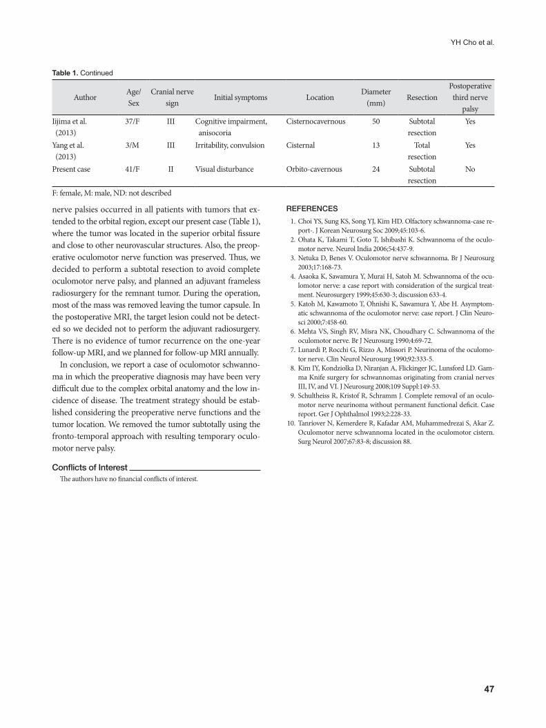
We reviewed 25 patients who received surgery for oculo-motor nerve schwannomas (Table 1). Preoperative oculomo-tor nerve palsy manifested in 19 cases out of 25, and optic nerve dysfunction was shown in only 2 patients, including the present case. The tumor extended to the orbit region in 4 cases, and 2 of 4 patients manifested optic nerve dysfunction. Regardless of the radicality of the resection, postoperative oculomotor nerve palsy occurred in patients with oculomo-tor schwannomas in the orbital region, excluding our case.
Because of the tumor’s benign property, total resection of the tumor is the treatment modality and adjunctive therapy is not needed. The oculomotor nerve is known to be fragile and is easily injured [4]. Previous studies reported that total
resection of the tumor results in complete oculomotor nerve palsy [5-7]. Worsening of oculomotor nerve function may occur after subtotal or partial resection of the tumor [2,4]. The oculomotor nerve contains somatic motor fibers to many of the orbital muscles, but also carries parasympathetic fibers to the papillary muscle. Thus, oculomotor nerve palsy may worsen the patient’s quality of life. It is difficult to decide on the best treatment strategy for oculomotor schwannomas. According to each case, several treatment strategies have been reported by researchers. Katoh et al. [5] recommend ‘wait-and-see’ policy for asymptomatic patients with oculo-motor schwannoma. According to Kim et al. [8], Gamma Knife radio-surgery may be an effective and minimally inva-sive treatment modality without risk of cranial nerve palsy in treatment of patients with schwannomas originating from the oculomotor, trochlear, and abducence nerves. Radical re-section inevitably results in worsened oculomotor function, almost invariably in complete palsy. Thus, Asaoka et al. [4] recommend subtotal resection except large tumors that cause intractable symptoms. On the other hand, there were some reports of total resection of oculomotor schwannoma with-out permanent nerve palsy [3,9]. It’s location influences the radicality of tumor resection. The chance of oculomotor nerve injury after surgical resection may increase as the re-section proceeds more anteriorly toward the superior orbital fissure [10]. In our review, past postoperative oculomotor
A
BFig. 4. Photograph demonstrating post-surgical function of oculomotor nerve. A: There were medial and down gaze limitation and ptosis of left eye. B: The nerve function was improved after four months.
46 Brain Tumor Res Treat 2014;2(1):43-47
Oculomotor Schwannoma
Table 1. Survey of oculomotor schwannomas treated by surgery in the literatures
AuthorAge/Sex
Cranial nerve sign
Initial symptoms LocationDiameter
(mm)Resection
Postoperative third nerve
palsyOkamoto et al. (1985)
52/F III Exothlamos, seizure Orbito- cisternocavernous
ND Subtotalresection
Yes
Katsumata et al. (1990)
47/M III Diplopia, ptosis Cisternal 15 Totalresection
ND
Lunardi et al. (1990)
60/F III Hemiparesis, ptosis Cisternal ND Totalresection
Yes
Mehta et al. (1990)
19/F III Gait disturbance Cisternocavernous 50 Subtotalresection
Yes
Takano et al. (1990)
65/M III Ptosis, diplopia Cavernous 25 Partialresection
Yes
Kurokawa et al. (1992)
55/M III, V, VI Double vision Cavernous ND Totalresection
Yes
Kadota et al. (1993)
41/M III Ptosis, diplopia Cisternal 20 Totalresection
ND
Schultheiss et al. (1993)
65/M No Incidental Cisternal 8 Totalresection
No
Niazi and Boggan (1994)
13/M III Hemiparesis, diplopia, headache, dysarthria
Cisternocavernous ND Subtotalresection
Yes
Kachhara et al. (1998)
55/F III, IV, V Headache, diplopia Cavernous ND Totalresection
Yes
61/F II, III, IV Defective vision Orbito- cisternocavernous
ND Totalresection
Yes
Asaoka et al. (1999)
64/F No Headache Cisternal 15 Subtotalresection
No
Mariniello et al. (1999)
8/F III Diplopia Cavernous 10 Totalresection
Yes
Katoh et al. (2000)
66/F No Asymptomatic Cisternocavernous ND Subtotalresection
Yes
Lingawi (2000)
53/M No Headache Cisternal 5 Totalresection
No
Sarma et al. (2002)
36/F III Diplopia Cavernous ND Totalresection
Yes
Hatakeyama et al. (2003)
33/M III, V Diplopia, ptosis Cisternocavernous 40 Totalresection
No
Netuka et al. (2003)
12/F V Headache Cisternal 28 Totalresection
No
Ohata et al. (2006)
63/F III Diplopia, ptosis, eye pain, chemosis
Orbito-cavernous 30 Partialresection
Yes
Tanriover et al. (2007)
34/F III Anisocoria, ptosis, headache, exotropia
Cisternal 20 Subtotalresection
No
Prabhu and Bruner (2009)
38/F III Headache, diplopia, ptosis, dizziness
Cisternocavernous 35 Totalresection
Yes
Goel and Shah (2010)
32/M III Headache, diplopia, ptosis Cisternal ND Subtotalresection
No
16/M III, IV, V Headache, ptosis Cisternocavernous ND Totalresection
Yes
YH Cho et al.
47
nerve palsies occurred in all patients with tumors that ex-tended to the orbital region, except our present case (Table 1), where the tumor was located in the superior orbital fissure and close to other neurovascular structures. Also, the preop-erative oculomotor nerve function was preserved. Thus, we decided to perform a subtotal resection to avoid complete oculomotor nerve palsy, and planned an adjuvant frameless radiosurgery for the remnant tumor. During the operation, most of the mass was removed leaving the tumor capsule. In the postoperative MRI, the target lesion could not be detect-ed so we decided not to perform the adjuvant radiosurgery. There is no evidence of tumor recurrence on the one-year follow-up MRI, and we planned for follow-up MRI annually.
In conclusion, we report a case of oculomotor schwanno-ma in which the preoperative diagnosis may have been very difficult due to the complex orbital anatomy and the low in-cidence of disease. The treatment strategy should be estab-lished considering the preoperative nerve functions and the tumor location. We removed the tumor subtotally using the fronto-temporal approach with resulting temporary oculo-motor nerve palsy.
Conflicts of InterestThe authors have no financial conflicts of interest.
REFERENCES
1. Choi YS, Sung KS, Song YJ, Kim HD. Olfactory schwannoma-case re-port-. J Korean Neurosurg Soc 2009;45:103-6.
2. Ohata K, Takami T, Goto T, Ishibashi K. Schwannoma of the oculo-motor nerve. Neurol India 2006;54:437-9.
3. Netuka D, Benes V. Oculomotor nerve schwannoma. Br J Neurosurg 2003;17:168-73.
4. Asaoka K, Sawamura Y, Murai H, Satoh M. Schwannoma of the ocu-lomotor nerve: a case report with consideration of the surgical treat-ment. Neurosurgery 1999;45:630-3; discussion 633-4.
5. Katoh M, Kawamoto T, Ohnishi K, Sawamura Y, Abe H. Asymptom-atic schwannoma of the oculomotor nerve: case report. J Clin Neuro-sci 2000;7:458-60.
6. Mehta VS, Singh RV, Misra NK, Choudhary C. Schwannoma of the oculomotor nerve. Br J Neurosurg 1990;4:69-72.
7. Lunardi P, Rocchi G, Rizzo A, Missori P. Neurinoma of the oculomo-tor nerve. Clin Neurol Neurosurg 1990;92:333-5.
8. Kim IY, Kondziolka D, Niranjan A, Flickinger JC, Lunsford LD. Gam-ma Knife surgery for schwannomas originating from cranial nerves III, IV, and VI. J Neurosurg 2008;109 Suppl:149-53.
9. Schultheiss R, Kristof R, Schramm J. Complete removal of an oculo-motor nerve neurinoma without permanent functional deficit. Case report. Ger J Ophthalmol 1993;2:228-33.
10. Tanriover N, Kemerdere R, Kafadar AM, Muhammedrezai S, Akar Z. Oculomotor nerve schwannoma located in the oculomotor cistern. Surg Neurol 2007;67:83-8; discussion 88.
Table 1. Continued
AuthorAge/Sex
Cranial nerve sign
Initial symptoms LocationDiameter
(mm)Resection
Postoperative third nerve
palsyIijima et al. (2013)
37/F III Cognitive impairment, anisocoria
Cisternocavernous 50 Subtotalresection
Yes
Yang et al. (2013)
3/M III Irritability, convulsion Cisternal 13 Totalresection
Yes
Present case 41/F II Visual disturbance Orbito-cavernous 24 Subtotalresection
No
F: female, M: male, ND: not described