ocular toxoplasmosis - british journal of...
TRANSCRIPT
Brit. J. Ophthal. (I973) 57, 1
Communications
Ocular toxoplasmosis
E. S. PERKINS
Department of Experimental Ophthalmology, Institute of Ophthalmology, University of London
A large amount of research has been done on toxoplasmosis in the last 30 years, and theworld-wide incidence of the infection, and its clinical manifestations, are recognized anddocumented in an extensive literature. New discoveries concerning the life-cycle of theorganism (Frenkel, I970; Hutchison, Dunachie, Siim, and Work, I970) promise to solvemany of the problems of the transmission of infection and will give added stimulus toepidemiological studies. In the field of ophthalmology the importance of toxoplasmosis isnow established, but some problems still remain.The ocular lesions in infants with congenital toxoplasmosis are well recognized as
pathogenic, but whether similar chorioretinal lesions in adults, although now acknowledgedto be due to toxoplasmosis, result from recurrences of congenital infection or follow apostnatally acquired infection is still controversial. Other types of uveitis have beenattributed to toxoplasmosis, and if this is correct it is important to recognize such cases.The present study attempts to clarify these problems by a review of the relevant literaturesupported by clinical studies in patients with uveitis. A further problem which will bediscussed is whether congenital toxoplasmosis results from a chronic maternal infectionor from one acquired during pregnancy: although this is not strictly an ophthalmologicalproblem, ophthalmologists are often asked to advise on the prognosis for future preg-nancies.
Review of the literature
No attempt is made here to provide a comprehensive general review of the literature ontoxoplasmosis, but a list of recent reviews is given in the Bibliography. Attention herewill be concentrated on the incidence of ocular complications in acquired systemicinfections with clinical manifestations, the evidence for acquired infection in cases ofuveitis, and the type ofmaternal infection causing toxoplasmosis.
INCIDENCE OF OCULAR COMPLICATIONS IN RECENTLY ACQUIRED TOXOPLASMOSIS
In reviewing the literature on this subject, an immediate problem is that of deciding on the criteriawhich should be accepted as evidence ofinfection. Most authors have relied on one or a combinationof three methods: the detection of high levels of antibody, particularly the demonstration of risingtitres; the observation of organisms in biopsy specimens or tissue fluids; and the demonstration ofinfection in animals after the inoculation oftissue extracts.The demonstration in biopsy or post mortem specimens of organisms morphologically resembling
Toxoplasma is not entirely reliable, as nuclear fragments (Duke-Elder, Ashton, and Brihaye-vanGeertruyden, I953) and even pine pollen (Langer, I966) can cause confusion. Even if the organism
Received for publication March 21, 1972Address for reprints: Institute of Ophthalmology, Judd St., London, WCIH 9QS
copyright. on 23 S
eptember 2018 by guest. P
rotected byhttp://bjo.bm
j.com/
Br J O
phthalmol: first published as 10.1136/bjo.57.1.1 on 1 January 1973. D
ownloaded from

E. S. Perkins
can be identified unequivocally, this is not definite evidence of a recently acquired infection. It isinteresting to note that, in the original series described by Wilder (1952), cerebral calcification wasfound in three of six patients X-rayed, indicating that the infection was almost certainly congenitalin origin. If the organism can be found in the ocular tissues of the adult as a result of congenitalinfection there seems no reason why they should not also be found in other tissues (brain, lymphnodes, muscles, etc.) after congenital infection. Organisms have been found in patients withcirculating antibodies absent or at very low levels (Hogan, Kimura, and O'Connor, I964; Frances-chetti and Engelbrecht, I964; Tanaka, Takada, Sakamoto, Takasu, Sano, and Uemichi, I965),which again suggests very long-standing or congenital infection.
LABORATORY INFECTION
A number of accidental laboratory infections have been reported, and as these are usually welldocumented it is worth considering them in some detail. In 2 I cases* no ocular disease was reportedexcept for conjunctival hyperaemia in one (Strom, I95 I). Conjunctivitis accompanied the infectionin two cases (Strom, I951; Straub, I962). Matsubayashi, Kioke, Uyemura, Soh, and Hamano (i 96I)and Mikuni (I968) each reported the infection of a pathologist during an autopsy: both patientsdeveloped an exudative retinitis and organisms were recovered from the subretinal fluid. Inneither were the ocular lesions typical ofthe focal chorioretinitis seen in congenital infection.
In summary, therefore, uveitis occurred in only two of 26 laboratory infections, and in neither casewere the lesions similar to those seen in the congenital disease. The infection of six volunteers(Fair and Walls, I962) did not result in any ocular signs or symptoms.
ACQUIRED TOXOPLASMOSIS WITHOUT OCULAR DISEASE
In reviewing the literature I have found accounts of over I,6oo cases of acquired toxoplasmosisreported over the last io years in which the clinical diagnosis was supported by serological, and insome cases histological, evidence.t This does not include cases in which the clinical signs wereconfined to the eyes; these will be discussed later.
It is clear from this review that the commonest clinical manifestation of acquired toxoplasmosis is alymphadenopathy, often with fever, and sometimes a skin rash, hepatosplenomegaly, or involvementof other systems. Such cases account for 89 -2 per cent. of those reviewed (Table I). Cases in whichthe signs and symptoms were mainly confined to the central nervous system accounted for 4x3 percent., myocarditis and pericarditis 1 .4 per cent., and pulmonary disease o-8 per cent. The remaining4x2 per cent. consisted of a variety of conditions including hepatitis, polymyositis, psychiatric dis-orders, skin lesions, etc. The evidence for the diagnosis of toxoplasmosis was often less convincing inthis miscellaneous group of cases.
It is possible that ocular lesions were not recognized in some of these cases, but many authorsdescribed others in their series with ocular signs (see below) and it is unlikely that many with activeuveitis would have been overlooked.
Table I Systemic toxoplasmosis without Table H Ocular manifestations in acquiredocular complications toxoplasmosis
Type No. of Per cent.cases of total
Lymphadenopathy I,46I 89-2CNS disease 71 4.3Myocarditis 23 1.4Pulmonary disease I3 o-8Miscellaneous conditions 69 4-2
Total 1,637
VNo. of Per cent.cases of total
Focal chorioretinitis 44 63.8Conjunctivitis 2 3-oPapillitis and optic atrophy 8 iis6Pan-uveitis 8 II*6Other 7 IO*I
Total 69
*Bengtsson (I950), Magnusson (i95i), Strom ('95'), Giroud, Le Gac, Roger, and Gaillard O 953), Sexton, Eyles, and Dillman (I953),Thalhammer (1954), Beverley, Skipper, and Marshall (1955), Hormann (i955), Wettingfeld, Rowe, and Eyles (1956), Brown andJacobs (1956), Kayhoe, Jacobs, Beye, and McCullough (1957), van Soestbergen (0957), Rawal (g959), Frenkel, Weber, and Lunde(196o), IJmdenstock, Mandoul, and Pestre-Alexandre (i965), Neu (1967), Remington, Miller, and Brownlee (X968).tA list of the references to cases without ocular signs included in this part ofthe review can be obtained from the author.
2copyright.
on 23 Septem
ber 2018 by guest. Protected by
http://bjo.bmj.com
/B
r J Ophthalm
ol: first published as 10.1136/bjo.57.1.1 on 1 January 1973. Dow
nloaded from
Ocular toxoplasmosis
ACQUIRED TOXOPLASMOSIS WITH SYSTEMIC AND OCULAR SIGNS
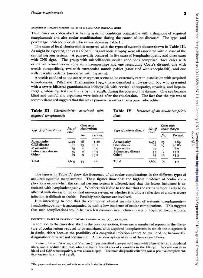
These cases were described as having systemic conditions compatible with a diagnosis of acquiredtoxoplasmosis and also ocular manifestations during the course of the disease.* The type andpercentage incidence ofocular disease are shown in Table II.The cases of focal chorioretinitis occurred with the types of systemic disease shown in Table III.
As might be expected, the cases of papillitis and optic atrophy were all associated with disease of thecentral nervous system. A pan-uveitis occurred in five cases of lymphadenopathy and three caseswith CNS signs. The group with miscellaneous ocular conditions comprised three cases withexudative retinal lesions (one with haemorrhage and one resembling Coats's disease), one withuveitis (unspecified), two with extraocular muscle palsies (associated with encephalitis), and onewith macular oedema (associated with hepatitis).A uveitis confined to the anterior segment seems to be extremely rare in association with acquired
toxoplasmosis. Pillat and Thalhammer (I957) have described a 12-year-old boy who presentedwith a severe bilateral granulomatous iridocyclitis with cervical adenopathy, myositis, and hepato-megaly, whose dye test rose fron I :64 to I: I6,384 during the course of the disease. One eye becameblind and painful and organisms were isolated after the enucleation. The fact that the eye was soseverely damaged suggests that this was a pan-uveitis rather than a pure iridocyclitis.
Table III Chorioretinitis associated with Table IV Incidence of all ocular complica-acquired toxoplasmosis tions
Cases withType of systemic disease No. of chorioretinitis
casesNo. Per cent.
Adenopathy 1,479 i8 I*22CNS disease 8i I3 i6-iMyocarditis 25 2 8-oPulmonary disease I5 2 13.3Other 69 9 I3-o
Total 1,669 44 2-6
Cases withType of systemic disease No. of ocular changes
casesNo. Per cent.
Adenopathy 1,479 25 i *69CNS disease 81 27 30-86Myocarditis 25 2 8-oPulmonary disease 15 2 I3.3Other 69 IO 14'5Total 1,669 66 4-2
The figures in Table IV show the frequency of all ocular complications in the different types ofacquired systemic toxoplasmosis. These figures show that the highest incidence of ocular com-plications occurs when the central nervous system is affected, and that the lowest incidence is as-sociated with lymphadenopathy. Whether this is due to the fact that the retina is more likely to beaffected with disease of the central nervous system, or whether it is only a reflection of a more severeinfection, is difficult to decide. Possibly both factors are involved.
It is interesting to note that the commonest clinical manifestation of systemic toxoplasmosis-lymphadenopathy-is accompanied by such a low incidence of ocular complications. This suggeststhat such complications would be even less common in subclinical cases of acquired toxoplasmosis.
DOUBTFUL CASES OF SYSTEMIC TOXOPLASMOSIS WITH OCULAR SIGNS
In addition to the cases described in the previous section, there are a number of reports in the litera-ture of ocular lesions reputed to be associated with acquired toxoplasmosis in which the diagnosis isin doubt, either because the possibility of a congenital infection cannot be excluded, or because thediagnostic criteria are not convincing. A briefdescription ofsome of these cases follows.
Brennan, Brown, Warren, and Vranian (I949) described a 30-year-old man with bilateral iritis, a duodenalulcer, and a nodular skin rash who also had a healed area of choroiditis in the left eye. Inoculations fromblood and CSF were negative, as was a skin biopsy. The main diagnostic criterion was a positive complement-fixation test in a titre of I :I28.
*The papers reviewed are marked with an asterisk in the list of References.
B
3copyright.
on 23 Septem
ber 2018 by guest. Protected by
http://bjo.bmj.com
/B
r J Ophthalm
ol: first published as 10.1136/bjo.57.1.1 on 1 January 1973. Dow
nloaded from

E. S. Perkins
Frugoni (I95i) described a patient with meningeal signs who developed bilateral exudative retinal detach-ments going on to blindness with optic atrophy and pigmentary degeneration of the retina. The diagnosis oftoxoplasmosis was made on the positive dye test in a titre of I :128.Wollheim (I952) reviewed fourteen cases with positive dye tests in titres of I :36 to i :I44, including one with
choroiditis.Seitelberger and Spiel (x953) described two brothers aged 6 and 7 years with encephalitis. What were
thought to be toxoplasmic organisms were found at autopsy in the brain of one child, but no serologicaltests were carried out. The dye test was negative in the second child.
Rieger (I955) described three patients with extraocular muscle paresis and a positive dye test (I:64 in twoand I:i,024 in the third), and one with optic neuritis and a dye test positive at 1:256.
Paulley, Green,Jones, and Kane (I954) described a patient with myocarditis who developed papillitis and ahemiplegia. The dye test was positive at I:64.
Koeze and Klingon (i964) described a 67-year-old patient with anaemia and signs of chronic disease of thecentral nervous system who had flame-shaped haemorrhages in the fundi. Toxoplasma granulomas werefound post mortem but the retinal haemorrhages could have resulted from the anaemia.
Lalisse, Mises, and Durand (i964) described five patients aged 3 to io years with positive serology who hadabnormal EEGs. One child had had a recent chorioretinitis but there was no convincing evidence of recentsystemic infection.
As Straub has pointed out, it is well recognized that congenital ocular lesions can become reacti-vated in childhood or adult life, and there is no reason why toxoplasmic lesions of congenital origin inother tissues should not similarly recur. Lavat (I962), for example, quoted a case in which congenitalocular lesions were observed at 2 months and an encephalitis developed at 8 years, and a rathersimilar case was reported by Saraux, Seringe, Bach, and Le Besnerais ( I 962).
Martinelli and Rossi (i 964) described abnormal EEG findings in fourteen cases, in eight of which there wasa chorioretinitis with a positive dye test. The other patients had optic atrophy, extraocular muscle pareses,dystrophia adiposogenitale, and spastic tetraplegia. The dye test titres were less then I:500 in all but twocases and it is not certain that the condition described resulted from acquired infection.
Harris (X967) reported a 2-year-old child with stomatitis and adenopathy and a healed chorioretinallesion in one eye. The dye test was positive at I :512, but this could have been a recurrence of congenitalinfection.
Manissadjian, D'Oliveira Penna, Costa Vaz, Ramos, Borges, and Schvartsman (i967) reported 26 childrento have aquired toxoplasmosis. Six of them showed ocular lesions: nystagmus in two (associated with bilateraloptic atrophy, with chorioretinitis in one and chorioretinal scars in the other); bilateral optic atrophy andchorioretinal scars in two further cases; macular pallor in one, and bilateral chorioretinal lesions in another.All had high dye test titres and all except one had hepatomegaly or signs of pulmonary disease. The two withnystagmus probably had defective vision from an early age, but it is possible that the condition was acquired inthe other four cases.Macchi (i968) described a 12-year-old child with an enlarged heart who had been found to have chorio-
retinal scars at the age of 6 and so had most probably had a congenital infection.Ohkawa, Yonekura, and Ito (i969) described a 38-year-old woman with hemiparaesthesiae, fits, and
dysphagia, who was found to have bilateral small cystic lesions in the fundi. Toxoplasma organisms wereisolated from a mouse inoculated with CSF but were not isolated from further passages. The dye test waspositive at I :256. The fundus lesions in this case were unlike any previously described as resulting fromtoxoplasmic infection.
SUMMARY OF THE OCULAR COMPLICATIONS OF ACQUIRED SYSTEMIC TOXOPLASMOSIS
Uveitis has been described in 52 out of I,669 cases of acquired toxoplasmosis (3 'I per cent.), but thehighest incidence of uveitis is associated with the relatively rare cases of toxoplasmic encephalitis.Papillitis and optic atrophy may also occur in these cases. The uveitis is usually a focal chorio-retinitis, but some cases ofpan-uveitis or exudative retinitis have been described.
OCULAR DISEASE AS THE ONLY MANIFESTATION OF ACQUIRED TOXOPLASMOSIS
This is a particularly difficult group to assess; even if the diagnosis of toxoplasmic uveitis is well sub-stantiated, it is difficult to prove that the condition results from an acquired infection. The isolationof the organism from the eye of an adult does not exclude congenital infection: for example, the
4copyright.
on 23 Septem
ber 2018 by guest. Protected by
http://bjo.bmj.com
/B
r J Ophthalm
ol: first published as 10.1136/bjo.57.1.1 on 1 January 1973. Dow
nloaded from
Ocular toxoplasmosis
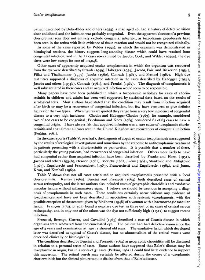
patient described by Duke-Elder and others (I953), a man aged 40, had a history of defective visionsince childhood and the infection was probably congenital. Even the apparent absence of a previouschorioretinal scar does not entirely exclude congenital infection, as toxoplasmic pseudocysts havebeen seen in the retina with little evidence of tissue reaction and would not be recognized clinically.
In some of the cases reported by Wilder (I952), in which the organism was demonstrated inhistological sections, the history suggests long-standing disease which could have resulted fromcongenital infection, and in the 21 cases re-examined by Jacobs, Cook, and Wilder (I 954a), the dyetitres were low except for one of I :2,048.
Other cases of apparently acquired ocular toxoplasmosis in which the organism was recoveredfrom the eye were described by Straub (1949), Habegger (X954), Jacobs, Fair, and Bickerton (I954),Pillat and Thalhammer (1957), Jacobs (I960), Conrads (I96I), and Frenkel (I962). High dyetest titres supported a diagnosis of acquired infection in the cases described by Habegger (1954),Jacobs and others (I954b), Conrads (I96I), and Frenkel (I962). The diagnosis of toxoplasmosis iswell substantiated in these cases and an acquired infection would seem to be responsible.Many papers have now been published in which a toxoplasmic aetiology for cases of chorio-
retinitis in children and adults has been well supported by statistical data based on the results ofserological tests. Most authors have stated that the condition may result from infection acquiredafter birth or may be a recurrence of congenital infection, but few have ventured to give definitefigures for the two types. When figures are quoted they range from a very low incidence of congenitaldisease to a very high incidence. Chodos and Habegger-Chodos (I963), for example, consideredtwo of IOO cases to be congenital; Friedmann and Knox (I969) considered 6I of 63 cases to have acongenital origin. I have always felt that acquired infection was a rare cause of toxoplasmic chorio-retinitis and that almost all cases seen in the United Kingdom are recurrences of congenital infection(Perkins, I 96 I).
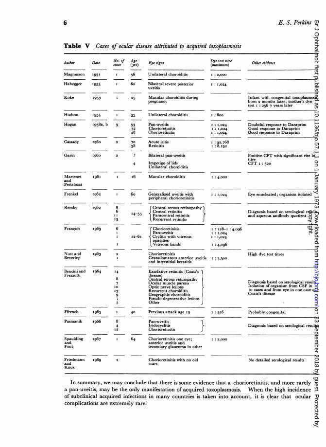
In the case reports (Table V, overleaf), the diagnosis of acquired ocular toxoplasmosis was suggestedby the results of serological investigations and sometimes by the response to antitoxoplasmic treatmentin patients presenting with a chorioretinitis or pan-uveitis. It is possible that a number of these,particularly the young patients, had recurrences of congenital infection. Others more likely to havehad congenital rather than acquired infection have been described by Franke and Horst (I951),Jacobs and others (I 954b), Hewson (i 96 I), Boericke (i 962), Genz (I963), Stankovic and Milojkovic(I963), Engelbrecht and Franceschetti (I963), Franceschetti and Engelbrecht (I964), and Jones,Kean, and Kimball (I969).
Table V shows that not all cases attributed to acquired toxoplasmosis presented with a focalchorioretinitis. Remky (i962), Bencini and Frezzotti (1964) both described cases of centralserous retinopathy, and the latter authors also included cases of geographic choroiditis and exudativemacular lesions without inflammatory signs. I believe we should be cautious in accepting a diag-nosis of toxoplasmosis in such cases. These conditions certainly occur without any evidence oftoxoplasmosis and have not been described in association with systemic toxoplasmosis, with thepossible exception of the account given by Binkhorst (I 948) ofa woman with a haemorrhagic macularlesion. Francois (I963, p. 405) found a negative dye test in three out of six cases of central serousretinopathy, and in only one of the others was the dye test sufficiently high (I :5 I 2) to suggest recentinfection.
Frezzotti, Berengo, Guerra, and Cavallini (I965) described a case of Coats's disease in whichorganisms were recovered from the enucleated eye. The patient had had defective vision since theage of 4 years and examination at age I I showed old scars. The exudative lesion which developedlater was described as typical of Coats's disease, but no abnormalities of the retinal vessels weredescribed clinically or histologically.The condition described by Bencini and Frezzotti (1964) as geographic choroiditis will be discussed
in relation to a personal series of cases. Some authors have suggested that Eales's disease may betoxoplasmic in origin, but in a series of 31 cases (Perkins, I 96 I) I could find little evidence to supportthis suggestion. The retinal vessels may certainly be affected during the course of a toxoplasmicchorioretinitis but the clinical picture is quite distinct from that of Eales's disease.
5copyright.
on 23 Septem
ber 2018 by guest. Protected by
http://bjo.bmj.com
/B
r J Ophthalm
ol: first published as 10.1136/bjo.57.1.1 on 1 January 1973. Dow
nloaded from
6
Table V Cases of ocular disease attributed to acquired toxoplasmosis
Author Date
Magnusson 1951
Habegger 1953
Koke 1953
Hudson 1954
Hogan 1958a, b
Cassady I 960
Garin 1960
Martenet 1961andPestalozzi
Frenkel 1962
Remky 1962
Fransois I963
Nutt and 1963Beverley
Bencini and I964Frezzotti
Ffrench I965
Pasmanik I966
Spaulding 1967andFont
FriedmannandKnox
I 969
ao. ofcases
Age(yrs)
56
6o
25
I 35
3 533248
2 7038
2 ?
4
I I6
86
I I
'3
6II
2I
I4
8
7Io
I3975
8
412
2
Eye signs
Unilateral choroiditis
Bilateral severe posterioruveitis
Macular choroiditis duringpregnancy
Unilateral choroiditis
Pan-uveitisChorioretinitisChorioretinitis
Acute iritisRetinitis
Bilateral pan-uveitis
Impetigo of lidsUnilateral choroiditis
Macular choroiditis
6o Generalized uveitis withperipheral chorioretinitis
Central serous retinopathy43Central retinitis
I4-55 Paracentral retinitisRecurrent retinitis
r ChorioretinitisPan-uveitis
I2-6I ^ Cyclitis with vitreousopacitiesLVitreous bands
ChorioretinitisGranulomatous anterior uveitisand interstitial keratitis
Exudative retinitis (Coats'sdisease)Central serous retinopathyOcular muscle paresisOptic nerve lesionsRecurrent choroiditisGeographic choroiditisPseudo-degenerative lesionsOther
40 Previous attack age I9
Pan-uveitisIridocyclitisChorioretinitis
64 Chorioretinitis one eye;anterior uveitis andsecondary glaucoma in other
Chorioretinitis with no oldscars
Dye test tttre(maximum)
I: 2,000
I : I,024
I: 800
I : 1,0241: I,024
I : 1,024
I 32,768I:8,192
Other evidence
Infant with congenital toxoplasmosisborn 2 months later; mother's dyetest i: 256 7 years later
Good response to DaraprimGood response to Daraprim
Positive CFT with significant rise intitreCFT I: 320
I: 4,000
I: 1,024 Eye enucleated; organism isolated
Diagnosis based on serological resultsand aqueous antibody quotient
I 28-I : 4,o96I : 1,024I : 1,024
I 4,o96
I: 2,500High dye test titres
Diagnosis based on serological results.Isolation of organism from CSF inio cases and from eye in one case ofCoats's disease
I 256 Probably congenital
Diagnosis based on serological results
I 2,000
No detailed serological results
E. S. Perkins
In summary, we may conclude that there is some evidence that a chorioretinitis, and more rarelya pan-uveitis, may be the only manifestation of acquired toxoplasmosis. When the high incidenceof subclinical acquired infections in many countries is taken into account, it is clear that ocularcomplications are extremely rare.
copyright. on 23 S
eptember 2018 by guest. P
rotected byhttp://bjo.bm
j.com/
Br J O
phthalmol: first published as 10.1136/bjo.57.1.1 on 1 January 1973. D
ownloaded from
Ocular toxoplasmosis
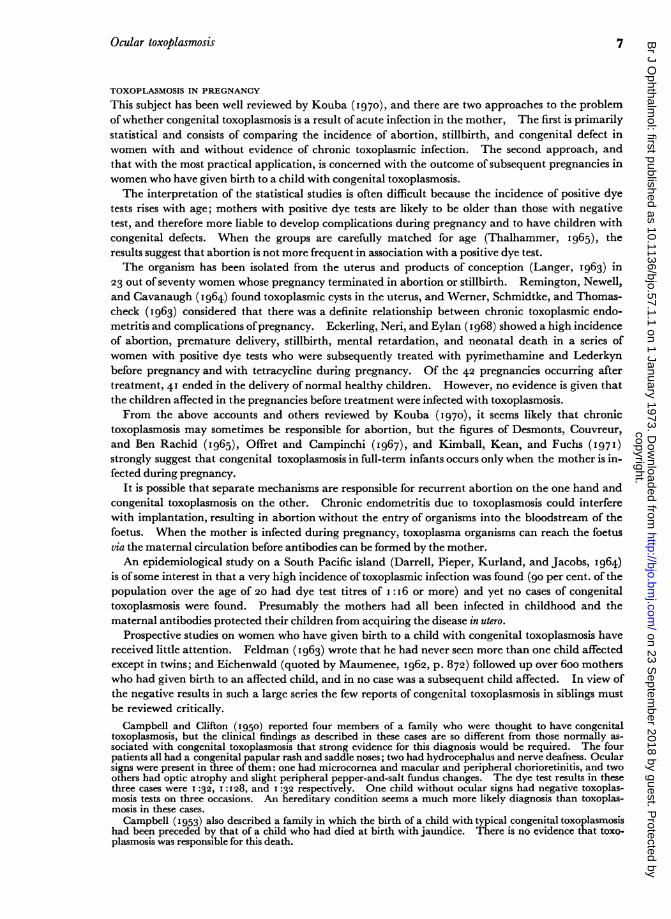
TOXOPLASMOSIS IN PREGNANCY
This subject has been well reviewed by Kouba (I970), and there are two approaches to the problemof whether congenital toxoplasmosis is a result of acute infection in the mother, The first is primarilystatistical and consists of comparing the incidence of abortion, stillbirth, and congenital defect inwomen with and without evidence of chronic toxoplasmic infection. The second approach, andthat with the most practical application, is concerned with the outcome of subsequent pregnancies inwomen who have given birth to a child with congenital toxoplasmosis.The interpretation of the statistical studies is often difficult because the incidence of positive dye
tests rises with age; mothers with positive dye tests are likely to be older than those with negativetest, and therefore more liable to develop complications during pregnancy and to have children withcongenital defects. When the groups are carefully matched for age (Thalhammer, 1965), theresults suggest that abortion is not more frequent in association with a positive dye test.The organism has been isolated from the uterus and products of conception (Langer, I963) in
23 out of seventy women whose pregnancy terminated in abortion or stillbirth. Remington, Newell,and Cavanaugh (i 964) found toxoplasmic cysts in the uterus, and Werner, Schmidtke, and Thomas-check (I963) considered that there was a definite relationship between chronic toxoplasmic endo-metritis and complications ofpregnancy. Eckerling, Neri, and Eylan (1968) showed a high incidenceof abortion, premature delivery, stillbirth, mental retardation, and neonatal death in a series ofwomen with positive dye tests who were subsequently treated with pyrimethamine and Lederkynbefore pregnancy and with tetracycline during pregnancy. Of the 42 pregnancies occurring aftertreatment, 41 ended in the delivery of normal healthy children. However, no evidence is given thatthe children affected in the pregnancies before treatment were infected with toxoplasmosis.From the above accounts and others reviewed by Kouba (1970), it seems likely that chronic
toxoplasmosis may sometimes be responsible for abortion, but the figures of Desmonts, Couvreur,and Ben Rachid (I965), Offret and Campinchi (I967), and Kimball, Kean, and Fuchs (I97I)strongly suggest that congenital toxoplasmosis in full-term infants occurs only when the mother is in-fected during pregnancy.
It is possible that separate mechanisms are responsible for recurrent abortion on the one hand andcongenital toxoplasmosis on the other. Chronic endometritis due to toxoplasmosis could interferewith implantation, resulting in abortion without the entry of organisms into the bloodstream of thefoetus. When the mother is infected during pregnancy, toxoplasma organisms can reach the foetusvia the maternal circulation before antibodies can be formed by the mother.An epidemiological study on a South Pacific island (Darrell, Pieper, Kurland, and Jacobs, I964)
is of some interest in that a very high incidence of toxoplasmic infection was found (go per cent. of thepopulation over the age of 20 had dye test titres of I:I6 or more) and yet no cases of congenitaltoxoplasmosis were found. Presumably the mothers had all been infected in childhood and thematernal antibodies protected their children from acquiring the disease in utero.
Prospective studies on women who have given birth to a child with congenital toxoplasmosis havereceived little attention. Feldman (I963) wrote that he had never seen more than one child affectedexcept in twins; and Eichenwald (quoted by Maumenee, I962, p. 872) followed up over 6oo motherswho had given birth to an affected child, and in no case was a subsequent child affected. In view ofthe negative results in such a large series the few reports of congenital toxoplasmosis in siblings mustbe reviewed critically.
Campbell and Clifton (I950) reported four members of a family who were thought to have congenitaltoxoplasmosis, but the clinical findings as described in these cases are so different from those normally as-sociated with congenital toxoplasmosis that strong evidence for this diagnosis would be required. The fourpatients all had a congenital papular rash and saddle noses; two had hydrocephalus and nerve deafness. Ocularsigns were present in three of them: one had microcornea and macular and peripheral chorioretinitis, and twoothers had optic atrophy and slight peripheral pepper-and-salt fundus changes. The dye test results in thesethree cases were I :32, I :128, and I :32 respectively. One child without ocular signs had negative toxoplas-mosis tests on three occasions. An hereditary condition seems a much more likely diagnosis than toxoplas-mosis in these cases.
Campbell (I953) also described a family in which the birth of a child with typical congenital toxoplasmosishad been preceded by that of a child who had died at birth with jaundice. There is no evidence that toxo-plasmosis was responsible for this death.
7copyright.
on 23 Septem
ber 2018 by guest. Protected by
http://bjo.bmj.com
/B
r J Ophthalm
ol: first published as 10.1136/bjo.57.1.1 on 1 January 1973. Dow
nloaded from

8 E. S. Perkins
Crothers (I943) reported two cases of siblings thought to have toxoplasmosis. In the first, one boy in afamily had convulsions, choroiditis, and cerebral calcification, and a younger brother had choroiditis, hemi-plegia, mental retardation, and cerebral calcification. Serological tests for toxoplasmosis were said to bepositive. In another family, two children had convulsions, choroiditis, and cerebral calcification: themother and a sister also had cerebral calcification. This would imply that the mother had congenital toxo-plasmosis and transmitted the infection to the children. The details of the serological tests are not sufficientfor their significance to be assessed.
Langer (I963) reported the isolation of toxoplasma organisms from a stillbirth and again from an abortion5 months later. He also reported the case of a mother who had had three abortions followed by a prematureinfant which died and in whom organisms were isolated. There is no direct evidence however that theprevious abortions were caused by toxoplasmosis.Remington and others (I964) reported one case of spontaneous abortion in which mouse inoculation from
uterine scrapings was positive for toxoplasmosis. The mother's dye test was positive at a titre of I :256 on twoseparate occasions, suggesting chronic infection. In a further case an abortion at 5 weeks when the dye testin the mother was positive at I :5I2 was followed by a further abortion with isolation of Toxoplasma one yearlater. The dye test titre was the same and the previous positive test excluded infection of the mother duringthe second pregnancy.Coppola and Tondi (I965) described three children in the same family who had signs of the Laurence-
Moon-Biedl syndrome with positive dye tests of I :250, I :5o and I :500 respectively. There is, however, noevidence that this syndrome is caused by toxoplasmosis.
Ciulla, Zanibelli, and Privitera (I964) described a family in which a child aged 3 years had motor distur-bances, cerebral calcification, and a dye test positive to I :I,ooo; a brother aged i8 months had a superioroblique paresis and a dye test positive to I :250; a sister aged I3 years showed mental retardation, a chorio-retinal scar, and a dye test positive to i :io; and another sister aged 14 years had cerebral calcification but anegative dye test. A diagnosis of toxoplasmosis is possible in the three siblings with positive dye tests butother causes cannot be excluded.
Rieger (1966) reported six families in which two or more siblings were thought to have toxoplasmosis on thebasis of positive dye tests. The ocular findings included congenital defects, such as microphthalmos andcoloboma of the iris, but no typical chorioretinal lesions. The diagnosis of toxoplasmosis was made on thebasis of positive dye tests, but in only two cases was the titre above I :64. In one case, a second sibling, toxo-plasma organisms were seen histologically. Unless further confirmation can be obtained that toxoplasmosisis responsible for these congenital anomalies, I feel that the results should be accepted with reservations.
Garcia (i968) reported the case of a woman who had a febrile illness at the fifth month of pregnancy andwhose child was born at the seventh month by Caesarean section. The child died after 24 hours and toxo-plasma organisms were seen in the foetal tissues and placenta. Toxoplasma skin tests in the mother werenegative. A subsequent pregnancy ended in abortion at 5 months and pseudocysts were seen in the adrenalsand in the placenta; I5 months later the mother's dye test was positive I :64, and a year after it was positiveI :10,24.Khanna, Singh, Chowdhry, and Om Prakash (1969) described an I I -month-old infant in whom the haemag-
glutination test was positive I :256 and who showed retinal oedema. Parasites were isolated by mouseinoculation from the CSF. This child was the sixth and only surviving child, all the previous children havingdied of CNS disease before the age of 6 years. The diagnosis of toxoplasmosis was presumptive only, as far asthe earlier children were concerned.
Miller, Aronson, and Remington (i969) reported an infant who died at birth and from whom toxoplasmaorganisms were recovered. The mother's dye test rose from I:4,o96 to I :32,768 after 14 months, stronglysuggesting a recently acquired infection. A second child was born with jaundice but no other evidence ofcongenital toxoplasmosis.
My personal impression of these reports is that they provide some evidence that abortion andstillbirth can be caused by maternal toxoplasmosis, but that no cases of typical congenital toxo-plasmosis in surviving siblings have been proved to occur. If this view is correct, and it is stronglysupported by some of the statistical studies, congenital toxoplasmosis results from acute infection ofthe mother during pregnancy and is extremely unlikely to result from chronic infection.The salient points of this review ofthe literature can be summarized as follows:
(i) A uveitis complicating laboratory infection with toxoplasmosis has been reported in two out of26 cases.
(2) Lymphadenopathy is the commonest clinical manifestation ofacquired toxoplasmosis, occurringin 89 per cent. of cases.
copyright. on 23 S
eptember 2018 by guest. P
rotected byhttp://bjo.bm
j.com/
Br J O
phthalmol: first published as 10.1136/bjo.57.1.1 on 1 January 1973. D
ownloaded from
Ocular toxoplasmosis
(3) Uveitis occurs in I *5 per cent. of cases of toxoplasmic lymphadenopathy, and in I9 per cent. ofthe rarer cases in which the central nervous system is involved. Papillitis and optic atrophy alsooccur in the latter cases.(4) There is some evidence that a chorioretinitis, and more rarely a pan-uveitis, may be the onlymanifestation of acquired toxoplasmosis.(5) Congenital toxoplasmosis only occurs when the mother is infected during that pregnancy.
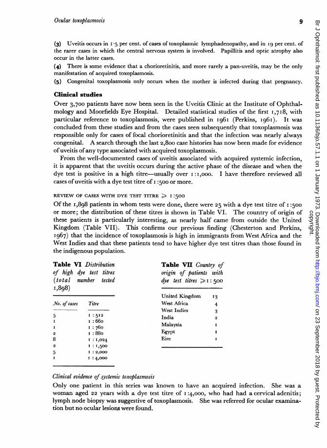
Clinical studiesOver 3,700 patients have now been seen in the Uveitis Clinic at the Institute of Ophthal-mology and Moorfields Eye Hospital. Detailed statistical studies of the first 1,7I8, withparticular reference to toxoplasmosis, were published in I96I (Perkins, I96I). It wasconcluded from these studies and from the cases seen subsequently that toxoplasmosis wasresponsible only for cases of focal chorioretinitis and that the infection was nearly alwayscongenital. A search through the last 2,800 case histories has now been made for evidenceof uveitis ofany type associated with acquired toxoplasmosis.From the well-documented cases of uveitis associated with acquired systemic infection,
it is apparent that the uveitis occurs during the active phase of the disease and when thedye test is positive in a high titre-usually over I :i,ooo. I have therefore reviewed allcases ofuveitis with a dye test titre of I :500 or more.
REVIEW OF CASES WITH DYE TEST TITRE > I :500
Of the I,898 patients in whom tests were done, there were 25 with a dye test titre of I :500or more; the distribution of these titres is shown in Table VI. The country of origin ofthese patients is particularly interesting, as nearly half came from outside the UnitedKingdom (Table VII). This confirms our previous finding (Chesterton and Perkins,I967) that the incidence of toxoplasmosis is high in immigrants from West Africa and theWest Indies and that these patients tend to have higher dye test titres than those found inthe indigenous population.
Table VI Distribution Table VII Country ofof high dye test titres origin of patients with(total number tested dye test titres > : 5001,898)
United Kingdom 13No. of cases Titre West Africa 4
West Indies 35 I :512 India 2
1i:66o Malaysia II I: 7602 I: 88o Egypt8 I : I,024 Eire I2 1 :1,5005 1:2,000I I :4,000
Clinical evidence of systemic toxoplasmosisOnly one patient in this series was known to have an acquired infection. She was awoman aged 22 years with a dye test titre of I :4,ooo, who had had a cervical adenitis;lymph node biopsy was suggestive of toxoplasmosis. She was referred for ocular examina-tion but no ocular lesions were found.
9copyright.
on 23 Septem
ber 2018 by guest. Protected by
http://bjo.bmj.com
/B
r J Ophthalm
ol: first published as 10.1136/bjo.57.1.1 on 1 January 1973. Dow
nloaded from

E. S. Perkins
Two other patients gave a history of systemic disease which could possibly have been dueto toxoplasmosis although there was no direct evidence for this apart from the positiveserology. One was a man aged 5I (No. 3435, Table IX) with a dye test titre of 1:I,024,who had an anterior uveitis with oedema of the posterior pole but no focal lesions; I I yearspreviously he had developed diplopia due to an external rectus paresis; he also had cardiacenlargement and occlusive arterial disease in the right leg. The other patient was awoman aged 54 (No. 2I6I, Table IX) with a dye test titre of I :8oo who had a generalizeduveitis with one area of focal choroiditis. She gave a history of anaemia, eczema, andchronic bronchitis with asthma.Although the systemic symptoms in the two latter patients could conceivably have been
due to toxoplasmosis, there is little to support such a diagnosis. The one with confirmedtoxoplasmic adenitis (No. 3347, Table IX) had no ocular lesions.The remaining 22 cases can be grouped into those with recurrences of a previous chorio-
retinitis, with or without other evidence suggesting that the disease was congenital inorigin, and those with ocular lesions of apparently recent origin.
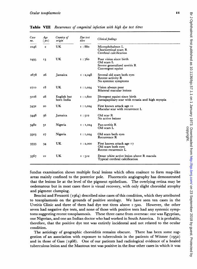
Recurrences of congenital toxoplasmosisThere were eleven cases in which the ocular lesions were typical of recurrences of a con-genital infection, and of these four were known to have had poor vision since birth (TableVIII). Previous recurrences were recorded in two others, and all but one had chorio-retinal scars. The exception, No. 3567, is particularly interesting as at the first examina-tion a dense white area of activity was seen above the macular region of the right eye andno old scars were apparent. There were, however, typical areas of cerebral calcification,so that the ocular lesion was almost certainly the result ofcongenital infection.
Other cases with dye test titres of I :500 or moreNo clear pattern emerges from the small number of cases, details of which are given inTable IX (overleaf). In Nos. 14I8 and I769 the infection could have been congenital,and the only other case of typical focal chorioretinitis was No. 3557, in which the dyetest titre of I :2,ooo was suggestive ofrecent infection. There was no other clinical evidenceof toxoplasmosis, but the possibility remains that the small area of chorioretinitis was theonly manifestation of systemic infection. In four cases (Nos. 2529, 33 I 5, 3476, and 3497)the only evidence of toxoplasmosis was the positive dye test, and the association with theocular lesions was most probably coincidental. The three cases of geographic choroiditiswill be discussed below.
ConclusionHigh dye test titres can be found in adults with recurrences of congenital infection, par-ticularly in immigrants from West Africa and the West Indies. High dye test titres mayoccasionally be found in other types of uveitis, but the rarity of this finding suggests thatin the absence of other evidence of a recent systemic infection this finding is purely coin-cidental.
GEOGRAPHIC CHOROIDITIS
This interesting condition has been described by a number of authors (Witmer, 1952;Maumenee, I968; Gass, I968; Schlaegel, I969; Krill and Archer, I971; Perkins andDobree, 1972). Affected patients complain of the sudden onset of defective vision, and
IOcopyright.
on 23 Septem
ber 2018 by guest. Protected by
http://bjo.bmj.com
/B
r J Ophthalm
ol: first published as 10.1136/bjo.57.1.1 on 1 January 1973. Dow
nloaded from
Ocular toxoplasmosis
Table VII Recurrences of congenital infection with high dye test titres
Age Country of(yrs) origin
2 UK
Dye testtitre
I: 880
Clinicalfindings
Microphthalmos LChorioretinal scars RCerebral calcification
2495 13 UK
2678 26 Jamaica
27Io I8 UK
31o8 28 English butborn India
3432 20 UK
3448 36 Jamaica
3482 31 Nigeria
3503 27 Nigeria
3533 34 UK
3567 21 UK
I : 760 Poor vision since birthOld scars LSevere generalized uveitis RConvergent squint
I : 2,048 Several old scars both eyesRecent activity RNo systemic symptoms
I: 1,024 Vision always poor
Bilateral macular lesions
I : I,6oo Divergent squint since birthJuxtapapillary scar with ectasia and high myopia
I : 1,024 First known attack age I IMacular scar with recurrence L
I :512 Old scar RNo active lesions
I: 1,024 Pan-uveitis ROld scars L
I: 1,024 Old scars both eyes
Recurrence R
I 2,000 First known attack age 17Old scars both eyesRecent recurrence L
I : 512 Dense white active lesion above R maculaTypical cerebral calcification
fundus examination shows multiple focal lesions which often coalesce to form map-likeareas mainly confined to the posterior pole. Fluorescein angiography has demonstratedthat the lesions lie at the level of the pigment epithelium. The overlying retina may beoedematous but in most cases there is visual recovery, with only slight choroidal atrophyand pigment clumping.
Bencini and Frezzotti (1 964) described nine cases of this condition, which they attributedto toxoplasmosis on the grounds of positive serology. We have seen ten cases in theUveitis Clinic and three of them had dye test titres above I :500. However, the otherseven had negative dye tests, and none of those with positive tests had any systemic symp-
toms suggesting recent toxoplasmosis. These three came from overseas: one was Egyptian,one Nigerian, and one an Indian doctor who had worked in South America. It is probable,therefore, that the positive dye test was entirely incidental and not related to the ocularcondition.The aetiology of geographic choroiditis remains obscure. There has been some sug-
gestion of an association with exposure to tuberculosis in the patients of Witmer (1952)and in those of Gass (I968). One of our patients had radiological evidence of a healedtuberculous lesion and the Mantoux test was positive in the four other cases in which it was
Caseno.
2246
IIcopyright.
on 23 Septem
ber 2018 by guest. Protected by
http://bjo.bmj.com
/B
r J Ophthalm
ol: first published as 10.1136/bjo.57.1.1 on 1 January 1973. Dow
nloaded from
E. S. Perkins
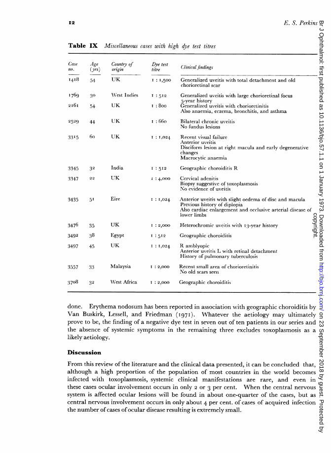
Table IX Miscellaneous cases with high dye test titres
Case Age Country qfno. (yrs) origin
I418 54 UK
1769 30
2I6I 54
Dye testtitre
I : I,500
Clinical findings
Generalized uveitis with total detachment and oldchorioretinal scar
WN'est Indies I:5I2 Generalized uveitis with large chorioretinal focus5-year history
UK i 8oo Generalized uveitis with chorioretinitisAlso anaemia, eczema, bronchitis, and asthma
2529 44 UK
33I5 6o UK
3345 32 India
3347 22 UK
3435 5I Eire
I: 66o Bilateral chronic uveitisNo fundus lesions
I: I,024 Recent visual failureAnterior uveitisDisciform lesion at right macula and early degenerativechangesMacrocytic anaemia
I 512 Geographic choroiditis R
I 4,000 Cervical adenitisBiopsy suggestive of toxoplasmosisNo evidence of uveitis
I 1,024 Anterior uveitis with slight oedema of disc and maculaPrevious history of diplopiaAlso cardiac enlargement and occlusive arterial disease oflower limbs
Heterochromic uveitis with 13-year history
Geographic choroiditis
R amblyopicAnterior uveitis L with retinal detachmentHistory of pulmonary tuberculosis
3557 33 Malaysia
3708 32 WNest Africa
I: 2,000 Recent small area of chorioretinitisNo old scars seen
I: 2,000 Geographic choroiditis
done. Erythema nodosum has been reported in association with geographic choroiditis byVan Buskirk, Lessell, and Friedman (I97I). Whatever the aetiology may ultimatelyprove to be, the finding of a negative dye test in seven out of ten patients in our series andthe absence of systemic symptoms in the remaining three excludes toxoplasmosis as a
likely aetiology.
Discussion
From this review of the literature and the clinical data presented, it can be concluded that,although a high proportion of the population of most countries in the world becomesinfected with toxoplasmosis, systemic clinical manifestations are rare, and even inthese cases ocular involvement occurs in only 2 or 3 per cent. When the central nervous
system is affected ocular lesions will be found in about one-quarter of the cases, but as
central nervous involvement occurs in only about 4 per cent. of cases of acquired infectionthe number ofcases ofocular disease resulting is extremely small.
34763492
3497
35
38
45
UK
Egypt
UK
I : 2,000
I :512
I : 1,024
12
copyright. on 23 S
eptember 2018 by guest. P
rotected byhttp://bjo.bm
j.com/
Br J O
phthalmol: first published as 10.1136/bjo.57.1.1 on 1 January 1973. D
ownloaded from

Ocular toxoplasmosis
If uveitis is such an uncommon complication of overt systemic infection with toxo-plasmosis, it is likely to be even more uncommon in association with subclinical infection.Population surveys have always shown that the incidence of infection increases with age,and if toxoplasmic chorioretinitis resulted from chronic acquired infection, as manyauthors have suggested, the number of cases would rise with age. This is not so; toxo-plasmic chorioretinitis occurs most frequently in the second and third decade of life and israre after the age of 50 (Perkins, I 96 I; Hogan and others, I 964).
If the ocular lesions result from acquired infection, it would be expected that the patientswould have higher levels of circulating antibody than asymptomatic members of thepopulation. High dye test titres are, however, exceptional in toxoplasmic uveitis andeven when found are not conclusive evidence of acquired infection, as some cases ofundoubted congenital infection may have dye test titres of over I :500 in adult life. Hoganand others (i 964) considered that only forty of 240 cases of toxoplasmic uveitis werecongenital in origin, and yet 20I of these cases had dye test titres of I :256 or less. Threeof the cases with high dye test titres had signs and symptoms of recent systemic infectionwhich would account for the high titres.
In a personal series of I,7I8 cases of uveitis, i88 were considered to be due to toxo-plasmosis (Perkins, I96I) and in only one of these was there evidence of possible recentsystemic infection. The possibility that the uveitis resulted from subclinical acquiredinfection could not be entirely excluded, but the presence of old scars in the fundus stronglysuggested that almost all cases were recurrences of congenital infection. On the otherhand it is not possible to exclude congenital infection in many of the cases described in theliterature (see above) as resulting from acquired infection without systemic signs or symp-toms.
I believe, therefore, that cases of focal chorioretinitis with positive serological evidence oftoxoplasmosis should be considered to result from congenital infection unless there is aclear history of recent systemic signs and symptoms of toxoplasmosis. This view is sup-ported by Schlaegel (I969), who stated that it was possible that all cases of toxoplasmicchorioretinitis were congenital in origin, and that when no previous scar was visible theinflammation arose from the rupture of cysts in the nerve-fibre layer of the retina whichwere not visible on ophthalmoscopic examination.When ocular complications do occur in systemic toxoplasmosis, focal chorioretinitis is
the typical manifestation, but extraocular muscle pareses, papillitis, and optic atrophy mayaccompany disease of the central nervous system. Apart from a few cases of pan-uveitiswhich have been described in association with systemic disease, there is no convincingevidence that anterior uveitis, retinal vasculitis, or geographic choroiditis are caused bytoxoplasmosis.Although it is possible that chronic uterine infection with toxoplasmosis is responsible
for abortion and stillbirth, congenital toxoplasmosis has never been confirmed in siblingsand results only from infection of the mother during pregnancy. It follows, therefore, thata woman with circulating antibodies to Toxoplasma will not have a child with congenitaltoxoplasmosis. There is, however, a serious risk to the child if the mother becomesinfected during pregnancy.
SummaryFrom a review of the literature and from clinical data presented, the following conclusionswere drawn:(i) Subclinical infection with toxoplasmosis is very common throughout the world but
13copyright.
on 23 Septem
ber 2018 by guest. Protected by
http://bjo.bmj.com
/B
r J Ophthalm
ol: first published as 10.1136/bjo.57.1.1 on 1 January 1973. Dow
nloaded from

E. S. Perkins14
systemic clinical manifestations are rare and even in these cases ocular involvementoccurs in only 2 to 3 percent.
(2) Ocular complications of systemic infection are more likely to occur when the centralnervous system is affected.
(3) Uveitis as the only sign of acquired toxoplasmosis is very rare.
(4) Almost all cases of toxoplasmic chorioretinitis seen in the United Kingdom are theresult of congenital infection.(5) There is no convincing evidence that anterior uveitis, retinal vasculitis, or geo-graphic choroiditis are caused by toxoplasmosis.(6) Chronic uterine infection may be responsible for abortion or stillbirth but congenitaltoxoplasmosis has never been confirmed in siblings and results only from infection of themother during pregnancy.
(7) A woman with circulating antibodies to Toxoplasma will not have a child withcongneital toxoplasmosis.
References*BACHMANN, F., KEISER, G., and MARTENET, A. C. (I962) Helv. med. Acta, 29, 74*BENCINI, A., and FREZZOTTI, R. (I964) "La toxoplasmosi". Pozzi, RomeBENGTSSON, E. (1950) Cardiologia (Basel), 17, 289BEVERLEY, J. K. A., SKIPPER, E., and MARSHALL, S. C. (1955) Brit. med. J., I, 577*BINKHORST, C. D. (1948) "Toxoplasmosis". Stenfert Kroese, LeydenBOERICKE, H. (I962) Z. Kinderheilk., 86, 306*BORIANI, P., MAEKELT, G. A., BOCCALANDRO, I., and ARISTIMUNO, J. (I969) Acta med. venez., I6, 15IBRENNAN, A. J., BROWN, T. McP., WARREN, j., and VRANIAN, G. (I949) Amer. J. Med., 7, 431
BROWN, j., and JACOBS, L. (I956) Ann. intern. Med., 44, 565CAMPBELL, A. M. G. (1953) Proc. roy. Soc. Med., 46, 89I
, and CLIFTON, F. (I950) Brain, 73, 281CASSADY, J. V. (I960) Trans. Amer. ophthal. Soc., 58, 392CHESTERTON, J. R., and PERKINS, E. S. (I967) Brit. J. Ophthal., 5I, 6I7CHODOS, J. B., and HABEGGER-CHODOS, H. E. (I963) Canad. med. Ass. J., 88, 505
CIULLA, M., ZANIBELLI, G., and PRIVITERA, A. (I964) Boll. Soc. med.-chir. Cremona, I8, 8iI*CONRADS, H. (I96I) Klin. Mbl. Augenheilk., 138,30COPPOLA, L., and TONDI, I. V. (I965) Minerva ginec., 17, 774CROTHERS, B. (1943) Arch. Neurol. Psychiat., 49, 315DARRELL, R. W., PIEPER, S., KURLAND, L. T., and JACOBS. L, (I964) Arch. Ophthal. (Chicago), 71, 63*DEGOS, R., TOURAINE, R., LEIBOWITCH, M., ESCANDE, j.-P., and TINTHOIN, J.-F. (I967) Bull. Soc.
franf. Derm. Syph., 74, 748*DESMONTS, G. (I966) Arch. Ophthal. (Chicago), 76, 839
, COUVREUR, j., and BEN RACHID, M.-S. (I965) Arch. frang. Pediat., 22, I 183DUKE-ELDER, S., ASHTON, N., and BRIHAYE-VAN GEERTRUYDEN, M. (I953) Brit. J. Ophthal., 37, 321ECKERLING, B., NERI, A., and EYLAN, E. (I968) Fertil. and Steril., I9, 883ENGELBRECHT, E., and FRANCESCHETTI, A. (I963) Path. et Microbiol. (Basel), 26, 731
FAIR, J. R., and WALLS, K.W. (I962) Invest. Ophthal., I, 418FELDMAN, H. A. (I963) New Engl. J. Med., 269, 1212FFRENCH, G. (I965) Surv. Ophthal., 10, 313FRANCESCHETTI, A., and ENGELBRECHT, E. (I964) Ophthalmologica (Basel), 147, 273*FRAN9OIS, J. (I963) "La toxoplasmose et ses manifestations oculaires". Masson, Paris*FRANKE, H. (I960) In "Human Toxoplasmosis", ed. J. C. Siim, p. I03. Munksgaard, Copenhagen
and HORST, H. G. (195I) Dtsch. med. Wschr., 76, 1049
copyright. on 23 S
eptember 2018 by guest. P
rotected byhttp://bjo.bm
j.com/
Br J O
phthalmol: first published as 10.1136/bjo.57.1.1 on 1 January 1973. D
ownloaded from
Ocular toxoplasmosis '5
*FRENKEL, J. K. (i962) In "Toxoplasmosis; with Special Reference to Uveitis", ed. A. E. Maumenee,p. 799. Williams and Wilkins, Baltimore
(I 97o) jt. infect. Dis., 122, 553,WEBER, R. W., and LUNDE, M. N. (i960) J. Amer. med. Ass., 173, 147I
FREZZOTTI, R., BERENGO, A., GUERRA, R., and CAVALLINI, F. (I965) Amer. J5. Ophthal., 59, 1099FRIEDMANN, C. T., and KNOX, D. L. (1969) Arch. Ophthal. (Chicago), 8I, 48IFRUGONI, C. (1951) Policlinico, Sez. prat., 58, 3GARCIA, A. G. P. (i968) Arch. Dis. Childh., 43, 705GARIN, J. P. (ig60) In "Human Toxoplasmosis", ed. J. C. Siim, p. 87. Munksgaard, CopenhagenGASS, J. D. M. (i968) Arch. Ophthal. (Chicago), 8o, I77GENZ, H. (i963) Internist (Berl.), 4, 4I7GIROUD, P., LE GAC, P., ROGER, F., and GAILLARD, J.-A. (1953) Sem. Hop. Paris, 29,4036*GUTZU, E., ATANASIU, M., TIMOFTE, V., LAUSCH, L., MACARIE, R., and OGHINA, B. (1970) Ibid., 46, 665HABEGGER, H. (1953) "Le reservoir biologique animal et sa relation avec l'infection toxoplasmiquehumaine". Thesis, Geneva
* (0954) Arch. Ophtal., n.s. 14, 470HARRIS, R. J. (1967) Acta paediat. scand., 56, 58oHEWSON, G. E. (196i) Irish J. med. Sci., 6th ser., No. 427, 323HORMANN, J. (1955) Z. ges. inn. Med., 1o, I50HOGAN, M. J. (0958a) Trans. Amer. Acad. Ophthal. Otolaryng., 62, 7
(I958b) Amer. J. Ophthal., 46, 467* , KIMURA, S. j., and O'CONNOR, G. R. (i964) Arch. Ophthal. (Chicago), 72, 592*HOMMES, 0. R. , and PRICK, J. J. G. (i965) Proc. kon. Ned. Akad. Wet. (Series C: Biol. med. Sci), 68,
360* , (i966) Psychiat. Neurol. Neurochir. (Amst.), 69, 24IHUDSON, J. R. (0954) Brit. J. Ophthal., 38, 179HUTCHISON, W. M., DUNACHIE, J. F., SIIM, j. c., and WORK, K. (I970) Brit. med. 5., I, 142*JACOBS, L. (ig60) In "Human Toxoplasmosis", ed. J. C. Siim, p. 149. Munksgaard, Copenhagen
- COOK, M. K., and WILDER, H. C. (1954a) Trans. Amer. Acad. Ophthal. Otolaryng., 58, I93- FAIR, J. R., and BICKERTON, J. H. (I954b) A.M.A. Arch. Ophthal., 52, 63
JONES, T. C., KEAN, B. H., and KIMBALL, A. C. (i969) N. r. St. J. Med., 69, 2237*KAYHOE, D. E., JACOBS, L., BEYE, H. K., and McCULLOUGH, N. B. (I957) New Engl. J. Med., 257, I247*KEEL, H. J., ROTH, W., KEISER, G., REUTTER, F., and MARZ, G. (1963) Schweiz. med. Wschr., 93, 1465KHANNA, K. K., SINGH, M., CHOWDHRY, P., and OM PRAKASH, (i969) Canad. med. Ass. 5., 100, 343KIMBALL, A. C., KEAN, B. H., and FUCHS, F. (197I) Amer. J. Obstet., III, 211
KOEZE, T. H., and KLINGON, G. H. (i964) Arch. Neurol. Psychiat., II, I9IKOKE, M. P. (I 953) Amer. 5'. Ophthal., 36, 845KOUBA, K. (1970) Cs- Gynek., 35, 234*KRAMER, W. (I963) Ned. T. Geneesk., 107, 1551*KREPLER, P., WEINGARTEN, K., and BRENNER, H. (I965) Z. Kinderheilk., 93, I55KRILL, A. E., and ARCHER, D. (I97I) Amer. 5. Ophthal., 72, 562LALISSE, A., MISES, J., and DURAND, C. H. (I964) Ann. Pediat., I1, 4ILANGER, H. (I963) Obstet. and Gynec., 21, 3I8
(I966) In "Toxoplasmose", ed. H. Kirchhoffand H. Kraubig. Thieme, StuttgartLAVAT, J. (I962) Bull. Soc. belge Ophtal., No. 130, p. 257*LELONG, M., BERNARD, J., DESMONTS, G., and COUVREUR, j. (I960) Arch. franf. Pidiat., 17, 28IMACCHI, V.. (I968) Minerva med., 59, 3868*MAFART, Y, BERENI, J., THOMAS, J., SAGNET, H., and ZIMMER, H. (I967) Marseille-mid., 104, I99*MAGNUSSON, J. H. (I951) Nord. Med., 45, 344MANISSADJIAN, A., D'OLIVEIRA PENNA, H. A., COSTA VAZ, F. A., RAMOS, J. L. A., BORGES, M. A. G., and
SCHVARTSMAN, S. (1967) Rev. Ass. mid. bras., I3, 395*MARTENET, c., and PESTALOZZI, D. (I96I) Ophthalmologica (Basel), 14I, 398
copyright. on 23 S
eptember 2018 by guest. P
rotected byhttp://bjo.bm
j.com/
Br J O
phthalmol: first published as 10.1136/bjo.57.1.1 on 1 January 1973. D
ownloaded from
E. S. Perkins
MARTINELLI, G., and ROSSI, R. (I964) Rass. Studi psichiat., 53, 456MATSUBAYASHI, H., KIOKE, T., UYEMURA, M., SOH, Y., and HAMANO, K., (I96I) Keio J. Med., 10, 209
MAUMENEE, A. E. (Ed.) (I962) "Toxoplasmosis: with Special Reference to Uveitis". Second SloanSymposium on Uveitis. Williams and Wilkins, Baltimore
(I968) In "Clinical Methods in Uveitis". Fourth Sloan Symposium on Uveitis, ed.S. B. Aronson, C. N. Gamble, E. K. Goodner, and G. R. O'Connor, p. 21. Mosby, St. Louis
MIKUNI, I. (I968) Rinsho Ganka, 22, 943MILLER, M. J., ARONSON, w. j., and REMINGTON, J. s., (I969) Ann. intern. Med., 71, I39NEU, H. C. (I967) J. Amer. med. Ass., 202, 844*NEUMANN, C. G., HILTON, c., and BARREDA, A. (I960) Amer. J. Dis. Child., Ioo, I I 7*NOETZEL, H. (I951) Beitr. path. Anat., III 4I9NUTT, A. B., and BEVERLEY, J. K. A. (I963) Trans. ophthal. Soc. Aust., 23, 35OFFRET, G., and CAMPINCHI, R. (I967) "XX Conc. ophthal. Germania I966. Acta", vol. I, p. 348
Int. Congr. Ser. No. 146. Excerpta Medica Foundation, AmsterdamOHKAWA, C., YONEKURA, Y., and ITO, K. (I969) Folia ophthal. jap., 20, 295*PASMANIK, S. (I966) Arch. chil. Oftal., 23, 3IPAULLEY, J. W., GREEN, W. P. D., JONES, R., and KANE, E. P. (1954) Lancet, 2, 624*PERKINS, E. S. (I96I) "Uveitis and Toxoplasmosis". Churchill, London
and DOBREE, J. H. (1972) "The Differential Diagnosis of Fundus Conditions".Kimpton, London
*PILLAT, A., and THALHAMMER, 0. (1957) v. Graefes Arch. Ophthal., 158, 403*PRICK, J. J. G., and PRICK-HOEFNAGELS, J. A. M. (I950) Folia psychiat. neerl., 53, 352*PRIOR, J. A., COLE, C. R., DOCTON, F. L., SASLAW, s., and CHAMBERLAIN, D. M. (1953) Arch. intern. Med.,
92 314
*RAMSELL, T. G., and GAMERO, B. A. (I967) Brit. J. Ophthal., 51, 282RAWAL, B. D. (1959) J. clin. Path., 12, 59REMINGTON, J. I., MILLER, M. j., and BROWNLEE, I. (1968) J. lab. clin. Med., 71, 855
, NEWELL, j. w., and CAVANAUGH, E. (I964) Obstet. and Gynec., 24, 25*REMKY, H. (I959) Bull. Soc. franf. Ophtal., 72, 274* (I962) "Toxoplasmosis: argumenta et documenta ophthalmologica". Lehmann,
MunichRIEGER, H. (1955) Ber. dtsch. ophthal. Ges., 59, I23
(I966) v. Graefes Arch. Ophthal., 170, 223*ROBINSON, P. (I 947) Ann. paediat. (Basel), z68, I 34*SARAUX, H., SERINGE, P., BACH, C. and LE BESNERAIS, Y. (I962) Bull. Soc. belge Ophtal., No. 130,
p. 257*SAUNDERS, s. j. and THATCHER, G. N. (I963) S. Afr. med. J., 37, 1026SCHLAEGEL, T. F. (I969) "Essentials of Uveitis". Churchill, LondonSEITELBERGER, F., and SPIEL, W. (1953) Wien. Z. Nervenheilk., 7, 298SEXTON, R. C., EYLES, D. E., and DILLMAN, R. E. (I953) Amer. J. Med., 14, 366*SHIMIZU, K., NAKAYAMA, I., and SANO. T. (I969) Acta Soc. ophthal. jap., 73, 172*SIIM, j. c. (I96I) Surv. Ophthal., 6, 781SOESTBERGEN, A. A. van (1957) Ned. T. Geneesk., 1OI I649*SPAULDING, A. G. and FONT, R. L. (I967) Surv. Ophthal., I2, i6STANKOVIC, I. and MILOJKOVIC, B. (I963) Riv. ital. Tracoma, 15, 14STRAUB, W. (1949) Ber. dtsch. ophthal. Ges., 55, 362
(I962) In "Toxoplasmose: Gottinger Symposion, I960", ed. H. Kirchhoff and H.Kraubig, p. 74. Thieme, Stuttgart
*STROM, J. (195I) Acta. med. scand., 139, 244TANAKA, H., TAKADA, S., SAKAMOTO, H., TAKASU, T., SANO, R., and UEMICHI, T. (I965) J. Osaka City
med. Center, 14, no. I-3, p. 159*TEJERO LAMARCA, j., and SOUTO CRESPO, J. M. (1970) Rev. clin. esp., 119, 445
copyright. on 23 S
eptember 2018 by guest. P
rotected byhttp://bjo.bm
j.com/
Br J O
phthalmol: first published as 10.1136/bjo.57.1.1 on 1 January 1973. D
ownloaded from
Ocular toxoplasmosis 17
*TERRAGNA, A., BRUSA, A., BRAITO, A., BERNABO BREA, G., and SCHENONE, M. A. (I 970) G. Mal.infett., 22, 2IO
THALHAMMER, 0. (I954) Lst. Z. Kinderheilk., IO, 3i6(I965) Arch. Gynak., 202, 96
UMDENSTOCK, R., MANDOUL, R., and PESTRE-ALEXANDRE, M. (1965) Bull. Soc. Path. exot., 58, 207
VAN BUSKIRK, E. M., LESSELL, s., and FRIEDMAN, E. (197I) Arch. Ophthal. (Chicago), 85, 369*VANEGAS BARRIOS, R., and VINOCOUR GRANADOS, R. (i 968) Arch. Inst. Cardiol. Mex., 38, 204*V.ERONESI, R., and CAMARGO, M. (I964) Rev. Inst. Med. trop. S. Paulo, 6, 75*VOISIN, J., MASSELIN, S., AUVERT, B., and BERTRAND, j.-j. (I965) Bull. Soc. Ophtal. Fr., 65, io66WERNER, H., SCHMIDTKE, L., and THOMASCHECK, G. (I963) Klin. Wschr., 41, 96WETTINGFELD, R. F., ROWE, j., and EYLES, D. E. (I956) Ann. intern. Med., 44, 557
WILDER, H. C. (1952) A. M. A. Arch. Ophthal., 47, 425; 48, 127*WILLEMSE, j. (I966) Psychiat. Neurol. Neurochir. (Amst.), 69, I5*WISING, P. (1952) Nord. Med., 47, 563WITMER, R. (1952) Ophthalmologica (Basel), 123, 353WOLLHEIM, E. (1952) Munch. med. Wschr., 94, I94
BibliographyDUKE-ELDER, S., and PERKINS, E. S. (I966) "Diseases of the Uveal Tract". "System of Ophthal-
mology", vol. 9, ed. S. Duke-Elder. Kimpton, LondonFRAN9OIS, j. (I963) "La toxoplasmose et ses manifestations oculaires". Masson, ParisJACOBS, L. (I967) Adv. Parasit., 5, IMAUMENEE, A. E. (ed.) (I962) "Toxoplasmosis: with special Reference to Uveitis". Second SloanSymposium on Uveitis. Williams and Wilkins, Baltimore
PERKINS, E. S. (i 96 I) "Uveitis and Toxoplasmosis". Churchill, London
copyright. on 23 S
eptember 2018 by guest. P
rotected byhttp://bjo.bm
j.com/
Br J O
phthalmol: first published as 10.1136/bjo.57.1.1 on 1 January 1973. D
ownloaded from