nucleus subputaminalis (ayala): the still …dementia.hiim.hr/1999 neuroscience.pdf · ·...
TRANSCRIPT
Pergamon
Neuroscience Vol. 89, No. 1, pp. 73 -89, 1999 Copyright © 1998 IBRO. Published by Elsevier Science Ltd
Printed in Great Britain. All rights reserved PII: S0306-4522(98)00304-2 0306-4522/99 $19.00+0.00
NUCLEUS SUBPUTAMINALIS (AYALA): THE STILL DISREGARDED MAGNOCELLULAR COMPONENT OF THE
BASAL FOREBRAIN MAY BE H U M A N SPECIFIC A N D CONNECTED WITH THE CORTICAL SPEECH AREA
G. SIMI(~,*§¶ L. M R Z L J A K , t A. FUQI(~,} B. WINBLAD,§ H. LOVRIC* and I. KOSTOVIC*
*Croatian Institute for Brain Research and Department of Anatomy, Section of Neuroanatomy, Zagreb University School of Medicine, Zagreb 10000, Croatia
?Section of Neurobiology, Yale University School of Medicine, 333 Cedar Street, New Haven, CT 06510, U.S.A.
:~Institute for Medical Research and Occupational Health, Zagreb 10000, Croatia
{}Department of Clinical Neuroscience and Family Medicine, Geriatric Section, Karolinska Institute, Huddinge University Hospital, S-14186 Huddinge, Sweden
Abstract--The small magnocellular group located within the rostrolateral extension of the basal forebrain was named and described as the nucleus subputaminalis in the human and chimpanzee brain by Ayala. Analysis of cytoarchitectonic and cytochemical characteristics of this cell group has been largely disregarded in both classical and more current studies. We examined the nucleus subputaminalis in 33 neurologically normal subjects (ranging from 15 weeks of gestation to 71 years-of-age) by using Nissl staining, choline acetyltransferase immunohistochemistry, acetyl cholinesterase histochemistry and nerve growth factor receptor immunocytochemistry. In addition, we applied reduced nicotinamide adenine dinueleotide phosphate-diaphorase histochemistry and calbindin-D28k immunocytochemistry in three neurologically normal subjects. At the most rostrolateral levels we describe the previously poorly characterized component of the lateral (periputaminal) subdivision of the subputaminal nucleus, which may be human specific since it is not described in non-human primates. Moreover, we find the human subputaminal nucleus best developed at the anterointermediate level, which is the part of the basal nucleus that is usually much smaller or missing in monkeys. The location of subputaminal cholinergic neurons within the frontal lobe, the ascension of their fibers through the external capsule towards the inferior frontal gyrus, the larger size of the subputaminal nucleus on the left side at the most rostral and anterointermediate levels and the most protracted development among all magnocellular aggregations within the basal forehrain strongly suggest that they may be connected with the cortical speech area.
These findings give rise to many hypotheses about the possible role of the subputaminal nucleus in various neurodegenerative, neurological and psychiatric disorders, particularly Alzheimer's disease and primary progressive aphasia. Therefore, future studies on the basal forebrain should more carefully investigate this part of the basal nucleus. © 1998 IBRO. Published by Elsevier Science Ltd.
Key words: Alzheimer's disease, cholinergic neurons, cortex, NGF receptor, nucleus basalis, primary progressive aphasia.
The most important cytoarchitectonic feature of the primate mediobasal forebrain is the presence of the
~I'o whom correspondence should be addressed. Abbreviations: ACHE, acetylcholinesterase; AD, Alzheimer's
disease; BuChE, butyrylcholinesterase; BW284C51, 1,5-bis(4-allyl-dimethylammonium-phenyl)-pentan-3-on dibromide; CalB, calbindin-D28k; Chl-4, designation for the subdivisions of the nucleus basalis complex as cholin- ergic cell groups; CHAT, choline acetyltransferase; DAB, 3,3'-diaminobenzidine; isoOMPA, tetraisopropyl- pyrophosporamide; NADPH-d, reduced nicotinamide adenine dinucleotide phosphate-diaphorase; NB, nucleus basalis; p75 NGFr, low-affinity nerve growth factor recep- tor; NSP, nucleus subputaminalis; PBS, phosphate- buffered saline; PPA, primary progressive aphasia; RT, room temperature; SPA, slowly progressive anarthria; TBS, Tris-buffered saline.
73
complex chain of the magnocellular nuclei which represent an extension of the brainstem reticular c o r e . 11'30'33"73'74'89'102 The largest cytoarchitectural
entity in the magnocellular basal forebrain system is the nucleus basalis (NB) which is situated below the lentiform nucleus. The well-known eponym "nucleus basalis of Meynert" was given by K611iker, 53 although Meynert 's work does not exactly show this cell group . 78'79 It was Brockhaus who realized that the magnocellular neurons in the Meynert 's nucleus are only one component of the whole complex of basal forebrain magnocellular cell groups. 11 The prominence of the NB in the primate brain may be explained by the extraordinary expansion of the cortical mantle which represents its main innervation target. 10"20'24'30"42"44"47'72'74'77'87'94'95'109 More than
74 G. Simi6 et al.
90% of the neurons in the NB are cholinergic in that their perikarya and axons contain choline acetyl- transferase (CHAT). 26'72f14'82'87,96 Their axons build
the most massive of all the subcallosal extra-thalamic afferent systems of the primate cerebral cor- tex. 44'50"74'77'87 Since neurons of the NB also receive a
cholinergic input from other cholinergic groups of the mediobasal forebrain and from the pontomesen- cephalic cholinergic nuclei, 43 most ChAT-positive neurons of the NB contain acetyl cholinesterase (ACHE). It has been suggested that AChE can be used as a very good marker for cholinergic neurons in the NB. 72
Besides ChAT and AChE co-localization, about 90% of all cholinergic neurons of the primate NB also display immunoreactivity for the low-affinity nerve growth factor receptor (p75 N G F r ) . 3'34"54,s°'gs There- fore, although indirect, p75 NGFr is also an excellent marker for cholinergic neurons . 54'57 In addition to AChE and NGFr, some cholinergic neurons may contain other cytochemical markers such as reduced nicotinamide adenine dinucleotide phosphate- diaphorase (NADPH-d), 2s the vitamin-D dependent calcium binding protein calbindin-D28k (CalB) 16 and others. However, due to the technical difficulties in visualization of such markers in human tissue, 2~'95 a more reliable differentiation of the NB from sur- rounding structures (especially for quantitation), can be obtained using classical Nissl stain. 31
The most widely accepted terminology for the primate magnocellular basal forebrain is based upon the topographical distribution of ChAT reactive cell bodies, as proposed by Mesulam and col- leagues. 74'7s'76 Although the human NB is larger and more complex, for the sake of consistency, this terminology is also used for human brain. According to this nomenclature, the main part of the human magnocellular basal forebrain, NB, is designated as the Ch4 group and is further subdivided into six sectors. Anterior part (Ch4a) is divided by vascula- ture into the anteromedial (Ch4am) and anterolateral (Ch4al) sectors, the anterointermediate part (Ch4ai) which spans the anterior and intermediate parts (and is not well-developed in monkeys), the inter- mediate part (Ch4i) which is divided by the ansa peduncularis into the intermediodorsal (Ch4id) and intermedioventral (Ch4iv) sectors. The posterior part occupies a sector designated as Ch4p. 72 Since this terminology is based on cholinergic neurons only it may not always be appropriate, especially for developmental studies when cytoarchitectonic boundaries of the basal forebrain are even less clear. Comparing the terminology of Mesulam and Geula, 72 with the classification given by Brockhaus, ~ which was based on a Nissl study, the following conclusions can be derived: the Ch4am group mainly corresponds to the pars diffusa, and Ch4i to the pars compacts of Brockhaus. The posterior group (Ch4p) of Mesulam and collegues corresponds to the several cell clusters that we have described
in human preterm infant and designated as pars aggregata. 59
Besides the "individual cytochemical signa- tures", 2s'71 different divisions of the NB have physiologically and morphologically heterogeneous neurons with discrete projectional patterns, indicating that the NB is composed of different organizational units. One cell group, which is topographically and cytoarchitectonically related to the NB, was named and described as the nucleus subputaminalis (NSP) many years ago in the human and anthropoid monkey brain by Giuseppe Ayala. s'9 Still, with the exception of three studies, 31'33"~15 neither classical j1'53 nor relatively modern studies 3'17'2°'56'72'82"8s'91"96 have mentioned
or classified this most lateral component of the magnocellular basal forebrain complex (see Discussion). We previously showed that this magno- cellular group of the basal forebrain shows distinct cholinesterase and cholinergic properties. 59'62 In the present study, we describe in greater detail the NSP and its components. We analysed the NSP by using Nissl staining, ChAT immunohistochemistry, AChE histochemistry and p75 NGFr immunocytochemistry in 33 neurologically normal subjects. In addition, we applied NADPH-d histochemistry and CalB immu- nocytochemistry in three normal subjects. Our main intention was to cytoarchitectonically and immuno- histochemically better describe and define this pro- spective source of cholinergic innervation of the cerebral cortex. The normative parameters obtained can be used in further studies of basal forebrain abnormalities, particularly in Alzheimer's disease (AD).
EXPERIMENTAL PROCEDURES
Preparation o[" tissue and Nissl staining
This study was based on 36 post mortem human brains from the Zagreb Collection of Human Brains. 6° All subjects had no history of neurological or psychiatric disease. The brains were obtained at routine autopsies in accordance with the Croatian law and under the control of the Faculty Ethical Committee at the Zagreb University School of Medicine. All brains were fixed within 24 h of death. Basic data about the patients from whom brains were obtained, the methods successfully applied, as well as the causes of death are shown in Tables 1 and 2.
After fixation in 4% formaldehyde buffered with 0.1 M phosphate buffer, the basal forebrain was removed from both hemispheres of each brain. Several tissue blocks were cut in horizontal and sagittal planes and all remaining blocks cut in a coronal plane. The blocks were dehydrated through a graded series of ethanol solutions (70%, 70%, 96%, 96%, 100%, and 100%; 12 h each) and ether-absolute alcohol solution (with ether and alcohol in equal parts) for 180rain twice. The blocks were then embedded in 2% celloidin (Cedukol, "Merck", cat. no. 4363) for 24 h, 4% celloidin for the next 24 h, and finally in 8% celloidin until adequately hardened. The serial sections of 25 p,m thickness were collected in 70% ethanol, put in 50% ethanol, then put in 5% ethanol for 2 min, then put in distilled water for 5 min, and finally in staining solution. Staining solutions comprised, one part 0.5% Cresyl Violet in distilled water mixed with four parts distilled water. The mounted sections
Nucleus subputaminalis (Ayala) 75
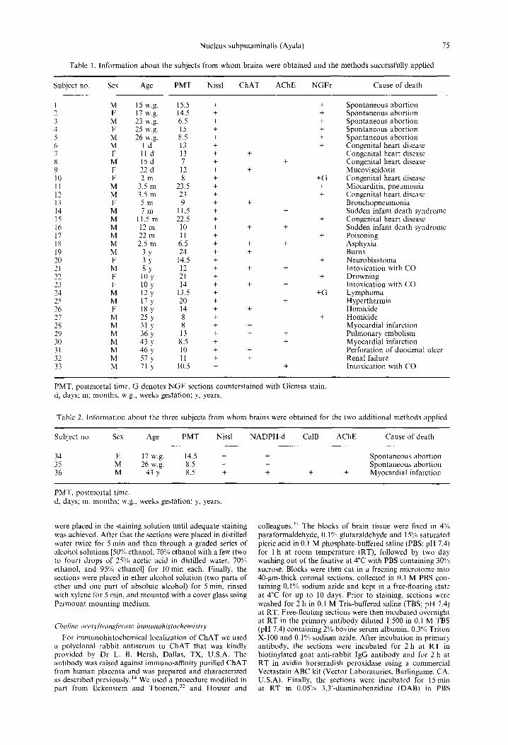
Table I. Information about the subjects from whom brains were obtained and the methods successfully applied
Subject no. Sex Age P M T Nissl ChAT AChE N G F r Cause of death
1 M 15 w.g. 15.5 + + Spontaneous abortion F 17 w.g. 14.5 + + Spontaneous abortion
3 M 23 w.g. 6.5 + + Spontaneous abortion 4 F 25 w.g. 15 + + Spontaneous abortion 5 M 26 w.g. 8.5 + + Spontaneous abortion 6 M 1 d 13 + + Congenital heart disease 7 F 11 d 13 + + Congenital heart disease 8 M 15 d 7 + + Congenital heart disease 9 F 22 d 12 + + Mucoviscidosis 10 F 2 m 8 + +G Congenital heart disease 11 M 3.5 m 23.5 + + Miocarditis, pneumonia 12 M 3.5 m 23 + + Congenital heart disease 13 F 5 m 9 + + Bronchopneumonia 14 M 7 m 11.5 + + Sudden infant death syndrome 15 M 11.5 m 22.5 + + Congenital heart disease 16 M 12 m 10 + + + Sudden infant death syndrome 17 M 22 m 11 + + Poisoning 18 M 2.5 m 6.5 + + + Asphyxia 19 M 3 y 24 + + Burns 20 F 3 y 14.5 + + Neuroblas toma 21 M 8 y 12 + + + Intoxication with CO 22 F 10 y 21 + + Drowning 23 F 10 y 14 + + + Intoxication with CO 24 M 12 y 13.5 + +G Lymphoma 25 M 17 y 20 + + Hyperthermia 26 F 18 y 14 + + Homicide 27 M 25 y 8 + + Homicide 28 M 31 y 8 + + Myocardial infarction 29 M 36 y 13 + + + Pulmonary embolism 30 M 43 y 8.5 + + Myocardial infarction 31 M 46 y 10 + + Perforation of duodenal ulcer 32 M 57 y 11 + + Renal failure 33 M 71 y 10.5 + + Intoxication with CO
PMT, postmortal time. G denotes N G F sections counterstained with Giemsa stain. d, days; m, months; w.g., weeks gestation; y, years.
Table 2. informat ion about the three subjects from whom brains were obtained for the two additional methods applied
Subject no. Sex Age P M T Nissl N A D P H - d CalB AChE Cause of death
34 F 17 w.g. 14.5 + + Spontaneous abortion 35 M 26 w.g. 8.5 + + Spontaneous abortion 36 M 43 y 8.5 + + + + Myocardial infarction
PMT, postmortal time. d, days: m, months; w.g., weeks gestation; y, years.
were placed in the staining solution until adequate staining was achieved. After that the sections were placed in distilled water twice for 5 rain and then through a graded series of alcohol solutions [50% ethanol, 70% ethanol with a few (two to four) drops of 25% acetic acid in distilled water, 70% ethanol, and 95% ethanol] for 10min each. Finally, the sections were placed in ether alcohol solution (two parts of ether and one part of absolute alcohol) for 5 min, rinsed with xylene for 5 rain, and mounted with a cover glass using Permount mount ing medium.
Choline aeetyltransjerase immunohistochemistry For immunohistochemical localization of C hAT we used
a polyclonal rabbit antiserum to C h A T that was kindly provided by Dr L. B. Hersh, Dallas, TX, U.S.A. The antibody was raised against immuno-affinity purified C h A T from human placenta and was prepared and characterized as described previously] 4 We used a procedure modified in part from Eckenstein and Thoenen, 22 and Houser and
colleagues. 37 The blocks of brain tissue were fixed in 4% paraformaldehyde, 0.1% glutaraldehyde and 15% saturated picric acid in 0.1 M phosphate-buffered saline (PBS; pH 7.4) for 1 h at room temperature (RT), followed by two day washing out of the fixative at 4°C with PBS containing 30% sucrose. Blocks were then cut in a freezing microtome into 40-gin-thick coronal sections, collected in 0.1 M PBS con- taining 0. l% sodium azide and kept in a free-floating state at 4°C for up to 10 days. Prior to staining, sections were washed for 2 h in 0.1 M Tris-buffered saline (TBS; pH 7.4) at RT. Free-floating sections were then incubated overnight at RT in the primary antibody diluted 1:500 in 0.1 M TBS (pH 7.4) containing 2% bovine serum albumin, 0.3% Triton X-100 and 0.1% sodium azide. After incubation in primary antibody, the sections were incubated for 2 h at RT in biotinylated goat anti-rabbit IgG antibody and for 2 h at RT in avidin horseradish peroxidase using a commercial Vectastain ABC kit (Vector Laboratories, Burlingame, CA, U.S.A). Finally, the sections were incubated for 15rain at RT in 0.05% 3,3'-diaminobenzidine (DAB) in PBS
76 G. Simi6 et al.
containing 0.01% hydrogen peroxide. Between each incu- bation step the sections were carefully rinsed for 10 min in four changes of 0.1 M TBS (pH 7.4). The reaction product was stabilized by the addition of nickel chloride to the DAB solution (0.4 mg/ml). After mounting on glass slides, sec- tions were dehydrated in a graded ethanol series, cleared in xylene and coverslipped with Entellan (Merck). Specificity of the ChAT labelling procedure was determined by the absence of immunochemical reaction in sections in which the primary antibody was omitted.
Acetyl cholinesterase histochemistry For AChE staining, tissue blocks were fixed by immersion
in 0.1 M phosphate buffer containing 1% glutaraldehyde and 2% paraformaldehyde at 4-6°C for 24-48 h. After fixation, the blocks were cut into 8-10 mm (for fetuses) or 1-2 cm (for older brains) thick slabs, and then serially sectioned on a freezing microtome at 70 or 80 gm. Free- floating sections were stained for AChE by using the Lewis's modification of the acetylthiocholine iodide method of Koelle and Friedenwald. s1'64'65 Since acetylcholine is a substrate for both AChE and butyrylcholinesterase (BuChE), specific inhibitors were used to block either or both enzymes. The histochemigal specificity was tested by treatment of adjacent sections in incubation medium con- taining eserine (10 4 M) to inhibit both AChE and BuChE, tetraisopropylpyrophosporamide (isoOMPA, 10 4M final concentration) for the inhibition of BuChE, 66'1°3 and 1,5- bis(4-allyl-dimethylammonium-phenyl)-pentan-3-on dibro- mide (BW284C51, 10 5, Sigma) for the inhibition of true ACHE. The reaction product was developed with sodium sulphide in 0.2 M acetic acid after incubation for up to 24 h. In all the brains examined, eserine inhibited all staining in the nucleus basalis complex. On the other hand, isoOMPA had little effect, indicating that the basal forebrain reaction is due to ACHE. Some sections were processed by the Karnovsky-Roots "direct coloring" method, 12'4s modified by Tago.l~3 Control tests were performed as described in our previous papers. 59"6~
Nerve growth factor receptor immunocytochemistry Coronal blocks containing whole basal forebrain were
fixed and cut on a freezing microtome into 90-ram-thick sections. Alternate sections were taken for immunocyto- chemistry and Nissl staining. The mouse monoclonal anti- body raised against human low-affinity p75 NGFr (gift of Dr M. Herlyn, Wistar Institute, PA, U.S.A.) was diluted 1:50 in 0.1 M PBS containing 1% horse serum, 5% bovine serum albumin and 5% sucrose. Sections were incubated in the primary antibody overnight at 40C and then, after washing in a 0.1 M TBS solution containing 0.05% Triton X-100, visualized using anti-mouse IgG Vectastain ABC kit (cat. no. PK-4002, Vector, Burlingame, CA, U.S.A.). One series of sections was processed by using incubation medium without the primary antibody to check for non-specific staining. The chromogen solution which completed the reaction consisted of 0.05% DAB, 0.005% H202, and 8% NiCI 2. Sections were mounted on gelatine-coated slides, dehydrated through graded series of alcohols (70%, 95%, 99%), cleared in xylenes and coverslipped with Permount mounting medium.
Reduced nicotinamide adenine dinucleotide phosphate- diaphorase histochemistry
After fixation in 4% paraformaldehyde buffered in 0.1 M PBS for 2448 h and cryoprotection in graded concen- trations of cold (4°C) 0.1 M buffered sucrose (12%, 16%, 18%; pH 7.4), tissue blocks were cut frozen at 50-70 gm and placed in 0.1 M PBS (pH 7.4) prior to NADPH-d histo- chemistry. Free-floating coronal sections were rinsed for 5 min in 0.1 M PBS (pH 7.4), and then incubated at 37°C in 50ml of 0.1M PBS (pH 8.0), containing l ml of 0.8%
Triton X-100 (Sigma, St Louis, MO, U.S.A.), 41.65 mg (1 mM) of reduced NADPH (Sigma), and 32.65mg (0.8 mM) of Nitroblue Tetrazolium (Sigma). The incubation period varied between 60-180 min. Trial sections for each case were monitored by intermittent microscopic examina- tion. The reaction was stopped by rinsing the sections in cold 0.1 M PBS (pH 7.4). Sections were then mounted, dried, dehydrated and coverslipped. Control sections were treated identically except for either the omission of the substrate for the NADPH-d reaction, the omission of the electron acceptor (Nitroblue Tetrazolium), or by heating the solution at 90°C for 5 min to denature the enzyme activity. In all control procedures, the specific NADPH-d histochemical reaction was eliminated.
Calbindin-D28k immunocvtochemistry After the cryoprotection and sectioning (80 gin), coronal
free-floating sections were processed according to the avidin biotin technique, 38 using the Vectastain ABC kit (Vector Laboratories, Burlingame, CA, U.S.A.). Primary monoclonal anti-calbindin-D28k antibody (Sigma, St Louis, MO, U.S.A.) was used in dilution 1:1000. For controls the primary antiserum was omitted.
RESULTS
Generally, in Nissl-stained preparations the NSP can be identified and described at the three main rostrocaudal (coronal) levels: anterior (septo- chiasmatic), intermediate (tubero-iufundibular), and posterior (premammillary-mammillary). The nucleus was readily discernible by the presence of the charac- teristic magnocellular and hyperchromatic neurons containing distinct nucleoli. An example of the NSP in a larger series of Nissl-stained sections of one brain is given in Fig. 1. An example of N G F r immuno- reactivity of NSP on selected levels is given in Fig. 2.
Anterior ( septal-chiasmatic ) level
At this level several (usually three) cell groups were found to be in close vicinity of the putamen: one or more (usually two) of these cell clusters were smaller and situated ventral to the putamen and formed medial (or subputaminal) division of the NSP, while one bigger cell group was located lateral to the putamen and formed lateral (or periputaminal) divi- sion of the NSP (Fig. 1D). The medial cell groups were not well delineated and were composed of the large multipolar, polymorphic perikarya. Analysis of serial sections through the more caudal coronal levels revealed that these cell groups continue into the subputaminal cell groups situated lateral to the pos- terior limb of the anterior commissure. The lateral periputaminal cell group was present in all cases analysed. It occupied an elongated and flattened disc-shaped territory situated along the ventrolateral border of the putamen and was actually incorporated in the basal origin of the external capsule. It was, however, clearly different from other interstitial com- ponents of the basal forebrain where neuronal peri- karya do not form the nucleus. Within its territory there was usually one main spindle-shaped cell
Nucleus subputaminalis (Ayala) 77
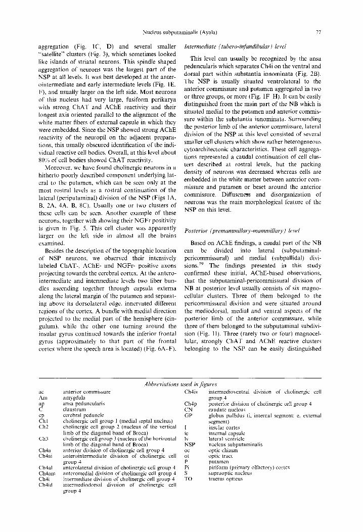
aggregation (Fig. I C, D) and several smaller "satellite" clusters (Fig. 3), which sometimes looked like islands of striatal neurons. This spindle shaped aggregation of neurons was the largest part of the NSP at all levels. It was best developed at the anter- ointermediate and early intermediate levels (Fig. 1E, F), and usually larger on the left side. Most neurons of this nucleus had very large, fusiforrn perikarya with strong ChAT and AChE reactivity and their longest axis oriented parallel to the alignment of the white matter fibers of external capsule in which they were embedded. Since the NSP showed strong AChE reactivity of the neuropil on the adjacent prepara- tions, this usually obscured identification of the indi- vidual reactive cell bodies. Overall, at this level about 80% of cell bodies showed ChAT reactivity.
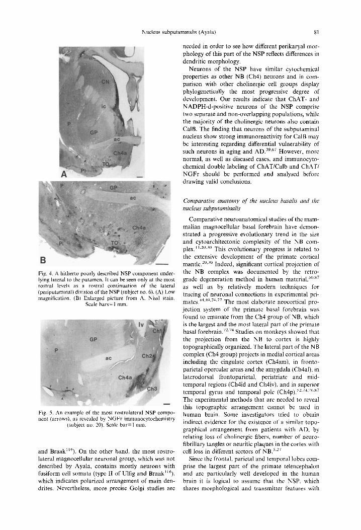
Moreover, we have found cholinergic neurons in a hitherto poorly described component underlying lat- eral to the putamen, which can be seen only at the most rostral levels as a rostral continuation of the lateral (periputaminal) division of the NSP (Figs 1A, B, 2A, 4A, B, 8C). Usually one or two clusters of these cells can be seen. Another example of these neurons, together with showing their NGFr positivity is given in Fig. 5. This cell cluster was apparently larger on the left side in almost all the brains examined.
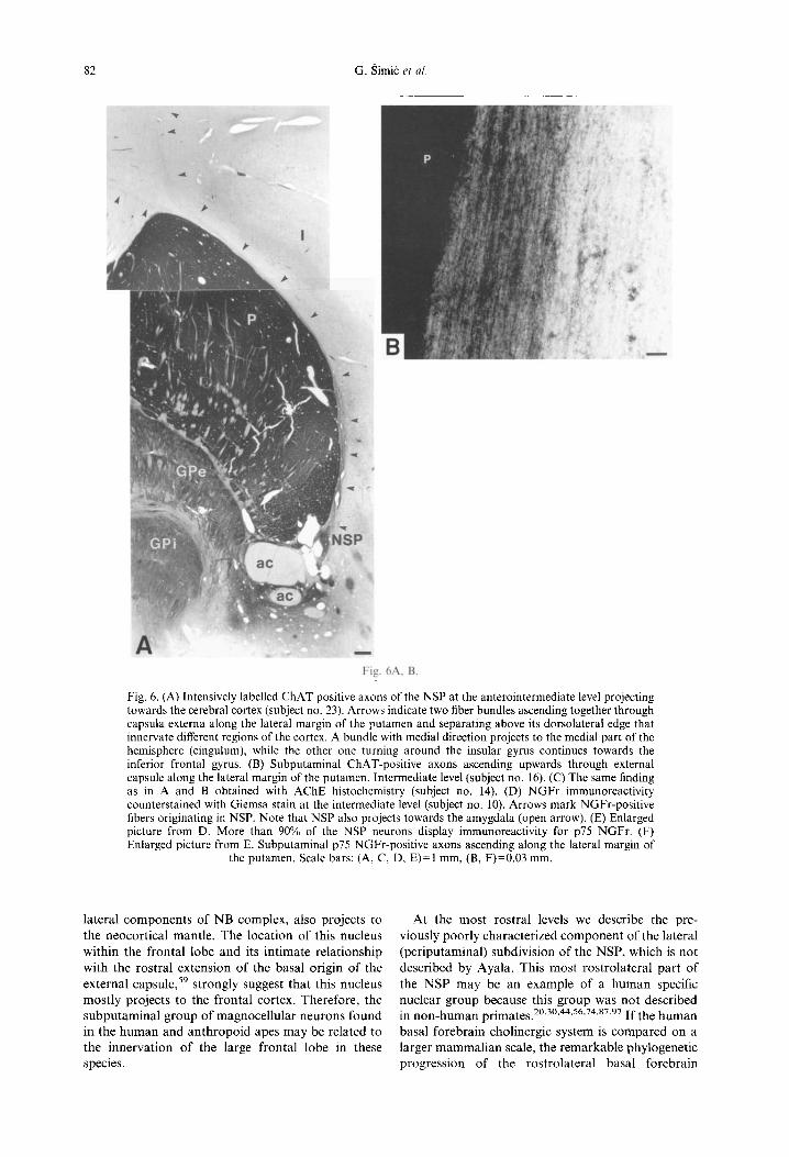
Besides the description of the topographic location of NSP neurons, we observed their intensively labeled CHAT-, ACHE- and NGFr- positive axons projecting towards the cerebral cortex. At the antero- intermediate and intermediate levels two fiber bun- dles ascending together through capsula externa along the lateral margin of the putamen and separat- ing above its dorsolateral edge, innervated different regions of the cortex. A bundle with medial direction projected to the medial part of the hemisphere (cin- gulum), while the other one turning around the insular gyrus continued towards the inferior frontal gyrus (approximately to that part of the frontal cortex where the speech area is located) (Fig. 6A-F).
Intermediate ( tubero-inJundibular ) level
This level can usually be recognized by the ansa peduncularis which separates Ch4i on the ventral and dorsal part within substantia innominata (Fig. 2B). The NSP is usually situated ventrolateral to the anterior commissure and putamen aggregated in two or three groups, or more (Fig. IF-H). It can be easily distinguished from the main part of the NB which is situated medial to the putamen and anterior commis- sure within the substantia innominata. Surrounding the posterior limb of the anterior commissure, lateral division of the NSP at this level consisted of several smaller cell clusters which show rather heterogeneous cytoarchitectonic characteristics. These cell aggrega- tions represented a caudal continuation of cell clus- ters described at rostral levels, but the packing density of neurons was decreased whereas cells are embedded in the white matter between anterior com- missure and putamen or beset around the anterior commissure. Diffuseness and disorganization of neurons was the main morphological feature of the NSP on this level.
Posterior (premammillary-mammillary) level
Based on AChE findings, a caudal part of the NB can be divided into lateral (subputaminal- pericommissural) and medial (subpallidal) divi- sions. 59 The findings presented in this study confirmed these initial, AChE-based observations, that the subputaminal-pericommissural division of NB at posterior level usually consists of six magno- cellular clusters. Three of them belonged to the pericommissural division and were situated around the mediodorsal, medial and ventral aspects of the posterior limb of the anterior commissure, while three of them belonged to the subputaminal subdivi- sion (Fig. 11). Three (rarely two or four) magnocel- lular, strongly ChAT and AChE reactive clusters belonging to the NSP can be easily distinguished
a c
Am ap C cp Chl Ch2
Ch3
Ch4a Ch4ai
Ch4al Ch4am Ch4i Ch4id
Abbreviations used in figures anterior commissure Ch4iv amygdala ansa peduncularis Ch4p claustrum CN cerebral peduncle GP cholinergic cell group 1 (medial septal nucleus) cholinergic cell group 2 (nucleus of the vertical I limb of the diagonal band of Broca) ic cholinergic cell group 3 (nucleus of the horizontal lv limb of the diagonal band of Broca) NSP anterior division of cholinergic cell group 4 oc anterointermediate division of cholinergic cell ot group 4 P anterolateral division of cholinergic cell group 4 Pi anteromedial division of cholinergic cell group 4 S intermediate division of cholinergic cell group 4 TO intermediodorsal division of cholinergic cell group 4
intermedioventral division of cholinergic cell group 4 posterior division of cholinergic cell group 4 caudate nucleus globus pallidus (i, internal segment; e. external segment) insular cortex internal capsule lateral ventricle nucleus subputaminalis optic chiasm optic tract putamen piriform (primary olfactory) cortex supraoptic nucleus tractus opticus
78 G. Simid et al.
£
Fig. 1A-H.
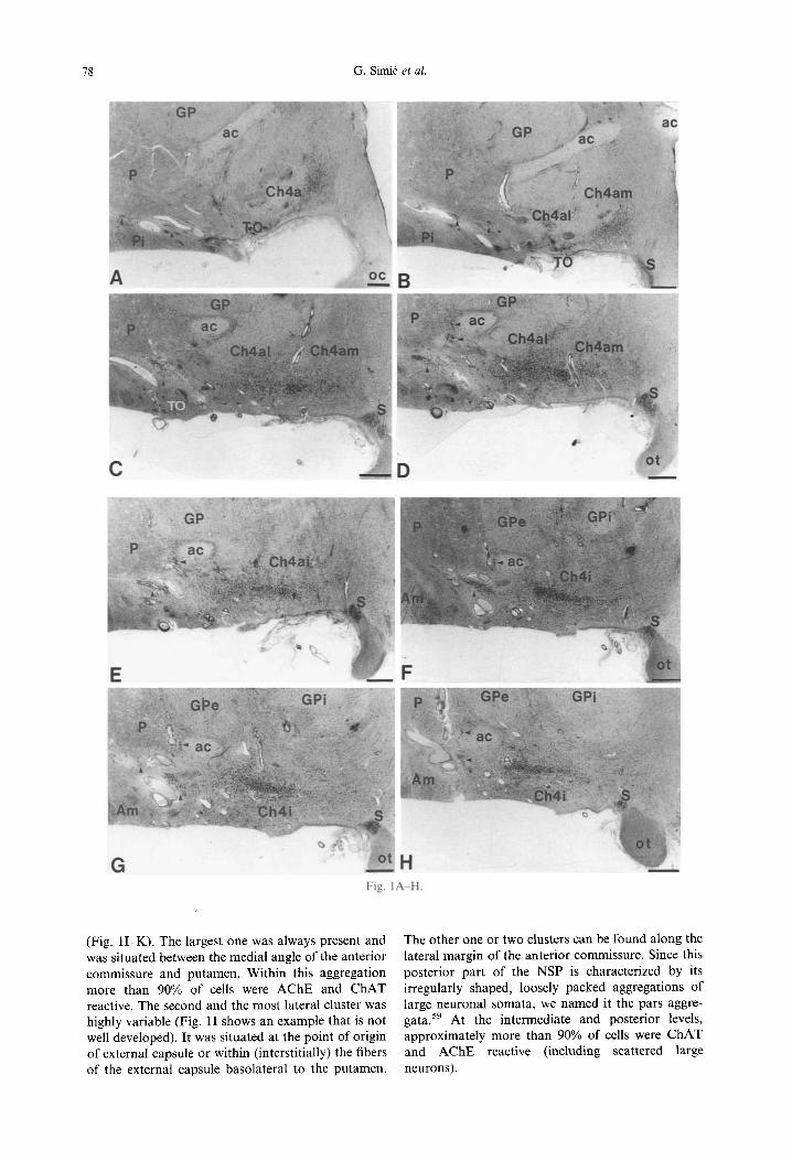
(Fig. I I -K). The largest one was always present and was situated between the medial angle of the anterior commissure and putamen. Within this aggregation more than 90% of cells were AChE and ChAT reactive. The second and the most lateral cluster was highly variable (Fig. 11 shows an example that is not well developed). It was situated at the point of origin of external capsule or within (interstitially) the fibers of the external capsule basolateral to the putamen.
The other one or two clusters can be found along the lateral margin of the anterior commissure. Since this posterior part of the NSP is characterized by its irregularly shaped, loosely packed aggregations of large neuronal somata, we named it the pars aggre- gata. 59 At the intermediate and posterior levels, approximately more than 90% of cells were ChAT and AChE reactive (including scattered large neurons).
Nucleus subputaminalis (Ayala) 79
Fig. l I K.
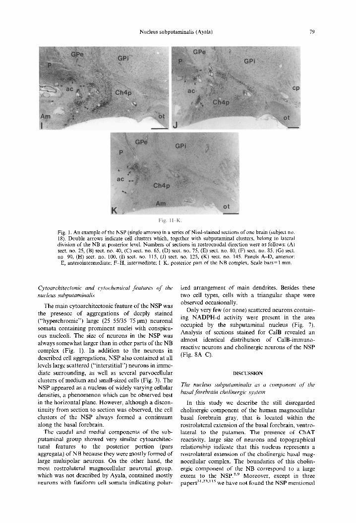
Fig. 1. An example of the NSP (single arrows) in a series of Nissl-stained sections of one brain (subject no. 18). Double arrows indicate cell clusters which, together with subputaminal clusters, belong to lateral division of the NB at posterior level. Numbers of sections in rostrocaudal direction were as follows: (A) sect. no. 25, (B) sect. no. 40, (C) sect. no. 65, (D) sect. no. 75, (E) sect. no. 80, (F) sect. no. 85, (G) sect. no 90, (H) sect. no. 100, (I) sect. no. 115, (J) sect. no. 125, (K) sect. no. 145. Panels A-D, anterior;
E, anterointermediate; F-H, intermediate; I K, posterior part of the NB complex. Scale bars= 1 mm.
Cytoarchitectonic and cytochemical features of the nucleus subputaminalis
The main cytoarchitectonic feature of the NSP was the presence of aggregations of deeply stained ("hyperchromic") large (25-55/35 75 gm) neuronal somata containing prominent nuclei with conspicu- ous nucleoli. The size of neurons in the NSP was always somewhat larger than in other parts of the NB complex (Fig. 1). In addition to the neurons in described cell aggregations, NSP also contained at all levels large scattered ("interstitial") neurons in imme- diate surrounding, as well as several parvocellular clusters of medium and small-sized cells (Fig. 3). The NSP appeared as a nucleus of widely varying cellular densities, a phenomenon which can be observed best in the horizontal plane. However, although a discon- tinuity from section to section was observed, the cell clusters of the NSP always formed a continuum along the basal forebrain.
The caudal and medial components of the sub- putaminal group showed very similar cytoarchitec- tural features to the posterior portion (pars aggregata) of NB because they were mostly formed of large multipolar neurons. On the other hand, the most rostrolateral magnocellular neuronal group, which was not described by Ayala, contained mostly neurons with fusiform cell somata indicating polar-
ized arrangement of main dendrites. Besides these two cell types, cells with a triangular shape were observed occasionally.
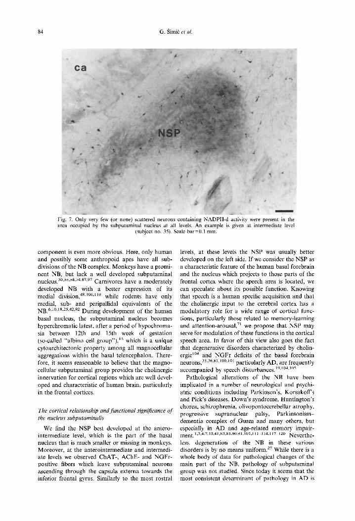
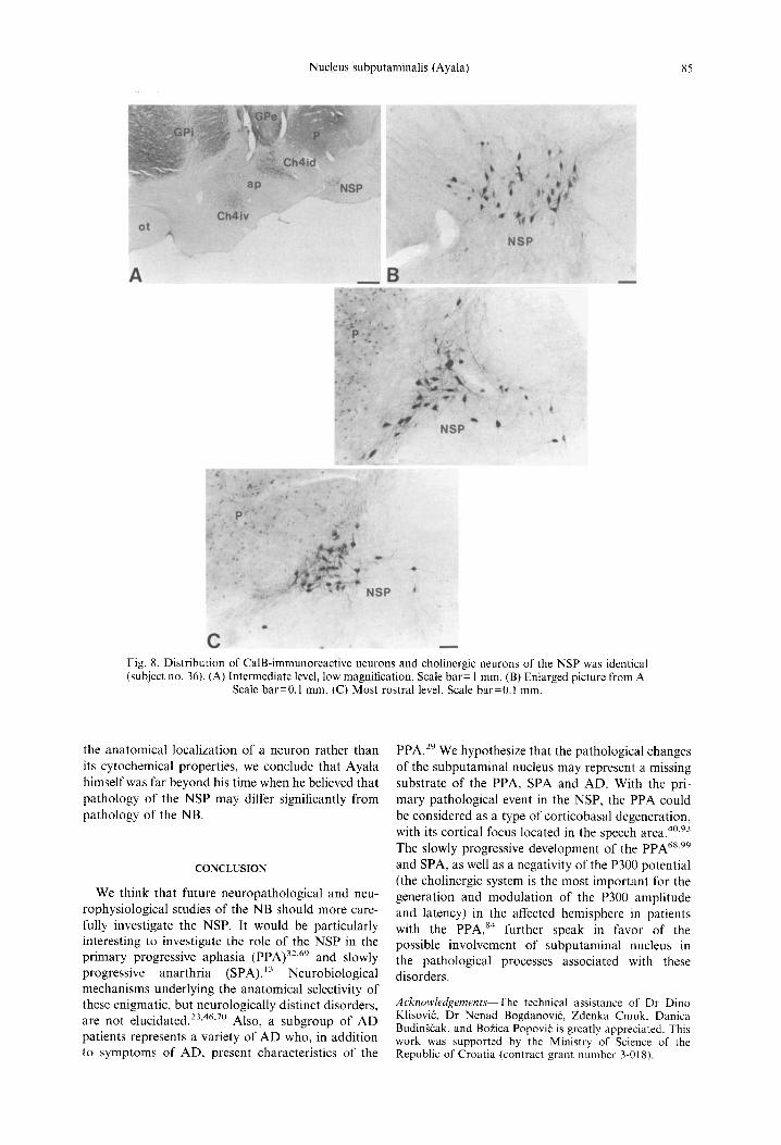
Only very few (or none) scattered neurons contain- ing NADPH-d activity were present in the area occupied by the subputaminal nucleus (Fig. 7). Analysis of sections stained for CalB revealed an almost identical distribution of CalB-immuno- reactive neurons and cholinergic neurons of the NSP (Fig. 8A-C).
DISCUSSION
The nucleus subputaminalis as" a component of the basal forebrain cholinergic system
In this study we describe the still disregarded cholinergic component of the human magnocellular basal forebrain gray, that is located within the rostrolateral extension of the basal forebrain, ventro- lateral to the putamen. The presence of ChAT reactivity, large size of neurons and topographical relationship indicate that this nucleus represents a rostrolateral extension of the cholinergic basal mag- nocellular complex. The boundaries of this cholin- ergic component of the NB correspond to a large extent to the NSP. 8'9 Moreover, except in three papers 31'33'1~5 we have not found the NSP mentioned
80 G. Simi6 et al.
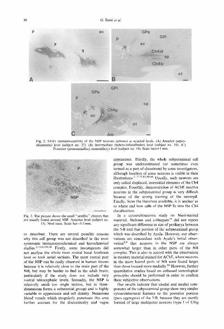
Fig. 2. NGFr immunoreactivity of the NSP neurons (arrows) at selected levels. (A) Anterior (septo- chiasmatic) level (subject no. 27). (B) Intermediate (tubero-infundibular) level (subject no. 19). (C)
Posterior (premammillary-mammillary) level (subject no. 19). Scale bars= 1 mm.
~i!i~ ! ~ ~i ~i!!
Fig. 3. The picture shows the small "satellite" clusters that are usually found around NSP. Anterior level (subject no.
13). Nissl stain. Scale bar=0.5 ram.
or described. There are several possible reasons why this cell group was not described in the most systematic immunocytochemical and histochemical studies. 2'72's2'88'96 Firstly, some investigators did not analyse the whole most rostral basal forebrain level or took serial sections. The most rostral part of the NSP can be easily observed in human fetuses because it is relatively close to the main part of the NB, but may be harder to find in the adult brain, particularly if the study does not include very rostral telencephalic levels. Secondly, the NSP is relatively small (on single section, but in three- dimensions forms a substantial group) and is highly variable in appearance and cell density. Numerous blood vessels which irregularly punctuate this area further account for the discontinuity and vague
appearance. Thirdly, the whole subputaminal cell group was underestimated (or sometimes even termed as a part of claustrum) by some investigators, although location of some neurons is visible in their i l l u s t r a t i o n s . 2"17"72"s2"88"96 Usually, such neurons are
only called displaced, interstitial elements of the Ch4 complex. Fourthly, demonstration of AChE reactive neurons in the subputaminal group is very difficult because of the strong staining of the neuropil. Finally, from the literature available, it is unclear as to where and how cells of the NSP fit into the Ch4 classification.
In a cytoarchitectonic study on Nissl-stained material, Hedreen and colleagues 33 did not report any significant difference in size of perikarya between the NB and that portion of the subputaminal group which was described by Ayala. However, our obser- vations are concordant with Ayala's initial obser- vation s'9 that neurons in the NSP are always somewhat larger than in other parts of the NB complex. This is also in accord with the observation in monkey material stained for ACHE, where neurons in the more lateral parts of NB were found larger than those located more medially, s6 However, further quantitative studies based on unbiased stereological principles should be performed in order to confirm these subjective observations.
Our results indicate that caudal and medial com- ponents of the subputaminal group show very similar cytoarchitectural features to the posterior portion (pars aggregata) of the NB, because they are mostly formed of large multipolar neurons (type I of Ulfig
Nucleus subputaminalis (Ayala) 81
iiiiii!ilil i ~!!i ~ iii:;iiiii~
needed in order to see how different perikaryal mor- phology of this part of the NSP reflects differences in dendritic morphology.
Neurons of the NSP have similar cytochemical properties as other NB (Ch4) neurons and in com- parison with other cholinergic cell groups display phylogenetically the most progressive degree of development. Our results indicate that CHAT- and NADPH-d-positive neurons of the NSP comprise two separate and non-overlapping populations, while the majority of the cholinergic neurons also contain CalB. The finding that neurons of the subputaminal nucleus show strong immunoreactivity for CalB may be interesting regarding differential vulnerability of such neurons in aging and A D . 39'67 However, more normal, as well as diseased cases, and immunocyto- chemical double labeling of ChAT/Calb and CHAT/ N G F r should be performed and analysed before drawing valid conclusions.
B
Fig. 4. A hitherto poorly described NSP component under- lying lateral to the putamen. It can be seen only at the most rostral levels as a rostral continuation of the lateral (periputaminal) division of the NSP (subject no. 6). (A) Low magnification. (B) Enlarged picture from A. Nissl stain.
Scale bars= 1 ram.
Fig. 5. An example of the most rostrolateral NSP compo- nent (arrows), as revealed by NGFr immunocytochemistry
(subject no. 20). Scale bar= 1 ram.
and Braak ~6). On the other hand, the most rostro- lateral magnocellular neuronal group, which was not described by Ayala, contains mostly neurons with fusiform cell somata (type II of Ulfig and Braak ~ ~6), which indicates polarized arrangement of main den- drites. Nevertheless, more precise Golgi studies are
Comparative anatomy of the nucleus basalis and the nucleus subputaminalis
Comparative neuroanatomical studies of the mam- malian magnocellular basal forebrain have demon- strated a progressive evolutionary trend in the size and cytoarchitectonic complexity of the NB com- plex. 11,20,30 This evolutionary progress is related to the extensive development of the primate cortical mantle. 2°'3° Indeed, significant cortical projection of the NB complex was documented by the retro- grade degeneration method in human ma te r i a l9 "s7 as well as by relatively modern techniques for tracing of neuronal connections in experimental pri- mates. 44'49'74'77 The most elaborate neocortical pro- jection system of the primate basal forebrain was found to emanate from the Ch4 group of NB, which is the largest and the most lateral part of the primate basal forebrain. 72"74 Studies on monkeys showed that the projection from the NB to cortex is highly topographically organized. The lateral part of the NB complex (Ch4 group) projects in medial cortical areas including the cingulate cortex (Ch4am), in fronto- parietal opercular areas and the amygdala (Ch4al), in laterodorsal frontoparietal, peristriate and mid- temporal regions (Ch4id and Ch4iv), and in superior temporal gyrus and temporal pole (Ch4p). 52"74"76"s7 The experimental methods that are needed to reveal this topographic arrangement cannot be used in human brain. Some investigators tried to obtain indirect evidence for the existence of a similar topo- graphical arrangement from patients with AD, by relating loss of cholinergic fibers, number of neuro- fibrillary tangles or neuritic plaques in the cortex with cell loss in different sectors of NB. 5'27
Since the frontal, parietal and temporal lobes com- prise the largest part of the primate telencephalon and are particularly well developed in the human brain it is logical to assume that the NSP, which shares morphological and transmitter features with
82 G. ~;imi6 et al.
ii~ i i:
Fig. 6A, B.
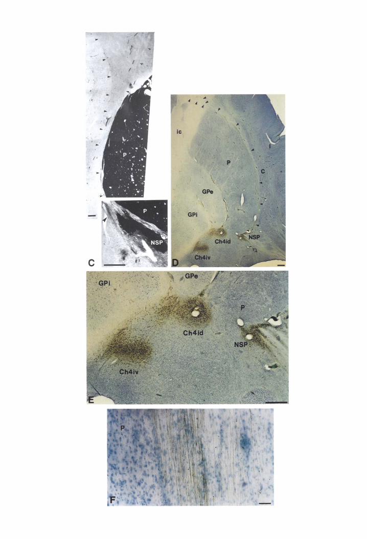
Fig. 6. (A) Intensively labelled ChAT positive axons of the NSP at the anterointermediate level projecting towards the cerebral cortex (subject no. 23). Arrows indicate two fiber bundles ascending together through capsula externa along the lateral margin of the putamen and separating above its dorsolateral edge that innervate different regions of the cortex. A bundle with medial direction projects to the medial part of the hemisphere (cingulum), while the other one turning around the insular gyrus continues towards the inferior frontal gyrus. (B) Subputaminal ChAT-positive axons ascending upwards through external capsule along the lateral margin of the putamen. Intermediate level (subject no. 16). (C) The same finding as in A and B obtained with AChE histochemistry (subject no. 14). (D) NGFr immunoreactivity counterstained with Giemsa stain at the intermediate level (subject no. 10). Arrows mark NGFr-positive fibers originating in NSP. Note that NSP also projects towards the amygdala (open arrow). (E) Enlarged picture from D. More than 90°/,, of the NSP neurons display immunoreactivity for p75 NGFr. (F) Enlarged picture from E. Subputaminal p75 NGFr-positive axons ascending along the lateral margin of
the putamen, Scale bars: (A, C, D, E)=I mm, (B, F)=0.03 mm.
lateral components of NB complex, also projects to the neocortical mantle. The location of this nucleus within the frontal lobe and its intimate relationship with the rostral extension of the basal origin of the external capsule, 59 strongly suggest that this nucleus mostly projects to the frontal cortex. Therefore, the subputaminal group of magnocellular neurons found in the human and anthropoid apes may be related to the innervation of the large frontal lobe in these species.
At the most rostral levels we describe the pre- viously poorly characterized component of the lateral (periputaminal) subdivision of the NSP, which is not described by Ayala. This most rostrolateral part of the NSP may be an example of a human specific nuclear group because this group was not described
2 0 3 0 4-4 5 6 7 4 8 7 9 7 in non-human primates. • . . . . . I f the human basal forebrain cholinergic system is compared on a larger mammal ian scale, the remarkable phylogenetic progression of the rostrolateral basal forebrain
84 G. Simid et al.
Fig. 7. Only very few (or none) scattered neurons containing NADPH-d activity were present in the area occupied by the subputaminal nucleus at all levels. An example is given at intermediate level
(subject no. 35). Scale bar=0.1 mm.
component is even more obvious. Here, only human and possibly some anthropoid apes have all sub- divisions of the NB complex. Monkeys have a promi- nent NB, but lack a well developed subputaminal nucleus. 30'55'58'74'87"97 Carnivores have a moderately
developed NB with a better expression of its medial division, 4s:°6:1° while rodents have only medial, sub- and peripallidal equivalents of the N B . 6'10'18'25'42"92 During development of the human
basal nucleus, the subputaminal nucleus becomes hyperchromatic latest, after a period of hypochroma- sis between 12th and 15th week of gestation (so-called "albino cell group"), 63 which is a unique cytoarchitectonic property among all magnocellular aggregations within the basal telencephalon. There- fore, it seems reasonable to believe that the magno- cellular subputaminal group provides the cholinergic innervation for cortical regions which are well devel- oped and characteristic of human brain, particularly in the frontal cortices.
The cortical relationship and functional significance of the nucleus subputaminalis
We find the NSP best developed at the antero- intermediate level, which is the part of the basal nucleus that is much smaller or missing in monkeys. Moreover, at the anterointermediate and intermedi- ate levels we observed CHAT-, ACHE- and NGFr- positive fibers which leave subputaminal neurons ascending through the capsula externa towards the inferior frontal gyrus. Similarly to the most rostral
levels, at these levels the NSP was usually better developed on the left side. If we consider the NSP as a characteristic feature of the human basal forebrain and the nucleus which projects to those parts of the frontal cortex where the speech area is located, we can speculate about its possible function. Knowing that speech is a human specific acquisition and that the cholinergic input to the cerebral cortex has a modulatory role for a wide range of cortical func- tions, particularly those related to memory-learning and attention-arousal, w we propose that NSP may serve for modulation of these functions in the cortical speech area. In favor of this view also goes the fact that degenerative disorders characterized by cholin- ergic 1°8 and N G F r deficits of the basal forebrain neurons,35'36'81,100,101 particularly AD, are frequently accompanied by speech disturbances) 9:°4:°5
Pathological alterations of the NB have been implicated in a number of neurological and psychi- atric conditions including Parkinson's, Korsakoff's and Pick's diseases, Down's syndrome, Huntington's chorea, schizophrenia, olivopontocerebellar atrophy, progressive supranuclear palsy, Parkinsonism- dementia complex of Guam and many others, but especially in AD and age-related memory impair- ment.l,3,4,7,15,41,83,85,90,91,107,111 114,117-120 Neverthe-
less, degeneration of the NB in these various disorders is by no means u n i f o r m s While there is a whole body of data for pathological changes of the main part of the NB, pathology of subputaminal group was not studied. Since today it seems that the most consistent determinant of pathology in AD is
Nucleus subputaminalis (Ayala/ 85
Fig. 8. Distribution of CalB-immunoreactive neurons and cholinergic neurons of the NSP was identical (subject no. 36). (A) Intermediate level, low magnification. Scale bar= 1 mm. (B) Enlarged picture from A.
Scale bar=0.1 mm. (C) Most rostral level. Scale bar--0.1 mm.
the anatomical localization of a neuron rather than its cytochemical properties, we conclude that Ayala himself was far beyond his time when he believed that pathology of the NSP may differ significantly from pathology of the NB.
CONCLUSION
We think that future neuropathological and neu- rophysiological studies of the NB should more care- fully investigate the NSP. It would be particularly interesting to investigate the role of the NSP in the primary progressive aphasia (PPA) 32'69 and slowly progressive anarthria (SPA).13 Neurobiological mechanisms underlying the anatomical selectivity of these enigmatic, but neurologically distinct disorders, are not elucidated. 23'46"7° Also, a subgroup of AD patients represents a variety of AD who, in addition to symptoms of AD, present characteristics of the
PPA. 29 We hypothesize that the pathological changes of the subputaminal nucleus may represent a missing substrate of the PPA, SPA and AD. With the pri- mary pathological event in the NSP, the PPA could be considered as a type of corticobasal degeneration, with its cortical focus located in the speech area. 4°-93 The slowly progressive development of the P P A 68'99
and SPA, as well as a negativity of the P300 potential (the cholinergic system is the most important for the generation and modulation of the P300 amplitude and latency) in the affected hemisphere in patients with the PPA, 84 further speak in favor of the possible involvement of subputaminal nucleus in the pathological processes associated with these disorders.
Acknowledgements The technical assistance of Dr Dino Klisovi6, Dr Nenad Bogdanovid, Zdenka Cmuk, Danica Buding6ak, and Bo2ica Popovid is greatly appreciated. This work was supported by the Ministry of Science of the Republic of Croatia (contract grant number 3-018).
86 G. Simid et al.
REFERENCES
1. Allen S. J., Dawbarn D., MacGowan S. H., Wilcock G. K., Treanor J. J. S. and Moss T. H. (1990) A quantitative morphometric analysis of basal forebrain neurons expressing beta-NGF receptors in normal and Alzheimer's disease brains. Dementia 1, 125-137.
2. Allen S. J., Dawbarn D., Spillantini M. G., Goedert M., Wilcock G. K., Moss T. H. and Semenenko F. M. (1989) Distribution of ~-nerve growth factor receptors in the human basal forebrain. J. comp. Neurol. 289, 626-640.
3. Allen S. J., Dawbarn D. and Wilcock G. (1988) Morphometric immunohistochemical analysis of neurons in the nucleus basalis of Meynert in Alzheimer's disease. Brain Res. 454, 275 281.
4. Arendt T., Bigl V., Arendt A. and Tennstedt A. (1983) Loss of neurons in the human nucleus basalis of Meynert in Alzheimer's disease, paralysis agitans and KorsakolTs disease. Acta neuropath. 61, 101-108.
5. Arendt T., Bigl V., Tennstedt A. and Arendt A. (1985) Neuronal loss in different parts of the nucleus basalis is related to neuritic plaque formation in cortical target areas in Alzheimer's disease. Neuroscience 14, 1 14.
6. Armstrong D. M., Saper C. B., Levey A. I., Wainer B. H. and Terry R. D. (1983) Distribution of cholinergic neurons in rat brain demonstrated by the immunocytochemical localization of choline acetyltransferase. J. eomp. Neurol. 216, 53-68.
7. Averback P. (1981) Nucleus ansae peduncularis in neuropsychiatric disease. Archs Neurol. 38, 230-235. 8. Ayala G. (1915) A hitherto undifferentiated nucleus in the forebrain (nucleus subputaminalis). Brain 37, 433-438. 9. Ayala G. (1924) Weitere untersuchungen tiber den nucleus subputaminalis. J. Psychol. Neurol. 30, 285-299.
10. Bigl V., Woolf N. J. and Butcher L. L. (1982) Cholinergic projections from the basal forebrain to frontal, parietal, temporal, occipital and cingulate cortices: a combined fluorescent tracer and acetylcholinesterase analysis. Brain Res. Bull. 8, 727-739.
11. Brockhaus H. (1942) Vergleichend-anatomische untersuchungen fiber den basalkernkomplex. J. Psychol. Neurol. 51, 57-95.
12. Broderson S. H., Westrum L. E. and Sutton A. E. (1974) Studies on the direct coloring thiocholine method for localizing cholinesterase activity. Histochemistry 40, 13-23.
13. Broussolle E., Bakchine S., Tommasi M., Laurent B., Bazin B., Cinotti L., Cohen L. and Chazot G. (1996) Slowly progressive anarthria with late anterior opercular syndrome--a variant of form of frontal cortical atrophy syndromes. J. neurol. Sci. 144, 44-58.
14. Bruce G., Wainer B. H. and Hersh L. B. (1985) Immunoaffinity purification of human choline acetyltransferase: comparison of the brain and placental enzymes. J. Neurochem. 45, 611-620.
15. Casanova M. F., Walker L. C., Whitehouse P. J. and Price D. L. (1985) Abnormalities of the nucleus basalis in Down's syndrome. Ann. Neurol. 18, 310-313.
16. Celio M. R. and Norman A. W. (1985) Nucleus basalis Meynert neurons contain the vitamin D-induced calcium binding protein (Calbindin-D 28K). Anat. Embryol. 173, 143-148.
17. Chen E.-Y., Mufson E. J. and Kordower J. H. (1996) TRK and p75 neurotrophin receptor systems in the developing human brain. J. comp. Neurol. 369, 591-618.
18. Cuello A. C. and Sofroniew M. V. (1984) The anatomy of the CNS cholinergic neurons. Trends" Neurosci. 7, 74-78.
19. Cummings J. L., Benson D. F., Hill M. A. and Read S. (1985) Aphasia in dementia of the Alzheimer's type. Neurology 35, 394-397.
20. Divac I. (1975) Magnocellular nuclei of the basal forebrain project to neocortex, brainstem and olfactory bulb. Review of some functional correlates. Brain Res. 93, 385-398.
21. Eckenstein F. and Baughman R. W. (1987) Cholinergic innervation in cerebral cortex. In Cerebral Cortex (eds. Jones E. G. and Peters A.), Vol. 6, pp. 129 160. Plenum, New York.
22. Eckenstein F. and Thoenen H. (1982) Production of specific antisera and monoclonal antibodies to choline acetyltransferase: characterization and use for identification of cholinergic neurons. Eur. molec. Biol. Org. J. 1, 363 368.
23. Espert R., Navarro J. F., Deus J., Gadea M. and Chirivella J. (1996) A review of primary progressive aphasia (Mesulam syndrome) (1982-1996). Psicol. Conduct. 4, 437-452.
24. Everitt E., Sirkia T. E., Roberts A. C., Jones G. H. and Robbins T. W. (1988) Distribution and some projections of cholinergic neurons in the brain of the common marmoset Callithrixjacchus. J. comp. Neurol. 271, 533 558.
25. Fibiger H. C. (1982) The organization and some projections of cholinergic neurons of the mammalian forebrain. Brain Res. Rev. 4, 327-388.
26. German D. C., Bruce G. and Hersh L. B. (1985) Immunohistochemical staining of cholinergic neurons in the human brain using a polyclonal antibody to human choline acetyltransferase. Neurosci. Lett. 61, 1 5.
27. Geula C. and Mesulam M.-M. (1994) Cholinergic systems and related neuropathological predilection patterns in Alzheimer's disease. In Alzheimer's Disease (eds Terry R. D., Katzman R. and Bick K. L.), pp. 263 291. Raven, New York.
28. Geula C., Shatz C. R. and Mesulam M.-M. (1993) Differential localization of NADPH-diaphorase and Calbindin- D2s k within the cholinergic neurons of the basal forebrain, striatum and brainstem in the rat, monkey, baboon and human. Neuroseience 54, 461-476.
29. Goldblum M. C., Tzortzis C., Michot J. L., Panisset M. and Boller F. (1994) Language impairment and rate of cognitive decline in Alzheimer's disease. Dementia 5, 334-338.
30. Gorry J. D. (1963) Studies on the comparative anatomy of the ganglion basale of Meynert. Acta anat. 55, 51 104. 31. Halliday G. M., Cullen K. and Cairns M. J. (1993) Quantitation and three-dimensional reconstruction of Ch4
neurons in the human basal forebrain. Synapse 15, 1-16. 32. Heath P. D., Kennedy P. and Kapur N. (1983) Slowly progressive aphasia without generalized dementia. Ann.
Neurol. 13, 687-688. 33. Hedreen J. C., Struble R. G., Whitehouse P. J. and Price D. L. (1984) Topography of the magnoceUular basal
forebrain system in human brain. J. Neuropath. exp. Neurol. 43, 1 21. 34. Hefti F., Hartikka J., Salvatierra A., Weiner W. J. and Mash D. C. (1986) Localization of nerve growth factor
receptors in cholinergic neurons of the human basal forebrain. Neurosci. Lett. 69, 3741.
Nucleus subputaminalis (Ayala) 87
35. Henderson Z. (1996) Responses of basal forebrain cholinergic neurons to damage in the adult brain. Prog. Neurobiol. 48, 219-254.
36. Higgins G. A. and Mufson E. J. (1989) NGF receptor gene expression is decreased in the nucleus basalis in Alheimer's disease. Expl Neurol. 106, 222 236.
37. Houser C. R., Crawford G. D., Barber R. P., Salvaterra P. M. and Vaughn J. E. (1983) Organization and morphological characteristics of cholinergic neurons: an immunocytochemical study with a monoclonal antibody to choline acetyltransferase. Brain Res. 266, 97-119.
38. Hsu S.-M., Raine L. and Fanger H. (1981) Use of avidin-biotin-peroxidase complex (ABC) in immunoperoxidase techniques: a comparison between ABC and unlabelled antibody (PAP) procedures. J. Histochem. Cvtochem. 29, 577-580.
39. Iacopino A. M. and Christakos S. (1990) Specific reduction of calcium-binding protein (28-kilodalton calbindin D) gene expression in aging and neurodegenerative diseases. Proc. natn. Acad. Sci. U.S.A. 87, 4078-4082.
40. Ikeda K., Akiyama H., Iritani S., Kase K., Arai T., Niizato K., Kuroki N. and Kosaka K. (1996) Corticobasal degeneration with primary progressive aphasia and accentuated cortical lesion in superior temporal gyrus-mase report and review. Acta neuropath. 92, 534-539.
41. lraizoz I., de Lacalle S. and Gonzalo L. (1991) Cell loss and nuclear hypertrophy in topographical subdivisions of the nucleus basalis of Meynert in Alzheimer's disease. Neuroscience 41, 33-40.
42. Johnston M. V., McKinney M. and Coyle J. T. (1979) Evidence for a cholinergic projection to neocortex from neurons in basal forebrain. Proc. natn. Acad. Sci. U.S.A. 76, 5392-5396.
43. Jones B. and Cuello A. C. (1989) Afferents to the basal forebrain cholinergic cell area from pontomesencephalic- catecholamine, serotonin and acetylcholine-neurons. Neuroscience 31, 37-61.
44. Jones E. G., Burton H., Saper C. B. and Swanson L. W. (1976) Midbrain, diencephalic and cortical relationships of the basal nucleus of Meynert and associated structures in primates. J. comp. Neurol. 167, 385-419.
45. Karnovsky M. S. and Roots L. (1964) A "direct coloring" thiocholine method for cholinesterases. J. Histochem. Cytochem. 12, 219 221.
46. Kertesz A. and Munoz D. G. (1997) Primary progressive aphasia. Clin. Neurosci. 4, 95 102. 47. Kievet J. and Kuypers H. G. J. M. (1975) Basal forebrain and hypothalamus connections to frontal and parietal
cortex in the rhesus monkey. Science 187, 660-662. 48. Kimura H., McGeer P. L., Peng J. H. and McGeer E. G. (1981) The central cholinergic system studied by choline
acetyltransferase immunocytochemistry in the cat. J. comp. Neurol. 200, 151-201. 49. Kitt C. A., Price D. L., DeLong M. R., Struble R. G., Mitchell S. J. and Hedreen J. C. (1982) The nucleus basalis
of Meynert: projection to cortex, amygdala, and hippocampus. Soc. Neurosci. Abstr. 8, 212. 50. Kodama S. Z. (1929) Uber die sogenannten basal ganglien (morphologische und pathologisch-anatomische
untersuchungen). II. Pathologisch-anatomische untersuchungen mit bezug auf die sogenannten basal ganglien und ihre adnexe. B. lJber die faserverbindungen zwischen den basal ganglien und ihre adnexen, sowie den fibrigen subkortikalen kerngebieten beim menschen, nebst einigen experimentallen mitteilungen Schweizer Arch. Neurol. P~D,chiatr. 23, 38 100.
51. Koelle G. B. and Friedenwald J. S. (1949) A histochemical method for localizing cholinesterase activity. Proc. Soc. exp. Biol. Med. 70, 617-622.
52. Koliatsos V. E., Martin L. J., Walker L. C., Richardson R. T., DeLong M. R. and Price D. L. (1988) Topographic, non-collateralized basal forebrain projections to amygdala, hippocampus and anterior cingulate cortex in the rhesus monkey. Bruin Res. 463, 133 139.
53. K611iker K. (1896) Handbuch der Gewebelehre des Menschen. Fiir Arzte und Studirende. Nervensystem des Menschen und der Thiere, Vol. 2. Engelmann, Leipzig.
54. Kordower J. H., Bartus R. T., Bothwell M., Schatterman G. and Gash D. M. (1988) Nerve growth factor receptor immunoreactivity in the nonhuman primate (Cebus apella): distribution, morphology, and colocalization with cholinergic enzymes. J. comp. Neurol. 277, 465-486.
55. Kordower J. H., Bartus R. T., Marciano F. F. and Gash D. M. (1989) Telencephalic cholinergic system of the new world monkey (Cebus apella): morphological and cytoarchitectonic assessment and analysis of the projection to the amygdala, a( comp. Neurol. 279, 528 545.
56. Kordower J. H., Chen E.-Y., Sladek J. R. and Mufson E. J. (1994) TRK-immunoreactivity in the monkey central nerw~us system: forebrain. J. comp. Neurol. 349, 20-35.
57. Kordower J. H., Gash D. M., Bothwell M., Hersh L. and Mufson E. J. (1989) Nerve growth factor receptor and choline acetyltransferase remain colocalized in the nucleus basalis (Ch4) of Alzheimer's patients. Neurobiol. Aging 10, 287 294.
58. Kordower J. H. and Rakic P. (1990) Neurogenesis of the magnocellular basal nuclei in the rhesus monkey. J. comp. Neurol. 291,637-653.
59. Kostovi6 I. (1986) Prenatal development of nucleus basalis complex and related fiber systems in man: a histochemical study. Neuroscience 17, 1047-1077.
60. Kostovi6 I., Juda~ M., Kostovi6-Kne~evi6 L. J., Simi6 G., Delale I., Chudy D., Sajin B. and Petanjek Z. (1991) Zagreb research collection of human brains for developmental neurobiologists and clinical neuroscientists. Int. J. dev. Biol. 35, 215 230.
61. Kostovi6 1., Skavi6 J. and Strinovi6 D. (1988) Acetylcholinesterase in the human frontal associative cortex during the period of cognitive development: early laminar shifts and late innervation of pyramidal neurons. Neurosci. Lett. 90, 107 112.
62. Kostovi6 I., Stefulj-Fu~i6 A. and Mrzljak L. (1986) Nucleus subputaminalis--a characteristic magnocellular component of the cholinergic basal forebrain complex in man. Neurosci. Lett. 26 Suppl., 226.
63. Kra~un I., R6sner H. (1986) Early cytoarchitectonic development of the anlage of the basal nucleus of Meynert in the human fetus. Int. J. dev. Neurosci. 4, 143-149.
64. Krnjevic K. and Silver A. (1965) A histochemical study of cholinergic fibers in the cerebral cortex. J. Anat. 99, 711-759.
65. Krnjevic K. and Silver A. (1966) Acetylcholinesterase in the developing forebrain. J. Anat. 100, 63 89.
88 G. Simid et al.
66. Main A. R. (1976) Structure and inhibitors ofcholinesterase. In Biology o f Cholinergic Function (eds Goldberg A. and Hanin I.), pp. 269-353. Raven, New York.
67. Mattson M. P., Rychlik B., Chu C. and Christakos S. (1991) Evidence for calcium-reducing and excitoprotective roles for the calcium-binding protein calbindin-D28k in cultured hippocampal neurons. Neuron 6, 41 51.
68. Mazzoni M., Orsucci M. P. and Giraldi C. (1996) Primary progressive aphasia--description of a clinical case with nine years follow-up, ltal. J. neurol. Sci. 17, 161 165.
69. Mesulam M.-M. (1982) Slowly progressive aphasia without generalized dementia. Ann. Neurol. 11, 592 598. 70. Mesulam M.-M. (1987) Primary progressive aphasia--differentiation from Alzheimer's disease. Ann. Neurol. 22,
533-534. 7t. Mesulam M.-M. (1995) The cholinergic contribution to neuromodulation in the cerebral cortex. Semin. Neurosci. 7,
297 307. 72. Mesulam M.-M. and Geula C. (1988) Nucleus basalis (Ch4) and cortical cholinergic innervation in the human brain:
observations based on the distribution of acetylcholinesterase and choline acetyltransferase. J. comp. Neurol. 275, 216 240.
73. Mesulam M.-M., Geula C., Bothwell M. A. and Hersh L. B. (1989) Human reticular formation: cholinergic neurons of the pedunculopontine and laterodorsal tegmental nuclei and some cytochemical comparisons to forebrain cholinergic neurons. J. comp. Neurol. 281,611 633.
74. Mesulam M.-M., Mufson E. J., Levey A. I. and Wainer B. H. (1983) Cholinergic innervation of cortex by the basal forebrain: cytochemistry and cortical connections of the septal area, diagonal band nuclei, nucleus basalis (substantia innominata) and hypothalamus in the rhesus monkey. J. comp. Neurol. 214, 170-197.
75. Mesulam M.-M., Mufson E. J., Levey A. i. and Wainer B. H. (1984) Atlas ofcholinergic neurons in the forebrain and upper brainstem of the Macaque based on monoclonal choline acetyttransferase immunocytochemistry and acetylcholinesterase histochemistry. Ncuroscience 12, 669-686.
76. Mesulam M.-M., Mufson E. J. and Wainer B. H. (1986) Three-dimensional representation and cortical projection topography of the nucleus basalis (Ch4) in the macaque: concurrent demonstration of choline acetyltransferase and retrograde transport with stabilized tetramethylbenzidine method for horse-radish peroxidase. Brain Res. 367, 301 308.
77. Mesulam M.-M. and Van Hoesen G. W. (1976) Acetylcholinesterase-rich projections from the basal forebrain of the rhesus monkey to the neocortex. Brain Res. 109, 152 157.
78. Mettler F. A. (1968) Anatomy of the basal ganglia. In Handbook o f Clinical Neurology (eds Vinken P. J. and Bruyn G. W.), Vol. 6, pp. 1-55. North-Holland Publishing Company, Amsterdam.
79. Meynert T. (1872) Vom Gehirn der Saugetiere. In Handbuch der Lehre yon den Geweben des Menschen und Thiere: translated into English in A Manual o f Histology by S. Stricker (Wood, New York, 1872), pp. 694-808. Engelmann, Leipzig.
80. Mufson E. J., Bothwell M., Hersh L. B. and Kordower J. H. (1989) Nerve growth factor receptor immunoreactive profiles in the normak aged human basal forebrain: colocalization with cholinergic neurons. J. comp. Neurol. 285, 196 217.
81. Mufson E. J., Bothwell M. and Kordower J. H. (1989) Loss of nerve growth factor receptor-containing neurons in Alzheimer's disease: a quantitative analysis across subregions of the basal forebrain. Expl Neurol. 105, 221-232.
82. Nagai T., Pearson T., Peng G., McGeer E. G. and McGeer P. L. (1983) lmmunocytochemicat staining of the human forebrain with monoclonal antibody to human choline acetyltransferase. Brain Res. 265, 300-306.
83. Nakano I. and Hirano A. (1983) Neuron loss in the nucleus basalis of Meynert in Parkinsonism dementia complex of Guam. Ann. Neurol. 13, 87 91.
84. Onofrj M., Fulgente T., Thomas A., Locatelli T. and Comi G. (1995) P300 asymmetries in focal brain lesions are reference dependent EEG Clin. Neurophysiology 94, 432~,39.
85. Oyanagi K., Takahashi H., Wakabayashi K. and Ikuta F. (1989) Correlative decrease of large neurons in the neostriatum and basal nucleus of Meynert in Alzheimer's disease. Brain Res. 504, 354-357.
86. Parent A., Poirier L. J., Boucher R. and Butcher L. L. (1977) Morphological characteristics of AChE-containing neurons in the CNS of DFP-treated monkeys. Part 2. Diencephalic and medial telencephalic structures. J. neurol. Sci. 32, 9 28.
87. Pearson R. C. A., Gatter K. C. and Powell T. P. S. (1983) Retrograde cell degeneration in the basal nucleus in monkey and man. Brain Res. 261,321-326.
88. Perry R. H., Candy J. M., Perry E. K., Thompson J. and Oakley A. E. (1984) The substantia innominata and adjacent regions in the human brain: histochemical and biochemical observations. J. Anat. 138, 713 732.
89. Ramon-Moliner E. and Nauta W. J. H. (1966) The isodendritic core of the brainstem. Z comp. Neurol. 126, 311 336.
90. Rogers J. D., Brogan D. and Mirra S. S. (1985) The nucleus basalis of Meynert in neurological disease: a quantitative morphological study. Ann. Neurol. 17, 163 170.
91. Rossor M. N., Garett N. J., Johnson A. L., Mountjoy C. Q., Roth M. and Iversen L. L. (1982) A post-mortem study of the cholinergic and GABA systems in senile dementia. Brain 105, 313 330.
92. Rye D. B., Wainer B. H., Mesulam M.-M., Mufson E. J. and Saper C. B. (1984) Cortical projections arising from the basal forebrain: a study of cholinergic and noncholinergic components employing combined retrograde tracing and immunohistochemical localization of choline acetyltransferase. Neuroscience 13, 627 643.
93. Sakurai Y., Hashida H., Uesugi H., Arima K., Murayama S., Bando M., Iwata M., Momose T. and Sakuta M. (1996) A clinical profile of corticobasal degeneration presenting as primary progressive aphasia. Eur. Neurol. 36, 134-137.
94. Saper C. B. (1987) Diffuse cortical projection systems: anatomical organization and role in cortical function. In Handbook o f Physiology The Nervous System (ed. Plum F.), pp. 16%210. American Physiological Society, Bethesda.
95. Saper C. B. (1990) Cholinergic system. In The Human Nervous System (ed. Paxinos G.), pp. 1095-1114. Academic, San Francisco.
96. Saper C. B. and Chelimsky T. C. (1984) A cytoarchitectonic and histochemical study of nucleus basalis and associated cell groups in the normal human brain. Neuroscienee 13, 1023-1037.
Nucleus subputaminalis (Ayala) 89
97. Satoh K. and Fibiger H. C. (1985) Distribution of central cholinergic neurons in the baboon (Papio papio) I. General morphology. J. comp. Neurol. 236, 197-214.
98. Schatterman G. C., Gibbs L., Lanahan A. A., Claude P. and Bothwell M. (1988) Expression of NGF receptor in the developing and adult primate central nervous system. J. Neurosci. 8, 860-873.
99. Scheltens P., Ravid R. and Kamphorst W. (1994) Pathologic findings in a case of primary progressive aphasia. Neurology 44, 279-282.
100. Scott S. A. and Crutcher K. A. (1994) Nerve growth factor and Alzheimer's disease. Rev. Neurosci. 5, 179-211. 101. Scott S. A., Mufson E. J., Weingartner J. A., Skau K. A. and Crutcher K. A. (1995) Nerve growth factor in
Alzheimer's disease: increased levels throughout the brain coupled with declines in nucleus basalis. J. Neurosci. 15, 6213-6221.
102. Shute C. C. D. and Lewis P. R. (1967) The ascending cholinergic reticular system: neocortical, olfactory and subcortical projections. Brain 90, 497-520.
103. Silver A. (1974) The Biology of Cholinesterases. North Holland, Amsterdam. 104. Sj6gren H. (1950) Twenty-four cases of AD. Acta med. scand. 245 Suppl., 225-233. 105. Sj6gren T., Sj6gren H. and Lindgren A. (1952) Morbus Alzheimer and morbus Pick: a genetic, clinical and
patho-anatomical study. Actapsychiat. neurol, scand. 82 Suppl., 1-152. 106. Steriade M., Parent A., Pare D. and Smith Y. (1987) Cholinergic and non-cholinergic neurons of cat basal forebrain
project to reticular and mediodorsal thalamic nuclei. Brain Res. 408, 372-376. 107. Stevens J. R. (1982) Neuropathology of schizophrenia. Archs gen. Psychiat. 39, 1131-1139. 108. Strada O., Vyas S., Hirsch E. C., Ruberg M., Brice A., Agid Y. and Javoy-Agid F. (1992) Decreased choline
acetyltansferase mRNA expression in the nucleus basalis of Meynert in Alzheimer's disease: an in situ hybridization study. Proc. natn. Acad. Sci. U.S.A. 89, 95494553.
109. Struble R. G., Lehmann J., Mitchell S. J., McKinney M., Price D. L., Coyle J. T. and DeLong M. R. (1986) Basal forebrain neurons provide major cholinergic innervation of primate neocortex. Neurosci. Lett. 66, 215 220.
110. St-Jacques R., Gorczyca W., Mohr G. and Schipper H. M. (1996) Mapping of the basal forebrain cholinergic system of the dog: a choline acetyltransferase immunohistochemical study. J. comp. Neurol. 366, 717-725.
111. Tag[iavini F. and Pilleri G. (1983) Basal nucleus of Meynert: a neuropathological study in Alzheimer's disease, simple senile dementia, Pick's disease and Huntington's chorea. J. neurol. Sci. 62, 243-260.
112. Tagliavini F. and Pilleri G. (1985) Neuronal loss in the basal nucleus of Meynert in a patient with olivopontocer- ebellar atrophy. Acta neuropath. 66, 127-133.
113. Tago H., Kimura H. and Maeda T. (1986) Visualization of detailed acetylcholinesterase fiber and neuron staining in rat brain by a sensitive histochemical procedure. J. Histochem. Cytochem. 34, 1431-1438.
114. Terry R. D. and Davis P. (1980) Dementia of the Alzheimer's type. A. Rev. Neurosci. 3, 77-95. 115. Ulfig N. (1989) Configuration of the magnocellular nuclei in the basal forebrain of the human adult. Acta anat. 134,
100--105. 1 t6. Ulfig N. and Braak H. (1989) Neuronal types and their percent distribution within the magnocellular nuclei of the
human basal forebrain. Acta anat. 134, 237-241. 117. Vogel O., Broere C., Lak H. T., Donkelaar H. T., Nieuwenhuys R. and Schultz P. (1990) Cell loss and shrinkage in
the nucleus basalis Meynert complex in Alzheimer's disease.. Neurobiol. Aging 11, 3-13. 118. Whitehouse P. J., Price D. L., Clark A. W., Coyle J. T. and DeLong M. R. (1981) Alzheimer's disease: evidence for
selective loss of cholinergic neurons in the nucleus basalis. Ann. Neurol. 10, 122-126. 119. Whitehouse P. J., Price D. L., Struble R. G., Clark A. W., Coyle J. T. and DeLong M. R. (1982) Alzheimer's disease
and senile dementia: loss of neurons in the basal forebrain. Science 215, 1237-1239. 120. Wilcock G. K., Esiri M. M., Bowen D. M. and Smith C. C. T. (1983) The nucleus basalis in Alzheimer's disease: cell
counts and cortical biochemistry. Neuropath. appl. Neurobiol. 9, 175 179.
(Accepted 19 May 1998)