nucleotides flanking a conserved taat core dictate the dna
TRANSCRIPT
MOLECULAR AND CELLULAR BIOLOGY, Apr. 1993, p. 2354-23650270-7306/93/042354-12$02.00/0Copyright C) 1993, American Society for Microbiology
Vol. 13, No. 4
Nucleotides Flanking a Conserved TAAT Core Dictate theDNA Binding Specificity of Three Murine
Homeodomain ProteinsKATRINA M. CATRON,1 NANCY ILER,12 AND CORY ABATE' 3*
Center for Advanced Biotechnology and Medicine, 679 Hoes Lane, 1 Graduate Program in Microbiology,Rutgers University, 2 and Department ofNeuroscience and Cell Biology, Robert Wood Johnson
Medical School,3 Piscataway, New Jersey, 08854-5638
Received 17 September 1992/Returned for modification 9 November 1992/Accepted 7 January 1993
Murine homeobox genes play a fundamental role in directing embryogenesis by controlling gene expressionduring development. The homeobox encodes a DNA binding domain (the homeodomain) which presumablymediates interactions of homeodomain proteins with specific DNA sites in the control regions of target genes.
However, the bases for these selective DNA-protein interactions are not well defined. In this report, we havecharacterized the DNA binding specificities of three murine homeodomain proteins, Hox 7.1, Hox 1.5, andEn-1. We have identified optimal DNA binding sites for each of these proteins by using a randomoligonucleotide selection strategy. Comparison of the sequences of the selected binding sites predicted a
common consensus site that contained the motif (C/G)TAATTG. The TAAT core was essential for DNAbinding activity, and the nucleotides flanking this core directed binding specificity. Whereas variations in thenucleotides flanking the 5' side of the TAAT core produced modest alterations in binding activity for all threeproteins, perturbations of the nucleotides directly 3' of the core distinguished the binding specificity of Hox 1.5from those of Hox 7.1 and En-1. These differences in binding activity reflected differences in the dissociationrates rather than the equilibrium constants of the protein-DNA complexes. Differences in DNA bindingspecificities observed in vitro may contribute to selective interactions of homeodomain proteins with potentialbinding sites in the control regions of target genes.
Studies aimed at elucidating the molecular processes thatregulate murine embryonic development have led to theidentification of numerous genes whose protein productsfunction to control gene expression during embryogenesis.These transcriptional regulatory proteins play a fundamentalrole in directing embryogenesis by establishing and main-taining the appropriate patterns of spatial and temporal geneexpression. At least 30 such genes have been isolated in mice(29). Many of these share a conserved motif termed thehomeobox which encodes a sequence-specific DNA bindingdomain (the homeodomain) (32, 47). The homeobox was firstidentified as a common feature of Drosophila genes thatcontrol pattern formation during embryogenesis and wassubsequently shown to be characteristic of numerous genesfrom a variety of species that regulate transcription duringdevelopment (5, 16, 29, 47, 51). Of the 60 amino acids thatcompose the homeodomain, some of the most highly con-served are those that contact DNA directly (30, 32, 41).Some of these conserved amino acids contact nucleotideswithin a TAAT motif, a critical component of many homeo-domain DNA binding sites. Other amino acids make addi-tional contacts with nucleotides flanking the TAAT motif,and such contacts are presumed to influence the specificityof the protein-DNA interaction (20, 21, 48).
Several distinct families of murine homeobox genes areexpressed in overlapping spatial and temporal patterns in thedeveloping embryo (25, 29). Members of these gene familiesshare sequence similarity with the homeobox regions ofdistinct Drosophila developmental regulatory genes. Themost prevalent group, the hox gene family, contains at least
* Corresponding author.
20 members that are clustered on four chromosomes, andthese are related to the Drosophila homeotic gene, antenna-pedia (5, 37, 47). The hox genes are expressed early in thedeveloping murine embryo (beginning from days 7.5 to 8.5)in overlapping patterns throughout the neural tube, thesomites, and the sclerotomes (25, 37). These genes have theintriguing property that their anterior boundaries of expres-sion correlate well with their linear organization along thechromosome cluster (13, 18, 37, 50). This colinearity ofchromosome organization and gene expression has beenconserved from Drosophila to Homo sapiens and is pro-posed to represent a molecular code that provides positionalinformation during development. Indeed, targeted disrup-tions of hox 1.5 or hox 1.6 produce gross developmentalabnormalities, highlighting a critical role for these genes inpattern formation during development (9, 33).
In addition to the hox genes, members of several otherhomeobox gene families are also expressed in overlappingspatial and temporal patterns during development. The mu-rine genes engrailed-I and hox 7.1, for example, are mem-bers of relatively small gene families (two or three mem-bers), and their homeobox sequences are more closelyrelated to those of the Drosophila genes engrailed and msh(for muscle-specific homeobox), respectively, than to that ofthe antennapedia gene (22, 28). Both engrailed-1 and hox7.1, similar to the antennapedia-like hox genes, are ex-pressed throughout the developing neural tube, but unlikethe hox genes, their anterior boundaries of expression ex-tend to the presumptive midbrain and diencephalon (10, 22,28, 34, 45). Their gene products, En-1 and Hox 7.1, have thehallmark features of transcriptional regulatory proteins andare presumed to function in the control of gene expressionduring embryogenesis.
2354
DNA BINDING SITES FOR Hox 7.1, Hox 1.5, AND En-i 2355
As a consequence of their overlapping, but distinct, pat-terns of expression, many different combinations of homeo-domain proteins may be present in developing cells. Presum-ably, the interactions of these various proteins with specifictarget genes are essential for maintaining appropriate geneexpression, thereby dictating proper cell development. Themost conserved feature among these proteins, the homeo-domain, is also a major contributor to their functionalspecificity, at least for certain Drosophila proteins (17, 31,35). Therefore, binding specificity and function may beintimately linked. A comprehensive analysis of their bindingproperties is likely to provide insight as to how theseproteins interact selectively with specific target genes, andthis is particularly relevant in situations in which numeroushomeodomain proteins are coexpressed. In this paper, wehave investigated the DNA binding specificities of threemurine homeodomain proteins, Hox 7.1, Hox 1.5, and En-i.Using a random oligonucleotide selection strategy, we haveidentified optimal DNA binding sites for each of theseproteins. We show that the homeodomain regions of Hox7.1, Hox 1.5, and En-1 bind to similar DNA sites that containa TAAT core flanked on either side by G or C residues [e.g.,(C/G)TAATTG]. Both the core and flanking nucleotides arerequired for high-affinity DNA binding. We also show thatthe specific nucleotides directly flanking the 3' side of thecore distinguish the binding specificities of Hox 7.1, Hox 1.5,and En-1. Subtle differences in DNA binding specificitiesobserved in vitro may contribute to selective interactions ofhomeodomain proteins with potential binding sites in thecontrol regions of target genes.
MATERLILS AND METHODS
Expression and purification of Hox 7.1, Hox 1.5, andEngrailed-1. DNA sequences corresponding to the ho-meobox regions of hox 7.1 and engrailed-1 were obtained bypolymerase chain reaction (PCR) with cDNA derived frommouse 9.5-day embryonic RNA (a generous gift of J. McMa-hon and A. McMahon, Roche Institute of Molecular Biolo-gy). The homeobox sequence of hox 1.5 was isolated by PCRamplification from an 11.5-day mouse embryonic cDNAlibrary (Clonetech). Oligonucleotides used for amplificationwere complementary to sequences encoding amino acids 157to 233 of Hox 7.1 (22), the homeodomain region of Hox 1.5(74 amino acids) (36), and the C-terminal region of En-grailed-1 (En-1) including the homeodomain (130 aminoacids) (28). The oligonucleotides also contained BamHI andHindIll restriction sites to facilitate cloning. The amplifiedfragments of hox 7.1, hox 1.5, and engrailed-I were clonedinto the bacterial expression vector pDS56 (3), and theirsequences were confirmed. This vector contains six histidinecodons, and the proteins were expressed as hexahistidinefusion polypeptides; in our experience, the hexahistidinefusion does not interfere with DNA binding activity (1-3).Bacterial cells containing pDS56-hox 7.1, pDS56-hox 1.5, orpDS56-engrailed-1 were grown to a density of 0.6 opticaldensity units, and protein expression was induced by theaddition of 1 mM isopropyl-f-D-thiogalactopyranoside. Af-ter 4 h, cells were harvested, protein lysates were preparedby extraction with 6 M guanidine-HCl, and the proteins werepurified from the bacterial cell lysates by nickel-affinitychromatography in the presence of 6 M guanidine-HCl aspreviously described (2, 3). The purified polypeptides wererenatured by extensive dialysis against 25 mM sodium phos-phate (pH 7.4), 50mM potassium chloride, 5 mM magnesiumchloride, 10% (vol/vol) glycerol, and 1 mM dithiothreitol.
For expression in mammalian cells, hox 7.1 was clonedinto the vector pCb6+ (42) and transfected into Cos-1 cellsby a standard DEAE-dextran procedure. Details of theseprocedures will be published elsewhere (26). Cell lysateswere prepared from Cos-1 cells expressing Hox 7.1, and theprotein was partially purified by nickel-affinity chromatogra-phy with an imidazole gradient for elution as previouslydescribed (27).
Oligonucleotide selection. Oligonucleotide selection wasperformed with a DNA fragment that contained a 14-bprandom sequence flanked on either side by 15 bases ofnonrandom sequence [5'- AGACGGATCCATTGCA(N14)CTGTAGGAATTCGGA-3'] (a generous gift of J. Morrisand F. J. Rauscher III, Wistar Institute). The single-strandedoligonucleotide containing the random sequence was madedouble stranded by filling in with the Klenow fragment ofDNA polymerase I with a template oligonucleotide that wascomplementary to the 3' nonrandom sequence [5'-TCCGAATTCCTACAG-3' (oligonucleotide SMB)]. Maxam-Gil-bert sequencing was performed to confirm that the probecontained a random sequence. PCR amplification of thedouble-stranded fragment was performed with the templateoligonucleotide (SMB) and a second oligonucleotide thatcorresponded to the 5' nonrandom sequence [5'-AGACGGATCCATTGCA-3' (oligonucleotide SMA)]. The double-stranded fragment was radiolabeled with T4 polynucleotidekinase in the presence of [y-32P]ATP and used as a probe ingel retardation assays. Binding reactions were performed byaddition of protein (1 to 2 ,uM) to buffer containing 10 mMTris-HCl (pH 7.5), 50 mM sodium chloride, 7.5 mM magne-sium chloride, 1 mM EDTA, 5% (vol/vol) glycerol, 5%(wt/vol) sucrose, 0.1% Nonidet P-40, 0.5 ,ug of bovine serumalbumin per jil, 0.5 jig of poly(dI-dC), and 5 mM dithiothre-itol. After incubation for 5 min at room temperature, theradiolabeled DNA probe was added, and the incubation wascontinued for an additional 5 min. The high levels of poly(dI-dC) and short incubation time were required to minimizeaggregation of DNA-protein complexes. The protein-DNAcomplexes were resolved from free DNA on 6.5% polyac-rylamide gels containing 0.5 x Tris-borate-EDTA. The DNAin the bound complexes was identified by autoradiographyand extracted from the gel. PCR amplification was per-formed with a standard reaction mixture (Perkin-ElmerCetus) and the following conditions: 93°C, 30 s; 45°C, 2 min;45 to 67°C, 2 min; and 67°C, 2 min (25 cycles) and 45°C, 10min; 45 to 67°C, 10 min; and 67°C, 10 min (1 cycle). Theamplified DNA was gel purified, radiolabeled with T4 poly-nucleotide kinase, and used as a probe in gel retardationassays. The selection procedure was repeated for a total offour rounds. In the initial rounds, less than 1% of the probewas shifted and by the fourth round of selection approxi-mately 50% of the probe was shifted. After the last round ofPCR amplification, the gel-purified DNA fragments werephosphorylated and cloned into the EcoRV site of pBlue-script (Stratagene). Clones containing single inserts wereidentified, and the sequences of the inserts were determined.To investigate the binding potential of the selected sites,DNA probes were prepared by PCR amplification of plas-mids containing the selected binding sites with radiolabeledoligonucleotide primers (SMA and SMB, as above). A totalof 1 ,ul of the PCR product was used as a probe in the bindingassay performed above.DNA binding assays. For binding assays with oligonucle-
otide probes, oligonucleotides were synthesized on an Ap-plied Biosystems DNA synthesizer model 391. The oligonu-cleotides were radiolabeled with T4 polynucleotide kinase
VOL. 13, 1993
2356 CATRON ET AL.
H-TERM ARM HELIX HELIX HELIX I
NRKPRTPF TTRQLLRLLERKFRQK QYLS IRERAEFSSSL SLT ETQUKIW.FONRRRKRKRLQSKRGRTRY TRPfLUELEKEFHFN RYLM RPRRUEIAMLL HLT ERQIKIWUFQNRR1KYKKDQ
EDKRPRTAF AE-LQRLKREFQRN RY3IT EQRRQTLRQEL SLN ESQIKIWUFONKRAKIKKA10 20 30 40 50 60
KR RT Y T QL L F Y R L L E QIKIUFQNRR K KK
Oligoruc eotides corresponding to homeocboxsecuences of hox 7. 1, hox 1.5, or en- I C 2 - Lfl
IE I- I
Iso ate hormecbox sequences by PCR usingmur re cDNA from 9.5 or .5 day embryos
3amrn (hox 71, hox 1 5, or en-Ih
Subcione 'Into pDS56 vector
ear (Hi (hox 7. 1, hox 1.5, or en-i)
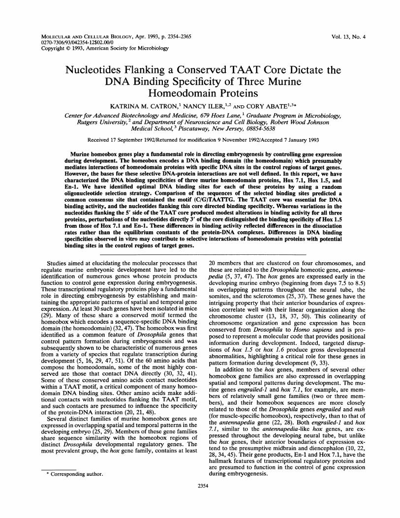
FIG. 1. Cloning and expression of the homeodomain regions of Hox 7.1, Hox 1.5, and En-1. (A) Predicted amino acid sequencescorresponding to the homeodomain regions of Hox 7.1 (22), Hox 1.5 (36), and En-1 (28) are indicated with the single-letter code; amino acidsconserved among the proteins are underlined. The consensus amino acids are those residues that are highly conserved among allhomeodomain proteins (47). The position of the N-terminal arm and helices I, II, and III are indicated, and positions of amino acids withinthe homeodomain are indicated numerically. (B) DNA sequences corresponding to the homeobox regions of hox 7.1, hox 1. 5, and en-i wereamplified by PCR with murine embryonic 9.5- or 11.5-day cDNA. Oligonucleotide primers were specific for the 5' or 3' sequences andcontained BamHI and HindIII restriction sites. The amplified DNA fragments were cloned into the E. coli expression vector pDS56 in framewith the initiator methionine and six histidine codons. (C) Polypeptides corresponding to the homeodomain regions of Hox 7.1, Hox 1.5, andEn-1 were expressed as hexahistidine fusion proteins in E. coli and purified by nickel-affinity chromatography. The purified proteins (2 ,ug)were resolved on an SDS-13.5% polyacrylamide gel and visualized by staining with Coomassie brilliant blue. Markers correspond tomolecular mass standards (Bio-Rad) in kilodaltons (bovine serum albumin, 68 kDa; ovalbumin, 46 kDa; carbonic anhydrase, 31 kDa; soybeantrypsin inhibitor, 20 kDa; and lysozyme, 14 kDa).
and annealed at 37°C for 1 h at equimolar concentrations.The binding reactions were performed as described above,with the exception that the protein-DNA complexes wereformed for 15 min at room temperature. DNA bindingactivity was quantitated with a Phosphor Imager (MolecularDynamics) and was calculated as the percentage of DNAbound divided by total DNA (bound/bound + free). EachDNA binding experiment was repeated a minimum of fourtimes, and representative gel shift assays are presented.For determination of equilibrium dissociation constants
(Kd), a constant amount of labeled DNA (1 x 10`0 M) wasincubated with various amounts of protein (5 x 10`0 to 10x 10-7M for Hox 7.1 and Hox 1.5 and 5 x 10⁻⁹ to 5 x 10⁻⁶M for En-1) for 30 min at room temperature (to allowprotein-DNA complexes to reach equilibrium) in reactionbuffer minus poly(dI-dC). The reaction mixtures were elec-trophoresed, under the conditions described above, in theabsence of loading dye. Bound and free DNAs were quan-titated with a Phosphor Imager. The Kd was calculated withthe equation Kd = [D][P]/[DP], where [D] is the concentra-tion of free DNA, [P] is the concentration of free protein,and [DP] is the concentration of the DNA-protein complex.The DNA concentration was limiting relative to the proteinconcentration to allow the approximation [DP] = [DPtotalI.The dissociation rate constants (kd) and complex half-lives
(t1/2) were determined by incubating the proteins with limit-
ing concentrations of radiolabeled DNA in binding reactionmixture [without poly(dI-dC)]. After incubation at roomtemperature for 15 min, a 50-fold molar excess of coldcompetitor DNA was added and the reaction mixtures wereincubated for the additional times indicated prior to electro-phoresis. The bound and free DNAs were quantitated with a
PhosphorImager, and the values for kd were determined withthe formula ln(fraction ofDNA bound) = -kdt. t112 (the timerequired for half of the protein-DNA complexes to dissoci-ate) was calculated with the equation t112 = -ln(O.5)Ikd.
RESULTS
Expression and purification of the murine homeodomainproteins Hox 7.1, Hox 1.5, and Engrailed-1. The homeo-domain regions of Hox 7.1, Hox 1.5, and En-1 share manyamino acids that are highly conserved among all homeo-domain proteins (Fig. 1A). These similarities are primarilyconcentrated within helix III, and some of these conservedamino acids have been shown by structural studies tocontact DNA directly (30, 41). The conservation is morelimited throughout the other helices, and overall, the homeo-domains of Hox 7.1, Hox 1.5, and En-1 share approximately50% homology at the amino acid level (Fig. 1A). Therefore,we reasoned that these proteins share sufficient sequencehomology to facilitate a comparative analysis of their DNA
AHox 7.1Hox 1.5En-i
CONSENSUS
B
LU
MOL. CELL. BIOL.
DNA BINDING SITES FOR Hox 7.1, Hox 1.5, AND En-i 2357
binding specificities. To investigate their binding propertiesin vitro, purified polypeptides were obtained by overexpres-sion of the homeobox sequences of hox 7.1, hox 1.5, anden-1 in Escherichia coli. The appropriate sequences wereisolated from mouse embryonic cDNA and cloned into thebacterial expression vector pDS56 (Fig. 1B). This vectorcontained six histidine codons after the initiator methioninecodon, such that the recombinant polypeptides were ex-pressed as hexahistidine fusion proteins (2, 3). This histidinefusion facilitated subsequent purification by nickel-affinitychromatography. Protein lysates were prepared from bacte-rial cells expressing the recombinant Hox 7.1, Hox 1.5, orEn-1 polypeptides by extraction with buffer containing 6 Mguanidine. The lysates were chromatographed in the pres-ence of guanidine on a nickel-affinity column, and thepurified proteins were renatured by extensive dialysis. Theproteins were soluble at relatively high concentrations (Hox7.1, 800 ,uM; Hox 1.5, 70 ,uM; and En-i, 350 ,uM) and werepurified to apparent homogeneity as evident by Coomassieblue staining of a sodium dodecyl sulfate (SDS)-polyacryl-amide gel (Fig. 1C).
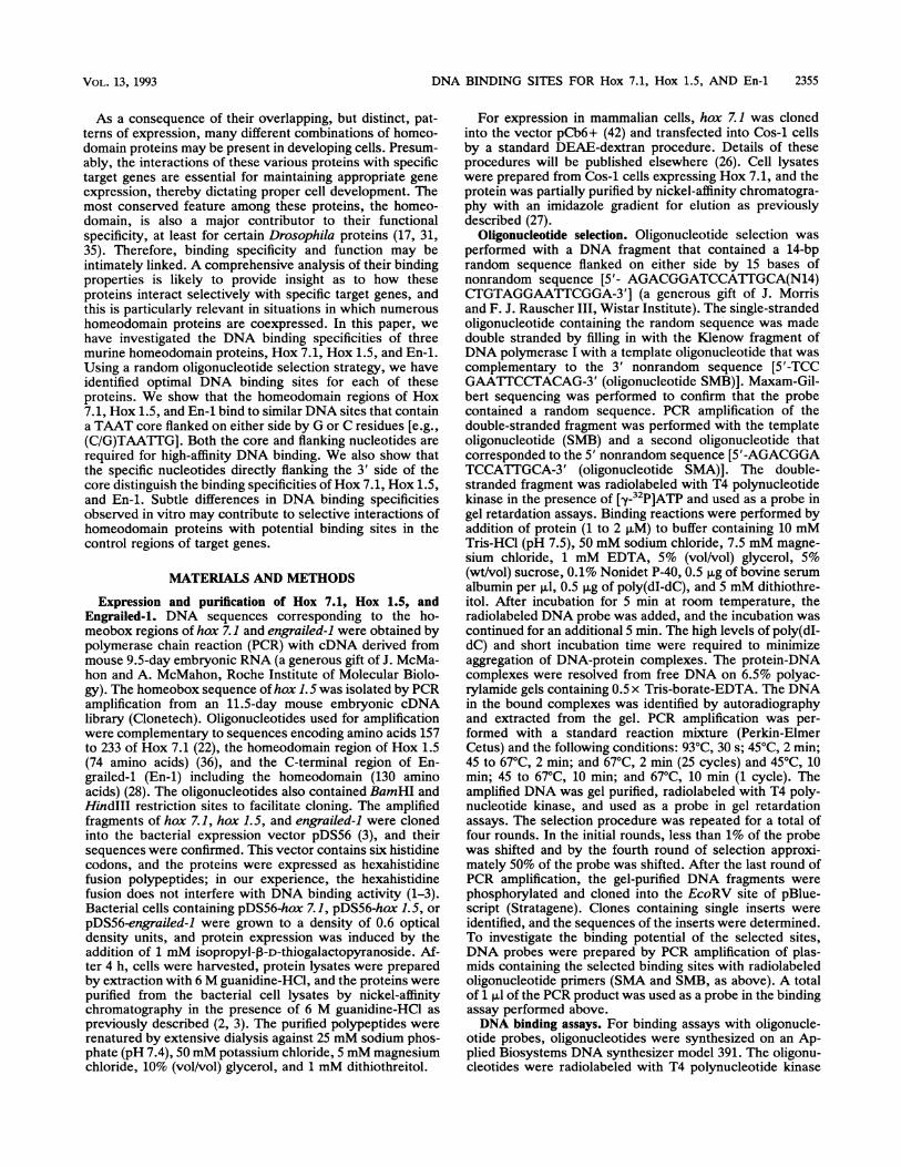
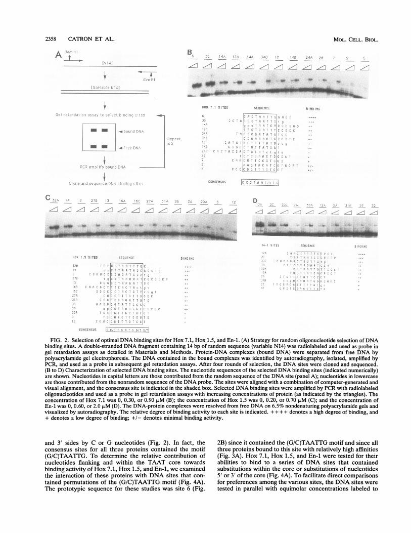
Selection of DNA binding sites for Hox 7.1, Hox 1.5, andEn-1. The random oligonucleotide selection procedure hasbeen used successfully to identify optimal DNA binding sitesfor a variety of transcriptional regulatory proteins (6, 14, 15,40, 43). This strategy relies on the ability of a protein tointeract with specific DNA sites selected from among arandom population of potential binding sites. In this report,we have used this procedure to characterize the DNAbinding specificities of Hox 7.1, Hox 1.5, and En-1 (Fig. 2A).Binding selection was performed with a double-strandedDNA fragment that contained a 14-bp random sequence. TheDNA fragment was used as a probe in gel retardation assays,and those fragments that interacted with the purified Hox7.1, Hox 1.5, or En-1 polypeptides were separated from thepopulation of random sites by gel electrophoresis (Fig. 2A).In the initial round of selection, a 1 to 3 ,uM concentration ofeach protein was used in the gel retardation assays and lessthan 1% of the probe was retained in the bound complex. Insubsequent rounds of selection, the protein concentrationwas reduced to 0.5 F.M, and by the fourth round approxi-mately 50% of the probe was shifted by the proteins. Afterthe fourth round, the DNA sites obtained by selection withHox 7.1, Hox 1.5, and En-1 were cloned and sequenced. Thesequences of selected sites and the relative binding affinitiesof Hox 7.1, Hox 1.5, and En-1 for each of these sites aresummarized in Fig. 2B to D.Hox 7.1 binding specificity. Each of 13 sites selected by
Hox 7.1 from the random pool was capable of binding thecognate protein (Fig. 2B). Although all of these sites inter-acted with Hox 7.1, they exhibited marked differences in thedegree of binding, and these differences were most evidentwhen binding assays were performed at lower protein con-centrations (Fig. 2B). Therefore, using the selection proce-dure, we obtained a series of potential DNA sites for Hox 7.1that included relatively high-affinity (e.g., sites 6 and 35),intermediate-affinity (e.g., 14A, 12A, 34A, 34B, 10, and14B), and low-affinity (e.g., 24A, 26, 7, 2, and 5) binding sites(Fig. 2B). The presence of such low-affinity sites, even afterfour rounds of selection, is presumably due to the inherentnonspecific binding activity of Hox 7.1 (7). Many of theselected sites contained AT-rich sequences that were flankedby G or C nucleotides (e.g., Fig. 2B, sites 6, 35, and 34B).Alignment of these sequences (with a combination of com-puter-generated and visual alignment) identified the consen-sus binding site, CXGTAAT(A/T)G (Fig. 2B). Interestingly,
the sequence of the consensus site was more similar to thatof the highest-affinity binding sites (e.g., sites 6 and 35) thanthe lower-affinity sites (e.g., sites 7, 2, and 5) (Fig. 2B).Hox 1.5 binding specificity. Each of the binding sites
recovered with Hox 1.5 as a substrate in the selectionprocedure interacted with the cognate protein in vitro (Fig.2C). However, Hox 1.5 differed markedly in its apparentaffinity for each of these binding sites, analogous to thesituation with Hox 7.1 (Fig. 2C). Thus, the population ofHox 1.5 binding sites included relatively high-affinity (e.g.,sites 32A and 14), intermediate-affinity (e.g., sites 2, 27B, 13,16A, 16C, 27A, 31A, 35, and 34), and low-affinity (e.g., 20A,3, and 12) binding sites (Fig. 2C). As with the Hox 7.1 sites,the Hox 1.5 sites contained AT-rich sequences flanked by Gor C nucleotides (compare Fig. 2B and C). However, thepredicted consensus sequence for Hox 1.5 [C(C/G)TAATX(G/T)(G/T)] was somewhat different from the consensussite for Hox 7.1 and had more variability for G or Tnucleotides on the 3' side of the TAAT core (compare Fig.2B and C). This may reflect a greater flexibility of Hox 1.5 tointeract with DNA sites that contain variations of the 3'flanking nucleotides. The sequence of the Hox 1.5 consensussite was more similar to that of the highest-affinity bindingsites (e.g., sites 32A and 14) than the lower-affinity sites(e.g., 20A, 3, and 12) (Fig. 2C).En-1 binding specificity. By using En-1 as a substrate in the
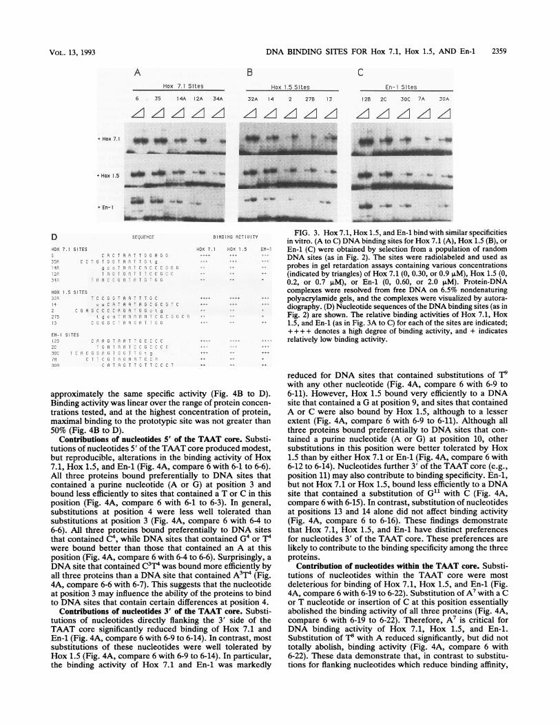
binding selection procedure, several DNA binding sites wereidentified (Fig. 2D). However, although selection with En-1yielded some relatively high-affinity sites (e.g., 12B, 2C, and30C), a majority of the selected sites were of relatively lowaffinity (Fig. 2D). We have observed that twofold-higherconcentrations of En-1 (2 ,uM) are required for efficient DNAbinding compared with either Hox 7.1 (0.9 ,uM) or Hox 1.5(0.7 ,uM) (7). It is likely that the reduced efficiency of theselection procedure was due to an overall lower affinity ofEn-1 for DNA. The sequences of the binding sites selectedfor En-1 were similar to those selected for Hox 7.1 and Hox1.5 in that they contained AT-rich sequences flanked by G orC nucleotides (Fig. 2D). Although fewer sites were obtainedwith En-1, comparison of these sequences indicated a ten-tative consensus binding site, GTAATXG (Fig. 2D).Hox 7.1, Hox 1.5, and En-1 have related binding specificities
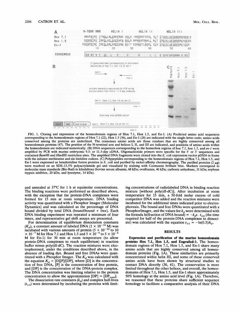
in vitro. The highest-affinity binding sites selected for Hox7.1, Hox 1.5, and En-1 were closely related (compare Fig.2B, C, and D). In particular, these sites each contained aTAAT core flanked on the 5' and 3' sides by C or Gnucleotides. These findings suggested that the homeo-domains of Hox 7.1, Hox 1.5, and En-1 bind to similar DNAsites in vitro. Indeed, it has been shown that rather divergentDrosophila homeodomains interact with the same sites invitro (11, 24). Hox 7.1, Hox 1.5, and En-1 each interactedwith sites that had been selected for binding to the otherproteins and bound to these other sites with approximatelythe same relative affinity as the cognate protein (Fig. 3A toD). Therefore, all three proteins interacted most efficientlywith sites 6, 35, 32A, and 12B and less efficiently with sites12A, 34A, 13, and 30A (Fig. 3A to D). In contrast, otherDNA binding proteins, such as Fos and Jun, did not bind tothese sites, although these other proteins also containedhexahistidine fusion polypeptides (7). Thus, the home-odomain regions of Hox 7.1, Hox 1.5, and En-1 exhibitsimilar DNA binding specificities in vitro.
Nucleotides flanking the TAAT core contribute to bindingaffinity of Hox 7.1, Hox 1.5, and En-i. The selected bindingsites for Hox 7.1, Hox 1.5, and En-1 were biased towardthose containing TAAT core nucleotides flanked on the 5'
VOL. 13, 1993
2358 CATRON ET AL.
t~~~~~~~~~: Zt
.V r .
*4 '_A4.-''22 4A
z zG zz i10
t
- - -4
4-4,.,,
-a fyS
~._ 2'- 35- 20A 3
., I , -1 A 35 2z, A 3 /1
_. Uwd0 . .,-
_~~~~~~~~~~~~~~~~~~~~~~~~~~~~~~~~~~~~~.
HCX .5 SIlTES SEQUENCE
,r cWu H1
G. .rsF.C 'CC,i T Pz^f '-Tf
:7i ;i5CGr. f
CCNSEhSUS S
e ND NO
FIG. 2. Selection of optimal DNA binding sites for Hox 7.1, Hox 1.5, and En-1. (A) Strategy for random oligonucleotide selection ofDNAbinding sites. A double-stranded DNA fragment containing 14 bp of random sequence (variable N14) was radiolabeled and used as probe ingel retardation assays as detailed in Materials and Methods. Protein-DNA complexes (bound DNA) were separated from free DNA bypolyacrylamide gel electrophoresis. The DNA contained in the bound complexes was identified by autoradiography, isolated, amplified byPCR, and used as a probe in subsequent gel retardation assays. After four rounds of selection, the DNA sites were cloned and sequenced.(B to D) Characterization of selected DNA binding sites. The nucleotide sequences of the selected DNA binding sites (indicated numerically)are shown. Nucleotides in capital letters are those contributed from the random sequence of the DNA site (panel A); nucleotides in lowercaseare those contributed from the nonrandom sequence of the DNA probe. The sites were aligned with a combination of computer-generated andvisual alignment, and the consensus site is indicated in the shaded box. Selected DNA binding sites were amplified by PCR with radiolabeledoligonucleotides and used as a probe in gel retardation assays with increasing concentrations of protein (as indicated by the triangles). Theconcentration of Hox 7.1 was 0, 0.30, or 0.90 ,uM (B); the concentration of Hox 1.5 was 0, 0.20, or 0.70 p.M (C); and the concentration ofEn-1 was 0, 0.60, or 2.0 pLM (D). The DNA-protein complexes were resolved from free DNA on 6.5% nondenaturing polyacrylamide gels andvisualized by autoradiography. The relative degree of binding activity to each site is indicated. + ++ + denotes a high degree of binding, and+ denotes a low degree of binding; +/- denotes minimal binding activity.
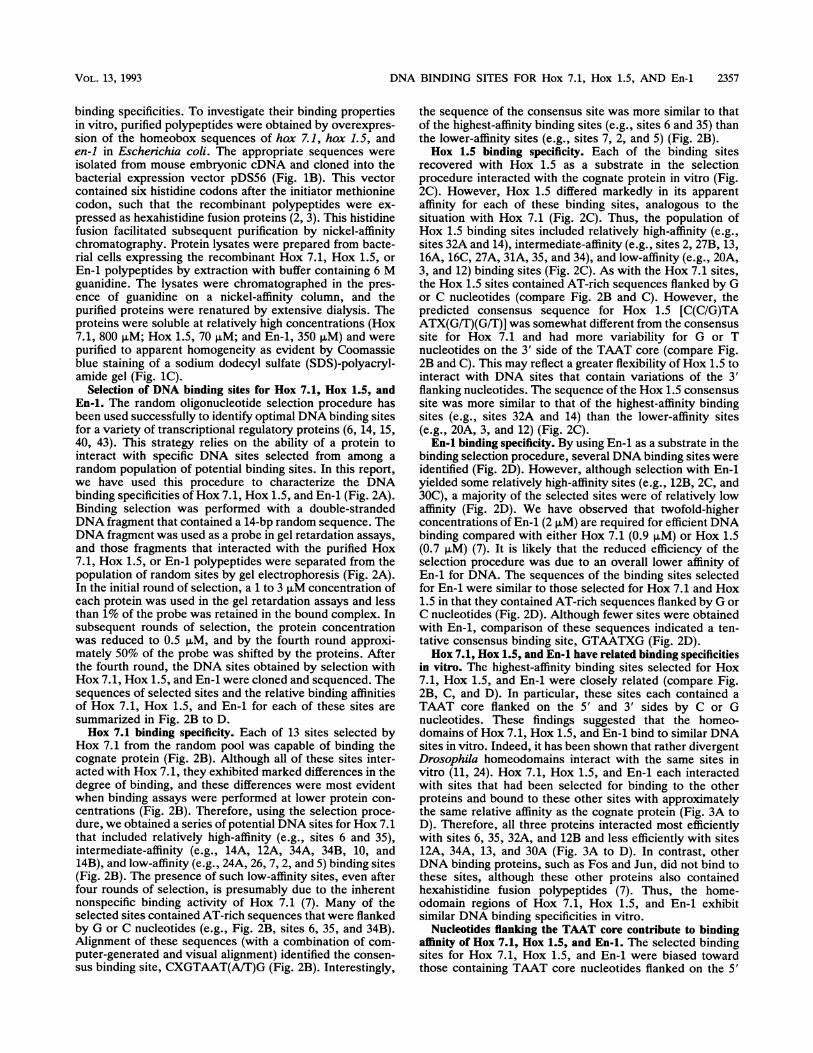
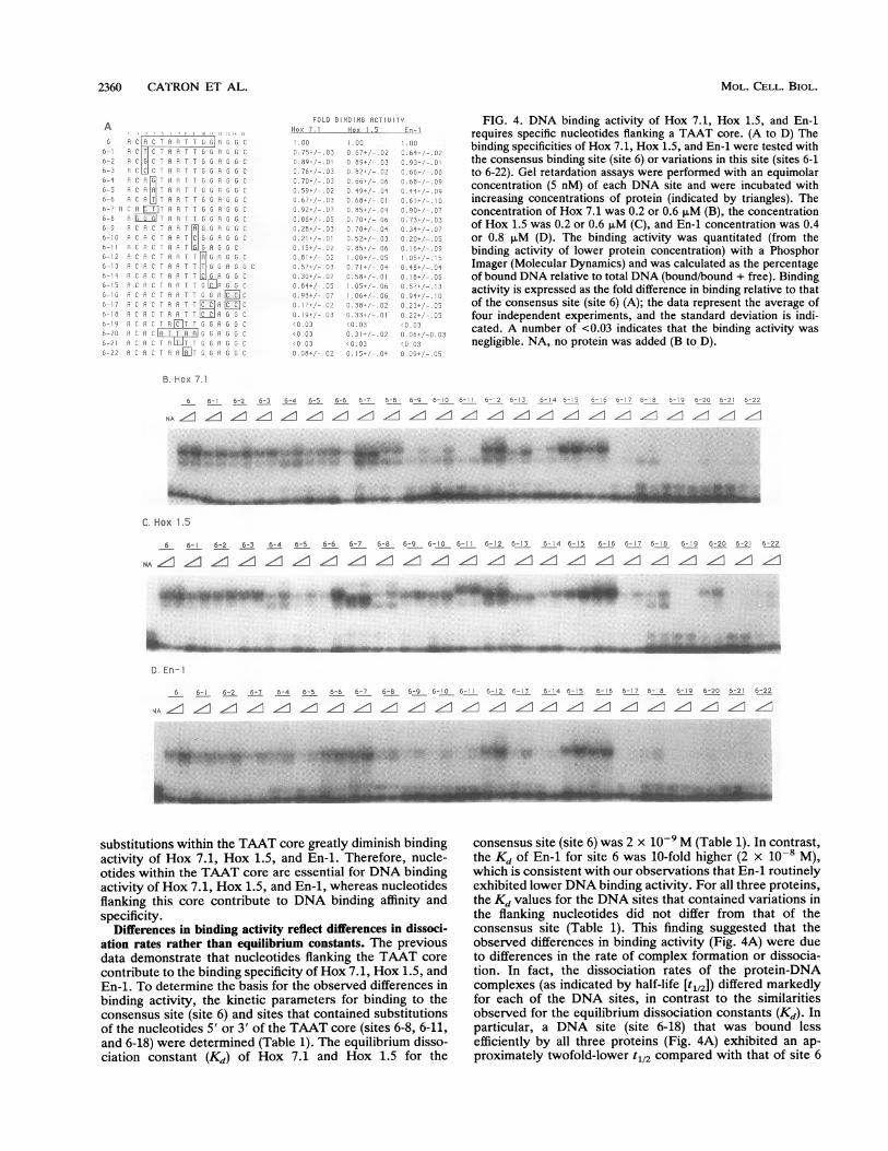
and 3' sides by C or G nucleotides (Fig. 2). In fact, theconsensus sites for all three proteins contained the motif(G/C)TAATTG. To determine the relative contribution ofnucleotides flanking and within the TAAT core towardsbinding activity of Hox 7.1, Hox 1.5, and En-1, we examinedthe interaction of these proteins with DNA sites that con-
tained permutations of the (G/C)TAATTG motif (Fig. 4A).The prototypic sequence for these studies was site 6 (Fig.
2B) since it contained the (G/C)TAATTG motif and since allthree proteins bound to this site with relatively high affinities(Fig. 3A). Hox 7.1, Hox 1.5, and En-1 were tested for theirabilities to bind to a series of DNA sites that containedsubstitutions within the core or substitutions of nucleotides5' or 3' of the core (Fig. 4A). To facilitate direct comparisonsfor preferences among the various sites, the DNA sites weretested in parallel with equimolar concentrations labeled to
HON 7,1 SITES S EQU EN CE 553 1I I G
C1.7
;P2
T
g. -' "~~~~C
CONS|ENSUS'_ 9
D
a, -..-
S ES SELQENE
MOL. CELL. BIOL.
DNA BINDING SITES FOR Hox 7.1, Hox 1.5, AND En-1 2359
A BHox 7.1 Sites
C
Hox 1.5 Sltes En-i Sites
6 35 14A 12A 34A
'ZIzI z.AzzI
32A 14 2 27B 13
Z LZI1 A A12B 2C 30C 7A 30A
Azl zA z z
+ Hox 7.1 _>M OM _4 _4r 40 * 41 to t.
AN,. ls A. L-;L
mimuwf m
o . Om
D SEQUENCE
HOX 7.1 SITES5 C i C TR A T GS A G
CP C T G T, 5 G A A T T 5 g
IfR ~~~gc a T A A T C R C C C JG GIA T G T G A T T T C C G C CS ar R T A R C CG ri T P T 5 T G G
HOX 1.5 SITES32X 7 C C GJT PA T T GC4 c a C A T A A T A G C 3 C G T C
2 C G A G C C C R R T G G c t g
275 a c A A A A A T C G C , C A13 - Lv 1G J v A A u A T T G G
EN-I SITES125 pAf RTAATT3CCCC2C P T Ri]TCr 3CCCC7SC T C A C G G A 3 T 5 T T E t g
X X G aTrT C R3 0RP 9 T T T - C 0 2
BINDING ACTIUITY FIG. 3. Hox 7.1, Hox 1.5, and En-1 bind with similar specificitiesin vitro. (A to C) DNA binding sites for Hox 7.1 (A), Hox 1.5 (B), or
HCX 7.1 HEX 1.5 EN-1 En-1 (C) were obtained by selection from a population of randomDNA sites (as in Fig. 2). The sites were radiolabeled and used as
probes in gel retardation assays containing various concentrations(indicated by triangles) of Hox 7.1 (0, 0.30, or 0.9 ,uM), Hox 1.5 (0,
++ _t 0.2, or 0.7 AM), or En-1 (0, 0.60, or 2.0 F±M). Protein-DNAcomplexes were resolved from free DNA on 6.5% nondenaturing
...ttt polyacrylamide gels, and the complexes were visualized by autora-...tF diography. (D) Nucleotide sequences of the DNA binding sites (as in
Fig. 2) are shown. The relative binding activities of Hox 7.1, Hox1.5, and En-1 (as in Fig. 3A to C) for each of the sites are indicated;+ ++ + denotes a high degree of binding activity, and + indicatesrelatively low binding activity.
approximately the same specific activity (Fig. 4B to D).Binding activity was linear over the range of protein concen-
trations tested, and at the highest concentration of protein,maximal binding to the prototypic site was not greater than50% (Fig. 4B to D).
Contributions of nucleotides 5' of the TAAT core. Substi-tutions of nucleotides 5' of the TAAT core produced modest,but reproducible, alterations in the binding activity of Hox7.1, Hox 1.5, and En-1 (Fig. 4A, compare 6 with 6-1 to 6-6).All three proteins bound preferentially to DNA sites thatcontained a purine nucleotide (A or G) at position 3 andbound less efficiently to sites that contained a T or C in thisposition (Fig. 4A, compare 6 with 6-1 to 6-3). In general,substitutions at position 4 were less well tolerated thansubstitutions at position 3 (Fig. 4A, compare 6 with 6-4 to6-6). All three proteins bound preferentially to DNA sitesthat contained C'4, while DNA sites that contained G4 or Twere bound better than those that contained an A at thisposition (Fig. 4A, compare 6 with 6-4 to 6-6). Surprisingly, aDNA site that contained C3T4 was bound more efficiently byall three proteins than a DNA site that contained A3T4 (Fig.4A, compare 6-6 with 6-7). This suggests that the nucleotideat position 3 may influence the ability of the proteins to bindto DNA sites that contain certain differences at position 4.
Contributions of nucleotides 3' of the TAAT core. Substi-tutions of nucleotides directly flanking the 3' side of theTAAT core significantly reduced binding of Hox 7.1 andEn-1 (Fig. 4A, compare 6 with 6-9 to 6-14). In contrast, mostsubstitutions of these nucleotides were well tolerated byHox 1.5 (Fig. 4A, compare 6 with 6-9 to 6-14). In particular,the binding activity of Hox 7.1 and En-i was markedly
reduced for DNA sites that contained substitutions of T9with any other nucleotide (Fig. 4A, compare 6 with 6-9 to6-11). However, Hox 1.5 bound very efficiently to a DNAsite that contained a G at position 9, and sites that containedA or C were also bound by Hox 1.5, although to a lesserextent (Fig. 4A, compare 6 with 6-9 to 6-11). Although allthree proteins bound preferentially to DNA sites that con-tained a purine nucleotide (A or G) at position 10, othersubstitutions in this position were better tolerated by Hox1.5 than by either Hox 7.1 or En-1 (Fig. 4A, compare 6 with6-12 to 6-14). Nucleotides further 3' of the TAAT core (e.g.,position 11) may also contribute to binding specificity. En-1,but not Hox 7.1 or Hox 1.5, bound less efficiently to a DNAsite that contained a substitution of G11 with C (Fig. 4A,compare 6 with 6-15). In contrast, substitution of nucleotidesat positions 13 and 14 alone did not affect binding activity(Fig. 4A, compare 6 to 6-16). These findings demonstratethat Hox 7.1, Hox 1.5, and En-i have distinct preferencesfor nucleotides 3' of the TAAT core. These preferences arelikely to contribute to the binding specificity among the threeproteins.
Contribution of nucleotides within the TAAT core. Substi-tutions of nucleotides within the TAAT core were mostdeleterious for binding of Hox 7.1, Hox 1.5, and En-1 (Fig.4A, compare 6 with 6-19 to 6-22). Substitution of A7 with a Cor T nucleotide or insertion of C at this position essentiallyabolished the binding activity of all three proteins (Fig. 4A,compare 6 with 6-19 to 6-22). Therefore, A7 is critical forDNA binding activity of Hox 7.1, Hox 1.5, and En-i.Substitution of T8 with A reduced significantly, but did nottotally abolish, binding activity (Fig. 4A, compare 6 with6-22). These data demonstrate that, in contrast to substitu-tions for flanking nucleotides which reduce binding affinity,
+ En-i
.iw,op, ,4 -''0 a
_M - w"
morm ,,
4 0 o -
41 -lw O
VOL. 13, 1993
+ Hox 1.5 aft NO go" -- oo ft,
2360 CATRON ET AL.
A; C.1 i A. F T G C
111 F- T1 5 *
6-- 3 C1i' H F- T !G q 3 6 C
nO RIP: HTP G]610AC~~~~~~~~69c-7Aa - R 'i - G. 9;;c '^
6 - 8 A A q G GJ .9 r6 9 A C P. A A T G6 ]irz AO hlC r.I G A G 6
6 P: P ;: P A G G C'H P:R. r, T TFo1 F cG c
C,- A Cf>r 1' i: q .IT :, G P; v Gb6- I: " :: C P 9 T_C G AR G5Cn 5 Ci' t' k1-1G [R o C
o; n1;2 ' oi ca ARc3 C6 9 A" h CT H[C^ C
6 -I18 i' R T T A G5jCs.
3 2;. 1'LP. T ,T A l6G]LJ u C35 : H: LIIT TCG r P c; CW
C. FIT G i
FOLD B16ND1G ACT; UiTYHox 1._ Hox -1.5 En
8-9 C
7.)
-
C3
3 92''--6 j
3.?8- C
865/
u 8 I 2(
O
C. SI7 0
C9 5.
i.C C
Oi3 - C
:. '; I.'- .C 6
!, i ,. ,
,. O
FIG. 4. DNA binding activity of Hox 7.1, Hox 1.5, and En-irequires specific nucleotides flanking a TAAT core. (A to D) Thebinding specificities of Hox 7.1, Hox 1.5, and En-1 were tested withthe consensus binding site (site 6) or variations in this site (sites 6-1to 6-22). Gel retardation assays were performed with an equimolarconcentration (5 nM) of each DNA site and were incubated withincreasing concentrations of protein (indicated by triangles). Theconcentration of Hox 7.1 was 0.2 or 0.6 ,uM (B), the concentrationof Hox 1.5 was 0.2 or 0.6 ,uM (C), and En-1 concentration was 0.4or 0.8 ,uM (D). The binding activity was quantitated (from thebinding activity of lower protein concentration) with a PhosphorImager (Molecular Dynamics) and was calculated as the percentageof bound DNA relative to total DNA (bound/bound + free). Bindingactivity is expressed as the fold difference in binding relative to thatof the consensus site (site 6) (A); the data represent the average offour independent experiments, and the standard deviation is indi-cated. A number of <0.03 indicates that the binding activity wasnegligible. NA, no protein was added (B to D).
B. hox 7. 1
6 6- 6-2 6-3 6-4 6-5 6-6 6-7 6-8 6-9 6-10 6-' 6- 2 5- 7 56-4 6-5 56- C. F-,- 7 5- B 6- Y 5 26 5-2 C -22
NA~ lfz lf1 lff lf! "I If! lfz If: lf! Iff lllfz -, Iff ll- 1z c
*F;R. . *
.;
C. Hox 1.5
6 6-1 6-2 6-3 6-4 6-5 6-6 6-7 6-8 6-9 6-! 0 6-! 6- 2 6-13 6-14 6-15 5-16 5- 17 6- 8 6-! 9 6-20 6-2 6-22
NA ff df ff ffl Iff Z!! -.- ZI t-- !-- f::GL' f::'fz ,I ,I f
adebwolicA&L', i. Imp 10 #iwAs. IWRRIIIIPFW"!. 11.1. **40, p1m; ...'V.lw,W
D. En-I
6 6-1 6-2 6-3 6-4 6-5 6-6 6-7 6-8 6-9 6-10 6-' - 2 6-3 6-4 6-5 6-16 5- 7 6- 8 6-19 6-20 6-21 6-22
4Ah .1 . =. Z1 Z 1Z Z. /1Z z z/1z.z. z z. . z z
substitutions within the TAAT core greatly diminish bindingactivity of Hox 7.1, Hox 1.5, and En-1. Therefore, nucle-otides within the TAAT core are essential for DNA bindingactivity of Hox 7.1, Hox 1.5, and En-1, whereas nucleotidesflanking this core contribute to DNA binding affinity andspecificity.
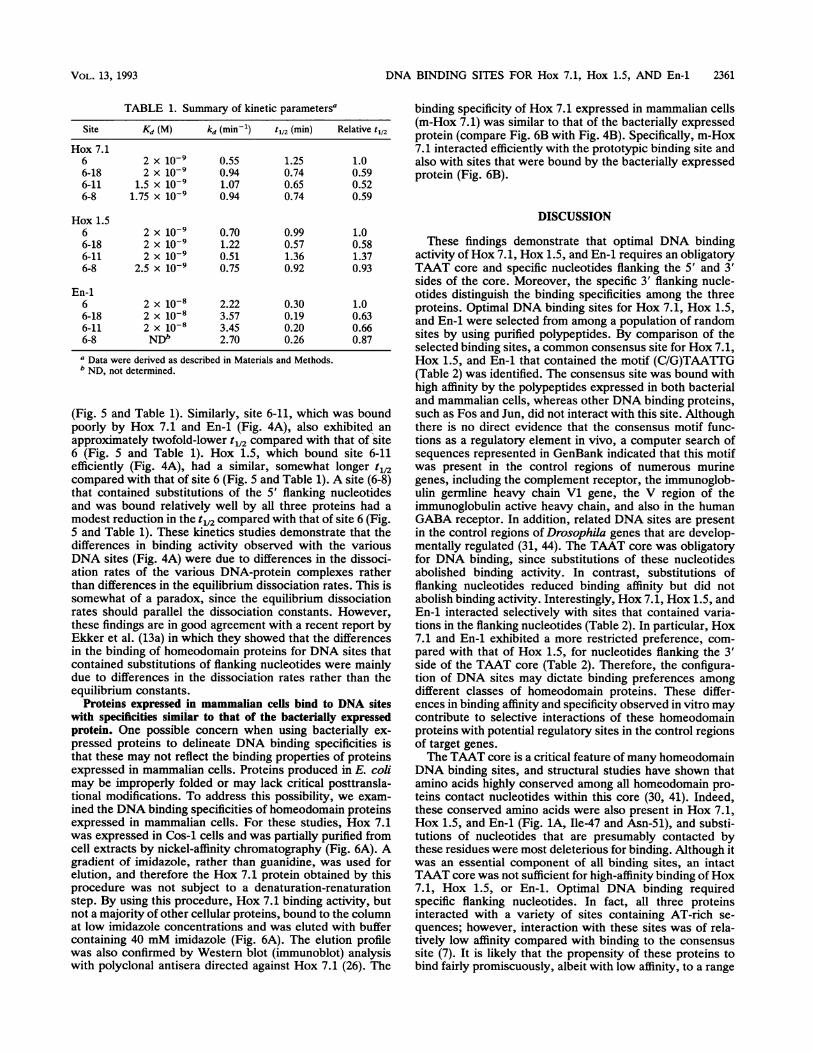
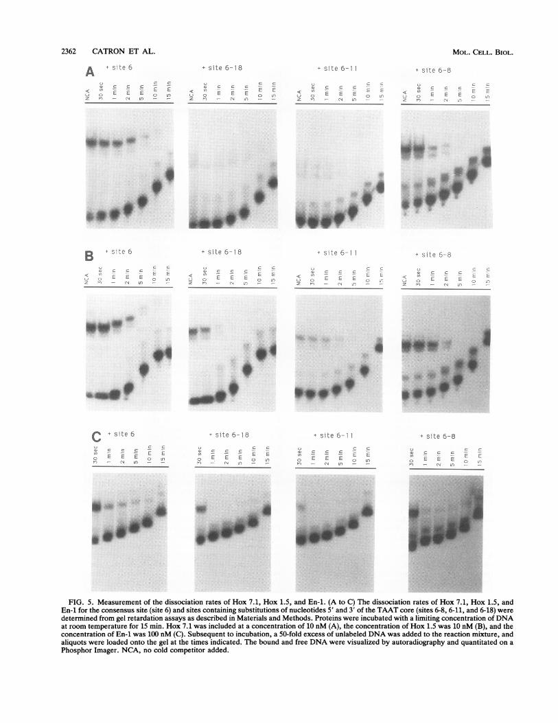
Differences in binding activity reflect differences in dissoci-ation rates rather than equilibrium constants. The previousdata demonstrate that nucleotides flanking the TAAT core
contribute to the binding specificity of Hox 7.1, Hox 1.5, andEn-1. To determine the basis for the observed differences inbinding activity, the kinetic parameters for binding to theconsensus site (site 6) and sites that contained substitutionsof the nucleotides 5' or 3' of the TAAT core (sites 6-8, 6-11,and 6-18) were determined (Table 1). The equilibrium disso-ciation constant (Kd) of Hox 7.1 and Hox 1.5 for the
consensus site (site 6) was 2 x 10⁻⁹ M (Table 1). In contrast,the Kd of En-i for site 6 was 10-fold higher (2 x 10⁻⁸ M),which is consistent with our observations that En-i routinelyexhibited lower DNA binding activity. For all three proteins,the Kd values for the DNA sites that contained variations inthe flanking nucleotides did not differ from that of theconsensus site (Table 1). This finding suggested that theobserved differences in binding activity (Fig. 4A) were dueto differences in the rate of complex formation or dissocia-tion. In fact, the dissociation rates of the protein-DNAcomplexes (as indicated by half-life [t1l2]) differed markedlyfor each of the DNA sites, in contrast to the similaritiesobserved for the equilibrium dissociation constants (Kd). Inparticular, a DNA site (site 6-18) that was bound lessefficiently by all three proteins (Fig. 4A) exhibited an ap-proximately twofold-lower t112 compared with that of site 6
MOL. CELL. BIOL.
DNA BINDING SITES FOR Hox 7.1, Hox 1.5, AND En-1 2361
TABLE 1. Summary of kinetic parametersa binding specificity of Hox 7.1 expressed in mammalian cells
Site Kd (M) kd (min-') t12 (min) Relative t112 (m-Hox 7.1) was similar to that of the bacterially expressedprotein (compare Fig. 6B with Fig. 4B). Specifically, m-Hox
Hox 7.1 7.1 interacted efficiently with the prototypic binding site and6 2 x 10⁻⁹ 0.55 1.25 1.0 also with sites that were bound by the bacterially expressed6-18 2 X 10⁻⁹ 0.94 0.74 0.59 protein (Fig. 6B).
I 1 I v"n-9 1 n7 n c n cl0-116-8
1. -I) X1.75 x
lui0-9
1.N/0.94
u.0j0.74 0.59
Hox 1.566-186-116-8
En-166-186-116-8
DISCUSSION2 x 10-9 0.702 x 10⁻⁹ 1.222 x 10⁻⁹ 0.51
2.5 x 10⁻⁹ 0.75
2 x 10-82 x 10-82 x 10-8NDb
2.223.573.452.70
0.990.571.360.92
0.300.190.200.26
1.00.581.370.93
1.00.630.660.87
a Data were derived as described in Materials and Methods.bND, not determined.
(Fig. 5 and Table 1). Similarly, site 6-11, which was boundpoorly by Hox 7.1 and En-1 (Fig. 4A), also exhibited anapproximately twofold-lower t112 compared with that of site6 (Fig. 5 and Table 1). Hox 1.5, which bound site 6-11efficiently (Fig. 4A), had a similar, somewhat longer t1,2compared with that of site 6 (Fig. 5 and Table 1). A site (6-8)that contained substitutions of the 5' flanking nucleotidesand was bound relatively well by all three proteins had amodest reduction in the t112 compared with that of site 6 (Fig.5 and Table 1). These kinetics studies demonstrate that thedifferences in binding activity observed with the variousDNA sites (Fig. 4A) were due to differences in the dissoci-ation rates of the various DNA-protein complexes ratherthan differences in the equilibrium dissociation rates. This issomewhat of a paradox, since the equilibrium dissociationrates should parallel the dissociation constants. However,these findings are in good agreement with a recent report byEkker et al. (13a) in which they showed that the differencesin the binding of homeodomain proteins for DNA sites thatcontained substitutions of flanking nucleotides were mainlydue to differences in the dissociation rates rather than theequilibrium constants.
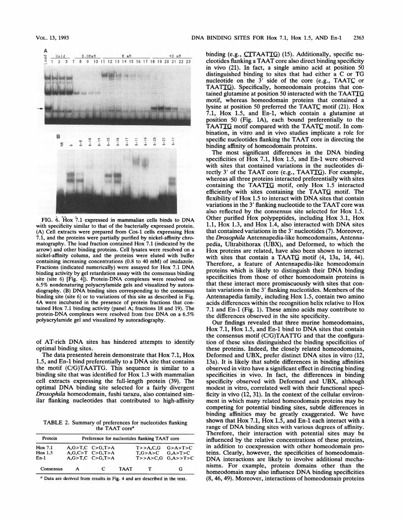
Proteins expressed in mammalian cells bind to DNA siteswith specificities similar to that of the bacterially expressedprotein. One possible concern when using bacterially ex-pressed proteins to delineate DNA binding specificities isthat these may not reflect the binding properties of proteinsexpressed in mammalian cells. Proteins produced in E. colimay be improperly folded or may lack critical posttransla-tional modifications. To address this possibility, we exam-ined the DNA binding specificities of homeodomain proteinsexpressed in mammalian cells. For these studies, Hox 7.1was expressed in Cos-1 cells and was partially purified fromcell extracts by nickel-affinity chromatography (Fig. 6A). Agradient of imidazole, rather than guanidine, was used forelution, and therefore the Hox 7.1 protein obtained by thisprocedure was not subject to a denaturation-renaturationstep. By using this procedure, Hox 7.1 binding activity, butnot a majority of other cellular proteins, bound to the columnat low imidazole concentrations and was eluted with buffercontaining 40 mM imidazole (Fig. 6A). The elution profilewas also confirmed by Western blot (immunoblot) analysiswith polyclonal antisera directed against Hox 7.1 (26). The
These findings demonstrate that optimal DNA bindingactivity of Hox 7.1, Hox 1.5, and En-1 requires an obligatoryTAAT core and specific nucleotides flanking the 5' and 3'sides of the core. Moreover, the specific 3' flanking nucle-otides distinguish the binding specificities among the threeproteins. Optimal DNA binding sites for Hox 7.1, Hox 1.5,and En-1 were selected from among a population of randomsites by using purified polypeptides. By comparison of theselected binding sites, a common consensus site for Hox 7.1,Hox 1.5, and En-1 that contained the motif (C/G)TAATTG(Table 2) was identified. The consensus site was bound withhigh affinity by the polypeptides expressed in both bacterialand mammalian cells, whereas other DNA binding proteins,such as Fos and Jun, did not interact with this site. Althoughthere is no direct evidence that the consensus motif func-tions as a regulatory element in vivo, a computer search ofsequences represented in GenBank indicated that this motifwas present in the control regions of numerous murinegenes, including the complement receptor, the immunoglob-ulin germline heavy chain Vi gene, the V region of theimmunoglobulin active heavy chain, and also in the humanGABA receptor. In addition, related DNA sites are presentin the control regions of Drosophila genes that are develop-mentally regulated (31, 44). The TAAT core was obligatoryfor DNA binding, since substitutions of these nucleotidesabolished binding activity. In contrast, substitutions offlanking nucleotides reduced binding affinity but did notabolish binding activity. Interestingly, Hox 7.1, Hox 1.5, andEn-1 interacted selectively with sites that contained varia-tions in the flanking nucleotides (Table 2). In particular, Hox7.1 and En-1 exhibited a more restricted preference, com-pared with that of Hox 1.5, for nucleotides flanking the 3'side of the TAAT core (Table 2). Therefore, the configura-tion of DNA sites may dictate binding preferences amongdifferent classes of homeodomain proteins. These differ-ences in binding affinity and specificity observed in vitro maycontribute to selective interactions of these homeodomainproteins with potential regulatory sites in the control regionsof target genes.The TAAT core is a critical feature of many homeodomain
DNA binding sites, and structural studies have shown thatamino acids highly conserved among all homeodomain pro-teins contact nucleotides within this core (30, 41). Indeed,these conserved amino acids were also present in Hox 7.1,Hox 1.5, and En-1 (Fig. 1A, Ile-47 and Asn-51), and substi-tutions of nucleotides that are presumably contacted bythese residues were most deleterious for binding. Although itwas an essential component of all binding sites, an intactTAAT core was not sufficient for high-affinity binding of Hox7.1, Hox 1.5, or En-1. Optimal DNA binding requiredspecific flanking nucleotides. In fact, all three proteinsinteracted with a variety of sites containing AT-rich se-quences; however, interaction with these sites was of rela-tively low affinity compared with binding to the consensussite (7). It is likely that the propensity of these proteins tobind fairly promiscuously, albeit with low affinity, to a range
VOL. 13, 1993
2362 CATRON ET AL.
A 4 site 6
Q,~~~~~~~~~~~~~~~~~~~~~~~~~~~~~~~~~~~c
:D C C C- -
u o E E E o nz n - CN In - -
+ site 6-18
o ~~C Ca) C C C -<n - E E
;j C) E E E 0 Cz n - A jn -..
+ site 6-11
a, c c c<1 ~ - L CC) G E E E o -.z -i - N dn --
+ site 6-8
C
C- E E E i' ) JEZ -1 4_ )
.." 0..^~~~~~~~..>__:
^ ~ 00
B site 6
Sa C C C< EES_C) E E E rIonz n - 1-1 In - -
+ site 6- 8
) c *c _ = -
< tn E E o r
z rl~.1
* site 6-1
< :S: E LL) E E t , £z n (N J'-:
+ site 6-8
}) =:C- C C
< n -_ ('J O E E ;:- ,,1Z n _- - ~n
I*q04~~~~~~~~J0
0 www'', - _
.;. . [email protected] _;_
C + site 6
U ~~~~~~~~~~ccv c c c -cn ~~~E Eo E E E o0 L
I- 'IN LaO -
+ site 6-18
LJ c C:D) C C C -- - E EO EEE 2 nn- N Ln _
+ site 6-1 1
u - cc C C- - - L E
0 E E E n-1) - r1 Un - -
+ S.e 6-8
C 2 _- £2.
E E Epn - CN J -
._ ...
E_...
W.-.
FIG. 5. Measurement of the dissociation rates of Hox 7.1, Hox 1.5, and En-1. (A to C) The dissociation rates of Hox 7.1, Hox 1.5, andEn-1 for the consensus site (site 6) and sites containing substitutions of nucleotides 5' and 3' of the TAAT core (sites 6-8, 6-11, and 6-18) weredetermined from gel retardation assays as described in Materials and Methods. Proteins were incubated with a limiting concentration ofDNAat room temperature for 15 min. Hox 7.1 was included at a concentration of 10 nM (A), the concentration of Hox 1.5 was 10 nM (B), and theconcentration of En-1 was 100 nM (C). Subsequent to incubation, a 50-fold excess of unlabeled DNA was added to the reaction mixture, andaliquots were loaded onto the gel at the times indicated. The bound and free DNA were visualized by autoradiography and quantitated on aPhosphor Imager. NCA, no cold competitor added.
;W - :w*''IM
,~,*g*600.
.**_
gE9
_ .
MOL. CELL. BIOL.
azk".,wA
DNA BINDING SITES FOR Hox 7.1, Hox 1.5, AND En-i 2363
AUo i d 0. 06m1 _ 1 _mo mml
o 2 3 7 6 9 10 11 12 13 1 15 16 17 18 19 20 21 22 23
L i,..k,... i*F-0
B o Ln,,, r- cp_ 3 z
Z 0 la 10e C s < -
*"
FIG. 6. Hox 7.1 expressed in mammalian cells binds to DNAwith specificity similar to that of the bacterially expressed protein.(A) Cell extracts were prepared from Cos-1 cells expressing Hox7.1, and the proteins were partially purified by nickel-affinity chro-matography. The load fraction contained Hox 7.1 (indicated by thearrow) and other binding proteins. Cell lysates were resolved on anickel-affinity column, and the proteins were eluted with buffercontaining increasing concentrations (0.8 to 40 mM) of imidazole.Fractions (indicated numerically) were assayed for Hox 7.1 DNAbinding activity by gel retardation assay with the consensus bindingsite (site 6) [Fig. 4]). Protein-DNA complexes were resolved on6.5% nondenaturing polyacrylamide gels and visualized by autora-diography. (B) DNA binding sites corresponding to the consensusbinding site (site 6) or to variations of this site as described in Fig.4A were incubated in the presence of protein fractions that con-tained Hox 7.1 binding activity (panel A; fractions 18 and 19). Theprotein-DNA complexes were resolved from free DNA on a 6.5%polyacrylamide gel and visualized by autoradiography.
of AT-rich DNA sites has hindered attempts to identifyoptimal binding sites.The data presented herein demonstrate that Hox 7.1, Hox
1.5, and En-1 bind preferentially to a DNA site that containsthe motif (C/G)TAATTG. This sequence is similar to abinding site that was identified for Hox 1.3 with mammaliancell extracts expressing the full-length protein (39). Theoptimal DNA binding site selected for a fairly divergentDrosophila homeodomain, fushi tarazu, also contained sim-ilar flanking nucleotides that contributed to high-affinity
TABLE 2. Summary of preferences for nucleotides flankingthe TAAT corea
Protein Preference for nucleotides flanking TAAT core
Hox 7.1 A,G>T,C C>G,T>A T>>A,C,G G>A>T>CHox 1.5 A,G,C>T C>G,T>A T,G>A>C G,A>T>CEn-i A,G>T,C C>G,T>A T>>A>C,G G,A>>T>C
Consensus A C TAAT T G
a Data are derived from results in Fig. 4 and are described in the text.
binding (e.g., (JJTAATT) (15). Additionally, specific nu-cleotides flanking a TAAT core also direct binding specificityin vivo (21). In fact, a single amino acid at position 50distinguished binding to sites that had either a C or TGnucleotide on the 3' side of the core (e.g., TAATC orTAATM). Specifically, homeodomain proteins that con-tained glutamine at position 50 interacted with the TAATIXmotif, whereas homeodomain proteins that contained alysine at position 50 preferred the TAATC motif (21). Hox7.1, Hox 1.5, and En-1, which contain a glutamine atposition 50 (Fig. 1A), each bound preferentially to theTAATjI motif compared with the TAATC motif. In com-bination, in vitro and in vivo studies implicate a role forspecific nucleotides flanking the TAAT core in directing thebinding affinity of homeodomain proteins.The most significant differences in the DNA binding
specificities of Hox 7.1, Hox 1.5, and En-i were observedwith sites that contained variations in the nucleotides di-rectly 3' of the TAAT core (e.g., TAATJX). For example,whereas all three proteins interacted preferentially with sitescontaining the TAATJ. motif, only Hox 1.5 interactedefficiently with sites containing the TAAT. motif. Theflexibility of Hox 1.5 to interact with DNA sites that containvariations in the 3' flanking nucleotide to the TAAT core wasalso reflected by the consensus site selected for Hox 1.5.Other purified Hox polypeptides, including Hox 3.1, Hox1.1, Hox 1.3, and Hox 1.4, also interacted with DNA sitesthat contained variations in the 3' nucleotides (7). Moreover,the Drosophila Antennapedia-like homeodomains, Antenna-pedia, Ultrabithorax (UBX), and Deformed, to which theHox proteins are related, have also been shown to interactwith sites that contain a TAAT£ motif (4, 13a, 14, 44).Therefore, a feature of Antennapedia-like homeodomainproteins which is likely to distinguish their DNA bindingspecificities from those of other homeodomain proteins isthat these interact more promiscuously with sites that con-tain variations in the 3' flanking nucleotides. Members of theAntennapedia family, including Hox 1.5, contain two aminoacids differences within the recognition helix relative to Hox7.1 and En-i (Fig. 1). These amino acids may contribute tothe differences observed in the site specificity.Our findings revealed that three murine homeodomains,
Hox 7.1, Hox 1.5, and En-l bind to DNA sites that containthe consensus motif (C/G)TAATTG and that the configura-tion of these sites distinguished the binding specificities ofthese proteins. Indeed, the closely related homeodomains,Deformed and UBX, prefer distinct DNA sites in vitro (12,13a). It is likely that subtle differences in binding affinitiesobserved in vitro have a significant effect in directing bindingspecificities in vivo. In fact, the differences in bindingspecificity observed with Deformed and UBX, althoughmodest in vitro, correlated well with their functional speci-ficity in vivo (12, 31). In the context of the cellular environ-ment in which many related homeodomain proteins may becompeting for potential binding sites, subtle differences inbinding affinities may be greatly exaggerated. We haveshown that Hox 7.1, Hox 1.5, and En-1 each interact with arange of DNA binding sites with various degrees of affinity.Therefore, their interaction with potential sites may beinfluenced by the relative concentrations of these proteins,in addition to coexpression with other homeodomain pro-teins. Clearly, however, the specificities of homeodomain-DNA interactions are likely to involve additional mecha-nisms. For example, protein domains other than thehomeodomain may also influence DNA binding specificities(8, 46, 49). Moreover, interactions of homeodomain proteins
VOL. 13, 1993
2364 CATRON ET AL.
with other proteins (19, 38) and competition among tran-scriptional regulatory proteins for overlapping or adjacentbinding sites (23) are likely to play a significant role indirecting DNA binding specificity. The studies presentedherein establish the appropriate foundation in which toexamine these possibilities.
ACKNOWLEDGMENTS
Preliminary experiments were performed in Tom Curran's labo-ratory, Roche Institute of Molecular Biology, and we are indebted tohim for his generous support, encouragement, and many helpfuldiscussions. We thank Jill McMahon and Andy McMahon, RocheInstitute of Molecular Biology, for the gift of the mouse embryoniccDNA and for helpful discussions, and Jennifer Morris and Frank J.Rauscher III, Wistar Institute, for the gift of the random oligonu-cleotide probe and for helpful discussions. We thank Zhigang Shang,Scott Holmes, and Peter Lobel for assistance in analyzing thekinetic data. We acknowledge Aaron Shatkin, Drew Vershon, MikeTocci, Guy Montelione, and Danny Reinberg for reading the manu-script and Janet Hansen and Alyson Broads for preparation.
This work was supported, in part, by funds to C.A. from the NewJersey Commission on Science and Technology and by a grant(HD29446-01) from the NIH; K.M.C. was supported by a postdoc-toral training grant from Hoffmann-La Roche, and N.I. was sup-ported by a predoctoral fellowship from Merck. C.A. is a recipientof a Sinscheimer Scholar award.
REFERENCES1. Abate, C. Unpublished data.2. Abate, C., D. Luk, and T. Curran. 1991. Transcriptional regu-
lation by Fos and Jun interaction among multiple activator andregulatory domains. Mol. Cell. Biol. 11:3624-3632.
3. Abate, C., D. Luk, R. Gentz, F. J. Rauscher III, and T. Curran.1990. Expression and purification of the leucine zipper andDNA-binding domains of Fos and Jun: both Fos and Jun contactDNA directly. Proc. Natl. Acad. Sci. USA 87:1032-1036.
4. Affolter, M., A. Percival-Smith, M. Muller, W. Leupin, andW. J. Gehring. 1990. DNA binding properties of the purifiedAntennapedia homeodomain. Proc. Natl. Acad. Sci. USA 87:4093-4097.
5. Akam, M. 1989. Hox and Hom: homologous gene clusters ininsects and vertebrates. Cell 57:347-349.
6. Blackwell, T. K., and H. Weintraub. 1990. Differences andsimilarities in DNA-binding preferences of MyoD and E2Aprotein complexes revealed by binding site selection. Science250:1104-1110.
7. Catron, K. M., and C. Abate. Unpublished data.8. Chalepakis, G., R. Fritsch, H. Fickenscher, U. Deutsch, M.
Goulding, and P. Gruss. 1991. The molecular basis of theundulatedlPax-1 mutation. Cell 66:873-884.
9. Chisaka, O., and M. R. Capecchi. 1991. Regionally restricteddevelopmental defects resulting from targeted disruption of themouse homeobox gene Hox-1.5. Nature (London) 350:473-479.
10. Davis, C. A., and A. L. Joyner. 1988. Expression patterns of thehomeobox-containing genes en-i and en-2 and the proto-onco-gene int-i diverge during mouse development. Genes Dev.2:1736-1744.
11. Desplan, C., J. Theis, and P. H. O'Farrell. 1988. The sequencespecificity of homeodomain-DNA interaction. Cell 54:1081-1090.
12. Dessain, S., C. T. Gross, M. A. Kuziora, and W. McGinnis. 1992.Anti-type homeodomains have distinct DNA binding specifici-ties that correlate with their different regulatory functions inembryos. EMBO J. 11:991-1002.
13. Dolle, P., J.-C. Izpisua-Belmonte, H. Falkenstein, A. Renucci,and D. Duboule. 1989. Coordinate expression of the murinehox-5 complex homeobox-containing genes during limb patternformation. Nature (London) 342:767-772.
13a.Ekker, S. C., D. P. von Kessler, and P. A. Beachy. 1992.Differential sequence recognition is a determinant of specificityin homeotic gene action. EMBO J. 11:4059-4072.
14. Ekker, S. C., K. E. Young, D. P. von Kessler, and P. A. Beachy.1991. Optimal DNA sequence recognition by the Ultrabithoraxhomeodomain of Drosophila. EMBO J. 10:1179-1186.
15. Florence, B., R. Handrow, and A. Laughon. 1991. DNA-bindingspecificity of the fushi tarazu homeodomain. Mol. Cell. Biol.11:3613-3623.
16. Gehring, W. J. 1987. Homeo boxes in the study of development.Science 235:1245-1252.
17. Gibson, G., A. Schier, P. LeMotte, and W. J. Gehring. 1990. Thespecificities of Sexcombs Reduced and Antennapedia are de-fined by a distinct portion of each protein that includes thehomeodomain. Cell 62:1087-1103.
18. Graham, A., N. Papalopulu, and R. Krumlauf. 1989. The murineand Drosophila homeobox gene complexes have common fea-tures of organization and expression. Cell 57:367-378.
19. Grueneberg, D. A., S. Natesan, C. Alexandre, and M. Z. Gilman.1992. Human and Drosophila homeodomain proteins that en-hance the DNA-binding activity of serum response factor.Science 257:1089-1095.
20. Hanes, S. D., and R. Brent. 1989. DNA specificity of the Bicoidactivator protein is determined by homeodomain recognitionhelix residue 9. Cell 57:1275-1283.
21. Hanes, S. D., and R. Brent. 1991. A genetic model for interac-tion of the homeodomain recognition helix with DNA. Science251:426-430.
22. Hill, R. E., P. F. Jones, A. R. Rees, C. M. Sime, M. J. Justice,N. G. Copeland, N. A. Jenkins, E. Graham, and D. R. Davidson.1989. A new family of mouse homeo box-containing genes:molecular structure, chromosomal location, and developmentalexpression of Hox-7.1. Genes Dev. 3:26-37.
23. Hoch, M., N. Gerwin, H. Taubert, and H. Jackie. 1992. Com-petition for overlapping sites in the regulatory region of theDrosophila gene Kruppel. Science 256:94-97.
24. Hoey, T., and M. Levine. 1988. Divergent homeo box proteinsrecognize similar DNA sequences in Drosophila. Nature (Lon-don) 332:858-861.
25. Holland, P. W. H., and B. L. M. Hogan. 1988. Expression ofhomeo box genes during mouse development: a review. GenesDev. 2:773-782.
26. Iler, N., and C. Abate. Unpublished data.27. Janknecht, R., G. de Martynoff, J. Lou, R. A. Hipskind, A.
Nordheim, and H. G. Stunnenberg. 1991. Rapid and efficientpurification of native histidine-tagged protein expressed byrecombinant vaccinia virus. Proc. Natl. Acad. Sci. USA 88:8972-8976.
28. Joyner, A. L., and G. R. Martin. 1987. en-i and en-2, two mousegenes with sequence homology to the Drosophila engrailedgene: expression during embryogenesis. Genes Dev. 1:29-38.
29. Kessel, M., and P. Gruss. 1990. Murine developmental controlgenes. Science 249:374-379.
30. Kissinger, C. R., B. Liu, E. Martin-Blanco, T. B. Kornberg, andC. 0. Pabo. 1990. Crystal structure of an Engrailed homeo-domain-DNA complex at 2.8 A resolution: a framework forunderstanding homeodomain-DNA interactions. Cell 63:579-590.
31. Kuziora, M. A., and W. McGinnis. 1989. A homeodomainsubstitution changes the regulatory specificity of the Deformedprotein in Drosophila embryos. Cell 59:563-571.
32. Laughon, A. 1991. DNA binding specificity of homeodomains.Biochemistry 30:11357-11367.
33. Lufldn, T., A. Dierich, M. LeMeur, M. Mark, and P. Chambon.1991. Disruption of the hox-i.6 homeobox gene results indefects in a region corresponding to its rostral domain ofexpression. Cell 66:1105-1119.
34. Mackenzie, A., G. L. Leeming, A. K. Jowett, M. W. J. Ferguson,and P. T. Sharpe. 1991. The homeobox gene hox-7. 1 has specificregional and temporal expression patterns during early murinecraniofacial embryogenesis, especially tooth development invivo and in vitro. Development 111:269-285.
35. Mann, R. S., and D. S. Hogness. 1990. Functional dissection ofUltrabithorax proteins in D. melanogaster. Cell 60:597-610.
36. McGinnis, W., R. L. Garber, J. Wirz, A. Kuroiwa, and W. J.Gehring. 1984. A homologous protein-coding sequence in Dros-
MOL. CELL. BIOL.
DNA BINDING SITES FOR Hox 7.1, Hox 1.5, AND En-1 2365
ophila homeotic genes and its conservation in other metazoans.Cell 37:403-408.
37. McGinnis, W., and R. Krumlauf. 1992. Homeobox genes andaxial patterning. Cell 68:283-302.
38. Mendel, D. B., P. A. Khavari, P. B. Conley, M. K. Graves, L. P.Hansen, A. Admon, and G. R. Crabtree. 1991. Characterizationof a cofactor that regulates dimerization of a mammalian home-odomain protein. Science 254:1762-1767.
39. Odenwald, W. F., J. Garbern, H. Arnheiter, E. Tournier-Lasserve, and R. A. Lazzarini. 1989. The Hox-1.3 homeo boxprotein is a sequence-specific DNA-binding phosphoprotein.Genes Dev. 3:158-172.
40. Oliphant, A. R., C. J. Brandl, and K. Struhl. 1989. Defining thesequence specificity of DNA-binding proteins by selecting bind-ing sites from random-sequence oligonucleotides: analysis ofyeast GCN4 protein. Mol. Cell. Biol. 9:2944-2949.
41. Otting, G., Y. Q. Qian, M. Billeter, M. Muller, M. Affolter,W. J. Gehring, and K. Wuthrich. 1990. Protein-DNA contacts inthe structure of a homeodomain-DNA complex determined bynuclear magnetic resonance spectroscopy in solution. EMBO J.9:3085-3092.
42. Patwardhan, S., A. Gashler, M. G. Siegel, L. C. Chang, L. G.Joseph, T. B. Shows, and M. M. Lebeau. 1991. egr3, a novelmember of the egr family of genes encoding immediate-earlytranscription factors. Oncogene 6:917-928.
43. Rauscher, F. J., III, J. F. Morris, 0. E. Tournay, D. M. Cook,and T. Curran. 1990. Binding of the Wilms' tumor locus zincfinger protein to the EGR-1 consensus sequence. Science 250:
1259-1262.44. Reguiski, M., S. Dessain, N. McGinnis, and W. McGinnis. 1991.
High-affinity binding sites for the Deformed protein are requiredfor the function of an autoregulatory enhancer of the deformedgene. Genes Dev. 5:278-286.
45. Robert, B., D. Sassoon, B. Jacq, W. Gehring, and M. Bucking-ham. 1989. hox-7, a mouse homeobox gene with a novel patternof expression during embryogenesis. EMBO J. 8:91-100.
46. Rosenfeld, M. G. 1991. POU-domain transcription factors: Pou-er-ful developmental regulators. Genes Dev. 5:897-907.
47. Scott, M. P., J. W. Tamkun, and G. W. Hartzell III. 1989. Thestructure and function of the homeodomain. Biochim. Biophys.Acta 989:25-48.
48. Treisman, J., P. Gonczy, M. Vashishtha, E. Harris, and C.Desplan. 1989. A single amino acid can determine the DNAbinding specificity of homeodomain proteins. Cell 59:553-562.
49. Treisman, J., E. Harris, and C. Desplan. 1991. The paired boxencodes a second DNA-binding domain in the Paired homeodomain protein. Genes Dev. 5:594-604.
50. Wilkinson, D. G., S. Bhatt, M. Cook, E. Bonicelli, and R.Krumlauf. 1989. Segmental expression of hox-2 homeobox-containing genes in the developing mouse hindbrain. Nature(London) 341:405-409.
51. Wright, C. V. E., K. W. Y. Cho, G. Oliver, and E. M.DeRobertis. 1989. Vertebrate homeodomain proteins: families ofregion-specific transcription factors. Trends Biochem. Sci. 14:52-56.
VOL. 13, 1993