nuclear physics and medical applications - ifcoelho/newman/newman26.pdf · 634 nuclear physics and...
TRANSCRIPT
N U C L E A R S I Z E , S T R U C T U R E , A N D F O R C E S 633
J. Newman, Physics of the Life Sciences, DOI: 10.1007/978-0-387-77259-2_26, © Springer Science+Business Media, LLC 2008
In this concluding chapter, we first summarize our knowledge of the atomic nucleusand discuss the types of nuclear radiation emitted by nuclei and how they can bedetected. The rest of the chapter focuses on a variety of applications of nuclear radi-ation in science and medicine. We start our discussion of applications by introducingthe half-life and its use in radioactive dating. Then we introduce some importantideas on dosimetry and the biological effects of radiation, as well as some ideas onnuclear medicine. Two methods (SPECT and PET) are discussed that use nuclearradiation to do imaging of the body, known as radiation tomography. The chapterconcludes with the processes of nuclear fission and fusion, two topics that should beunderstood at a basic level by everyone in this nuclear age.
1. NUCLEAR SIZE, STRUCTURE, AND FORCES
The nucleus is an extremely small dense object in an otherwise nearly empty atom.As we’ve seen, atomic sizes are about 0.1 nm. The nucleus is typically several fm(10�15 m), or about 100,000 times smaller than the atom. To appreciate these rela-tive sizes, imagine that we scale the size of an atom up to the size of a football field(100 yd ~ 100 m). On this scale the nucleus would have a relative size of 100 m/(100,000) � 1 mm, so that it would be like the head of a pin, not in a haystack,but on a football field. This is truly astounding because almost all of the mass ofan atom is located inside the nucleus. Matter consists of dense cores in mostlyempty space; the head of a pin located within an empty three-dimensional footballfield of space.
Remember that the nucleus contains the protons and neutrons (together known asnucleons) of the atom, representing nearly its entire mass, because protons and neu-trons each have more than 1800 times the mass of an electron. Unlike the electron,which appears to be pointlike, having no measurable size, nucleons have a finite sizeof about 1 fm. Neutral atoms have equal numbers of protons and electrons, with thisnumber known as the atomic number and represented by Z; the number of neutronsin a nucleus is known as the neutron number and represented by N. The total numberof protons and neutrons in a nucleus added together is known as the mass number A,where
A � Z � N. (26.1)
The integer mass number is approximately equal to the atomic mass. Remember thatatomic masses are measured in atomic mass units (u), defined as 1/12 the mass of thecarbon-12 atom, or 1 u � 1.66 � 10�27 kg.
26Nuclear Physics and Medical Applications
634 N U C L E A R P H Y S I C S A N D M E D I C A L A P P L I C AT I O N S
A particular nuclear species is called a nuclide, and is represented by thechemical symbol of its neutral atom together with its value of A written as a pre-superscript. For example, 13C represents the nuclide with 6 protons (because allcarbon atoms have six protons), and N (� A � Z) � 13 � 6 � 7 neutrons.Sometimes the Z value will be written explicitly as 13
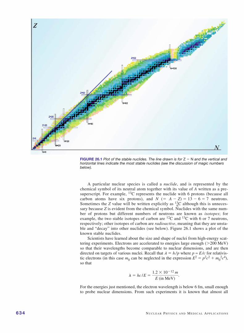
6C although this is unneces-sary because Z is evident from the chemical symbol. Nuclides with the same num-ber of protons but different numbers of neutrons are known as isotopes; forexample, the two stable isotopes of carbon are 12C and 13C with 6 or 7 neutrons,respectively; other isotopes of carbon are radioactive, meaning that they are unsta-ble and “decay” into other nuclides (see below). Figure 26.1 shows a plot of theknown stable nuclides.
Scientists have learned about the size and shape of nuclei from high-energy scat-tering experiments. Electrons are accelerated to energies large enough (�200 MeV)so that their wavelengths become comparable to nuclear dimensions, and are thendirected on targets of various nuclei. Recall that l � h /p where p ≈ E/c for relativis-tic electrons (in this case m0 can be neglected in the expression E2 � p2c2 � m0
2c4),so that
For the energies just mentioned, the electron wavelength is below 6 fm, small enoughto probe nuclear dimensions. From such experiments it is known that almost all
l � hc / E �1.2 � 10�12 m
E (in MeV).
FIGURE 26.1 Plot of the stable nuclides. The line drawn is for Z � N and the vertical andhorizontal lines indicate the most stable nuclides (see the discussion of magic numbersbelow).
B I N D I N G E N E R G Y A N D N U C L E A R S TA B I L I T Y 635
nuclei are nearly spherical (although many of the rare-earth element nuclei, thosewith Z � 57 � 71, are ellipsoidal) with somewhat fuzzy boundaries and effectiveradii R that depend on the mass number A according to
R � R0 A1/3, (26.2)
with R0 1.2 fm.Because the density of the nucleus is given by the ratio of its mass (proportional
to A) to its volume (proportional to R3, and thus, according to Equation (26.2), also toA), perhaps unexpectedly we see that the density of all nuclei is the same. We cantherefore calculate the nuclear density using A � 1, to find that � � 1.67 � 10�27kg/[(4�/3)(1.2 � 10�15 m)3] 2 � 1017 kg/m3. This is an extremely high density; notethat the density of common materials, and thus of atoms, is only on the order of 103
kg/m3, so that nuclei are 1014 times denser than atoms! Both the greater mass of anucleon compared to the electron and, even more, the tiny size of the nucleus com-pared to atoms are responsible for this.
Our picture of the nucleus as a dense ball of nucleons that are essentially in con-tact with one another leads to the striking question of why the nucleus is ever stable.After all, the protons, all with the same positive charge, are extremely close togetherin the nucleus and their electrical repulsive force is huge. Two protons that are 2 fmapart would experience an electrical repulsive force given by
(26.3)
where e is the proton charge and r is the 2 fm separation distance. This force is almostequal to 60 N (about 13 lb), a huge force that would instantly rip the nucleus apart ifit were the only force acting.
In fact, the nucleus is held together by the strong nuclear force, one of two veryshort-range nuclear forces (the other, known as the weak nuclear force, is involvedin radioactive decay). The strong force between two neighboring protons in anucleus provides an attractive force roughly 100 times stronger than the electricalrepulsion between the two. This attractive force is the same for all protons and neu-trons, independent of their electric charge, so that two neighboring neutrons, pro-tons, or a neutron and a proton all feel the same attractive force. However, thestrong force rapidly vanishes at distances of even a few fm within the nucleus, andcertainly outside the nucleus. A useful simple picture of the nucleus is the liquiddrop model in which the nucleus is pictured as a tiny drop of liquid. This analogyis appropriate because both the nucleus and a liquid drop have a uniform density,are incompressible, and are held together by large forces: surface tension forces inthe case of a liquid, strong forces in the nucleus. This model provides a useful wayto look at the process of nuclear fission later in this chapter as analogous to a dropof liquid breaking into two smaller drops.
2. BINDING ENERGY AND NUCLEAR STABILITY
The total energy of the nucleus is the sum of its kinetic and potential energy.Because the potential energy is negative and larger, in magnitude, than the kineticenergy, the total energy of the nucleus is negative, just as we have seen it is for aneutral atom. If the nucleus were disassembled into its constituent protons and neu-trons, their total energy would be more than that of the nucleus. This is just thesame as the case for atoms where energy is needed to ionize an atom, for example,in hydrogen to separate the electron and proton, so that the energy of the final sep-arated electron and proton have greater energy than that of the ground state atom.This difference is due to the binding energy of the atom or nucleus and, in the case
F �1
4pe0 e2
r2,
�
�
of the nucleus is a considerable amount of energy. For any nucleus of atomic andmass numbers Z and A, the (positive amount of) binding energy is given by
Nuclear Binding Energy � Zmpc2 � Nmnc2 � mc2, (26.4)
where mp, mn, and m are the masses of the proton, neutron, and nucleus, respectively.Because the energy equivalent of 1 atomic mass unit is (1 u)c2 � 931.5 MeV (foundfrom E � mc2 � (1.6605 � 10�27 kg)(2.9979 � 108 m/s)2(1 eV/1.6022 � 10�19 J) �9.315 � 108 eV � 931.5 MeV (with energy conversion to eV)), we see that thenucleons each have an energy equivalent of about 930 MeV, whereas a nucleus ofmass number A has an energy equivalent of about A � 930 MeV. A comparable cal-culation for an atom shows that the atomic binding energy is only on the order of atmost tens of eV.
636 N U C L E A R P H Y S I C S A N D M E D I C A L A P P L I C AT I O N S
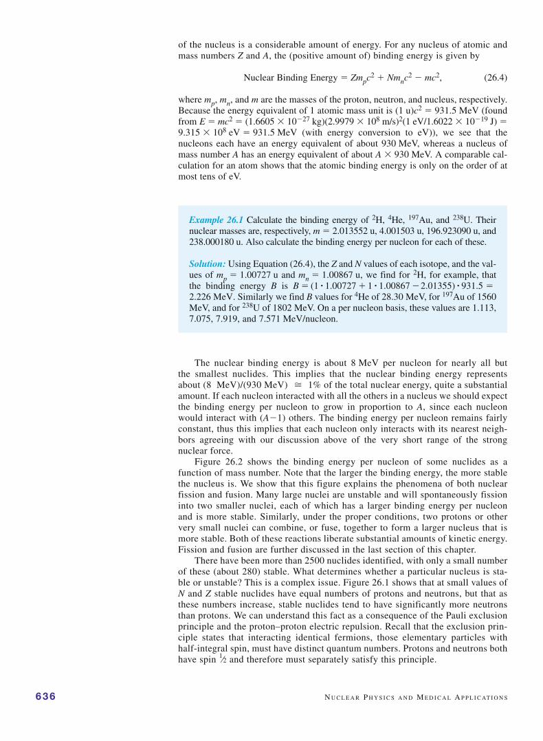
Example 26.1 Calculate the binding energy of 2H, 4He, 197Au, and 238U. Theirnuclear masses are, respectively, m � 2.013552 u, 4.001503 u, 196.923090 u, and238.000180 u. Also calculate the binding energy per nucleon for each of these.
Solution: Using Equation (26.4), the Z and N values of each isotope, and the val-ues of mp � 1.00727 u and mn � 1.00867 u, we find for 2H, for example, thatthe binding energy B is
Similarly we find B values for 4He of 28.30 MeV, for 197Au of 1560MeV, and for 238U of 1802 MeV. On a per nucleon basis, these values are 1.113,7.075, 7.919, and 7.571 MeV/nucleon.
2.226 MeV.B � (1 # 1.00727 � 1 # 1.00867 �2.01355) # 931.5 �
The nuclear binding energy is about 8 MeV per nucleon for nearly all butthe smallest nuclides. This implies that the nuclear binding energy representsabout (8 MeV)/(930 MeV) 1% of the total nuclear energy, quite a substantialamount. If each nucleon interacted with all the others in a nucleus we should expectthe binding energy per nucleon to grow in proportion to A, since each nucleonwould interact with (A�1) others. The binding energy per nucleon remains fairlyconstant, thus this implies that each nucleon only interacts with its nearest neigh-bors agreeing with our discussion above of the very short range of the strongnuclear force.
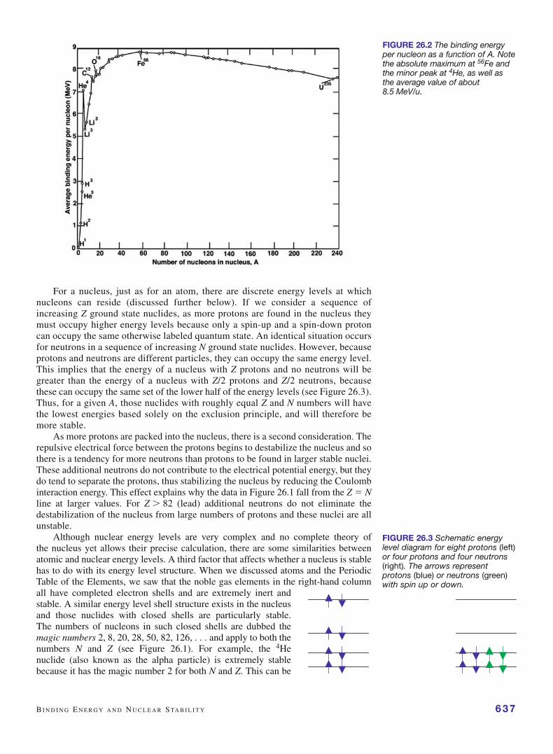
Figure 26.2 shows the binding energy per nucleon of some nuclides as afunction of mass number. Note that the larger the binding energy, the more stablethe nucleus is. We show that this figure explains the phenomena of both nuclearfission and fusion. Many large nuclei are unstable and will spontaneously fissioninto two smaller nuclei, each of which has a larger binding energy per nucleonand is more stable. Similarly, under the proper conditions, two protons or othervery small nuclei can combine, or fuse, together to form a larger nucleus that ismore stable. Both of these reactions liberate substantial amounts of kinetic energy.Fission and fusion are further discussed in the last section of this chapter.
There have been more than 2500 nuclides identified, with only a small numberof these (about 280) stable. What determines whether a particular nucleus is sta-ble or unstable? This is a complex issue. Figure 26.1 shows that at small values ofN and Z stable nuclides have equal numbers of protons and neutrons, but that asthese numbers increase, stable nuclides tend to have significantly more neutronsthan protons. We can understand this fact as a consequence of the Pauli exclusionprinciple and the proton–proton electric repulsion. Recall that the exclusion prin-ciple states that interacting identical fermions, those elementary particles withhalf-integral spin, must have distinct quantum numbers. Protons and neutrons bothhave spin 1⁄2 and therefore must separately satisfy this principle.
�
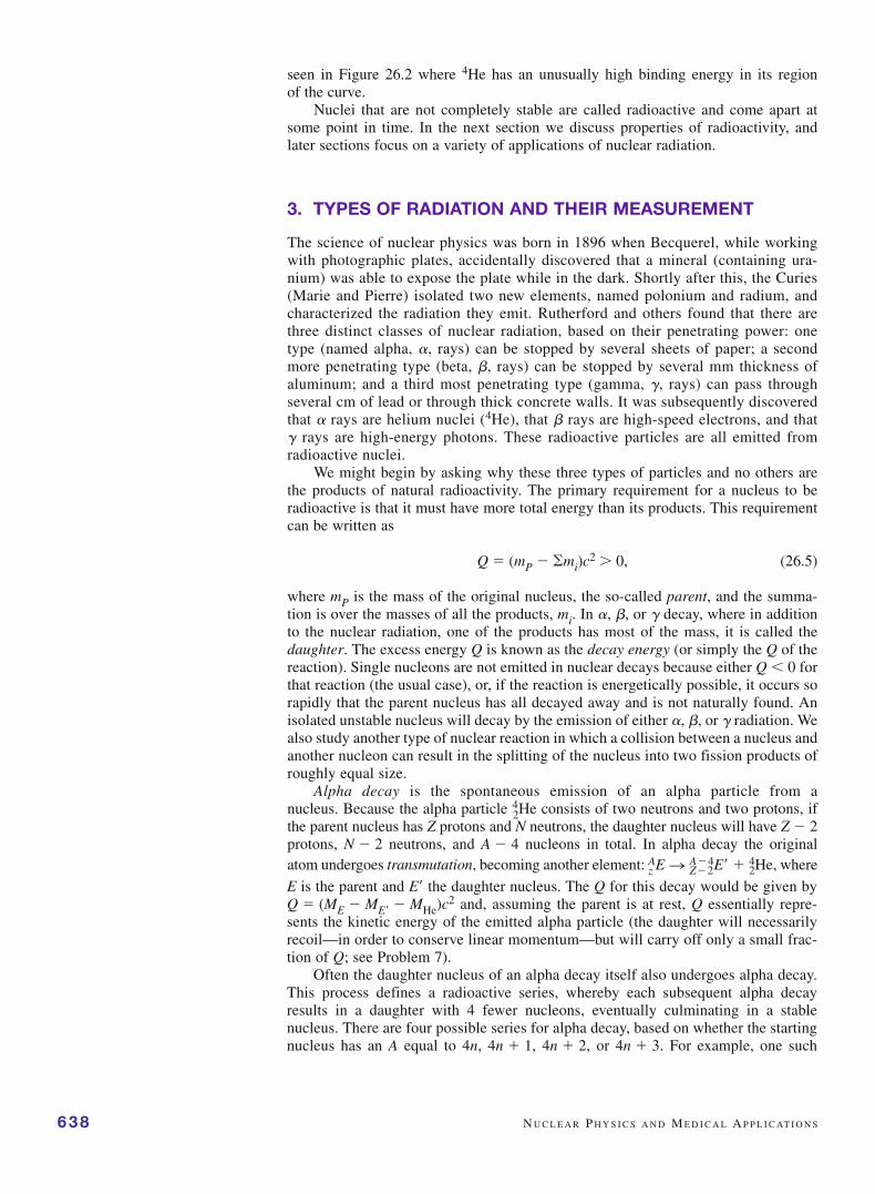
For a nucleus, just as for an atom, there are discrete energy levels at whichnucleons can reside (discussed further below). If we consider a sequence ofincreasing Z ground state nuclides, as more protons are found in the nucleus theymust occupy higher energy levels because only a spin-up and a spin-down protoncan occupy the same otherwise labeled quantum state. An identical situation occursfor neutrons in a sequence of increasing N ground state nuclides. However, becauseprotons and neutrons are different particles, they can occupy the same energy level.This implies that the energy of a nucleus with Z protons and no neutrons will begreater than the energy of a nucleus with Z/2 protons and Z/2 neutrons, becausethese can occupy the same set of the lower half of the energy levels (see Figure 26.3).Thus, for a given A, those nuclides with roughly equal Z and N numbers will havethe lowest energies based solely on the exclusion principle, and will therefore bemore stable.
As more protons are packed into the nucleus, there is a second consideration. Therepulsive electrical force between the protons begins to destabilize the nucleus and sothere is a tendency for more neutrons than protons to be found in larger stable nuclei.These additional neutrons do not contribute to the electrical potential energy, but theydo tend to separate the protons, thus stabilizing the nucleus by reducing the Coulombinteraction energy. This effect explains why the data in Figure 26.1 fall from the Z � Nline at larger values. For Z � 82 (lead) additional neutrons do not eliminate thedestabilization of the nucleus from large numbers of protons and these nuclei are allunstable.
Although nuclear energy levels are very complex and no complete theory ofthe nucleus yet allows their precise calculation, there are some similarities betweenatomic and nuclear energy levels. A third factor that affects whether a nucleus is stablehas to do with its energy level structure. When we discussed atoms and the PeriodicTable of the Elements, we saw that the noble gas elements in the right-hand columnall have completed electron shells and are extremely inert andstable. A similar energy level shell structure exists in the nucleusand those nuclides with closed shells are particularly stable.The numbers of nucleons in such closed shells are dubbed themagic numbers 2, 8, 20, 28, 50, 82, 126, . . . and apply to both thenumbers N and Z (see Figure 26.1). For example, the 4Henuclide (also known as the alpha particle) is extremely stablebecause it has the magic number 2 for both N and Z. This can be
B I N D I N G E N E R G Y A N D N U C L E A R S TA B I L I T Y 637
FIGURE 26.2 The binding energyper nucleon as a function of A. Notethe absolute maximum at 56Fe andthe minor peak at 4He, as well asthe average value of about 8.5 MeV/u.
FIGURE 26.3 Schematic energylevel diagram for eight protons (left)or four protons and four neutrons(right). The arrows representprotons (blue) or neutrons (green)with spin up or down.
seen in Figure 26.2 where 4He has an unusually high binding energy in its regionof the curve.
Nuclei that are not completely stable are called radioactive and come apart atsome point in time. In the next section we discuss properties of radioactivity, andlater sections focus on a variety of applications of nuclear radiation.
3. TYPES OF RADIATION AND THEIR MEASUREMENT
The science of nuclear physics was born in 1896 when Becquerel, while workingwith photographic plates, accidentally discovered that a mineral (containing ura-nium) was able to expose the plate while in the dark. Shortly after this, the Curies(Marie and Pierre) isolated two new elements, named polonium and radium, andcharacterized the radiation they emit. Rutherford and others found that there arethree distinct classes of nuclear radiation, based on their penetrating power: onetype (named alpha, , rays) can be stopped by several sheets of paper; a secondmore penetrating type (beta, , rays) can be stopped by several mm thickness ofaluminum; and a third most penetrating type (gamma, �, rays) can pass throughseveral cm of lead or through thick concrete walls. It was subsequently discoveredthat rays are helium nuclei (4He), that rays are high-speed electrons, and that� rays are high-energy photons. These radioactive particles are all emitted fromradioactive nuclei.
We might begin by asking why these three types of particles and no others arethe products of natural radioactivity. The primary requirement for a nucleus to beradioactive is that it must have more total energy than its products. This requirementcan be written as
Q � (mP � �mi)c2 � 0, (26.5)
where mP is the mass of the original nucleus, the so-called parent, and the summa-tion is over the masses of all the products, mi. In , , or � decay, where in additionto the nuclear radiation, one of the products has most of the mass, it is called thedaughter. The excess energy Q is known as the decay energy (or simply the Q of thereaction). Single nucleons are not emitted in nuclear decays because either Q � 0 forthat reaction (the usual case), or, if the reaction is energetically possible, it occurs sorapidly that the parent nucleus has all decayed away and is not naturally found. Anisolated unstable nucleus will decay by the emission of either , , or � radiation. Wealso study another type of nuclear reaction in which a collision between a nucleus andanother nucleon can result in the splitting of the nucleus into two fission products ofroughly equal size.
Alpha decay is the spontaneous emission of an alpha particle from anucleus. Because the alpha particle 4
2He consists of two neutrons and two protons, ifthe parent nucleus has Z protons and N neutrons, the daughter nucleus will have Z � 2protons, N � 2 neutrons, and A � 4 nucleons in total. In alpha decay the originalatom undergoes transmutation, becoming another element: whereA
z E n A�4Z�2E¿ � 4
2He,
638 N U C L E A R P H Y S I C S A N D M E D I C A L A P P L I C AT I O N S
E is the parent and E the daughter nucleus. The Q for this decay would be given byQ � (ME � ME � MHe)c
2 and, assuming the parent is at rest, Q essentially repre-sents the kinetic energy of the emitted alpha particle (the daughter will necessarilyrecoil—in order to conserve linear momentum—but will carry off only a small frac-tion of Q; see Problem 7).
Often the daughter nucleus of an alpha decay itself also undergoes alpha decay.This process defines a radioactive series, whereby each subsequent alpha decayresults in a daughter with 4 fewer nucleons, eventually culminating in a stablenucleus. There are four possible series for alpha decay, based on whether the startingnucleus has an A equal to 4n, 4n � 1, 4n � 2, or 4n � 3. For example, one such
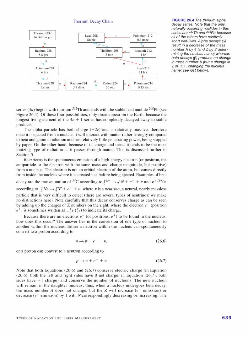
series (4n) begins with thorium 232Th and ends with the stable lead nuclide 208Pb (seeFigure 26.4). Of these four possibilities, only three appear on the Earth, because thelongest living element of the 4n � 1 series has completely decayed away to stableproducts.
The alpha particle has both charge (�2e) and is relatively massive, thereforeonce it is ejected from a nucleus it will interact with matter rather strongly comparedto beta and gamma radiation and has relatively little penetrating power, being stoppedby paper. On the other hand, because of its charge and mass, it tends to be the mostionizing type of radiation as it passes through matter. This is discussed further inSection 5.
Beta decay is the spontaneous emission of a high-energy electron (or positron, theantiparticle to the electron with the same mass and charge magnitude, but positive)from a nucleus. The electron is not an orbital electron of the atom, but comes directlyfrom inside the nucleus where it is created just before being ejected. Examples of beta
decay are the transmutation of 14C according to and of 19Ne
according to where � is a neutrino, a neutral, nearly massless
particle that is very difficult to detect (there are several types of neutrinos; we makeno distinctions here). Note carefully that this decay conserves charge as can be seenby adding up the charges or Z numbers on the right, where the electron e� (positrone�) is sometimes written as to indicate its charge.
Because there are no electrons e� (or positrons, e�) to be found in the nucleus,how does this occur? The answer lies in the conversion of one type of nucleon toanother within the nucleus. Either a neutron within the nucleus can spontaneouslyconvert to a proton according to
(26.6)
or a proton can convert to a neutron according to
(26.7)
Note that both Equations (26.6) and (26.7) conserve electric charge (in Equation(26.6), both the left and right sides have 0 net charge; in Equation (26.7), bothsides have �1 charge) and conserve the number of nucleons. The new nucleonwill remain in the daughter nucleus; thus, when a nucleus undergoes beta decay,the mass number A does not change, but the Z will increase (e� emission) ordecrease (e� emission) by 1 with N correspondingly decreasing or increasing. The
p n n � e� � n
n n p � e� � n,
�10 e (1
0 e)
1019 Ne n 9
19 F � e� � n,614 C n 7
14 N � e� � n
T Y P E S O F R A D I AT I O N A N D T H E I R M E A S U R E M E N T 639
FIGURE 26.4 The thorium alphadecay series. Note that the onlynaturally occurring nuclides in thisseries are 232Th and 208Pb becauseall of the others have relativelyshort half-lives. Alpha decays ()result in a decrease of the massnumber A by 4 (and Z by 2 deter-mining the nucleus name) whereasbeta decays () produce no changein mass number A (but a change inZ of � 1, changing the nucleusname; see just below).
Thorium-23214 Billion yrs
Radium-2285.8 yrs
Actinium-2286 hrs
Thorium-2281.9 yrs
Radium-2243.7 days
Radon-22456 sec
Polonium-2160.15 sec
Lead-21211 hrs
Bismuth 2121 hr
Thallium-2083 min
Polonium-2120.3 μsec
Lead-208Stable
Thorium Decay Chain
α
α αα
α
β
ββ
βα
ββα

ejected beta particle (e� or e�) and neutrino together acquire essentially the totalkinetic energy Q released in the decay (Q � (MParent � MDaughter)c
2, because thebeta particle has negligible mass), so that the electron can have any energybetween essentially 0 and Q, whereas the neutrino gains the balance of Q inkinetic energy. The beta particle is identical to any electron, but is so named sim-ply to indicate it originates in a nucleus.
When beta decay was first characterized, the variable energy of the emittedbeta particle was not understood because the neutrino had not been detected. Inaddition to an apparent violation of conservation of energy, the laws of conserva-tion of momentum and angular momentum appeared to be violated as well. In 1934Enrico Fermi worked out a detailed theory of beta decay, proposing not only theexistence of the neutrino, but a fourth type of fundamental force in nature knownas the weak nuclear force. It was not until 1953 that direct laboratory evidence forthe neutrino was obtained, but it had been accepted long before based on scientists’belief in the fundamental conservation laws. It is currently thought that neutrinosare the most ubiquitous of all particles in the universe. In 1998 the first experi-mental evidence was obtained for a very small, but nonzero, neutrino mass by aninternational team of 120 scientists working in Japan. These experiments are verydifficult and still a bit controversial. If nonzero, even if extremely small, the vastnumbers of neutrinos in the universe would contribute substantially toward the totalmass of the universe.
640 N U C L E A R P H Y S I C S A N D M E D I C A L A P P L I C AT I O N S
Example 26.2 Calculate the Q for the following reactions: (i) the alpha decay of238U to 234Th; and (ii) the � decay of 234Th to 234Pa. Use the following data: thenuclear masses of m(238U) � 238.00018 u, m(4He) � 4.00150 u, m(234Th) �233.99409 u, and m(234Pa) � 233.99325 u, and m() � (9.11 � 10�31/1.66 �10�27) � 0.00055 u.
Solution: (i) The alpha decay products of 238U are 4He � 234Th. We calculatethe Q for this reaction, to be Q � [m(238U) � m(234Th) � m(4He)]c2 �(0.00459)(931.5) � 4.28 MeV.
(ii) In this case the reaction is 234Th → 234Pa � � � �, where the protactinium(Pa) nucleus has one more proton formed in the beta decay. Here we can calcu-late the Q of the reaction ignoring the neutrino produced. Doing this, we find thatQ � (233.99409 � 233.99325 � 0.00055)(931.5) � 0.270 MeV. This is themaximum kinetic energy the electron can have because otherwise the neutrinomay carry off some energy as well.
Gamma decay, the third type of radioactivity, is the emission of a high-energyphoton from a nucleus. The gamma ray is emitted when a nucleus makes adownward transition between two nuclear energy levels, just as a photon is emit-ted from an atom when it makes a downward transition between atomic energylevels. A major difference is that, because of the much larger energy spacingbetween nuclear energy levels, a gamma ray has a much higher energy, about amillion times more than a photon from an atomic transition. This much largerenergy corresponds to a much shorter wavelength for gamma rays, on the order of10�12 � 10�15 m. Typically, gamma rays are emitted by daughter nuclei thatare left in excited states after or decays as they relax back to their groundstate.
Because gamma rays have no charge, they are the most penetrating of the threetypes of radiation. Medical imaging techniques that use radioactive isotopesrequire the emitted radiation to escape from the body in order to be detected. These

techniques use gamma emitters because or rays have such short penetratingdistances that they will not escape from the body. This is discussed further inSection 7 below.
We conclude this section with a discussion of the detection of nuclear radiation.There are several general methods to detect individual radiation particles as well asseveral methods to visualize the trajectory of these particles. One basic class ofdetectors is the ion collection detector, consisting of a high Z gas (typically xenon)filled chamber with a thin window through which radiation enters (Figure 26.5).Inside are two electrodes (a negative cathode and positive anode) across which ahigh voltage is applied. Ionizing radiation that enters the tube interacts with the gasto create ion pairs that travel to the electrodes and make up a current. If the appliedvoltage is high enough, the current generated is proportional to the amount of ion-izing radiation. Such detectors are called proportional counters. At even higherapplied voltages, a single ionization event will trigger an avalanche of subsequentionizations of the gas and under these conditions the detector is called aGeiger–Muller counter (sometimes a Geiger tube or counter). Geiger counters areexcellent for detecting small amounts of radiation because of the large degree ofamplification. In general, ionization detectors have limited application in nuclearmedicine because they have poor efficiency for gamma rays which are the primaryinformation-containing decay product, as mentioned above.
A second type of radiation monitor is a scintillation detector, consisting of ascintillator coupled to a photomultiplier tube (Figure 26.6). The scintillator, or phos-phor, is a material (typically NaI crystals, plastics, or a liquid) that emits visible lightwhen excited by radiation. These are dense materials that are very efficiently excitedby radiation, including gamma rays, and have relatively fast response times. Thenumber of photons produced is proportional to the energy of the incident radiationand the light produced is then detected by the photomultiplier tube (see the photo-electric effect discussion in Chapter 24) whose output photocurrent can be analyzedto determine the energy of the incident radiation.
Semiconductor detectors that use p–n junctions (see Chapter 25) to detectionization due to radioactive particles are a third type of detector. Electron–hole pairscreated in the p–n junction by radioactive particles constitute an electric current pro-portional to the radiation energy.
A number of devices allow one to visualize the path of a single charged particle.The simplest is a photographic emulsion in which a chemical change along theparticle’s trajectory can be developed to visualize the path. Two other devices, thecloud chamber and bubble chamber, make use of either a supercooled gas (that isready to condense on any ionized particle) or a superheated liquid (that is ready toboil along the path of an ion), respectively, to visualize the trajectory of a high-energy ion. Usually a magnetic field within the chamber causes the chargedparticles to travel in helical paths. (Do you remember why?) Photographs of the
T Y P E S O F R A D I AT I O N A N D T H E I R M E A S U R E M E N T 641
Current pulse to electronics
Metal chamber atnegative high voltage window
Wire electrode at positive high voltage
Gas-filled tube radiation
Ionized gas tracks
FIGURE 26.5 Schematic of aGeiger counter used to measurethe presence of radiation.
photomultiplier
scintillator
radiation
Current pulseto electronics
FIGURE 26.6 A scintillation detector, converting radiation tolight in the scintillator, the light thenbeing detected by a photomultiplierand converted to an electric current signal.

charge track (Figure 26.7) can then be used to measure theradii of curvature to deduce the momentum and sign of thecharge of the particle.
4. HALF-LIFE AND RADIOACTIVE DATING
In a macroscopic collection of radioactive nuclei, eachnucleus decays independently of all the others. In factbecause each nucleus is shielded by its atomic electrons,even environmental conditions of pressure, temperature,and the like do not affect radioactivity. It is impossible topredict when any particular nucleus will undergo radioac-tive decay. The radioactive decay process is a purely ran-dom one. We can, however, make statistical predictionsabout the fraction of nuclei that will decay in a given timeinterval based on an assumption that the probability for adecay is the same in every equal time interval up until thenucleus actually does decay. Once the parent transmutes tothe daughter nuclide, that particular nucleus cannot repeatthe process. Only if the daughter is itself radioactive can itdecay further, but that process is described by a differentprobability.
This statistical notion allows us to write that the decrease�N in the total number of N nuclei in a sample (�N equal tothe number of radioactive decays) in a short time interval �tis proportional to the time interval and to the total number ofnuclei in the sample. In symbols we have that
(26.8)
where the proportionality constant � is called the decay constant whose valuedepends on the particular radioactive nuclide. Equation (26.8) can be solved for thenumber of nuclei N at any time t using calculus (see box) to find
(26.9)
where N0 is the number of nuclei at time t � 0. Equation (26.9), plotted in Figure 26.8normalized to the fraction remaining, is known as the law of radioactive decay. Thetime � � 1/� is known as the lifetime of the decay and represents the time forthe number of parent nuclei to decay to N0/e � N0/2.718, as shown in the figure. The
N(t) � N0 e�lt,
¢N � � lN¢t,
642 N U C L E A R P H Y S I C S A N D M E D I C A L A P P L I C AT I O N S
FIGURE 26.7 Bubble chamber photo showing several interaction sites (vertices where tracks meet) and spirals indicating long-lived charged particles undergoing energy loss.
0
0.2
0.4
0.6
0.8
1
1.2
0 2 4 6
time (in lifetimes)
frac
tio
n r
emai
nin
g
1/e
1/e2
τ1/2
–6
log
frac
tion
rem
aini
ng
–5
–4
–3
–2
–1
0
0 1 2 3 4 5 6
time
1
FIGURE 26.8 Radioactive decaylaw, normalized to the fractionremaining after some time. Thehalf-life and one and two lifetimesare indicated on the figure. Theinsert shows that a semilog plot ofthe natural logarithm of the fractionremaining is plotted versus time,the data decrease linearly.

number of parent nuclei decreases exponentially with time. In subse-quent equal time intervals �, the number of parent nuclei will continueto decrease by the same ratio of 1/e as indicated in the figure, so thatafter two lifetimes there will be N0/e 2 nuclei left, after three lifetimesN0/e3, and so on.
More commonly the rate of decay is specified by the half-life,defined as the time for the number of parent nuclei to decrease by a fac-tor of two, rather than a factor of e (see Figure 26.8). Using Equation(26.9), we can substitute N(t) � N0/2,
and then solve for t1/2 by taking the logarithm of both sides to find that
(26.10)
After one half-life there are N0/2 nuclei remaining, after two half-livesthere are (N0/2)/2 � N0/22 � N0/4 remaining, after three half lives(N0/4)/2 � N0/23 � N0/8 remaining, and so on. The half-lives of variousradioactive isotopes are listed in Table 26.1. Half-lives in nature varyfrom vanishingly short (10�22 s) to nearly everlasting (1021 years).
t1/2 �loge 2
l�
0.693
l.
N0 /2 � N0 e�lt1/2
H A L F -L I F E A N D R A D I O A C T I V E DAT I N G 643
Writing the �s in Equation (26.8) asdifferentials, we have
Dividing by N and integrating both sidesfrom time 0 with N0 nuclei to some arbi-trary time t with N(t) nuclei we have
Remembering that the integral on the left isthe natural logarithm of N, we have
Then, using the definition of the logarithm,we can rewrite this as Equation (26.9). Alsonote that by differentiating Equation (26.9)we can obtain Equation (26.11) for theactivity,
dN
dt��N0le
�lt � cdN
dtd0 e�lt,
� �lt.
loge(N(t)) � loge(N0) � logeaN(t)
N0b
LN0
N(t) dN
N� �l L
t
0dt.
dN ��lNdt.
Table 26.1 Half-Lives of Some Radioactive Nuclides
Isotope Symbol Half-Life Radioactivity
Uranium-238 238U 4.5 � 109 years , �
Carbon-14 14C 5730 years
Radium-226 226Ra 1600 years , �
Strontium-90 90Sr 29 years , �
Cobolt-60 60Co 5.3 years , �
Iodine-131 131I 8 days , �
Fluorine-18 18F 1.8 h
Barium-141 141Ba 18.3 min , �
Krypton-92 92Kr 1.8 s , �
Polonium-214 214Po 164 �s , �
where we have used the first equation inthis box in the second step.
The rate at which radioactive nuclei decay, �N/�t, is called the activity and ismeasured in disintegrations/s, or bequerel (Bq), where 1 Bq � 1 disintegration/s. Amore common unit of activity is the curie (Ci), with 1 Ci � 3.7 � 1010 Bq. The curieis a rather large unit of activity in nuclear medicine and the mCi and �Ci are oftenused. Activity can be directly measured by detection of the decay products. Becausethe number of decays in a short time interval is proportional to the number N of par-ent nuclei (see Equation (26.8)), the activity also decays exponentially with timeaccording to
(26.11)
where the subscript again indicates the zero-time value. This should make intuitivesense; if after 10 half-lives there are 1/210 fewer radioactive nuclei, then the rate atwhich decays occur would also be expected to be smaller by the same factor.
¢N
¢t� a¢N
¢tb
0e�lt,
One application of radioactivity is the dating of ancient materials. A commonlyused method is 14C dating (carbon-14 dating) of the age of once living organisms. Allliving plants and animals are carbon-based. There are two stable isotopes of carbonwith 12C representing close to 99% and 13C about 1%. Carbon-14, a beta emitter witha half-life of 5730 years, is formed in the upper atmosphere by the interaction of cos-mic rays with nitrogen in the air. The amount of 14C is very small, roughly 1.3 � 10�12
times as much as 12C, but its net amount has remained stable over many thousands ofyears due to the balance in its production in the atmosphere and its radioactive decay.All living material incorporates 14C, ultimately by the absorption of CO2 in the air dur-ing photosynthesis in plants. Animals incorporate 14C on eating plants or other animalsthat have eaten plants earlier in the food chain. However, when an organism dies, nonew 14C is further incorporated so that the ratio of 14C to 12C steadily declines with ageafter death, by a factor of two for every 5730 years. Measurement of 14C activity canthus be used to date the age of the remains of such organisms.
For objects older than about 60,000 years, carbon dating does not work becausethere is too little 14C activity left to measure accurately. By using other isotopes withmuch longer half-lives, such as 238U, the geological age of rock formations can bedetermined in much the same way. A measurement of the parent to daughter ratio canbe used to date materials back billions of years. Dating the oldest rocks found, theage of the Earth has been measured to be about 4 billion years. The oldest fossilsfound date from about 3 billion years ago. Radioactive dating has been critical in ahost of geological and evolutionary studies.
644 N U C L E A R P H Y S I C S A N D M E D I C A L A P P L I C AT I O N S
Example 26.3 Suppose that you wish to authenticate animal skin remains fromone of the earliest known collections of animals, that of Shulgi, a Sumerian rulerof a territory now in Iraq, dating back to 2094 BC. You take a small sample ofthe skin and chemically analyze it for carbon. From a 10 g sample of carbon,what activity would you expect to measure if the sample is indeed authentic?
Solution: First we need to find the number of carbon nuclei present in the 10 gsample. We do this by assuming that essentially all the carbon is 12C so that thereare (10 g)(1 mol/12 g)(6.02 � 1023 nuclei/mol) � 5.0 � 1023 nuclei present.Thus, when alive, the animal would have had about (1.3 � 10�12)(5 � 1023) �6.5 � 1011 14C nuclei present. In that case, from Equations (26.8) and (26.10)and the fact that 5730 y � 1.81 � 1011 s, we know the initial activity was �N0 �(ln 2/�1/2)N0 � (0.693/1.81 � 1011)(6.5 � 1011) � 2.49 Bq. If the animal skin isindeed authentic, it would be nearly 4100 years old. According to Equation(26.11) then, the expected count rate would be
Note that this rate would need to be measured very precisely by averaging overlong times to ensure a reliable value because the count rate is so low.
¢N
¢t� 12.49 Bq21e�4100/57302 � 1.22 Bq.
Our discussion of radioactive decay in this section thus far has been limited toa single radioactive species decaying away according to Equation (26.9). In a moretypical situation, there are several radioactive nuclides that decay successivelyfrom one to another in a radioactive series such as the thorium decay chain dis-cussed in the previous section. In this case the parent nuclide will decay accordingto Equation (26.9), but each of the other nuclides in the series will be produced bythe preceding decay and so the populations of these nuclides need to be found fromtheir production rates, shown schematically in Figure 26.9.

Using a similar analysis to that above for Equation (26.8), the change in first daugh-ter population will be given by
(26.12)
where the first term on the right-hand side is the rate at which the population of firstdaughters increases from decays of parent nuclides, and the second term on the rightis the rate at which first daughters decrease from its own decay at rate �1. A similarequation will hold for each subsequent daughter population.
These equations can be solved for a variety of interesting cases, but the mostcommon situation is one in which the parent decay rate is the slowest. Then over verylong times, the parent population will decrease exponentially, according to Equation(26.9). But over much shorter times, the parent population N0 will essentially remainconstant and will thus supply the first daughter population at a constant rate. Nowbecause the first daughter decay rate is much faster, its population N1 will remainconstant at a value controlled by the parent supply of first daughters. Under this con-dition, after a sufficient equilibrium time, all the N(t) will be constant in time, so thatthe left-hand side of Equation (26.12) becomes equal to zero. Then we find that
(26.13)
and we can find the first daughter population to be a constant N1 � (�0/�1)N0 in termsof the constant parent population N0. The same story will follow for the second and allother daughter populations in terms of that of the first, or previous, daughter population.
This analysis explains why it is possible to have naturally occurring very shortlifetime alpha emitting nuclei, such as are found in the radioactive series discussed inthe previous section. If you look back at Figure 26.4 you will see, for example, thatpolonium-212 decays to lead-208 by alpha emission with a half-life of 0.3 �s. Whyshould there be any Po212 left in naturally occurring ores mined on the Earth? Theanswer is that Po212 is a daughter in the radioactive series that has thorium-232, witha 14 billion year lifetime, as parent. The series of nuclei produced from Th232 con-tinually produce new Po212 at essentially a constant rate.
5. DOSIMETRY AND BIOLOGICAL EFFECTS OF RADIATION
The interaction of nuclear radiation with matter leads to ionization; in fact, nuclearradiation (as well as uv and x-ray photons) is sometimes also referred to as ionizingradiation. Because energies of only tens of electron volts are sufficient to ionizeatoms, , , and � particles, with energies of MeV, are each able to ionize manythousands of atoms before losing their energy. It is this ionization that makes nuclearradiation dangerous to living organisms. Here we introduce various units to measureexposure, and discuss those doses and the relative biological effects of radiation.
A unit of exposure, the roentgen (R), was first introduced to define the extent of ion-ization produced by x-rays, but is also used for gamma radiation. Defined as the totalnumber of ion pairs produced in a volume of 1 cm3 of dry air under standard conditions(0°C and 1 atmosphere of pressure), one roentgen is given by 1 R � 2.58 � 10�4 C/kgair. This is a unit of exposure, giving the ionization level in air, but it does not give anyinformation about absorption of radiation by living tissue or its effects on that tissue.
A measure of the absorbed dose of radiation, the absorbed energy per unit mass, isthe gray (Gy), where 1 Gy � 1 J/kg. An older unit, still commonly used today, is therad, where 1 rad � 0.01 Gy. For a given exposure, the absorbed dose will vary greatlydepending on the absorption characteristics of the material and the type of radiation.
N0l0 � N1l1
¢N1 (t) � [N0 (t)l0 � N1 (t)l1 ]¢t,
D O S I M E T RY A N D B I O L O G I C A L E F F E C T S O F R A D I AT I O N 645
Parent: No(t)
First Daughter: N1(t)
λ 0 λ 1 Second Daughter: N2(t)
λ 2
FIGURE 26.9 Schematic for series of radioactive decays, where theN’s are the populations and the �’s are the decay constants.
646 N U C L E A R P H Y S I C S A N D M E D I C A L A P P L I C AT I O N S
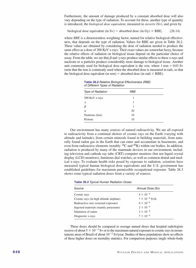
Furthermore, the amount of damage produced by a constant absorbed dose will alsovary depending on the type of radiation. To account for these, another type of quantityis introduced, the biological dose equivalent, measured in sieverts (Sv), and given by
biological dose equivalent (in Sv) � absorbed dose (in Gy) � RBE, (26.14)
where RBE is a dimensionless weighting factor, named for relative biological effective-ness, that depends on the type of radiation. Values for RBE are given in Table 26.2.These values are obtained by considering the dose of radiation needed to produce thesame effect as a dose of 200 KeV x-rays. Their exact values are somewhat fuzzy, becausethe relative effects of radiation on biological tissue depend on the particular choice ofassay. From the table, we see that and � rays produce similar effects to these x-rays andnucleons or particles produce considerably more damage to biological tissue. Anotherunit commonly used for biological dose equivalent is the rem, where 1 rem � 0.01 Sv(note that the rem is commonly used when the absorbed dose is measured in rads, so thatthe biological dose equivalent (in rem) � absorbed dose (in rad) � RBE).
Table 26.2 Relative Biological Effectiveness (RBE) of Different Types of Radiation
Type of Radiation RBE
200 KeV x-rays 1
� 1
1
20
Neutrons (fast) 10
Protons 10
Our environment has many sources of natural radioactivity. We are all exposedto radioactivity from a continual shower of cosmic rays on the Earth (varying withaltitude and latitude), from certain minerals found in building materials, from natu-rally found radon gas in the Earth that can enter and accumulate in basements, andeven from radioactive elements (notably 14C and 40K) within our bodies. In addition,radiation is produced by many of the manmade devices in our environment, includ-ing television and cathode ray tube (CRT) computer monitors (but not liquid crystaldisplay (LCD) monitors), luminous dial watches, as well as common dental and med-ical x-rays. To evaluate health risks posed by exposure to radiation, scientists havemeasured typical human biological dose equivalents and the U.S. government hasestablished guidelines for maximum permissible occupational exposure. Table 26.3shows some typical radiation doses from a variety of sources.
Table 26.3 Typical Human Radiation Doses
Source Annual Dose (Sv)
Cosmic rays 4 � 10�4
Cosmic rays (in high altitude airplane) 7 � 10�6 Sv/h
Radioactive ores (external exposure) 6 � 10�4
Ingested materials (mainly potassium) 2 � 10�4
Inhalation of radon 2 � 10�4
Diagnostic x-rays 7 � 10�4
These doses should be compared to average annual doses that hospital radiologistsreceive of about 5 � 10�3 Sv or to the maximum natural exposure to cosmic rays in moun-tainous areas of Brazil of about 10�2 Sv/year. Studies of these populations show no effectsof these higher doses on mortality statistics. For comparison purposes single whole-body

radiation doses at higher levels do have significant effects at levels over 0.50 Sv. At levelsup to about 2 Sv there is a significant reduction in blood platelet and white cell counts.Above this level there is severe blood damage, nausea, hair loss, hemorrhage, and short-term death in many cases. Whole-body doses between 4 and 5 Sv result in death to about50% of such a population, and doses over 6 Sv result in nearly universal death. Long-termeffects of radiation can be due to short-term high exposure or to accumulated chronic low-level exposure. Federal standards indicate an individual maximum annual exposure of 5 �10�3 Sv, excluding medical sources. This is increased a factor of 10 for people who workwith radiation sources, such as radiation technologists.
It is thought that radiation kills cells by damaging their DNA so that the cells can-not reproduce or by causing sufficient other damage to prevent the cell’s normal repairmechanisms from working effectively. In medicine, radiation is often used to destroycancer cells in a limited area of the body. Of course radiation will also kill healthy cells,particularly those that turn over rapidly, such as blood platelets and white cells or thecells lining the intestinal wall. That’s why the typical symptoms of radiation sickness areGI problems due to effects on the intestinal wall, immunological suppression due towhite cell kill-off, and general weakness due to red cell and platelet kill-off. By givingradiation over a period of time in repeated smaller doses, it is often possible to minimizedamage to normal cells while still killing tumor cells. The chemical changes induced byradiation are caused by the formation of free radicals, enhanced by the presence of oxy-gen. Therefore the oxygen content of a particular tissue or cancer type will affect the suc-cess of the radiation treatment. In the following two sections we discuss nuclearmedicine further, focusing on the use of radioisotopes for both therapy and diagnostics.
6. RADIOISOTOPES AND NUCLEAR MEDICINE
The key to understanding the use of radioactive isotopes (radioisotopes) in biologicalstudies and in medicine is the fact that chemistry and radioactivity are completely inde-pendent processes. Chemistry is based on valence electron interactions and does notdepend at all on nuclear properties. As an example of this, hydrogen and its isotope deu-terium (an atom made from a single electron and a nucleus with one neutron in additionto a single proton) have exactly the same chemistry. The only difference in these two istheir mass difference of nearly a factor of two. Because of this deuterium is often used inscience experiments (in various types of spectroscopy) as an indicator of the location ofhydrogen atoms because they bind in the same way chemically. Incorporation of radioac-tive isotopes in cells or in the body at very low doses does not directly change the normalsequence of chemical events that occurs. This fact allows radiolabeling (also known astagging or tracer studies) to follow a particular type of molecule in its pathway throughan organism. In this section we discuss several aspects of nuclear medicine, including theproduction and types of radioisotopes in use, tracer studies and detection methods in bio-logical research, and various diagnostic tests in medicine using radioisotopes.
In order to safely use radioisotopes in medicine, not only must the dose be wellcontrolled, but the half-life of the isotope must be relatively short so that the radioac-tivity is quickly reduced, causing no long-term problems. The typical dose used in diag-nostic tests is so low (~10�8 Sv/h) that there is no danger from radiation. Somecommonly used radioisotopes are listed in Table 26.4. Technetium(Tc)-99m is the mostcommon of these and can be combined with many different molecules to act as a radio-pharmaceutical. It has a half-life of only 6 h so that in order to have sufficient amountsavailable for hospital studies it must be freshly extracted from molybdenum-99, itselfhaving a 67 h half-life—a useful life span of about a week—and itself usually preparedin-house in a major hospital as discussed just below. The 99Mo is bound to a solidmatrix in a chromatography column and as the technetium-99m forms by beta decay itis washed from the column and then can be used directly or as a radiopharmaceuticalwhen labeling another molecule. Technetium-99m does not emit beta particles and itsgamma emission is at an energy of 140 keV, a relatively low energy so that many escapethe body to be detected. Furthermore, it has a very versatile chemistry and can be
R A D I O I S O T O P E S A N D N U C L E A R M E D I C I N E 647
When radiopharmaceuticals are used in human diagnostic studies, there are twoimportant characteristic times to consider. First, there is the physical half-life of the par-ent radioisotope, �1/2, as discussed above, that is due solely to nuclear decay. A secondtime constant is also important in these studies, the biological half-life �b equal to thetime for the body to wash out half of the pharmaceutical. This latter time constant is notof the same well-defined character as the radioactive half-life, but has considerablevariability. These two processes occur simultaneously so that the effective decay rate inthe body is given by the sum of the two different rate constants. This should make sensesince both paths, physical radioactivity and elimination from the body, act to decreaseradioactivity within the body and hence the effective rate constant should be their sum.The rate constant is the reciprocal of the corresponding time constants, therefore theoverall effective half-life �e is given by a “parallel” combination of time constants (sim-ilar to the effective resistance of parallel combinations of resistors),
(26.15)
Thus, the effective half-life is shorter than either the physical or biological half-life,just as the effective net resistance is less than either resistance in parallel. The mostdangerous of environmental sources of radiation are those that are ingested and havelong effective half-lives. An example is strontium-90 that can replace calcium inbones. It has a long biological half-life (45 years) as well as a long physical half-life(29 years), with a corresponding effective half-life of over 17 years.
The fact that radioisotopes used in medicine need to have short half-lives means thatthey must be constantly replenished for use in hospitals and other medical facilities (theyreally have a built-in shelf life!). Major hospitals have special supply arrangements oreven in-house facilities for their production. Two methods are used to produce radioiso-topes: nuclear reactors or accelerators. In nuclear reactors, either neutron beams are usedto produce radioisotopes with excessive numbers of neutrons that primarily decay bybeta, followed by gamma, emission, or the reactor fission products are isolated andpurified. This latter method is the primary source for 99Mo, the parent nucleus fortechnetium-99m, the most often used radioisotope. Cyclotrons (see Problem 21 inChapter 17) and linear accelerators with proton beams are used to produce proton-richradioisotopes. The production sources of the radioisotopes listed in Table 26.4 are indicated.
Medical research often uses radioactive tracers as an in vitro tool. When used intest tube studies, radioisotopes provide a variety of methods in cellular andsubcellular work. Some of the earliest uses of tracers were to map out biochemicalpathways. Radioactive tracers can be used to determine rates of metabolic processes,
1te
�1t1/2
�1tb
.
648 N U C L E A R P H Y S I C S A N D M E D I C A L A P P L I C AT I O N S
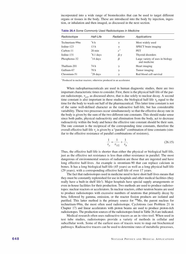
Table 26.4 Some Commonly Used Radioisotopes in Medicine
Radioisotope Half-Life Radiation Applications
Technetium-99m *6 h � Most widely used
Iodine-123 13 h � SPECT brain imaging
Carbon-11 20 min e� PET
Iodine-131 *8.1 days ,� Thyroid disorders
Phosphorus-32 *14 days Large variety of uses in biology and medicine
Thallium-201 74 h � Heart imaging
Gallium-67 78 h � Tumor imaging
Chromium-51 *28 days � Red blood cell survival
*Produced in nuclear reactors; otherwise produced in an accelerator.
incorporated into a wide range of biomolecules that can be used to target differentorgans or tissues in the body. These are introduced into the body by injection, inges-tion, or inhalation and then imaged, as discussed in the next section.

predominant pathways for biosynthesis and metabolism reactions, as well as spatiallocalization information. These are done by various chemical testing methods com-bined with measuring radioactivity levels at various stages in separations.
Tracers can also be used in amounts too small for chemical testing. For example,a radioimmunological assay can determine the amount of an antigen present even intiny amounts (~nanograms). In this technique a minute measured amount of radiola-beled antigen is added to the sample along with a measured small amount ofantibody, small enough that it is all fully bound with antigen (see Figure 26.10). Theantigen will bind to the antibody independent of whether it is labeled. When cen-trifuged, the antibody–antigen complex can be physically separated from theunbound antigen and the activity of each fraction can be determined. Therefore theratio of labeled-to-unlabeled antigen bound to the antibody will reflect the same ratioas found in solution. Because the amount of labeled antigen added is known, theamount of antigen in the original sample can simply be computed from that ratio.There are radioimmunological tests for literally hundreds of drugs or proteins foundin the blood, urine, and other bodily fluids. These are available in kits that arecommonly used in clinical laboratories.
In radioassays, it is important to record as much of the radioactivity as possible. Thebest detector used in biological research is one in which the sample is directly immersedin the detector itself, in the technique of liquid scintillation counting (Figure 26.11). Inthis method, the sample is dissolved or suspended in a mixture of a special solvent anda fluorescent liquid, together known as a scintillation cocktail. A radioactive particleemitted from the sample will produce a brief flash of light that is then detected by a sen-sitive photomultiplier tube, whose output electrical current is then a measure of theradioactivity. But more than this, if two different radioisotopes are present in the cock-tail they will result in different amplitude current pulses making up the output electriccurrent. A so-called pulse-height analysis of the output current of the photomultipliertube allows the relative amounts of the two isotopes to be determined.
7. SPECT AND PET: RADIATION TOMOGRAPHY
In this section we discuss two different imaging methods that are based on radioiso-topes: single photon emission computer tomography (SPECT) and positron emissiontomography (PET). Both of these methods give time-dependent three-dimensionalimages of the location of radioisotopes.
Earlier imaging methods, using gamma ray cameras, give two-dimensional projec-tions of the locations of radioactive sources within the body. The gamma ray cameras areplane arrays of scintillator/photomultiplier detectors, each with a lead collimating channelto only allow radiation directed toward it to be detected. Lead shielding stops all other radi-ation so that the detected intensity at each photomultiplier is a measure of the net amountof radioisotope along its axis (see Figure 26.12), giving a projected image of the “object”or location of radioisotopes within the body. These images are relatively poor compared to
SPECT A N D PET: R A D I AT I O N T O M O G R A P H Y 649
spin
+
antibodyAntigen
Red = labeledAntibody-Antigen Complex forms
FIGURE 26.10 Radioimmunological assay to determine the amountof antigen present. Known amounts of radiolabeled antigen andunlabeled antibody are combined and spun to separate theantibody–antigen complex from the antigen. From the ratio ofcounts in the pellet to that in the supernatant, the amount of antigenoriginally present can be found.
FIGURE 26.11 Liquid scintillationcounting. Radioactive decayparticles produce light in a scintilla-tion cocktail; the light is collectedand detected by a photomultiplier.
CT or MRI pictures, with resolution limited by multiple scattering ofgamma rays as they leave the body and by limited detector resolutionto about 1 cm at best. On the other hand, by monitoring the timedependence of the images, information on the metabolism of theradiopharmaceutical can be obtained. Examples of such uses includeimages of the heart, kidneys, lungs, urinary tract, and so on to deter-mine fluid flow volumes. For imaging, the best radioisotopes aregamma emitters since these will effectively escape the body to bedetected.
SPECT uses an imaging system similar to that of CT scans. Either multidetector orrotating gamma ray camera systems are used to capture a series of two-dimensionalimages, although each image uses a focused collection arrangement to improve resolu-tion and contrast (or ratio of the signal-to-noise of the background radiation). Data areback-projected to reconstruct the three-dimensional image, allowing sequential slicesto be imaged with a spatial resolution of about 5 mm at best, compared to the 1 mm res-olution of CT scans. Although the resolution is better in CT images, they measure onlyx-ray absorption through the body, which then must be interpreted in terms of structureof internal organs. SPECT examines images of the distribution of radiopharmaceuticalsand the time dependence of the radioactivity signal as well. Because this spatial distrib-ution is determined by the specific binding of the drug to which the radioisotope isattached, clearly these images are directly related to function and not simply to structure.
Most major hospitals have facilities to do SPECT and it is increasingly used sincethe advent of better detectors and radioisotopes. Some of the organs imaged most oftenusing SPECT include the brain, heart, circulatory system, bones, and tumors, in gen-eral. In combination with MRI and CT, this technique offers doctors an excellent toolin making diagnoses.
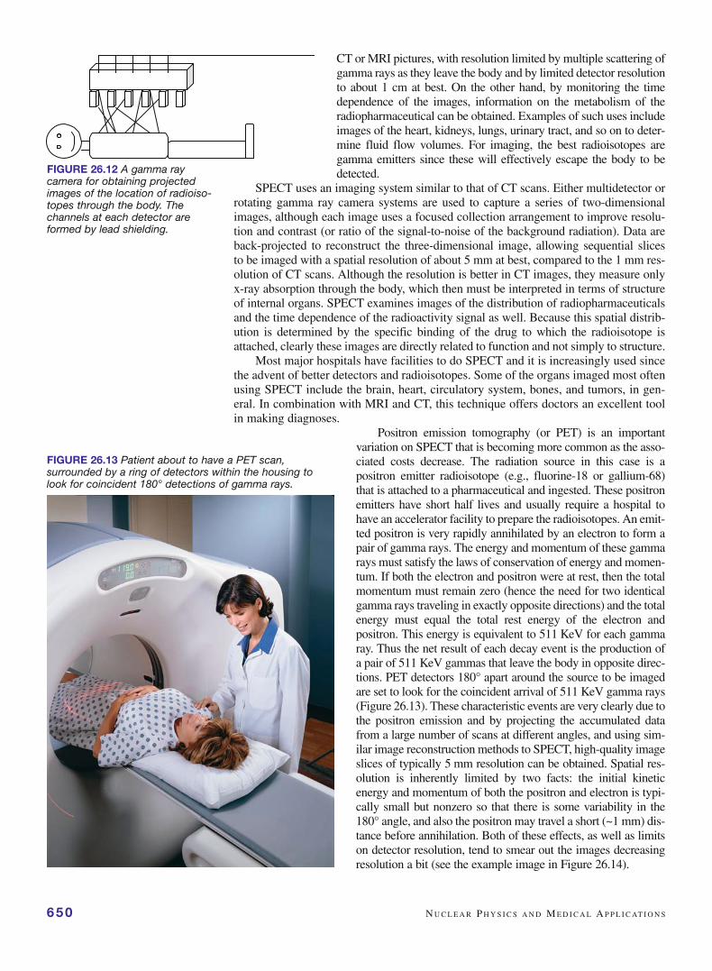
Positron emission tomography (or PET) is an importantvariation on SPECT that is becoming more common as the asso-ciated costs decrease. The radiation source in this case is apositron emitter radioisotope (e.g., fluorine-18 or gallium-68)that is attached to a pharmaceutical and ingested. These positronemitters have short half lives and usually require a hospital tohave an accelerator facility to prepare the radioisotopes. An emit-ted positron is very rapidly annihilated by an electron to form apair of gamma rays. The energy and momentum of these gammarays must satisfy the laws of conservation of energy and momen-tum. If both the electron and positron were at rest, then the totalmomentum must remain zero (hence the need for two identicalgamma rays traveling in exactly opposite directions) and the totalenergy must equal the total rest energy of the electron andpositron. This energy is equivalent to 511 KeV for each gammaray. Thus the net result of each decay event is the production ofa pair of 511 KeV gammas that leave the body in opposite direc-tions. PET detectors 180° apart around the source to be imagedare set to look for the coincident arrival of 511 KeV gamma rays(Figure 26.13). These characteristic events are very clearly due tothe positron emission and by projecting the accumulated datafrom a large number of scans at different angles, and using sim-ilar image reconstruction methods to SPECT, high-quality imageslices of typically 5 mm resolution can be obtained. Spatial res-olution is inherently limited by two facts: the initial kineticenergy and momentum of both the positron and electron is typi-cally small but nonzero so that there is some variability in the180° angle, and also the positron may travel a short (~1 mm) dis-tance before annihilation. Both of these effects, as well as limitson detector resolution, tend to smear out the images decreasingresolution a bit (see the example image in Figure 26.14).
650 N U C L E A R P H Y S I C S A N D M E D I C A L A P P L I C AT I O N S
FIGURE 26.12 A gamma raycamera for obtaining projectedimages of the location of radioiso-topes through the body. Thechannels at each detector areformed by lead shielding.
FIGURE 26.13 Patient about to have a PET scan,surrounded by a ring of detectors within the housing tolook for coincident 180° detections of gamma rays.
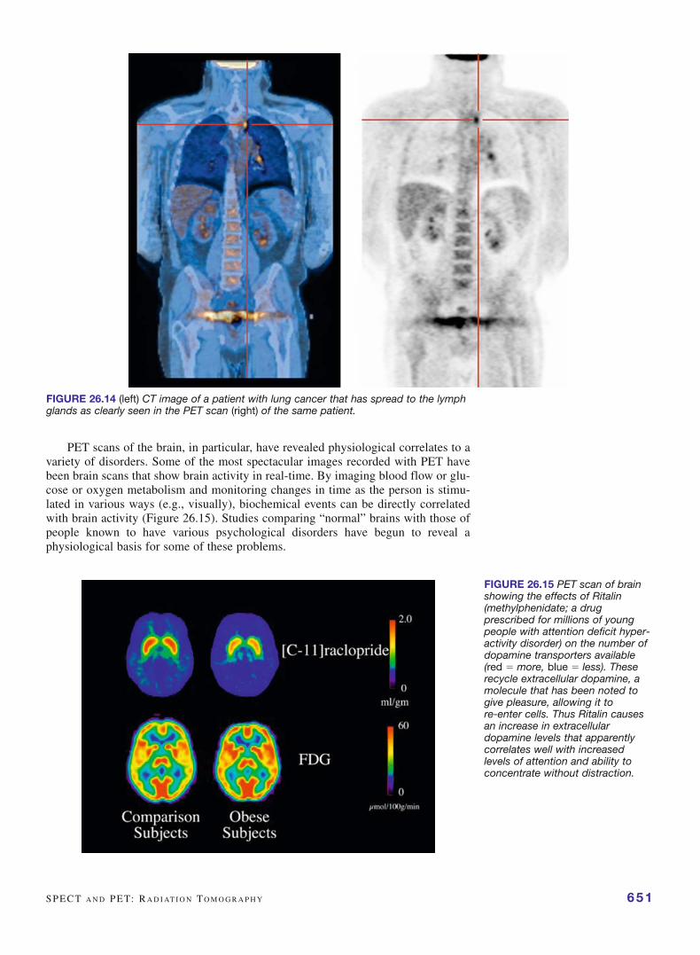
PET scans of the brain, in particular, have revealed physiological correlates to avariety of disorders. Some of the most spectacular images recorded with PET havebeen brain scans that show brain activity in real-time. By imaging blood flow or glu-cose or oxygen metabolism and monitoring changes in time as the person is stimu-lated in various ways (e.g., visually), biochemical events can be directly correlatedwith brain activity (Figure 26.15). Studies comparing “normal” brains with those ofpeople known to have various psychological disorders have begun to reveal aphysiological basis for some of these problems.
SPECT A N D PET: R A D I AT I O N T O M O G R A P H Y 651
FIGURE 26.14 (left) CT image of a patient with lung cancer that has spread to the lymphglands as clearly seen in the PET scan (right) of the same patient.
FIGURE 26.15 PET scan of brainshowing the effects of Ritalin(methylphenidate; a drugprescribed for millions of youngpeople with attention deficit hyper-activity disorder) on the number ofdopamine transporters available(red � more, blue � less). Theserecycle extracellular dopamine, amolecule that has been noted togive pleasure, allowing it tore-enter cells. Thus Ritalin causesan increase in extracellulardopamine levels that apparentlycorrelates well with increasedlevels of attention and ability toconcentrate without distraction.
8. FISSION AND FUSION
In Section 2 we saw in Figure 26.2 that the binding energy of nuclides with A numbersnear iron (56) have more binding energy, and are therefore more stable, than either verylow A or very high A nuclides. In most larger nuclei, such as uranium, the long-rangeCoulomb repulsion of the protons is in a precarious balance with the short-range strongnuclear attractive force between adjacent nucleons. If such a nucleus is perturbed, forexample, through a collision with an external nucleon, a new short-lived “excited” nucleusforms. The added energy causes the “liquid drop” nucleus to begin to elongate and oncethe nucleus becomes sufficiently asymmetric, the Coulomb repulsion of the two portionscauses the nucleus to be unstable and decay by dividing into two roughly equal fissionproducts. The difference in net binding energy between the higher-energy original nucleusand the total lower energy of the products is given off as kinetic energy of the fission prod-ucts. This energy is substantial; for example, uranium has a binding energy per nucleon ofabout 7.6 MeV/nucleon (remember that these binding energies are actually negative, sothat a smaller binding energy means a higher energy state), whereas the fission productshave values of close to 8.5 MeV/nucleon. The difference of 0.9 MeV/nucleon amounts toabout 100 MeV of kinetic energy for each of the two fission products.
Fission was first discovered in 1938 by Hahn and Strassmann, who bombarded ura-nium with a beam of neutrons and found two fission products, barium and krypton. Foreach starting nucleus, there are many different pairs of possible fission products, mostof them radioactive. One example of a fission reaction for uranium-235 is the reaction
(26.16)
The fact that there are often additional neutrons emitted, with an average of 2–3 perfission, caused scientists early on to propose that a chain reaction of neutron-acti-vated fission could occur. Each fission would lead to two or three neutrons released,some of which would produce further fissions so that there would be a positive feed-back and rapid growth in the energy released in fission products. By 1942 Fermi haddemonstrated such a chain reaction in the first nuclear reactor.
The first use of nuclear fission was in the form of two atomic bombs droppedover Hiroshima and Nagasaki to end World War II with Japan in 1945. War hadunited many of Europe’s finest scientists with those of the United States in a secreteffort to develop the atomic bomb at Los Alamos, New Mexico. Although it is gen-erally agreed that the use of these bombs shortened the war and reduced the totalnumber of deaths, some of the leading scientists who worked on the development ofthe atomic bomb believed, in retrospect, that it was a mistake and spent much of theirsubsequent efforts in attempts to bring about nuclear disarmament.
Enrico Fermi’s first nuclear reactor had as its main initial function the production ofplutonium to be used in two atomic bombs. Today there are about 450 nuclear power reac-tors used to generate electricity in about 30 countries around the world. Although there areseveral different designs of these reactors, they all basically use nuclear energy to generateheat, producing steam then used to drive turbines, thereby generating electricity.
There are several key problems to producing controlled nuclear fission in a nuclearreactor. The predominant uranium-238 isotope (representing over 99% of naturally occur-ring U) is relatively stable against fission, whereas uranium-235 (only about 0.7% abun-dance) undergoes fission very efficiently when slow neutrons are absorbed. Sometimesuranium ore is processed to enrich the 235U component to a few percent to provide a“richer” fuel. A minimum amount of fuel, the critical mass, typically on the order of kg,is needed to have a self-sustaining nuclear reaction. A second problem is that of the two orthree neutrons produced in a single fission, only one is needed to sustain a controlled reac-tion. If more than one neutron from each fission leads to additional fissions, the reactionwill “run away,” as in a nuclear bomb, whereas if this number is less than 1.0, the reactionwill eventually die out. Only by maintaining this number very near to 1.0, by the escape orabsorption of excess neutrons in a special device known as a control rod, can the reactionbe kept at a steady rate. Control rods are made from materials that very effectively absorb
01 n�92
235 U n 56141 Ba�36
92 Kr�301 n.
652 N U C L E A R P H Y S I C S A N D M E D I C A L A P P L I C AT I O N S

neutrons without undergoing fission. Neutron absorption in 235U leading to fission is mosteffective for slow thermal neutrons, those that have lost energy often making numerouscollisions in a special purpose material known as a moderator. Moderators are designed toeffectively slow neutrons. Water is commonly used as a moderator in nuclear reactors, withheavy (deuterated) water sometimes used because it absorbs fewer neutrons eliminatingthe need to enrich the uranium.
Despite huge investments in safety features, there have been two significant acci-dents at nuclear power plants: one at Three Mile Island, in Pennsylvania in 1979which was contained, and one at Chernobyl in Ukraine in 1986 where 31 people wereinitially killed, most from radiation. The Chernobyl accident released about 3–4% ofits radioactive material resulting in about 130,000 people receiving significant radia-tion doses leading to a sharp increase in thyroid cancer among children in that region,with other long-term health effects still unclear. Apart from safety issues of nuclearpower plants, there are also literally tons of highly radioactive waste products pro-duced in these plants that need to be safely and securely isolated from our environ-ment for thousands of years. Because of these safety and environmental concerns,alternative sources of electricity other than nuclear fission power are needed. Alongwith solar, wind, hydroelectric, and other “green” sources of power, a possible long-term solution involves a second type of nuclear reaction.
According to Figure 26.2, two very low mass number nuclides with a small bind-ing energy per nucleon can fuse together to produce a larger nuclide with a muchgreater binding energy per nucleon, thus releasing a large amount of energy. Thisprocess, known as nuclear fusion, releases much more energy per nucleon than fission,as can be seen from the steep initial slope in the binding energy per nucleon curve inFigure 26.2. In other words, the magnitude of the energy of the larger fused nucleus ismuch less than the sum of the energy of the lighter starting nuclei and the differenceis liberated in the fusion reaction. For example, in the fusion of deuterium and tritium,two isotopes of hydrogen, an alpha particle and a neutron, form according to
(26.17)
Calculating the net difference between the initial and final energies (using the massesof each and the equivalence of mass and energy; see the example just below) gives anet energy release of about 17 MeV for each fusion. Because there are only 5 nucle-ons involved in this reaction, the energy per nucleon is 3.4 MeV/nucleon, muchlarger than the 0.9 MeV/nucleon released in fission. On an energy per unit massbasis, fusion is a much more productive process than fission.
Nuclear fusion occurs naturally in stars, including our sun, at extremely high tem-peratures. These thermonuclear reactions in stars are believed to have been responsiblefor generating all of the larger mass nuclei in the universe starting from hydrogen. Webelieve that very early in the history of the universe the temperature was too hot foratoms to be stable. As the universe expanded and cooled, hydrogen atoms formed andthen condensed locally under gravity to form stars. As stars became more compact dueto the force of gravity, the interior temperatures and pressures increased, providingan environment in which nuclear fusion could occur. Stellar fusion first uses hydrogenas a fuel, but as hydrogen is depleted fusion of other light nuclei also occurs. Thus,all the other elements found on Earth and throughout the universe originated in suchstellar fusion reactions; we ourselves are therefore made of stellar material.
One fusion reaction is the so-called proton–proton cycle:
(26.18)
Net reaction: 411 H n 2
4 He � 2e� � 2n� 2g
23 H � 2
3 H n 24 He � 1
1 H � 11 H.
11 H � 1
2 H n 23 H � photon
11 H � 1
1 H n 12 H �e�� neutrino
12 H � 1
3 H :24 He � 0
1 n.
F I S S I O N A N D F U S I O N 653
The overall result of these reactions is that four protonshave fused to produce one alpha particle plus two each ofpositrons, neutrinos, and photons, with a net release of24.7 MeV. The positrons quickly annihilate with electronsto form four additional photons, each with 0.51 MeV, sothat the total energy released in the proton–proton cycleis (24.7 � 4 � 0.51) � 26.7 MeV per helium nucleusformed. In order for this reaction sequence to occur, pro-tons must be brought very close together at very hightemperature to overcome their mutual electrostatic repul-sion and fuse together. Central cores of stars, including oursun, have temperatures and pressures high enough forfusion to occur.
To produce fusion on the Earth, where the pressure ismuch lower than in the core of a star, even hotter tempera-tures are required. The first fusion reactions produced were
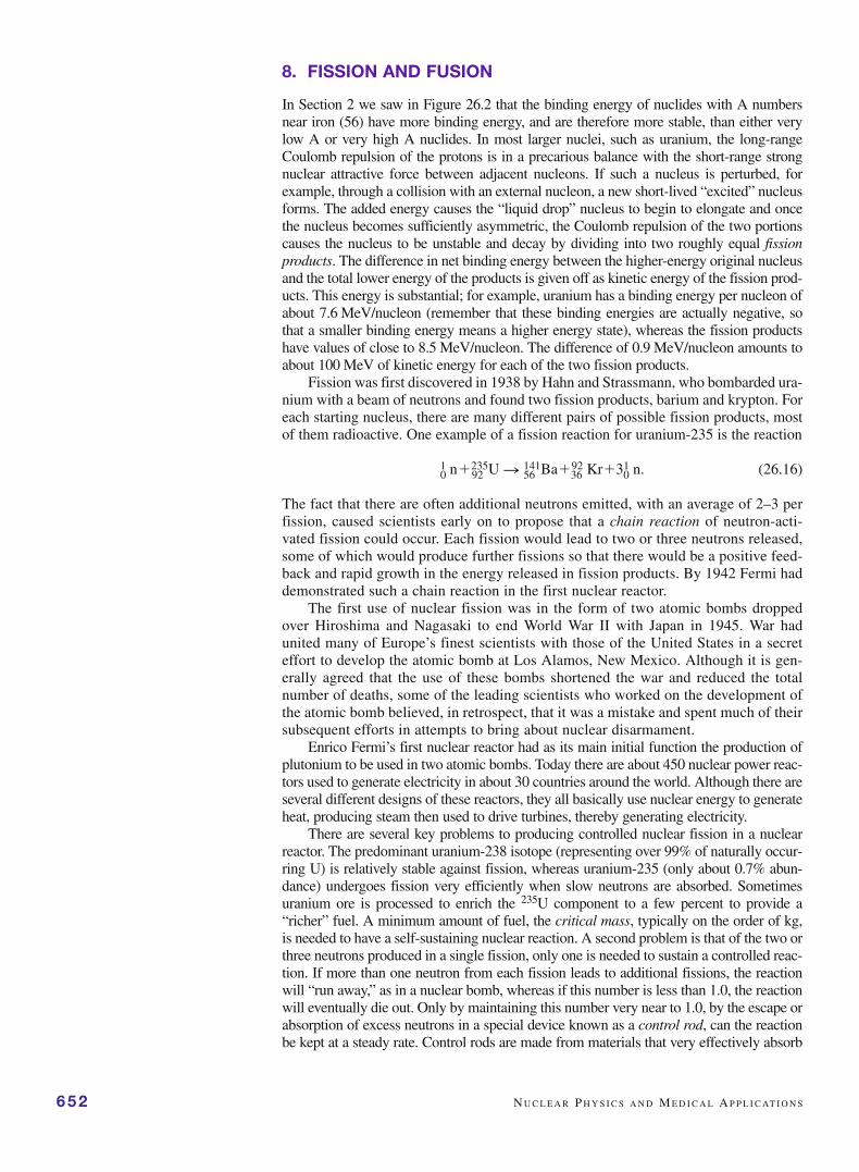
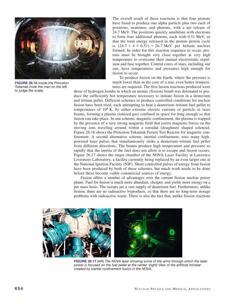
those of hydrogen bombs in which an atomic (fission) bomb was detonated to pro-duce the sufficiently hot temperature necessary to initiate fusion in a deuteriumand tritium pellet. Different schemes to produce controlled conditions for nuclearfusion have been tried, each attempting to heat a deuterium–tritium fuel pellet totemperatures of 108 K, by either extreme electric currents or particle or laserbeams, forming a plasma (ionized gas) confined in space for long enough so thatfusion can take place. In one scheme, magnetic confinement, the plasma is trappedby the presence of a very strong magnetic field that exerts magnetic forces on themoving ions traveling around within a toroidal (doughnut) shaped solenoid.Figure 26.16 shows the Princeton Tokamak Fusion Test Reactor for magnetic con-finement. A second alternative scheme, inertial confinement, uses many high-powered laser pulses that simultaneously strike a deuterium–tritium fuel pelletfrom different directions. The beams produce high temperature and pressure sorapidly that the inertia of the fuel does not allow it to escape and fusion occurs.Figure 26.17 shows the target chamber of the NOVA Laser Facility at LawrenceLivermore Laboratory, a facility currently being replaced by an even larger one atthe National Ignition Facility (NIF). Short controlled pulses of energy from fusionhave been produced by both of these schemes, but much work needs to be donebefore these become viable commercial sources of energy.
Fusion offers a number of advantages over the current fission nuclear powerplants. Fuel for fusion is much more abundant, cheaper, and yields more energy on aper mass basis. The oceans are a vast supply of deuterium fuel. Furthermore, unlikefission, there are no radioactive byproducts, so that there are no long-term storageproblems with radioactive waste. There is also the fact that, unlike fission reactions
654 N U C L E A R P H Y S I C S A N D M E D I C A L A P P L I C AT I O N S
FIGURE 26.16 Inside the PrincetonTokamak (note the man on the leftto judge the scale).
FIGURE 26.17 (left) The NOVA laser showing some of the arms through which the laserpower is focused on the fuel pellet at the center. (right) View of the artificial ministarcreated by inertial confinement fusion in the NOVA.

in which chain reactions can become uncontrolled if there is a malfunction of controlrods producing a melt-down as has happened at Chernobyl and Three Mile Island,failures in fusion reactors would lead to a shut-down of the fusion reactions them-selves and no possibility of an out-of-control chain reaction. For these reasons ifcommercially produced in a reactor, energy from fusion might be the ultimate cure tothe world’s energy problem.
C H A P T E R S U M M A RY 655
CHAPTER SUMMARYThe atomic nucleus contains Z protons and N neutronswith a total number of nucleons A � Z � N. Nuclei arevery small in size, having radii given by
R � R0A1/3, (26.2)
with R0 � 1.2 fm. A nucleus of mass m has a nuclearbinding energy given by
Nuclear Binding Energy�Zmpc
2�Nmnc2�mc2, (26.4)
and is typically about 8 MeV per nucleon in all but thesmallest nuclei.
Three types of nuclear radiation exist known as alpha,beta, and gamma radiation. Alpha radiation is the emissionof helium-4 nuclei (2 protons � 2 neutrons) from nucleithrough a process of tunneling. Beta emission comes fromthe production of electrons or positrons within the nucleusbecause of neutron or proton decay, respectively, and these“beta particles” are emitted from the nucleus along withneutrinos at high energy. Gamma emission comes fromtransitions from excited nuclear states giving rise to highenergy photons. Each of these types of radioactivity arecharacterized by their Q, or decay energy,
Q �(mP ��mi)c2 � 0, (26.5)
where P stands for parent nucleus and the sum is overall the products.
Radioactive decay is governed by an exponentialdecay of the numbers of radioactive nuclei N,
(26.9)
where N0 is the number of such nuclei at time zero and� is the decay rate for the process. The half-life of the
reaction is the time for 1/2 of the nuclei to decay and isrelated to the decay rate by
(26.10)
Measures of exposure to radioactivity include: theRoentgen (R) which is simply a measure of the number ofdecays per unit volume; the Gray (Gy; 1 Gy � 1 J/kg) orrad (1 rad � 0.01 Gy) which are measures of the absorbeddose of radiation; or the biological dose equivalent, mea-sured in sieverts (Sv), or in rem (1 rem � 0.01 Sv),
biological dose equivalent (in Sv)� absorbed dose (in Gy) � RBE, (26.14)
where RBE (relative biological effectiveness) is a dimen-sionless weighting factor describing the effectiveness ofdifferent radiation to be absorbed by the body.
Nuclear medicine involves the use of short-livedradioactive tracers (radiolabeling) to follow the path ofa particular molecule through the body either by in vivoor in vitro studies. Two imaging methods that use radio-tracers are SPECT (single photon emission computertomography) and PET (positron emission tomography).
Because the binding energy per nucleon for largenuclei is less in magnitude (~0.9 MeV) than for inter-mediate-sized nuclei, large nuclei can undergo fissionreleasing the excess energy of ~100 MeV for each of theproducts, along with several neutrons. Under controlledconditions this energy can be harnessed in nuclearpower plants, whereas if left uncontrolled, this serves asthe basis for a fission bomb. Fusion is the naturallyoccurring process in stars whereby hydrogen nuclei, orother small nuclei, are compressed and heated until theyfuse to form larger nuclei, releasing large amounts ofbinding energy (~3.4 MeV per nucleon) in the process.Current research is attempting to produce controlledfusion in the laboratory as a means of generating energythat would be much cleaner than fission nuclear powerplants. Very high power lasers are being used to attemptto achieve the very high temperatures and pressuresneeded to cause fusion.
t1/2 �loge 2
l�
0.693
l.
N(t) � N0 e�lt,
MULTIPLE CHOICE QUESTIONS1. The nucleus is about this many times as large as the
atom: (a) 10�2, (b) 10�3, (c) 10�4, (d) 10�5.2. The nuclide of iron has (a) 30 neutrons, (b) 56
neutrons, (c) 26 electrons, (d) 56 protons.3. The Nobelium nucleus, a very short-lived (3 min
half-life) manmade nuclide, has an effective radius ofabout (a) 1.2 fm, (b) 7.6 fm, (c) 5.6 fm, (d) 0.19 fm.
4. Without the strong nuclear force, carbon-based lifecould not exist. This is primarily because the strongnuclear force (a) permits nuclei to consist of morethan just a single proton, (b) keeps gravity from col-lapsing all matter into a single point, (c) keeps theelectric force from attracting all electrons into thenucleus, (d) is responsible for covalent bondsbetween carbon atoms.
5. Very large nuclei are radioactive because of all butthe following (a) the electrical repulsive force desta-bilizes them, (b) the total binding energy of twofission fragments is smaller than that of the originalnucleus, (c) the excess neutrons do not sufficientlyshield the repulsive force between protons, (d) it isrelatively easy for a proton to escape (tunnel) out ofthe nucleus.
6. A nucleus with 20 neutrons is unusually stablebecause (a) it has an unusually low binding energy,(b) it has a closed nuclear shell, (c) it has all spinpaired neutrons, (d) it has an unusually high propen-sity for decay.
7. The neutrino was first predicted in beta decaybecause of missing (a) spin, (b) angular momentum,(c) energy, (d) charge.
8. In a scintillation detector, incoming X’s are eventu-ally converted into outgoing Y’s where X and Y couldbe (a) gammas and visible photons, (b) electrons andelectrons, (c) electrons and visible photons, (d) visi-ble photons and electrons.
9. Starting with 1012 radioactive nuclei, after 4 half-livesabout (a) 2.5 � 1011, (b) 1.8 � 1010, (c) 108, (d) 6.3 �1010 nuclei will remain.
10. In radioactive decay, compared to the activity, thetotal number of radioactive nuclei decays (a) expo-nentially with the same half-life, (b) logarithmicallywith the same half-life, (c) exponentially with agreater half-life, (d) exponentially with a smallerhalf-life.
11. Carbon-14 dating relies on all of the followingassumptions except (a) the amount of 14C in the airhas remained constant, (b) no new 14C is taken inafter death, (c) 14C is the only radioactive form of car-bon with a long half-life, (d) 14C is an alpha emitterwith a 5730 year half-life.
12. One hundred hours of flying time in a high-altitudejet gives an equivalent radiation dose to a diagnosticx-ray. This large dose is primarily due to the fact that(a) there are fewer people at those altitudes to absorb
102255 No,
2656 Fe,
656 N U C L E A R P H Y S I C S A N D M E D I C A L A P P L I C AT I O N S
QUESTIONS1. Carefully distinguish between Z, A, and N. Which of
these change and which remain constant for , , and� decay?
2. Why does Equation (26.2) lead to a picture of thenucleus as a dense-packed ball of nucleons in contactwith each other? (Hint: How does the equation pre-dict the nuclear mass will vary with radius?)
3. Compare the notion of nuclear binding energy to theionization energy of an atom. How is it similar andhow is it different?
4. What three factors determine the stability ofnuclei?
5. Which of the following nuclei have net spin (indicatewhether due to protons or neutrons) in their groundstates: 1H, 12C, 13C, 15N, 31P?
6. What are magic numbers and how are they determined?7. Compare the , , and � decay particles in terms of
penetrating power and radiation damage produced.8. Complete the following nuclear processes by stating
what the nucleon X represents: (a)
9. Write the complete decay scheme for the � decay ofand of
10. Does it matter at what time a measurement starts inorder to measure the half-life of a radioactive sample?That is, suppose two experimenters take the samesample of a radioactive material with a 3 min half-lifeand each independently tries to measure the half-life.Does it matter whether they start their measurementsat the same time?
11. If a radioactive sample starts out with 1020 nuclei,how many will be left after 10 half-lives? After 10lifetimes?
12. What are the fortuitous circumstances that allow 14Cdating of once-living organisms?
13. What is the difference between exposure, absorbeddose, and biological dose equivalent? Which is mostimportant in determining health risks?
14. Which has the longer effective half-life when used asa radiopharmaceutical, an isotope with a 45 h half-life that takes twice as long to wash out of the bodyas a second isotope with a 75 h half-life and a 7 daybiological half-life?
15. In the radioimmunological assay, why is the ratio ofthe labeled-to-unlabeled antigen bound to the anti-body the same as that ratio found in the supernatant?
16. In PET, how is it known that a particular detectedgamma came from pair annihilation within the body?
17. In a nuclear fission power plant, what is the purposeof a control rod? A moderator?
18. Which would liberate more energy: assembling14 protons and 14 neutrons to make one 28Si nucleusor two 14N nuclei?
60Co.126Sn
(c) X n 230Th�4He
60Co n 60Ni �X; (b) X n 234Pa�b�;
the cosmic rays so each person gets a higher dose,(b) the Earth’s atmosphere shields us on the groundfrom most cosmic rays (c) the plane is that much closerto the sun, so the dose is higher, (d) cosmic rays inter-act with the plane’s metal and produce high doses ofsecondary radiation.
13. Which of the following is not a desired feature of aradiopharmaceutical? (a) A relatively short (hours)half-life, (b) relatively low energy radiation, (c) along biological half-life, (d) ability to bind to specifictarget tissue.
14. In liquid scintillation counting which of the followingis not true? (a) For each radioactive decay a singleelectron is detected, (b) a radioactive decay results ina photon of visible light, (c) different radioactiveemissions result in different amplitudes of detectedcurrent pulses, (d) the solvent that the sample is dis-solved or suspended in has a fluorescent component.
15. The detected particles in PET are all but the follow-ing. (a) They are an electron and positron, (b) they aremeasured 180° apart along a line, (c) their energy isequal to the rest mass of the electron, (d) they are pro-duced after ingesting a positron emitting source.
16. Essential features of a nuclear fission power plant includeall but the following. (a) Moderators, (b) control rods,(c) deuterium–tritium fuel pellets, (d) steam turbines.
PROBLEMS1. The largest stable nucleus has a mass number of 209.
Find the ratio of the radii, surface areas, and volumesof this largest nucleus to that of a hydrogen nucleus.
2. A neutron star has a diameter of about 20 km and hasa density roughly that of the nucleus. What is itsmass? How many solar masses is this (solar mass �2 � 1030 kg)? What is the mass number for the neu-tron star (i.e., how many nucleons does it contain)?
3. If the sun (mass �2 � 1030 kg, radius �7 � 108 m)collapsed until it had a density equal to that of nuclei,what would be its radius? (Actually, a star cannot col-lapse to nuclear densities unless its mass exceeds acritical mass, known as the Chandrasekhar mass, ofabout 1.4 solar masses to overcome the Pauli exclu-sion repulsion of the electrons.)
4. Using the numbers in Example 26.1 calculate thebinding energy of radium 226 (m �225.97709 u),radium 228 (m � 227.98275 u), and thorium 232 (m �231.98864 u). Also find their binding energy pernucleon.
5. Calculate Q for the -decay of using data in theprevious problem and in Example 26.1.
6. Calculate Q for the � decay of 24Na given the fol-lowing data: m(24Na) � 23.98492 u, m(24Mg) �23.97845 u, m(24Ne) � 23.98812 u, m(�) � 5.49� 10�4 u. What is the range of possible energies forthe emitted beta particle?
90232 Th
Q U E S T I O N S /P R O B L E M S 657
7. Show that in alpha decay from a stationary parentnuclide that conservation of energy and momentumlead to a relation between the Q for the nuclear reac-tion and the kinetic energy gained by the alpha parti-cle, KE, given by
Then look back at Example 26.2 and calculate thekinetic energy of the alpha emitted in the decay of 238U.
8. The first successful experiment to detect the neutrinowas done in 1953 by Reines who won the 1995 Nobelprize for this work. Neutrinos from the Hanfordnuclear reactor were incident on a tank of 200 L ofwater in which they very infrequently interacted withthe water protons to produce a neutron and a positronin the reaction: � � p → n � �. The positrons sub-sequently annihilated producing two signature gam-mas traveling 180° apart and the neutrons werecaptured by cadmium in the form of 40 kg of CdCl2salt added to the water to produce several additionalgammas. These gammas were detected by three scin-tillator layers and 110 photomultiplier tubes sur-rounding the water and Reines and co-workerscarried out numerous checks to ensure that thesegammas did originate from the above reaction and notfrom any other source. Although the neutrino fluxwas as much as 1013 /cm2-s, they detected on averageonly 0.027 events per hour per phototube in the entiredetector. Assuming the cross-sectional dimensions ofthe water tank to be 2 m on a side and the neutrinoflux to be uniform over this area, what fraction of theneutrinos interacted with the water in their detector,assuming 100% collection efficiency of the gammas?
9. Suppose that the phototube of a scintillation detectorhas a gain of 5 � 105, representing the average numberof electrons produced at the anode for each electronemitted at the photocathode. If a 10 �Ci gamma emit-ting radioactive source is detected with 5% efficiency,find the average output current from the phototube.
10. How long does it take for 90% of a 60Co sample orig-inally present to decay?
11. What mass of 90Sr is needed to have an activity of 1 mCi?How long will it take for the activity to decrease to0.25 mCi?
12. Iodine is selectively accumulated in the thyroid glandwhere it can build to dangerous levels. When radioac-tive materials have been released into the atmospherefrom either nuclear power plant accidents, such as themajor one at Chernobyl, or from nuclear testing, thesematerials tend to concentrate (through eating of plantsby animals) and show up in food products, such asmilk, a food which is preferentially eaten by children.Thus, even though the half-life of 131I is relatively
Q � KE £1�m(2
4 He)
m(daughter)≥ .
produced, and lead to serious health problems. Howlong would it take for all but 0.01% of a sample of90Sr to decay?
18. Calculate the activity (in Bq) of one gram of radium-226. (Hint: See Example 26.3; this is the definition ofone curie.)
19. An amateur archeologist finds a bone that he believesto be from a dinosaur. He sends a chip of it off to alaboratory for 14C dating. The lab finds that the chipcontains 5 g of carbon and has an activity of 0.5 Bq.How old is the bone? Could it be from a dinosaur?
20. An 85 kg person was exposed to a gamma source andreceived a whole body dose of 0.5 Sv. How muchenergy was deposited in the person’s body? Repeat thiscalculation if the radiation was from an alpha source.
21. What dose (in Gy) of gammas produces the same bio-logical effects as a 50 rad dose of alpha particles?
22. What fraction of a 1 g sample of sitting on a tablewill remain in 17 years? If the strontium had beeningested and all initially been absorbed into a person’sbones, what fraction would remain after 17 years?(The biological half-life of 90Sr is 45 years.)
23. A small amount of phosphorus-32 was accidentallyingested and its activity carefully monitored overtime. After 8 days, the activity had halved. The phys-ical half-life of phosphorus-32 is given in Table 26.4.Find its biological half-life.
24. In a radioimmunological assay 10 nM of a 125Ilabeled antigen and 1 nM of antibody were added toan unlabeled sample of the antigen. The solution wascentrifuged and the activity of the supernatant andpellet were measured and found to be in the ratio of5.4:1. How much antigen was originally present inthe solution?
25. Calculate the net energy released in each step of theproton–proton cycle shown in Equation (26.18). Thenadd up the net release for one step in the cycle beingsure to use a balanced net reaction; recall that the massof the neutrino and photon are zero. Check your resultby a direct calculation of the energy released in the netoverall reaction. Use the following masses: m(1H) �1.00728 u; m(2H) �2.01355 u; m(e�) � 5.49 � 10�4 u;m(3H) �3.01550 u; m(4He) �4.00151 u.
90Sr
658 N U C L E A R P H Y S I C S A N D M E D I C A L A P P L I C AT I O N S
short, this isotope has caused thyroid cancer in manychildren in affected areas. Some children received upto 1000 rem from 131I release at Chernobyl. By whatfactor is this above the maximum annual exposure rec-ommended? How long would it have taken for theradiation level to have decreased so that for the sameconsumption of milk the exposure would have been at0.1 times the maximum annual recommendation?
13. After the sudden release of radioactivity from theChernobyl nuclear reactor accident in 1986, theradioactivity of milk in Poland rose to 2000 Bq/L dueto iodine-131 present in the grass eaten by dairy cat-tle. Radioactive iodine, with a half-life 8.0 days, isparticularly hazardous because the thyroid gland con-centrates iodine.(a) What is the decay constant that characterizes the
decay of 131I if it has a half-life of 8 days?(b) What is the storage time needed to decrease the 131I
content of cheese produced from these cows’ milkto 15% of the original level?
14. A bone fragment is found in the desert. If it has amass of carbon (due to only 14C and 12C) of 200 g,how old is it if it has an activity of 15 decays per sec-ond? The ratio of 14C to 12C is 1.3 � 10�12.
15. To destroy a cancerous tumor, a dose of gamma radi-ation totaling an energy of 2.12 J is to be delivered in30.0 days from implanted sealed capsules containingpalladium-103. Assuming that this isotope has a half-life of 17.0 days and emits gamma rays of energy21.0 keV, which are entirely absorbed within thetumor, what is the initial activity of the set of cap-sules, and what total mass of radioactive palladiumshould these “seeds” contain?
16. 238U decays by the emission of an alpha particle.(a) What is the decay sequence?(b) What is the daughter nucleus?(c) What is the energy of the alpha particle (its mass
is 4.0026 u)?(d) What is the velocity of the particle?(e) Is the alpha particle relativistic?
17. Strontium is chemically similar to calcium and canreplace calcium in bones. The radiation from 90Srcan damage the bone marrow where blood cells are