no intrinsic depletion layer on a polystyrene thin film at a water interface
TRANSCRIPT

No Intrinsic Depletion Layer on a Polystyrene Thin Film at a WaterInterface
Young-Soo Seo* and Sushil Satija
NIST Center for Neutron Research, National Institute of Standards and Technology,Gaithersburg, Maryland 20899
ReceiVed March 19, 2006. In Final Form: June 27, 2006
Using neutron reflectivity, we found that there is no intrinsic depletion layer at a deuterated polystyrene (dPS) filmand deuterium oxide (D2O) interface. A spun-cast film is susceptible to contamination on its surface from its surroundingsduring sample preparation. A contamination layer of hydrogenated organic material will be detected as a reducedscattering length density layer at the interface. We demonstrate that, by careful treatment of the film, contaminationwould be the primary cause of the reduced scattering length density layer at the interface.
Introduction
The interfacial structure of hydrophobic surfaces in contactwith water has attracted a lot of interest in the past few years.The origin of hydrophobic forces has implications in many fields,including protein folding and colloidal systems. Theoretically,it has been proven that the hydrophobic force arises from a reduceddensity layer of water near large enough hydrophobic substrates.1
The thickness of the layer has been calculated to a few angstromsusing molecular dynamic simulation.2 This layer could beconsidered to be a precursor of air nanobubbles. Air nanobubbleformation on hydrophobic surfaces in aqueous media has becomewidely explored as a general phenomena. Nanobubble formationhas been recognized by observing strong and stepwise long-range attractive interactions between macroscopic hydrophobicsurfaces in aqueous media.3 Direct images of nanobubbles of20-30 nm height have been obtained using tapping mode AFMat the silane-modified glass-water interface.4 Nanobubbles werefound on various surfaces such as hydrophobic silane-modifiedsurfaces and also inorganic surfaces including gold,5 graphite,6
and mica.7
Recently, hydrophobic polymer surfaces were used to studyinterfacial structures at the water interface.8,9 Using neutronreflectivity, they found a few-nanometers-thick layer with reducedscattering length density (SLD) between a deuterated polystyrene(dPS) film on Si and the D2O interface. From the point of viewof neutron reflectivity experiments, a reduced SLD layer at adPS/D2O interface can be interpreted as either a lower waterdensity layer (i.e., depletion layer) or a hydrogenated contamina-tion layer adsorbed on the dPS film. Polymer films are susceptibleto contamination on their surfaces from the open air environment
during sample preparation. Therefore, careful treatment inpreparing the film would be necessary to find the origin of thereduced SLD layer at a dPS/D2O interface.
In this letter, we show that for carefully prepared dPS filmsthere is no evidence of a reduced SLD layer in neutron reflectivityexperiments at the dPS/D2O interface. The adsorption ofhydrogenated contaminants and/or oxidation on the dPS filmduring sample preparation might be responsible for the reducedSLD layer.
Experimental Section
Deuterated polystyrene (dPS) films were prepared by spin coatingeither from toluene or deuterated toluene (d-toluene) solution onfreshly cleaned Si blocks (105 mm diameter and 13 mm thickness).Si blocks were thoroughly cleaned in piranha solution. Coated filmthicknesses were about 500 Å. All of the films were carefully driedin a dish for an hour right after spin casting. To investigate theannealing effect, the dried films were annealed at 70°C for 2 h or160 °C for 24 h in an oil-free vacuum (∼10-3 Torr) oven. Thesamples were then placed in a homemade liquid cell filled with D2O.The cell was cleaned with deionized water and dried with nitrogengas before use. Visible air bubbles trapped inside the liquid cellwere carefully removed before neutron reflectivity measurements.The liquid cell temperature was controlled to(0.5 °C in thetemperature range of 20-50°C by a thermoset circulator during themeasurement.
Neutron reflectivity measurements were performed on the NG7horizontal reflectometer at the NIST (National Institute of Standardsand Technology) Center for Neutron Research (NCNR). The sampleand scattering geometry are schematically drawn in Figure 1a (lowerinset). The neutron scattering intensity was collected along thespecular reflection direction and plotted as a function of thez-directional scattering wave vector,Qz ) (4π/λ)sin θ where theneutron wavelength isλ ) 4.75 Å andθ is the incident angle to thesubstrate.
Results and Discussions
Neutron reflectivity is a very sensitive technique for detectinga low SLD layer at a dPS/D2O interface where the SLD of dPS(6.42 × 10⁻⁶ Å-2) is well matched to that of the bulk liquid(SLD of D2O ) 6.36× 10⁻⁶ Å-2). The calculated reflectivityshows only very weak Kiessig oscillations that result from thesmall difference between the SLDs of dPS and D2O as shownin Figure 1a. However, even a 2-Å-thick low SLD layer at theinterface gives rise to apparent Kiessig oscillation in thereflectivity profile where the corresponding layer SLD profileis depicted in Figure 1a (upper inset).
* Correspondence author. E-mail: [email protected]. Presentaddress: LG Chem. Research Park, Korea.
(1) Lum, K.; Chandler, D.; Weeks, J. D.J. Phys. Chem. B1999, 103, 4570-4577.
(2) Mamatkulov, S. I.; Khabibullaev, P. K.; Netz, R. R.Langmuir2004, 20,4756-4763.
(3) Parker, J. L.; Claesson, P. M.; Attard, P.J. Phys. Chem.1994, 98, 8468-8480.
(4) Tyrrell, J. W. G.; Attard, P.Phys. ReV. Lett. 2001, 87, 176104.(5) Holmberg, M.; Kuhle, A.; Garnas J.; Morch K. A.; Boisen A.Langmuir
2003, 19, 10510-10513.(6) Lou, S. T.; Gao, J. X.; Xiao X. D.; Li, X. J.; Li, G. L.; Zhang Y.; Li, M.
Q.; Sun, J. L.; Hu, J.Chin. Phys.2001, 10, S108-S110 Suppl. S.(7) Zhang, X. H.; Zhang, X. D.; Lou, S. T.; Zhang, Z. X.; Sun, J. L.; Hu J.
Langmuir2004, 20, 3813-3815.(8) Howse, J. R.; Steitz, R.; Pannek, M.; Simon, P.; Schubert, D. W.; Findenegg,
G. H. Phys. Chem. Chem. Phys. 2001, 3, 4044-4051.(9) Steitz, R.; Gutberlet, T.; Hauss, T.; Klosgen, B.; Krastev, R.; Schemmel,
S.; Simonsen, A. C.; Findenegg, G. H.Langmuir2004, 19, 2409-2418.
7113Langmuir2006,22, 7113-7116
10.1021/la060736v CCC: $33.50 © 2006 American Chemical SocietyPublished on Web 07/13/2006
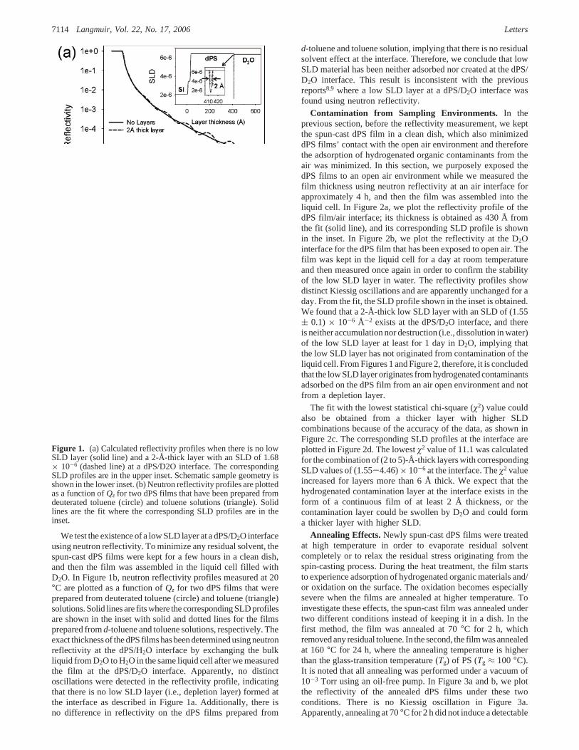
We test the existence of a low SLD layer at a dPS/D2O interfaceusing neutron reflectivity. To minimize any residual solvent, thespun-cast dPS films were kept for a few hours in a clean dish,and then the film was assembled in the liquid cell filled withD2O. In Figure 1b, neutron reflectivity profiles measured at 20°C are plotted as a function ofQz for two dPS films that wereprepared from deuterated toluene (circle) and toluene (triangle)solutions. Solid lines are fits where the corresponding SLD profilesare shown in the inset with solid and dotted lines for the filmsprepared fromd-toluene and toluene solutions, respectively. Theexact thickness of the dPS films has been determined using neutronreflectivity at the dPS/H2O interface by exchanging the bulkliquid from D2O to H2O in the same liquid cell after we measuredthe film at the dPS/D2O interface. Apparently, no distinctoscillations were detected in the reflectivity profile, indicatingthat there is no low SLD layer (i.e., depletion layer) formed atthe interface as described in Figure 1a. Additionally, there isno difference in reflectivity on the dPS films prepared from
d-toluene and toluene solution, implying that there is no residualsolvent effect at the interface. Therefore, we conclude that lowSLD material has been neither adsorbed nor created at the dPS/D2O interface. This result is inconsistent with the previousreports8,9 where a low SLD layer at a dPS/D2O interface wasfound using neutron reflectivity.
Contamination from Sampling Environments. In theprevious section, before the reflectivity measurement, we keptthe spun-cast dPS film in a clean dish, which also minimizeddPS films’ contact with the open air environment and thereforethe adsorption of hydrogenated organic contaminants from theair was minimized. In this section, we purposely exposed thedPS films to an open air environment while we measured thefilm thickness using neutron reflectivity at an air interface forapproximately 4 h, and then the film was assembled into theliquid cell. In Figure 2a, we plot the reflectivity profile of thedPS film/air interface; its thickness is obtained as 430 Å fromthe fit (solid line), and its corresponding SLD profile is shownin the inset. In Figure 2b, we plot the reflectivity at the D2Ointerface for the dPS film that has been exposed to open air. Thefilm was kept in the liquid cell for a day at room temperatureand then measured once again in order to confirm the stabilityof the low SLD layer in water. The reflectivity profiles showdistinct Kiessig oscillations and are apparently unchanged for aday. From the fit, the SLD profile shown in the inset is obtained.We found that a 2-Å-thick low SLD layer with an SLD of (1.55( 0.1) × 10-6 Å-2 exists at the dPS/D2O interface, and thereis neither accumulation nor destruction (i.e., dissolution in water)of the low SLD layer at least for 1 day in D2O, implying thatthe low SLD layer has not originated from contamination of theliquid cell. From Figures 1 and Figure 2, therefore, it is concludedthat the low SLD layer originates from hydrogenated contaminantsadsorbed on the dPS film from an air open environment and notfrom a depletion layer.
The fit with the lowest statistical chi-square (ø2) value couldalso be obtained from a thicker layer with higher SLDcombinations because of the accuracy of the data, as shown inFigure 2c. The corresponding SLD profiles at the interface areplotted in Figure 2d. The lowestø2 value of 11.1 was calculatedfor the combination of (2 to 5)-Å-thick layers with correspondingSLD values of (1.55-4.46)× 10-6 at the interface. Theø2 valueincreased for layers more than 6 Å thick. We expect that thehydrogenated contamination layer at the interface exists in theform of a continuous film of at least 2 Å thickness, or thecontamination layer could be swollen by D2O and could forma thicker layer with higher SLD.
Annealing Effects.Newly spun-cast dPS films were treatedat high temperature in order to evaporate residual solventcompletely or to relax the residual stress originating from thespin-casting process. During the heat treatment, the film startsto experience adsorption of hydrogenated organic materials and/or oxidation on the surface. The oxidation becomes especiallysevere when the films are annealed at higher temperature. Toinvestigate these effects, the spun-cast film was annealed undertwo different conditions instead of keeping it in a dish. In thefirst method, the film was annealed at 70°C for 2 h, whichremoved any residual toluene. In the second, the film was annealedat 160°C for 24 h, where the annealing temperature is higherthan the glass-transition temperature (Tg) of PS (Tg ≈ 100°C).It is noted that all annealing was performed under a vacuum of10-3 Torr using an oil-free pump. In Figure 3a and b, we plotthe reflectivity of the annealed dPS films under these twoconditions. There is no Kiessig oscillation in Figure 3a.Apparently, annealing at 70°C for 2 h did not induce a detectable
Figure 1. (a) Calculated reflectivity profiles when there is no lowSLD layer (solid line) and a 2-Å-thick layer with an SLD of 1.68× 10⁻⁶ (dashed line) at a dPS/D2O interface. The correspondingSLD profiles are in the upper inset. Schematic sample geometry isshown in the lower inset. (b) Neutron reflectivity profiles are plottedas a function ofQz for two dPS films that have been prepared fromdeuterated toluene (circle) and toluene solutions (triangle). Solidlines are the fit where the corresponding SLD profiles are in theinset.
7114 Langmuir, Vol. 22, No. 17, 2006 Letters
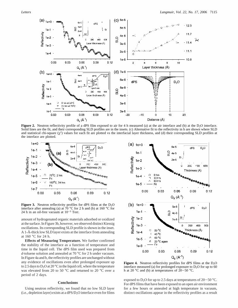
amount of hydrogenated organic materials adsorbed or oxidizedat the surface. In Figure 3b, however, we observed distinct Kiessigoscillations. Its corresponding SLD profile is shown in the inset.A 1-Å-thick low SLD layer exists at the interface from annealingat 160°C for 24 h.
Effects of Measuring Temperature.We further confirmedthe stability of the interface as a function of temperature andtime in the liquid cell. The dPS film used was prepared fromd-toluene solution and annealed at 70°C for 2 h under vacuum.In Figure 4a and b, the reflectivity profiles are unchanged withoutany evidence of oscillations even after prolonged exposure upto 2.5 days to D2O at 20°C in the liquid cell, where the temperaturewas elevated from 20 to 50°C and returned to 20°C over aperiod of 2 days.
Conclusions
Using neutron reflectivity, we found that no low SLD layer(i.e., depletion layer) exists at a dPS/D2O interface even for films
exposed to D2O for up to 2.5 days at temperatures of 20-50°C.For dPS films that have been exposed to an open air environmentfor a few hours or annealed at high temperature in vacuum,distinct oscillations appear in the reflectivity profiles as a result
Figure 2. Neutron reflectivity profile of a dPS film exposed to air for 4 h measured (a) at the air interface and (b) at the D2O interface.Solid lines are the fit, and their corresponding SLD profiles are in the insets. (c) Alternative fit to the reflectivity in b are shown where SLDand statistical chi-square (ø2) values for each fit are plotted vs the interfacial layer thickness, and (d) their corresponding SLD profiles atthe interface are plotted.
Figure 3. Neutron reflectivity profiles for dPS films at the D2Ointerface after annealing (a) at 70°C for 2 h and (b) at 160°C for24 h in an oil-free vacuum at 10-3 Torr.
Figure 4. Neutron reflectivity profiles for dPS films at the D2Ointerface measured (a) for prolonged exposure to D2O for up to 60h at 20°C and (b) at temperatures of 20-50 °C.
Letters Langmuir, Vol. 22, No. 17, 20067115

of a low SLD layer at the D2O interface, indicating that a lowSLD layer originates from hydrogenated contamination and/oroxidation of the dPS surface during sample treatment. It is noted
that chemical analysis such as X-ray photoelectron spectroscopy(XPS) would be necessary when scrutinizing airborne contami-nation and annealing-induced oxidation. This nonobservation ofan air layer at a water-hydrophobic solid interface agrees withthe ellipsometric results of Mao et al.10 and Takata et al.11
LA060736V
(10) Mao, M.; Zhang, J.; Yoon, R.-H.; Ducker, W. A.Langmuir2004, 20,1843-1849.
(11) Takata, Y.; Cho, J.-H. J.; Law, B. M.; Aratono, M.Langmuir2006, 22,1715-1721.
7116 Langmuir, Vol. 22, No. 17, 2006 Letters