nmr study on a novel mucin from jellyfish in natural abundance, qniumucin from aurelia...
TRANSCRIPT
NMR Study on a Novel Mucin from Jellyfish in Natural Abundance, Qniumucin from Aureliaaurita
Jun Uzawa,†,‡ Makoto Urai,† Takayuki Baba,† Hiroko Seki,‡ Kayoko Taniguchi,† and Kiminori Ushida*,†
Eco-Soft Materials Research Unit, AdVanced Science Institute, Riken, 2-1 Hirosawa, Wako, Saitama 351-0198 Japan, and Chemical AnalysisCenter, Chiba UniVersity, 1-33 Yayoi-cho, Inage-ku, Chiba 263-8522 Japan
ReceiVed September 24, 2008
A novel mucin (qniumucin), which we recently discovered in jellyfish, was investigated by several NMR techniques.Almost all the peaks in the 13C and proton NMR spectra were satisfactorily assigned to the amino acids in the mainchain and to the bridging GalNAc, the major sugar in the saccharide branches. The amino acid sequence in the tandemrepeat part (-VVETTAAP-) was reconfirmed by the cross-peaks between alpha protons and carbonyl carbons in theHMBC spectrum. A connectivity analysis around the O-glycoside bond (GalNAc-Thr) was also performed, and detailedinformation on the local configuration was obtained by the DPFGSE-NOE-HSD technique. The strategy and the resultsdescribed in this paper can be extended to the structural analysis of general O-glycan chains, which are more complexthan the present mucin. NMR analyses reveal the simple structure of qniumucin extracted by the present protocol, andthe homogeneity and purity of qniumucin are probably the result of it being extracted from jellyfish, a primitive animal.
Mucin is a family of glycoproteins found in almost all animalsas the main components of mucus, which is indispensable forsustaining life.1-11 It has a wide variety of functions, includingacting as a moisture holder, an antimicrobial, a counterpart oflectins, an adsorbent, and a surfactant. Mucin or “mucin domain”is defined as a polymer or a partial structure composed of a singleprotein main chain with branches of oligosaccharides connected atserine (Ser) or threonine (Thr) residues by O-glycoside bonds.
Although mucin itself is normally found as an extracellularsubstance, its complex structure has not yet been artificiallyreproduced by synthesis even when the main chain sequence ofamino acids has been identified from genes because O-glycosylationoccurs as a complex post-translational modification, which is aninterplay of numerous enzymes and organs (such as the Golgiapparatus) in cellular systems. Therefore, the extraction of mucinfrom animals or plants in natural abundance is the sole effectiveprocedure at present to provide mucins as mass producible materialsfor commerce.
Mucins extracted from domestic animals are commonly used aschemical reagents or food additives. They are divided into twocategories, submaxillary mucins and gastric mucins. Those of theformer group, i.e., bovine, ovine, and porcine submaxillary mucins(BSM, OSM, and PSM), have relatively high homogeneity with asimple main chain and simple glycochains, while those of the lattergroup are provided as mixtures of unresolved composition.
Most mucins have repeating partial sequences of amino acidsinvolved in the peptide main chain, which is called the “tandemrepeat” with various repeating periods. For human mucins, morethan 20 of which are known, MUC5AC, for example, has a shortperiod of 8 residues, while MUC6 has a long one of 169 residues.Although these tandem repeats are identified on human DNAs, mostnatural mucins discovered previously possess multiple componentsin the branching saccharides (glycoform) and extra domainsdifferent from the tandem repeat at both ends or in the middle ofthe protein chain. This structural complexity has prevented the studyof each mucin as a well-defined single substance. Therefore, whilestudies of the extraction of mucins from various natural sourceshave a long history in biological science, structural investigationsat the molecular level have not yet been performed extensively.
NMR is expected to bring a considerable advantage to thestructural analysis of mucins12-20 because the connectivity of theoligosaccharide and the nature of each monosaccharide can bedetermined without ambiguity, as has been confirmed for smallmoieties.14 For intact mucins in natural abundance, however, onlya few results have been presented to date, probably because of theabove-mentioned inhomogeneous property of mucins as largemacromolecules. Isotope concentration techniques are also difficultto apply to natural mucins. One exception is OSM, which has beenextensively studied in natural abundance by NMR owing to itsrelatively simple structure with short glycochains.15-19
We have recently extracted a novel mucin named qniumucin (Q-mucin) from jellyfish,21 whose total structure of the tandem repeatpart is shown in Scheme 1. Although the analysis of the branchedglycoside chain was not completed because of the existence ofglycoforms, no extra protein sequence was recognized other thanthis tandem repeat following amino acid analysis. Q-mucin, thestructure of which is as simple as that of OSM, has the potential tobe an inexpensive mass-producible chemical resource in contrastto OSM because extremely high amounts of jellyfish can becollected from the ocean as marine wastes accumulated at powerplants and fisheries. We expect that it can also be used as a startingmaterial for synthesizing designer mucins employing transglycosi-dases,22 and therefore, NMR analyses will be effective in supportingthis technology. The tandem repeat of Q-mucin is composed ofeight amino acid residues of five types, -VVETTAAP- with asmall amount of -VIETTAAP-. Its N-terminus is open and noextra domains are detected by either content or sequence analysisof amino acids.
In our previous paper,21 which was based on a simple composi-tion analysis of monosaccharides, the branched saccharide partsconnecting two Thr’s in the tandem repeat were found to be simple,and most of them were speculated to be -GalNAc (N-acetylgalactosamine) or -GalNAc-Gal (galactose). However, a variedcomposition of monosaccharides is detected after fractionation byion-exchange HPLC, thereby showing the existence of glycoforms.The composition changes with each extraction even from the samespecies. Moreover, one unidentified monosaccharide component,referred as “X” in this paper, was observed as a major component.
To clarify the composition and structure of the branchedglycoside chains, further investigation was considered indispensable.In this paper, we present new results obtained from the NMRanalysis of Q-mucin without any isotope labeling or concentration
* To whom correspondence should be addressed. Tel: +81-48-467-7963.Fax: +81-48-462-4668. E-mail: [email protected].
† Riken.‡ Chiba University.
J. Nat. Prod. 2009, 72, 818–823818
10.1021/np800601j CCC: $40.75 2009 American Chemical Society and American Society of PharmacognosyPublished on Web 04/16/2009
technique, which is difficult to apply to raw materials obtained fromnature. This study indicates the possibility of NMR analysis onglycoproteins including mucins as extracted and purified fromnatural resources. The results obtained from this simple but typicalmucin will provide useful information on the three-dimensionalstructure of the Thr-GalNAc part, which can be applicable to othermucins. Essential results from Q-mucin obtained by several NMRtechniques used here will be a guide for analyses of more complexmucins in natural abundance.
Results and Discussion
Because the monosaccharide composition fluctuates in sampleafter sample, we selected one sample from Aurelia aurita with thesimplest composition and with minimum content of the unidentifiedmonosaccharide. The main four saccharides were GalNAc, Gal,Ara (arabinose), and unidentified X. Similarly to the previousstudy,21 no sialic acids were found in the sugar composition. Weexpected from the composition summarized in Table 1 that themajority of the NMR signals belong to GalNAc or X and that thosefrom the two minor components, Ara and Gal, tend to fade intothe background.
The chemical shifts of protons and 13C’s in the peptide chainwere estimated by a combination of several techniques includingDQF-COSY, HMQC/HSQC, HMBC, DPFGSE (double pulsed fieldgradient spin echo)-TOCSY, and DPFGSE-NOE.23-26 In glyco-proteins, the signals from anomeric protons of saccharides andR-protons appear in a similar area of chemical shifts. The correlationanalyses with 13C’s for each of the protons were helpful to confirmthe assignments. However, full analysis of all cross-peaks appearingin these 2D spectra was impossible owing to the complexity of thespectra, the inhomogeneous property of mucin as a polymer sampleextracted from natural sources, and serious overlaps of peaks. Forthe present sample, any isotope labeling or concentration techniqueswould be difficult to use for magnifying the appropriate signals tohelp in the full analysis. Therefore, to avoid a very long period ofaccumulation and calculation, we chose significant peaks andperformed several 1D-type measurements to clarify two essentialstructures, i.e., the amino acid sequence and connectivity aroundthe O-glycoside bonds. The main content of this paper focuses onlyon the results obtained using this strategy.
The assignments of all the protons and 13C’s in the peptide chainwere satisfactorily obtained in reference to the results of the modelpeptide (MP), a peptide of eight amino acids identical to the singletandem repeat of Q-mucin but with no saccharide chains connected(R ) H and n ) 1 in Scheme 1). A comparison of the chemicalshift between Q-mucin and MP is summarized in Table S1 in theSupporting Information (SI). Although no large disagreements withclassical studies13-19 on OSM were found, the accuracy of thisstudy is improved because new pulse techniques were applied. Thesignals from duplicated residues at different positions (Ala, Thr,and Val) were well separated as numbered from the N-terminus,such as Val(1), Val(2) as shown in Scheme 1. The consistency ofall these assignments was also confirmed satisfactorily in all 2Dspectra.
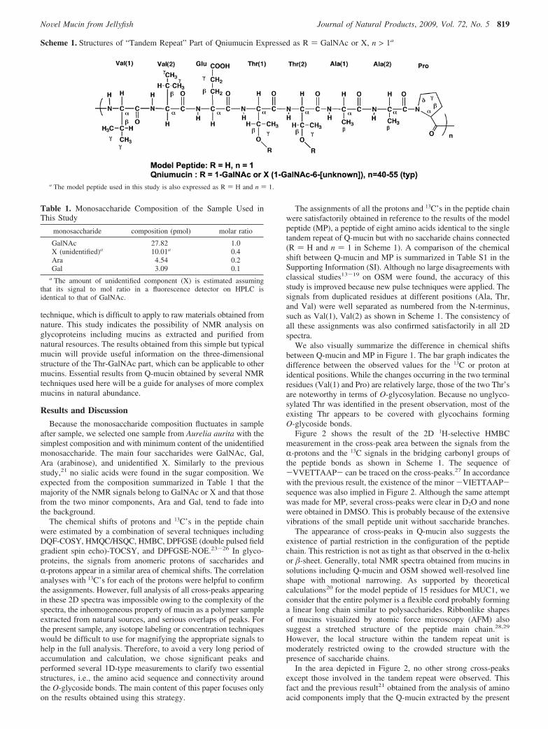
We also visually summarize the difference in chemical shiftsbetween Q-mucin and MP in Figure 1. The bar graph indicates thedifference between the observed values for the 13C or proton atidentical positions. While the changes occurring in the two terminalresidues (Val(1) and Pro) are relatively large, those of the two Thr’sare noteworthy in terms of O-glycosylation. Because no unglyco-sylated Thr was identified in the present observation, most of theexisting Thr appears to be covered with glycochains formingO-glycoside bonds.
Figure 2 shows the result of the 2D 1H-selective HMBCmeasurement in the cross-peak area between the signals from theR-protons and the 13C signals in the bridging carbonyl groups ofthe peptide bonds as shown in Scheme 1. The sequence of-VVETTAAP- can be traced on the cross-peaks.27 In accordancewith the previous result, the existence of the minor -VIETTAAP-sequence was also implied in Figure 2. Although the same attemptwas made for MP, several cross-peaks were clear in D2O and nonewere obtained in DMSO. This is probably because of the extensivevibrations of the small peptide unit without saccharide branches.
The appearance of cross-peaks in Q-mucin also suggests theexistence of partial restriction in the configuration of the peptidechain. This restriction is not as tight as that observed in the R-helixor �-sheet. Generally, total NMR spectra obtained from mucins insolutions including Q-mucin and OSM showed well-resolved lineshape with motional narrowing. As supported by theoreticalcalculations20 for the model peptide of 15 residues for MUC1, weconsider that the entire polymer is a flexible cord probably forminga linear long chain similar to polysaccharides. Ribbonlike shapesof mucins visualized by atomic force microscopy (AFM) alsosuggest a stretched structure of the peptide main chain.28,29
However, the local structure within the tandem repeat unit ismoderately restricted owing to the crowded structure with thepresence of saccharide chains.
In the area depicted in Figure 2, no other strong cross-peaksexcept those involved in the tandem repeat were observed. Thisfact and the previous result21 obtained from the analysis of aminoacid components imply that the Q-mucin extracted by the present
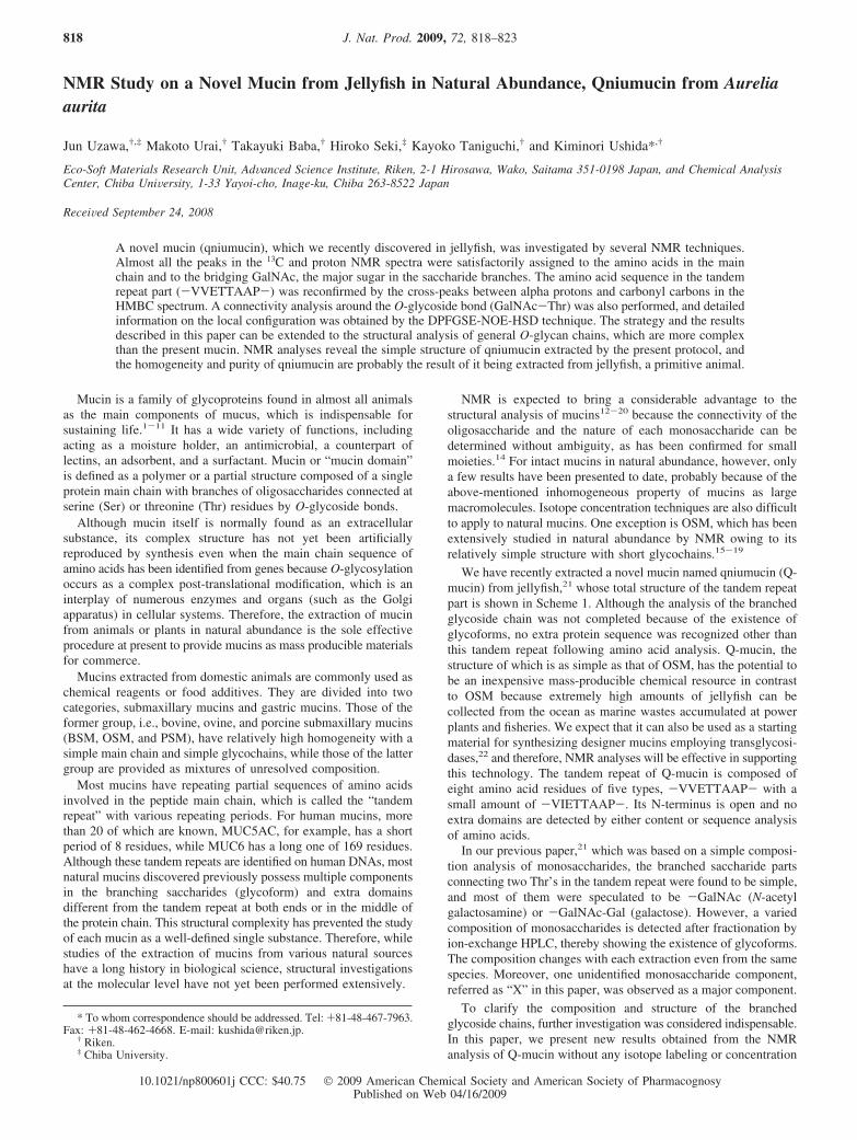
Scheme 1. Structures of “Tandem Repeat” Part of Qniumucin Expressed as R ) GalNAc or X, n > 1a
a The model peptide used in this study is also expressed as R ) H and n ) 1.
Table 1. Monosaccharide Composition of the Sample Used inThis Study
monosaccharide composition (pmol) molar ratio
GalNAc 27.82 1.0X (unidentified)a 10.01a 0.4Ara 4.54 0.2Gal 3.09 0.1
a The amount of unidentified component (X) is estimated assumingthat its signal to mol ratio in a fluorescence detector on HPLC isidentical to that of GalNAc.
NoVel Mucin from Jellyfish Journal of Natural Products, 2009, Vol. 72, No. 5 819
protocol is composed of only the sequential tandem repeat partdespite a wide distribution of molecular mass as previously shown.21
The results of Edman degradation in our previous paper21 showedthat the N-terminus started from the sequence of the tandem repeat.Moreover, we attempted to stain Q-mucin using general protocolsavailable for proteins, i.e., Coomassie brilliant blue (CBB) and silverstaining methods, which were unsuccessful, indicating the lack ofsensitive moieties for these methods in the total sequence. All theseresults support our speculation that Q-mucin is composed of amonotonic sequence of tandem repeat units. Different from themajority of mucins discovered to date, Q-mucin has an exceptionallyhigh homogeneity, which is an advantage if it is to be used as anindustrial material.
The simple structure of Q-mucin shows a sharp contrast to thoseof higher metazoa such as mammals. The apparent difference isthe lack of extra domains such as von Willebrand D (VWD) andcysteine-knot domains, which are responsible for the oligomeriza-tion of mucin molecules.30 Recent bioinformatic analysis suggests
that gel-forming mucins appeared early in metazoan evolution basedon the presence of their genes in a starlet sea anemone, whichbelongs to the same division, Cnidaria, as jellyfish.31 Therefore,without any information about the genome of the present jellyfish,we speculate that Q-mucin also belongs to the gel-forming mucinsas well.
For the NMR assignments of protons and 13C’s involved in thesaccharides, only those of GalNAc were clearly identified as shownin Table S2. Although two types of GalNAc were recognized, theyare found to connect with different threonines, Thr(1) and Thr(2)(Vide infra). The question is why we cannot identify any signalsfrom X separately. At this moment, we consider two possibilities:(1) X gives an NMR spectrum similar to that of GalNAc and (2)a large difference in relaxation time exists between GalNAc andX. We will discuss this at the end of the section.
Next, the connectivity between Thr and GalNAc in the glyco-branches was confirmed using various NMR techniques. A con-ventional HMBC measurement showed small cross-peaks for the
Figure 1. Bar graph indicating the difference in chemical shifts of various carbon-13 and protons in amino acid residues between modelpeptide (MP) and qniumucin (Q-mucin) (∆ ) δQ-mucin - δMP). Values in residues at both ends (Val(1) and Pro) and central Thr(1) andThr(2) show large differences.
Figure 2. 2D 1H selective HMBC spectrum of Q-mucin showing the interactions between R-protons and carbonyl 13C’s on the peptidebonds. The amino acid sequence -VVETTAAP- in the tandem repeat can be traced by connecting each correlation peak. A small amountof -VIETTAAP- sequence is also revealed.
820 Journal of Natural Products, 2009, Vol. 72, No. 5 Uzawa et al.
�-carbon of Thr versus H-1 in GalNAc and those for the �-protonof Thr versus C1 in GalNAc, indicating that both Thr residues areindependently coupled with a single GalNAc. All the NOEsobserved in the DPFGSE-NOE spectrum (2D NOESY spectrum isshown in Figure S3 in the SI) showed negative values. An NOEwas observed from the H-1 signal of GalNAc(1) to the H signal at4.38 ppm, which is assigned to the �-proton of Thr(1) on the basisof the results of the TOCSY experiment. Similarly, an NOE betweenthe H-1 signal of GalNAc(2) and the �-proton of Thr(2) was alsodetected. Each Thr has a methyl group, the proton of which showedNOEs with its own R-proton, its �-proton, and H-1 of GalNAc. Ofnote is the existence of the methyl group in Thr (in contrast toSer), which greatly helped us to trace the NOE through theO-glycoside bonds because the relaxation times of their protonsare appropriately short and close to that of the H-1 of GalNAc.
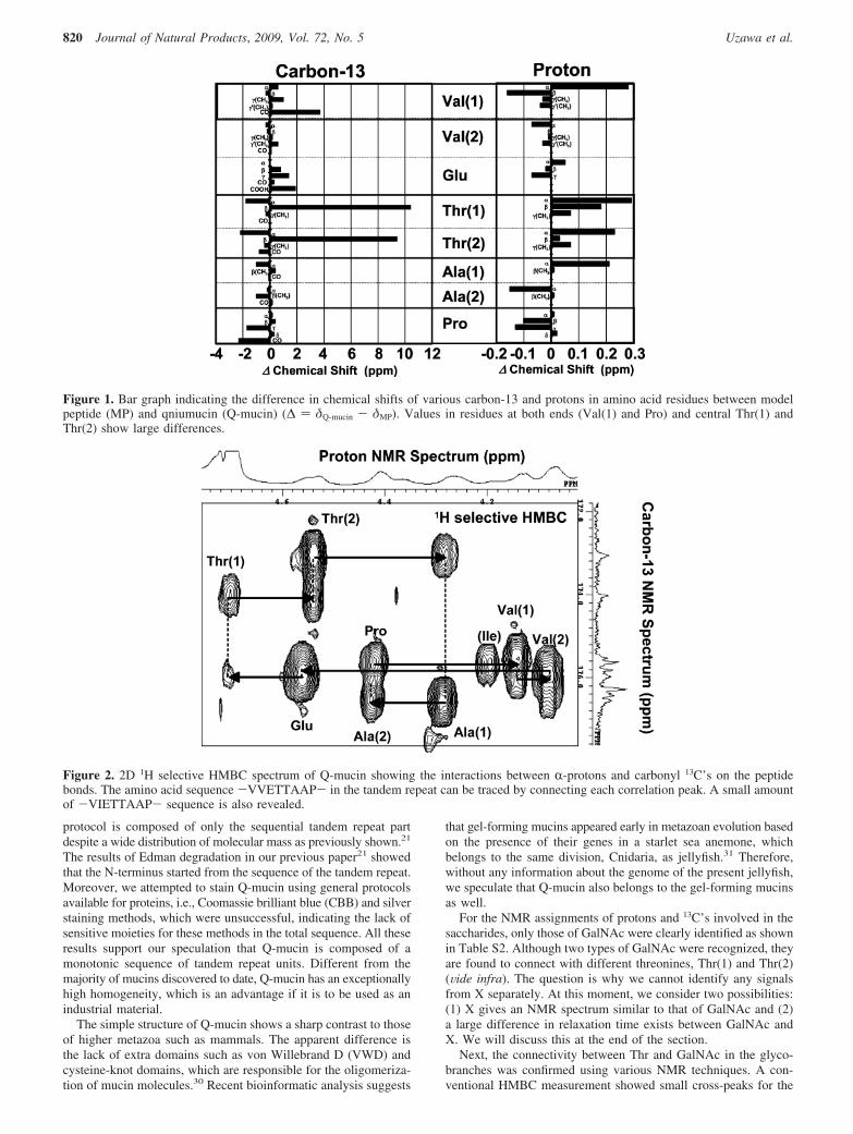
In the conventional 2D HMBC spectrum, however, several dayswere necessary to obtain only a minimally informative result, inwhich the two Thr-GalNAc pairs were not separated. To furtherconfirm the Thr-GalNAc connectivity, a type of 1D measurement,DPFGSE-NOE-HSD,32 was performed, as shown in Figure 3. Underthe experimental condition where NOE was observed between H-1and the �-proton, the protons in each methyl group were indepen-dently decoupled, resulting in narrowing of a single peak of theadjacent �-proton. Consistently, the spin coupling constants betweenR- and �-protons have been reported to be less than 2 Hz in theliterature.33 Because all the information was obtained in severalhours with this strategy, this approach is generally convenient foranalyzing unknown samples of O-glycans including mucins. Theresults of connectivity analyses are summarized in Figure 4 with amolecular structure. This is the first confirmation for Q-mucin thatthe connecting carbon of GalNAc to Thr is C1.
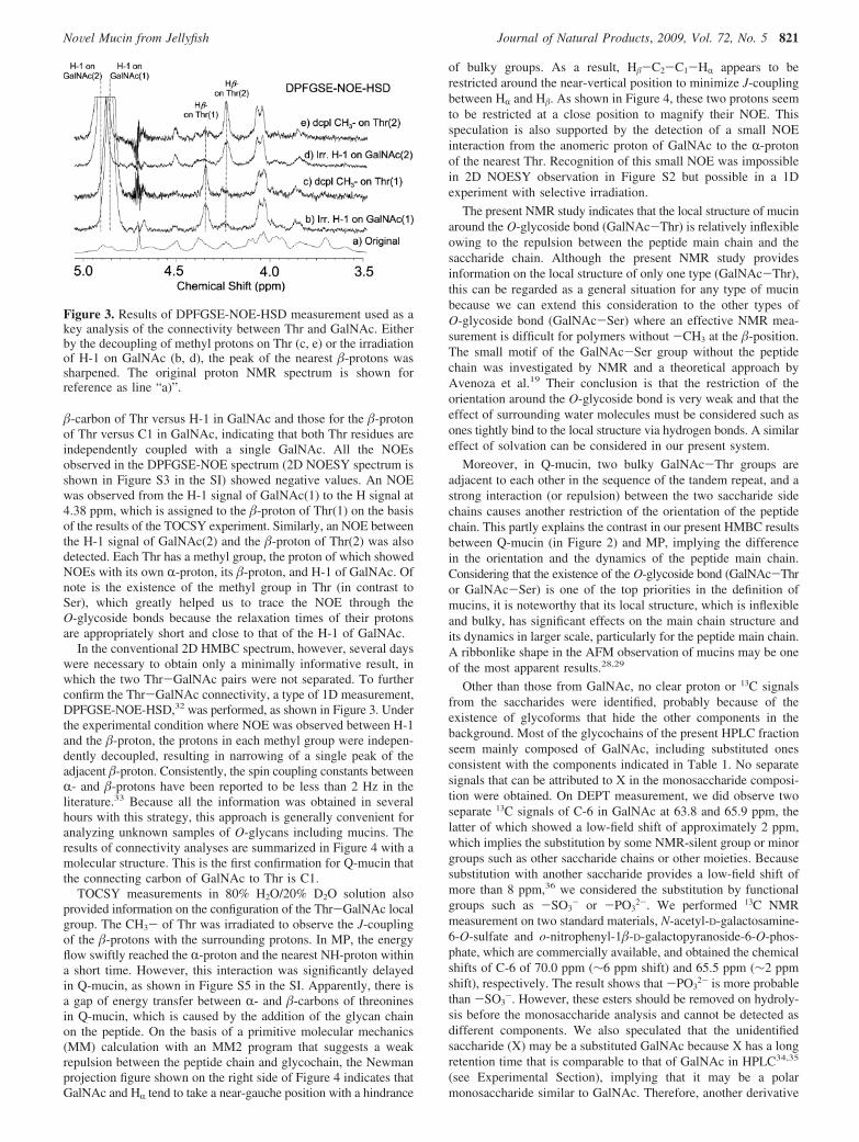
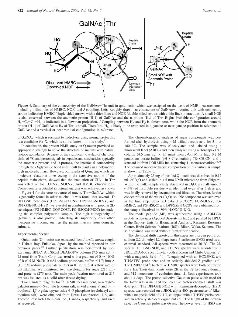
TOCSY measurements in 80% H2O/20% D2O solution alsoprovided information on the configuration of the Thr-GalNAc localgroup. The CH3- of Thr was irradiated to observe the J-couplingof the �-protons with the surrounding protons. In MP, the energyflow swiftly reached the R-proton and the nearest NH-proton withina short time. However, this interaction was significantly delayedin Q-mucin, as shown in Figure S5 in the SI. Apparently, there isa gap of energy transfer between R- and �-carbons of threoninesin Q-mucin, which is caused by the addition of the glycan chainon the peptide. On the basis of a primitive molecular mechanics(MM) calculation with an MM2 program that suggests a weakrepulsion between the peptide chain and glycochain, the Newmanprojection figure shown on the right side of Figure 4 indicates thatGalNAc and HR tend to take a near-gauche position with a hindrance
of bulky groups. As a result, H�-C2-C1-HR appears to berestricted around the near-vertical position to minimize J-couplingbetween HR and H�. As shown in Figure 4, these two protons seemto be restricted at a close position to magnify their NOE. Thisspeculation is also supported by the detection of a small NOEinteraction from the anomeric proton of GalNAc to the R-protonof the nearest Thr. Recognition of this small NOE was impossiblein 2D NOESY observation in Figure S2 but possible in a 1Dexperiment with selective irradiation.
The present NMR study indicates that the local structure of mucinaround the O-glycoside bond (GalNAc-Thr) is relatively inflexibleowing to the repulsion between the peptide main chain and thesaccharide chain. Although the present NMR study providesinformation on the local structure of only one type (GalNAc-Thr),this can be regarded as a general situation for any type of mucinbecause we can extend this consideration to the other types ofO-glycoside bond (GalNAc-Ser) where an effective NMR mea-surement is difficult for polymers without -CH3 at the �-position.The small motif of the GalNAc-Ser group without the peptidechain was investigated by NMR and a theoretical approach byAvenoza et al.19 Their conclusion is that the restriction of theorientation around the O-glycoside bond is very weak and that theeffect of surrounding water molecules must be considered such asones tightly bind to the local structure via hydrogen bonds. A similareffect of solvation can be considered in our present system.
Moreover, in Q-mucin, two bulky GalNAc-Thr groups areadjacent to each other in the sequence of the tandem repeat, and astrong interaction (or repulsion) between the two saccharide sidechains causes another restriction of the orientation of the peptidechain. This partly explains the contrast in our present HMBC resultsbetween Q-mucin (in Figure 2) and MP, implying the differencein the orientation and the dynamics of the peptide main chain.Considering that the existence of the O-glycoside bond (GalNAc-Thror GalNAc-Ser) is one of the top priorities in the definition ofmucins, it is noteworthy that its local structure, which is inflexibleand bulky, has significant effects on the main chain structure andits dynamics in larger scale, particularly for the peptide main chain.A ribbonlike shape in the AFM observation of mucins may be oneof the most apparent results.28,29
Other than those from GalNAc, no clear proton or 13C signalsfrom the saccharides were identified, probably because of theexistence of glycoforms that hide the other components in thebackground. Most of the glycochains of the present HPLC fractionseem mainly composed of GalNAc, including substituted onesconsistent with the components indicated in Table 1. No separatesignals that can be attributed to X in the monosaccharide composi-tion were obtained. On DEPT measurement, we did observe twoseparate 13C signals of C-6 in GalNAc at 63.8 and 65.9 ppm, thelatter of which showed a low-field shift of approximately 2 ppm,which implies the substitution by some NMR-silent group or minorgroups such as other saccharide chains or other moieties. Becausesubstitution with another saccharide provides a low-field shift ofmore than 8 ppm,36 we considered the substitution by functionalgroups such as -SO3
- or -PO32-. We performed 13C NMR
measurement on two standard materials, N-acetyl-D-galactosamine-6-O-sulfate and o-nitrophenyl-1�-D-galactopyranoside-6-O-phos-phate, which are commercially available, and obtained the chemicalshifts of C-6 of 70.0 ppm (∼6 ppm shift) and 65.5 ppm (∼2 ppmshift), respectively. The result shows that -PO3
2- is more probablethan -SO3
-. However, these esters should be removed on hydroly-sis before the monosaccharide analysis and cannot be detected asdifferent components. We also speculated that the unidentifiedsaccharide (X) may be a substituted GalNAc because X has a longretention time that is comparable to that of GalNAc in HPLC34,35
(see Experimental Section), implying that it may be a polarmonosaccharide similar to GalNAc. Therefore, another derivative
Figure 3. Results of DPFGSE-NOE-HSD measurement used as akey analysis of the connectivity between Thr and GalNAc. Eitherby the decoupling of methyl protons on Thr (c, e) or the irradiationof H-1 on GalNAc (b, d), the peak of the nearest �-protons wassharpened. The original proton NMR spectrum is shown forreference as line “a)”.
NoVel Mucin from Jellyfish Journal of Natural Products, 2009, Vol. 72, No. 5 821
of GalNAc, which is resistant to hydrolysis using normal protocols,is a candidate for X, which is still unknown in this study.37
In conclusion, the present NMR study on Q-mucin provided anappropriate strategy to solve the structure of mucins with naturalisotope abundance. Because of the significant overlap of chemicalshifts of 13C and proton signals in peptides and saccharides, typicallythe anomeric protons and R-protons, the interfacial connectivitythrough the O-glycoside bond is difficult to clarify in a polymer ofhigh molecular mass. However, our results of Q-mucin, which hasmoderate relaxation times owing to the extensive motion of thepeptide main chain, showed that the irradiation of CH3- in Thrwas effective for TOCSY, NOESY, and HMBC observations.Consequently, a detailed structural analysis was achieved as shownin Figure 4 for the core structure of mucin, Thr-GalNAc, whichis generally found in other mucins. It was found that several 1DDPFGSE techniques (DPFGSE-TOCSY, DPFGSE-NOESY, andDPFGSE-NOE-HSD) were useful in combination with popular 2Dtechniques (FG-HMBC, DQF-COSY, FG-HSQC/HMQC) for study-ing the complex polymeric samples. The high homogeneity ofQ-mucin is also proved, indicating its superiority over otherinexpensive mucins, such as the gastric mucins from domesticanimals.
Experimental Section
Qniumucin (Q-mucin) was extracted from Aurelia aurita caughtin Hakata Bay, Fukuoka, Japan, by the method reported in ourprevious paper.21 Further purification was performed by ion-exchange HPLC. A TSKgel DEAE-5PW column (7.5 mm i.d. ×75 mm) from Tosoh Corp. was used with a gradient of 0 f 100%of B (0.5 M NaCl/10 mM sodium phosphate buffer, pH 7) into A(10 mM sodium phosphate buffer) in 0-20 min at a flow rate of0.5 mL/min. We monitored two wavelengths for sugar (215 nm)and proteins (275 nm). The main peak fraction monitored at 215nm was isolated as a solid after freeze-drying.
Two standard reagents for 13C NMR measurement, N-acetyl-D-galactosamine-6-O-sulfate (sodium salt, mixed anomers) and o-ni-trophenyl-1�-D-galactopyranoside-6-O-phosphate (cyclohexyl am-monium salt), were obtained from Dexta Laboratories, UK, andToronto Research Chemicals Inc., Canada, respectively, and usedas received.
The chromatographic analysis of sugar components was per-formed after hydrolysis using 4 M trifluoroacetic acid for 3 h at100 °C. The sample was N-acetylated and labeled using afluorescent label (ABEE) and then analyzed using a Honenpak C18column (4.6 mm i.d. × 75 mm) from J-Oil Mills Inc., 0.2 Mpotassium borate buffer (pH 8.9) containing 7% CH3CN, and astandard kit from J-Oil Mills Inc. containing 11 monosaccharides.34,35
The obtained monosaccharide composition of this particular sampleis shown in Table 1.
Approximately 25 mg of purified Q-mucin was dissolved in 0.12mL of D2O and sealed in a 5 mm NMR microtube from Shigemi.While the bulk sample easily dissolved in D2O, a small amount(<5%) of insoluble residue was identified even after 7 days andwas finally removed by decantation and filtration. Therefore, furtherconcentration of the water (D2O)-soluble component was achievedin the final step. Some 2D data (FG-COSY, FG-NOESY, FG-HMBC, and FG-HSQC) and DPFGSE-TOCSY were obtained fromthe sample dissolved in 80% H2O/20% D2O.
The model peptide (MP) was synthesized using a ABI433Apeptide synthesizer (Applied Biosystems Inc.) and purified by HPLCat the Support Unit for Biomaterial Analysis, Research ResourceCenter, Brain Science Institute (BSI), Riken, Wako, Saitama. TheMP obtained was used without further purification.
The chemical shifts reported in this paper are those in ppm fromsodium 2,2-dimethyl-l,2-silapentane-5-sulfonate (DSS) used as anexternal standard. All spectra were measured at 30 °C. The 2Dspectra, DPFGSE-NOE, and TOCSY spectra were recorded on aJEOL ECA-600 spectrometer (both at Riken and Chiba University),with a magnetic field of 14 T, equipped with an HCX5FG2 andTH5ATFG probe head and an actively shielded Z-gradient coil.The HMBC and 1H-selective HMBC spectra were both optimizedfor 8 Hz. Their data points were 2K in the F2 frequency domainand 512 increments of evolution time, t1. Both experiments tookabout 4 days. The proton-selective Gaussian pulse width used forthe latter was 4 ms, and the selective proton chemical shift was4.43 ppm. The DPFGSE NOE with homospin-decoupling (HSD)spectra was recorded on a JEOL alpha-400 spectrometer of Rikenwith a magnetic field of 9.4 T, equipped with a TH5FG probe headand an actively shielded Z-gradient coil. The length of the proton-selective Gaussian pulse was 66 ms. The power level for HSD was
Figure 4. Summary of the connectivity of the GalNAc-Thr unit in qniumucin, which was assigned on the basis of NMR measurements,including indications of HMBC, NOE, and J-coupling. Left: Roughly drawn stereostructure of GalNAc-threonine unit with connectingarrows indicating HMBC (single-sided arrows with a thick line) and NOE (double-sided arrows with a thin line) interactions. A small NOEis also observed between the anomeric proton (H-1) of GalNAc and the R-proton (HR) of Thr. Right: Probable configuration aroundH�-C2-C1-HR is indicated in a Newman projection. J-Coupling between HR and H� is almost zero, while the NOE from the anomericproton (H-1) of GalNAc to HR of Thr is small. Therefore, HR is likely to be restricted in a gauche or near-gauche position in reference toGalNAc and a vertical or near-vertical configuration in reference to H�.
822 Journal of Natural Products, 2009, Vol. 72, No. 5 Uzawa et al.
the same as that of the original 1D HSD condition (8 kHz time-sharing decoupling). Mixing time in NOE was 400 ms. Repetitionrates were 4-5 s (acquisition time + pulse delay). The periodneeded to complete the total experiment was 50 min under eachcondition.
MM2 calculations were performed with a ChemOffice programon a personal computer.
Acknowledgment. We are indebted to Ms. R. Simizu and Ms. T.Momma for their help in extracting mucin from jellyfish. We also thankMs. Y. Kubota of Microbial Chemistry Research Center, Tokyo, forher technical assistance in NMR analyses. This research is partlysupported by Grants-in-Aid for Scientific Research (Kakenhi) No.17034067 in Priority Area “Molecular Nano Dynamics”, No. 17300166,No. 17651051, and No. 19590002 from the Ministry of Education,Culture, Sports, Science and Technology (MEXT) of Japan. Theresearch is also supported by the project to develop “innovative seeds”(Creation and Support Program for Start-ups from Universities) of theJapanese Science and Technology Agency.
Supporting Information Available: Table of assigned chemicalshifts, full 2D-NMR DQF-COSY, FG-NOESY, FG-HMBC, and FG-HSQC spectra, and the experimental results of DPFGSE-TOCSY areprovided. This material is available free of charge via the Internet athttp://pubs.acs.org.
References and Notes
(1) Taylor, M. E.; Drickamer, K. Introduction to Glycobiology; OxfordUniversity Press: London, 2003.
(2) Corfield, A. P., Ed. Glycoprotein Methods and Protocols, The Mucins(Methods in Molecular Biology); Humana Press, 2000.
(3) Sheehan, J. Biochem. Soc. Trans. 1995, 23, 795–851.(4) Corfield, A. P.; Carroll, D.; Myerscough, N.; Probert, C. S. J. Front.
Biosci. 2001, 6, d1321–1357.(5) Davis, B. G. Chem. ReV. 2002, 102, 579–601.(6) Hang, H. C.; Bertozzi, C. R. Bioorg. Med. Chem. 2005, 13, 5021–
5034.(7) Vliegenthart, J. F. G. FEBS Lett. 2006, 580, 2945–2950.(8) Singh, P. K.; Hollimgsworth, M. A. Trends Cell Biol. 2006, 16, 467–
476.(9) Harttrup, C. L.; Gendler, S. J. Annu. ReV. Physiol. 2008, 70, 431–
457.(10) Thronton, D. J.; Rousseau, K.; McGuckin, M. A. Annu. ReV. Physiol.
2008, 70, 459–486.(11) Linden, S. K.; Sutton, P.; Karlsson, N. G.; Korolik, V.; McGucklin,
M. A. Mucosal Immunol. 2008, 1, 183–197.(12) Gerken, T. A.; Dearborn, D. G. Biochemistry 1984, 23, 1485–1497.(13) Gerken, T. A. Arch. Biochem. Biophys. 1986, 247, 239–253.
(14) Gerken, T. A.; Butenhof, K. J.; Shogren, R. L. Biochemistry 1989,28, 5536–5543.
(15) Gerken, T. A.; Gupta, R.; Jentoft, N. Biochemistry 1992, 31, 639–648.
(16) Gerken, T. A.; Jentoft, N. Biochemistry 1987, 26, 4689–4699.(17) Live, D. H.; Williams, L. J.; Kuduk, S. D.; Schwarz, J. B.; Glunz,
P. W.; Chen, X.-T.; Sames, D.; Ajay Kumar, R.; Danishefsky, S. J.Proc., Natl. Acad. Sci. 1999, 96, 3489–3493.
(18) Coltart, D. M.; Royyuru, A. K.; Williams, L. J.; Glunz, P. W.; Sames,D.; Kuduk, S. D.; Schwarz, J. B.; Chen, X.-T.; Danishefsky, S. J.;Live, D. H. J. Am. Chem. Soc. 2002, 124, 9833–9844.
(19) Corzana, F.; Busto, J. H.; Jimenez-Oses, G.; Asensio, J. L.; Jimenez-Barbero, J.; Peregrina, J. M.; Avenoza, A. J. Am. Chem. Soc. 2006,128, 14640–14648.
(20) Kirnarsky, L.; Prakash, O.; Vogen, S. M.; Nomoto, M.; Hollingworth,M. A.; Sherman, S. Biochemistry 2000, 39, 12076–12082.
(21) Masuda, A.; Baba, T.; Dohmae, N.; Yamamura, M.; Wada, H.; Ushida,K. J. Nat. Prod. 2007, 70, 1089–1093.
(22) Taniguchi, N., Honke, K., Fukuda, M., Eds. Handbook of Glycosyl-transferases and Related Genes; Springer-Verlag: Tokyo, 2002.
(23) Hwang, T. L.; Shaka, A. J. J. Magn. Reson. 1995, A112, 275–279.(24) Stott, K.; Stonehouse, J.; Keeler, J.; Hwang, T. L.; Shaka, A. J. J. Am.
Chem. Soc. 1995, 117, 4199–4200.(25) Roumestand, C.; Mutzenhardt, P.; Delay, C.; Canet, D. Magn. Reson.
Chem. 1996, 34, 807–814.(26) Gradwell, M.; Kogelberg, H.; Frankel, T. A. J. Magn. Reson. 1997,
124, 267–270.(27) Bax, Ad.; Farley, K. A.; Walker, G. S. J. Magn. Reson. 1996, A119,
134–138.(28) McMaster, T.; Berry, M.; Corfield, A; Miles, M. Biophys. J. 1999,
77, 533–541.(29) Round, A. N.; McMaster, T. J.; Miles, M. J.; Corfield, A. P.; Berry,
M. Glycobiology 2007, 17, 578–585.(30) Perez-Vilar, J.; Hill, R. L. J. Biol. Chem. 1999, 274, 31751–31754.(31) Lang, T.; Hansson, G. C.; Samuelsson, T. Proc. Natl. Acad. Sci. U.S.A.
2007, 104, 16209–16214.(32) Uzawa, J.; Fujimoto, Y.; Yoshida, S. Magn. Reson. Chem. 2006, 44,
45–53.(33) Miura, Y.; Yamamoto, Y.; Inoue, Y.; Chujo, R. Int., J. Biol. Macromol.
1992, 14, 242–248.(34) Yasuno, S.; Murata, T.; Kokubo, K.; Yamaguchi, T.; Kamei, M. Biosci.
Biotech. Biochem. 1997, 61, 1944–1946.(35) Yasuno, S.; Kokubo, K.; Kamei, M. Biosci. Biotech. Biochem. 1999,
63, 1353–1359.(36) Guerardel, Y.; Balanzino, L.; Maes, E.; Leroy, Y.; Coddeville, B.;
Oriol, R.; Strecker, G. Biochem. J. 2001, 357, 167–182.(37) Recently, during revision of this paper, the authors and a co-worker
achieved the determination of species X using 31P NMR and high-resolution mass spectrometry on a fractionated sample from monosac-charide analysis in Table 1. The result will appear in a separatepublication. Urai, M; Nakamura, T.; Uzawa, J.; Baba, T.; Taniguchi,K.; Seki, H.; Ushida, K. Manuscript in preparation.
NP800601J
NoVel Mucin from Jellyfish Journal of Natural Products, 2009, Vol. 72, No. 5 823