neurotrophic sesquiterpene-neolignans from magnolia obovata: structure and neurotrophic activity
TRANSCRIPT

Turokdron Vol. 48, No. 3. Pp. 371392 1992 oo4o4cml92 $3.oc&.oo
PrintedinGrcuBrimin 0 1992 Paganon Press ptc
NEUROTROPHIC SESQUITERPENE-NEOLIGNANS FROM MAGNOLIA OBOVATA: STRUCTURE AND NEUROTROPHIC ACTIVITY
Yoshiyasu FUKUYAMA,* Yukio OTOSHI, Kumi MIYOSHI, Kazuhiko NAKAMURA,
Mitsuaki KODAMA,* Masakazu NAGASAWA,a Takashi HASEGAWA,a Hiroshi OKAZAKI,a
and Michihanr SUGAWARAa
Faculty of Pharmaceutical Sciences, Tokushima Btmri University, Yamashiro-cho, Tokushima 770, Japan
aOtsuka Pharmaceutical Co., Ltd., Kagasuno, Tokushima 771-01. Japan
(Received in Japan 7 October 1991)
ABSTRACT: Novel sesquiterpene-neolignans, eudesobovatols A (1) and B (2) eudesmagnolol (3) eudes- honokiols A (4) and B (5), clovanemagnolol(6). and caryolanemagnolol(7), have been isolated from the bark of Magnolia obovutu. Their structures were elucidated to be sesquiterpenes (eudesmol, 4,4,8-trimethyltricyclo 16.3.1.02,5] dodecane-1,9diol, and clovanediol) combined through ether bond with neolignans such as obovatol, honokiol, and magnolol on the basis of spectral data, degradation, and/or synthesis. Compounds 1, 6, and 7 were found to exhibit interesting neurotrophic activity on a neuronal cell culture system derived from fetal rat hemisphere.
The bark of Magnolia obovutu or M. ofJicinulis (Magnoliaceae) has long been used as traditional medicine
for neurosis and gastrointenstinal complaints in China, Korea, and Japan. The constituents of the species have
been investigated intensively becaus of these pharmacological interests and various type of compounds has
been isolated; i.e. neolignanst-3 (magnolol, honokiol, and obovatol), terpenes4v5 (a- and j3-pinens, camphene,
bornylacetate, a-, p-, and y-eudesmols, humulene oxide, caryophyllene, caryophyllene oxide), monoterpene-
neolignans6 and isoquinoline alkaloides 718 (magnocurarine, magnoflorine) in addition to phenylpropanoids
glycoside recently isolated.9vtn While magnolol and honokiol, the major components of M. obovutu and M.
o~$cirdis were claimed to be the active principle for the central depressant effect,*1 our preliminary studies
suggested the presence of neurotrophic active substances in the title plant and extensive studies on the minor
components led to the isolation of various sesquiterpenes linked to biphenyl- or biphenylether type neolignans,
named eudesobovatols A (1) and B (2), eudeshonokiols A (3) and B (4), eudesmagnolol (5) clovanemagnolol
(6) and caryolanemagnolol (7), some of which exhibited the activities accelerating neurite sprouting and
neuronal cell network formation as well as enhancing choline acetyltransferase activity in cultured neutonal
cell derived from fetal rat hemisphere.
11~ this paper we report the full accounts of the structures and neurotrophic activities of these novel sesqui-
terpene-neolignans isolated from M. obovutu.
Results and Discussions Isolation Since the bark of M. obovutu contains large amounts of neolignans, magnolol and honokiol, it
is essential to remove these major constituents effectively for the isolation of the minor components. Thus, the
ethyl acetate-soluble portion of methanolic extract was subjected to column chromatography using silica gel and
377

378 Y. FUKUYAMA et d.
R
Neurotrophic sesquiterpene-neolignans 379
Sephadex LH-20 repeatedly to remove these neolignans. Thus obtained fractions containing sesquiterpene-
neolignans were further separated by reverse-phase low-barr column chromatography (MPLC) and/or high-
performance liquid chromatography (HPLC) to give seven new sesquiterpene-neolignans l-7 (Fig. 1).
Structure of eudesobovatols A and B12 Eudesobovatol A (1) wss obtained as a viscous oil and
showed the molecular ion peak at m h 504 in the field desorption mass spectrum (FDMS) giving the molecular
formula C+4404 in combination with t3C NMR data summarized in Table I. UV and IR spectra indicated the
presence of hydroxyl groups (vatax 3600.3530 cm-t) and aromatic ring [ ktnax 208 ( E 5800), 274 ( E 7200),
28 1 (& 6700) nm; vmax 1600,150O cm-t]. Acetylation of 1 afforded a diacetate la, while treatment of 1 with
diazomethane gave a monomethyl ether lb indicating the presence of two hydroxyl groups, one of which
should be phenolic. tH NMR spectrum analyzed with the aid of 2D DQFCOSY and C/H COSY spectra
disclosed the presence of two ally1 groups [8 (CsDgN) 3.26 (2H, d. 3=6.8 Hz), 5.03 (dd. J=l7.2,2.0 Hz),
5.10 (dd, 5=10.3, 2.0 Hz), 5.91 (ddt, 5=17.2, 10.3, 6.8 Hz) and 3.31 (2H, d. J=6.4 Hz), 5.04 (dd, J=lO.3,
2.0 Hz), 5.08 (dd, 5=17.1,2.0 Hz). 6.01 (ddt, 3=17.1. 10.3, 6.4 Hz)], two AB type aromatic protons [8
7.05 (2H. d 5=8.8 Hz) and 7.11 (2H, d, 5=8.8 Hz)], and meru coupled aromatic protons [ 8 6.84 (d, 3=2.0
Hz) and 7.02 (d. 5=2.0 Hz)] as well as four tertiary methyl groups (8 0.93, 1.42 x 3), three of which must be
located on the carbon bearing oxygen function. The 1% NMR spectrum (Table I) was analyzed by using
C/H and long-range C/H COSYs. Comparison of these data with those of known neolignans isolated from M.
obovutu. coupled with the substitution pattern of aromatic rings deduced from IH NMR spectrum, clearly
revealed that 1 consists of obovatol3 (8) and a bicyclic sesquiterpene linked each other via an ether bond In
fact, in electron inpact mass spectrum (EIMS) prominent peaks were observed at m/z 282 (base peak) and 222 corresponding to 8 and terpenoid part, respectively. Careful analysis of 1H NMR spectrum of the terpenoid
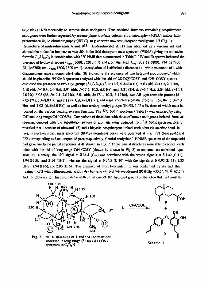
part gave rise to the partial structures A-D shown in Fig. 2. These partial structures were able to connect each
other with the aid of long-range C/H COSY (shown by arrows in Fig. 2) to construct an eudesmol type
structure. Namely, tbe 1% signal at 6 84.4 (C-4) was correlated with the proton signals at 8 1.42 (H-12), 1.94 (H-5). and 2.14 (H-3), whereas the signal at 6 34.5 (C-10) with the signals at 6 0.93 (H-11), 1.83
(H-8). 1.94 (H-5), and 2.95 (H-6). The presence of these two units in 1 was confumed by the fact that
treatment of 1 with trifluoroacetic acid in dry benzene yielded (+)-vudesmol(9) ([a]D +55.5’, lit. l3 62.5’ )
and 8 (Scheme 1). This result also revealed that one of the hydroxyl groups on the obovatol ring must be
1.01 1.29
1 .42
Fig. 2. Partial structures of 1 and C-H correlations observed in long-range (8 Hz) C/H COSY spectrum in CsDsN
7% OH
CF$OOH 9
+ OH
Scheme 1
380 Y. FLJKUYAMA et d.
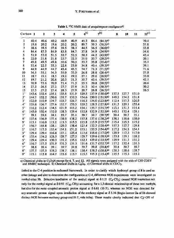
Table L *3C NMR data of sesquiterpene-neolignansa)
Carbon 1 2 3 4 5 6b) 7b) 8 10 11 12C)
1 40.4 40.6 40.2 40.9 40.9 45.3 84.4 (84.3)c) 70.4 2 19.6 20.2 19.6 20.2 20.2 89.7 39.3 (38.5)c) 37.8 3 38.6 38.5 37.6 39.5 38.3 44.5 36.5 (36.O)e) 33.8 4 84.4 87.5 84.9 83.8 84.7 37.8 34.9 (34.9)C) 34.6 5 51.9 53.0 51.5 53.7 52.5 50.3 44.2 (43.9)e) 43.4 6 22.0 21.8 21.8 22.7 22.5 21.0 20.7 (20.2)c) 20.1 7 49.8 49.5 49.8 50.6 50.5 33.2 35.8 (35.4)e) 35.1 8 22.4 22.5 22.3 22.8 23.0 34.8 40.4 (39.3)e) 39.1 9 44.9 44.9 44.7 45.4 45.5 74.7 71.3 (71.5)C) 71.6 10 34.5 35.1 34.3 35.0 35.0 26.8 28.8 (28.4)e) 27.8 11 18.7 19.1 18.5 19.2 19.2 27.1 29.6 (28.9)C) 33.0 12 19.7 21.2 20.8 20.3 21.3 35.7 40.4 (39.5)C) 42.1 13 70.9 72.6 70.9 71.4 71.5 25.5 20.8 (20.5)e) 20.6 14 27.3 26.5 27.2 27.7 27.9 31.3 30.4 (30.3)C) 30.2 15 27.5 27.2 27.4 28.3 27.9 28.7 26.8 (26.5)C) 26.5 1’ 143.6 132.4 135.1 133.8 131.9 129.5 133.9 (130.6)a) 137.5 127.7 131.0 2’ 144.7 152.2 150.6 131.7 132.3 154.6 150.0 (151.9)*) 149.2 154.2 131.8 3’ 122.0 110.9 124.7 133.7 126.7 116.3 124.0 (122.8)a) 112.4 117.3 126.9 4’ 129.6 136.7 127.6 152.7 155.5 129.2 128.5 (127.8)a) 131.3 129.1 155.4
5’ 116.2 112.4 134.5 121.9 115.2 134.1 135.7 (135.7)a) 113.1 131.3 115.4 6’ 145.0 150.4 132.0 128.3 129.4 132.8 132.9 (132.5)a) 145.1 132.4 129.1 7’ 39.1 39.8 39.1 35.7 35.1 39.7 39.7 (39.7)8) 39.4 39.7 35.1 8’ 137.6 136.9 137.4 138.0 138.2 137.8 137.6 (138.2)a) 138.1 138.6 138.0 9’ 115.1 116.0 115.2 115.3 115.3 115.8 115.9 (115.7)a) 115.4 115.3 115.2 1 I, 156.7 155.8 128.1 129.3 138.4 127.4 132.2 (128.4)a) 157.3 127.7 129.8
2” 116.7 117.5 153.4 154.1 151.2 153.1 153.5 (154.4)a) 117.2 154.2 154.1
3” 129.4 129.4 116.8 117.1 125.4 117.6 118.6 (117.O)a) 129.9 117.3 117.0 4” 133.4 134.2 128.5 128.7 127.5 129.7 129.6 (128.9)a) 133.8 129.1 128.2
5” 129.4 129.4 130.3 131.2 135.1 132.1 129.0 (133.5)a) 129.9 131.3 131.2
6” 116.7 117.5 131.9 131.3 131.3 131.6 131.7 (132.7)a) 117.2 132.4 131.3
7” 38.8 39.4 39.1 39.7 39.8 39.7 39.8 (39.8)a) 39.8 39.7 39.7
8” 137.5 137.5 138.2 138.7 138.1 138.4 138.5 (138.8)a) 138.3 138.6 138.7 9” 115.1 115.8 114.7 115.6 115.7 115.3 115.3 (115.4)a) 115.5 115.3 115.2
a) Chemical shifts in CsDsN except for 6,7, and 12. All signals were assigned with the aids of C/H COSY and HMBC technique. b) Chemical Shifts in C&j. c) Chemical shifts in CDCl3.
linked to the C-4 position in eudesmol framework. In order to clarify which hydroxyl group of 8 is used as
ether linkage and also to determine the configuration at C-4, dierence NOE experiments were investigated on
methyl ether lb. Selective irradiation of the methyl signal at 8 1.21 (C!4-CH3) caused NOE interaction not
only for the methyl signal at 6 0.92 (Cm-CH3) accounting for a L3diaxial relationship of these two methyls,
but also for the meta -coupled aromatic proton signal at 8 6.61 (H-3’), whereas no NOE was detected for
any aromatic proton signal upon irradiation of the methoxy signal at 8 3.74 (Regio-isomer 2a of lb showed
distinct NOE between methoxy group and H-3, vide infra). These results clearly indicated that Q-OH of

Neurotrophic sesquiterpene-neoligttans 381
8 was bonded to the C-4 ( u-equatorial) position of eudesmol framework. The a configuration of H-5 (6
1.94) was deduced on the basis of large coupling constant (J5,68=11.7 Hz) as well as the observation of NOE
between H-5 and H&z. Thus, the structure of eudesobovatol A was fully elucidated as 1.
Eudesobovatol B (2). viscous oil, exhibited a quasi-molecular ion peak due to [M-l]- at mlz 503 and
a base peak at m /z 282 in negative fast atom bombardment mass spectrum (FABMS) as in the case of 1. It formed a monomethyl ether 2a on treat-ment with diazomethane. The tH NMR spectrum was very similar to
that of 1 indicating that 2 has the same structure units, obovatol and eudesmol. as eudesobovatol A (1). The major difference was the methyl signal at 6 1.58 (H-12) and metu -coupled aromatic proton signals at 6 6.61
and 6.99 . The l3C NMR data of 2 (Table I) was again well corresponded to those of 1 except for the carbons
adjacent to the ether linkage, i.e. carbons-4, -1’. -2, -3, -4, -5, and -6’. Moreover, no NOE was observed
for any aromatic protons upon irradiation of C&H3 (6 1.58 ), although clear NOE was detected for the methyl
signal at 6 0.92 (Cto-CH3) indicating a 1,3-diaxal relationship of these two methyl groups as in 1. These
results revealed that 2 should be the regio isomer of 1 with respect to the position of ether linkage. This
proposal was verified by the observation of NOE for the meta -coupled proton resonance at 6 6.52 upon
irradiation of the methoxy signal at 6 3.81 in 2a. Thus, the structure of eudesobovatol B was represented as
the formula 2.
Structures of eudesmagnolo114 The molecular formula of eudesmagnolol (3) viscous oil, was
determined to be C33H4403 on the basis of FDMS (m/z 488 [Ml+) and 13C NMR data summarized in Table I
The presence of hydroxyl group and aromatic ring was again indicated by IR spectrum ( vmax 3610,3260,
1600, 1500 cm-t). Treatment of 3 with acetic anhydride in pyridine yielded a diacetate 3a. The IH NMR spectrum of 3 disclosed the presence of two 1,2,4-trisubstituted benzene rings [ 6 7.13 (dd, J=8.3, 2.0 Hz),
7.24 (d, 3=8.3 Hz), and 7.44 (d, 5=2.0 Hz); 7.19 (dd, J=8.3, 2.0 Hz), 7.26 (d, 3=8.3 Hz), and 7.35 (d, J=2.0
Hz)] and a set of ally1 groups [6 3.35 (2H, d, 3=6.8 Hz), 5.01 (dd. 5=10.3, 1.9 Hz), 5.10 (dd, J=17.1, 1.9
Hz), and 5.97 (ddt, 3=17.1, 10.3, 6.8 Hz) ; 6 3.45 (2H, d, J=6.8 Hz), 5.05 (dd, J= 10.3, 1.9 Hz), 5.14 (dd,
J=17.1, 1.9 Hz), 6.01 (ddt, J=17.1, 10.3, 6.8 Hz) in addition to a methyl group (6 0.81) on the quaternary
carbon and three methyl groups (6 1.22,1.43,1.45) on the carbon bearing oxygen function. Comparison of
tH and ‘3C NMR spectra to those of 1 and 2 revealed that 3 consists of eudesmol and a different type of
neolignan combined through an ether linkage. Since the FDMS of 3 showed a prominent ion peak at m/z
266 and the t3C NMR data were well corresponded (Table l), the neolignan incorporated in 3 was determined
to be magnololl (lo), one of the major component of M. obovntu. In fact, treatment of 3 with CF3COOH
afforded (+)-9 ( [o]D +55.5’ ) and 10 (Scheme 2). The configuration at C-4 in eudesmol part was determined
to be R since NOE was observed between Cd-CH3 and CIO-CH~ giving the full structure of eudesmagnolol(3).
H,O 0,H
3 -9+/\ /\ CF$OOH
P-Q - -
Scheme 2

382 Y, FuKUYA~~A et al.
Structure of eudeshonokiols AAl and B The molecular weight of eudeshonokiol A (4) was determined
as 488 by negative- (m /z 487 [M-l]-) and positive FABMS (m/z 511 [M+Na]+). The IR spectrum revealed
the presence of hydroxyl group (vatax 3670,355O cm-l) and aromatic ring (vatax 1600,148O cm-l). Treatment
of 4 with diazomethane afforded a monomethyl ether 4a. 1H and 13C NMR spectra were found to be similar to
those of 3, which suggested that 4 consists of eudesmol-type sesquiterpene and biphenyl-type neolignan.
However, the 1H NMR spectrum of aromatic unit was apparently different from that of 10. particularly
appearance of benzylic methylene protons as nonequivalent AB-type pattern [S 3.55 (dd, J~15.4.6.6 Hz) and
8 3.71 (dd, J=15.4, 6.6 Hz)]. These facts suggest that honokiol (11). a component of M. obovara., is
incorporated in 4. In fact, reaction of 4 with CF$OOH afforded (+)-9 ( [aID +45.3’ ) and honokiol (11) (Scheme 3). The Q-OH of the latter associated with an ether bond was verified by the observation of NOES
in 4a on H-l 1 (6 0.94 ) and H-S (8 7.01) upon irradiation of H-12 (6 1.33 ) and on H-3” (8 6.90) upon
irradiation of the methoxy signal (8 3.77 ) and was confirmed by the acid treatment of 4a to form 2-O -methyl-
honokiolts (11s) (Scheme 3) which has been isolated from the title plant by us and also derived from 11.
The UV and IR spectra and the pattern of 1H and 13C NMR (see Table I) spectra of eudeshonokiol B (5) were
very similar to those of 4 . Thus, it can be assumed readily that 5 is the regio-isomer of 4 in respective of the
hydroxyl group of 11 associated with the ether bond. This assumption was supported by the observation of the following NOES: C!4-CH3 (6 1.20 ) /one of the ortho -coupled aromatic protons (6 7.24; H-3”) in 5,OCH3
(6 3.86) /another ortho --coupled aromatic protons (6 6.87; H-5’) in methoxy derivative 5n. Finally, the
structure was confirmed by the cleavage of Sa with CF$!OOH to yield 4-O -methylhonokiol (llb) (Scheme
3), isolated from Magnolia grandiflora .15
OR
4,4a or Sa CF$OOH
+9+
11, R=R’=H lla, R=Me, R’=H
Scheme 3 llb; R=H, R’=Me
Structure of clovanemagnolol 16 The molecular formula of clovanemagnolol (6) was determined as
C33H~03 by high resolution EI mass spectrum (HREIMS) (m/z 486.3127) and its tH NMR spectrum
indicated the presence of an aromatic and sesquiterpene moieties. The aromatic part was readily assigned as
magnolol (10) since the ‘H NMR revealed the presence of two ally1 groups and two 1,2,4_trisubstituted
aromatic rings as well as the close similarity of t3C NMR data between them. However, the spectral data of
the terpene part were totally different from those of above-mentioned eudesmol-type structure. The DEPT
spectrum of 6 displayed the presence of fifteen carbons consisted of three methyl, six methylene, one methine,
1The name, eudeshonokiol, previously reported for 414 has now been corrected to eudeshonokiol A because
of the isolation of closely related compound (eudeshonokiol B) later on.
Neurotrophic sesquiterpene-neolignans 383
two oxygen bearing methine, and three quatemary carbons (Table I), wherease tH NMR spectrum (C&j)
contained three tertiary methyl signals (6 0.64,0.84, and 0.94) and two oxygenated methine signals (6 3.07
and 4.11). Acetylation of 6 to diacetate 6a caused a large down-field shift (A6 1.42) of the higher-field
proton signal (6 3.07 ). which indicated that the proton appeared at lower-field should be attached to the
carbon bearing ether bond Analysis of DQFCOSY and C/H CQSY spectra revealed that these two carbinyl
protons were involved in partial structures A and B, respectively, and the additional partial structures C
and an isolated methylene group were present in 6 in addition to three tertiary methyls. These partial structures
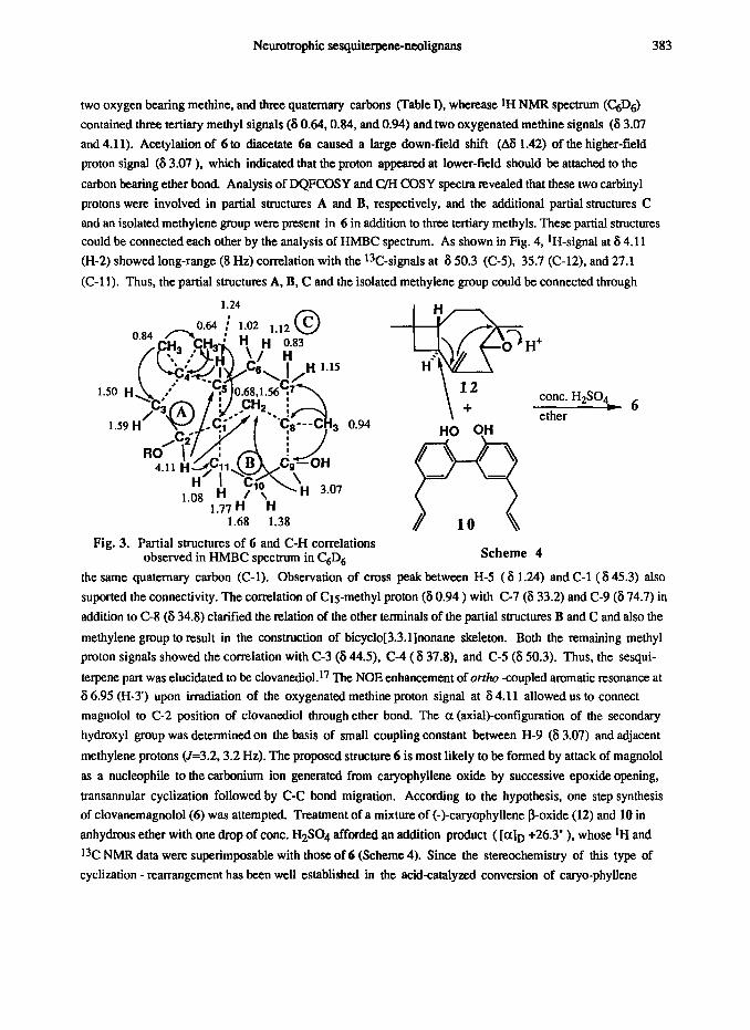
could be connected each other by the analysis of HMBC spectrum. As shown in Fig. 4. ‘H-signal at 6 4.11
(H-2) showed long-range (8 Hz) correlation with the %-signals at 6 50.3 (C-5), 35.7 (C-12), and 27.1
(C-l 1). Thus, the partial structures A, B, C and the isolated methylene group could be connected through
1.24
Fig. 3. Partial structures of 6 and C-H correlations observed in HMBC spectrum in C,D, Scheme 4
the same quaternary carbon (C-l). Observation of cross peak between H-5 (8 1.24) and C-l (6 45.3) also
suported the connectivity. The correlation of CtS-methyl proton (6 0.94 ) with C-7 (6 33.2) and C-9 (6 74.7) in
addition to C-8 (6 34.8) clarified the relation of the other terminals of the partial structures B and C and also the
methylene group to result in the construction of bicyclo[3.3.l]nonane skeleton. Both the remaining methyl
proton signals showed the correlation with C-3 (6 44.5), C-4 (6 37.8), and C-5 (6 50.3). Thus, the sesqui-
terpene part was elucidated to be clovanediol. l7 The NOE enhancement of orrho -coupled aromatic resonance at 6 6.95 (H-3’) upon irradiation of the oxygenated methine proton signal at 6 4.11 allowed us to connect
magnolol to C-2 position of clovanediol through ether bond. The a (axial)-configuration of the secondary
hydroxyl group was determined on the basis of small coupling constant between H-9 (6 3.07) and adjacent
methylene protons (J=3.2.3.2 Hz). The proposed structure 6 is most likely to be formed by attack of magnolol
as a nucleophile to the carbonium ion generated from caryophyllene oxide by successive epoxide opening,
transannular cyclization followed by C-C bond migration. According to the hypothesis, one step synthesis
of clovanemagnolol(6) was attempted. Treatment of a mixture of (-)-caryophyllene P-oxide (12) and 10 in
anhydrous ether with one drop of cont. H2SO4 afforded an addition product ( [aID +26.3’ ), whose ‘H and
t3C NMR data were superimposable with those of 6 (Scheme 4). Since the stereochemistry of this type of
cyclization - rearrangement has been well established in the acid-catalyzed conversion of caryo-phyllene
\
12 cont. H2S04
+ -6 ether

384 Y. FUKUYAMA et al.
a---_o- ._ .
Fig. 4. HMBC spectrum of 6 in ChDs
oxide to clovanediol,*8 the structure of clovanemagnolol including absolute configuration was unequivocally
established as the formula 6. In accord with the stereostructure, NOES shown in Fig. 5 were observed and p-
orientation of magnolol unit on cyclopentane ring could be confirmed
Fig. 5. NOES observed in NOESY spectrum of 6
Structure of caryolanemagnolol HREIMS of caryolanemagnolol (7), a colorless oiI, gave the
molecular formula as C+I&3 (m/z 486.3145). The presence of hydroxyl group and aromaic ring was again
indicated by IR (v, 3600,3300,1600, 1480 cm-l) and UV spectra [ A,,,ax 211(& 6800), 285 (E 8700)
nm]. The aromatic part could be easily identified as 10 because the lH NMR spectrum revealed the presence of
two 1,2,4-aisubstituted benzene rings and two ally1 groups and the lower-field t3C! NMR signals are closely
related with those of 10 (Table I) together with the observation of base ion peak at m/z 266 corresponding to

10 in EIMS. t H NMR spectrum showed the ~XWZICC of three tertiary methyls ( 6 0.7 LO.94, and 1. go) and
an oxygenated methine pmton ( 6 3.05) and was much different from those of eudesmol or clovanediol
type. On the basis of DQFCOSY and C&I aSY spectra ~XZ partial structures A and B (Fig. 6) could be
identifkd in 7 in addition to duet quaternary carbons. ‘l&se partial s&uctmes were combined with the analysis
of HMBC spectrum (Fig. 7). Namely, one of the tertiary methyl signals ( 6 0.71) was cormlated with C-7
(6 35.8) in B , a quaternary carbon C-8 (8 40.4). C-9 (6 71.3) in A, and a methylene carbon (6 40.4).
The other methyl signal (6 1.10) was correlated with the third methyl carbon ( 6 20.8), C-3 (36.5) and C-5 (8
44.2) in B, and C-4 (5 34.9). Furthermore, H-3p signal (6 2.03) had a cross peak with the oxygen-bearing
0.92n
/&+py* . .
Fig. 6. Partial structures of 7 and C-H correlations
1.85 t-i& ( 1.46 H’ “A
1.38 H’ *H 1.46
.7f
Scheme 5. Possible biosyn~etic route of - _
observed in HMBC spectrum in C,D,
Fig, 7. HMBC spectrum of 7 in QD,
386 Y. FUKUYAMA et al.
carbon C-l (6 84.4) and C-4, while H-lip signal (6 1.85) with C-l and C-2 (6 39.3). Thus obtained structure
containing four-membered ring is corresponding to the glycoll7 (12) derived from caryophyllene oxide. In fact,
13C chemical shifts of these compounds were almost same except for the carbons around C-l as can be seen in
Table I. Large down-field shift of C-l signal of 7 compared to 12 revealed that magnolol unit must be located at
this position. Observation of clear NOES between Me+3 (S 1.10) and H-5b (6 1.92) and between Me-4a (6
0.92) and H-2a (6 2.23) displayed touts nature of ring junction. The H-Q showed NOE interaction with one of
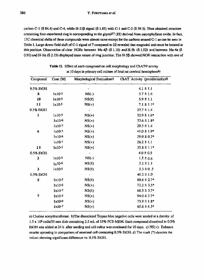
Table II. Effect of each compound on cell morphology and ChATa) actvity
at 10 days in primary cell culture of fetal rat cerebral hemisphere@
Compound Cone (M) Morphological Evaluationc) ChAT Activity (pmoi/min/dis)d)
0.5% EtOH 4.1 f 1.1
8 1x10⁻⁵ NV-1 3.7 f 1.6
10 1x10⁻⁵ NSW 5.9 f 1.1
11 1x10⁻⁵ NS(+) 7.1 f 1.1*
0.5% EtOH 23.7 f: 1.4
1 1x10⁻⁵ NV+) 32.9 f 1.6*
1x10-6 NS(+) 33.6 f. 1.8*
1x10-7 W+) 29.3 f 1.4
6 1x10⁻⁵ NS(+) 41.0 f 1.9*
1x10-6 NS(+) 29.0 f 0.2*
1x10-7 NS(+) 26.2 z!z 1.1
11 1x10⁻⁵ NS(+) 35.8 f l.l*
0.5% EtOH 4.0 + 0.5
2 1x10⁻⁵ NS(-) 1.5 f 0.6 1x10⁻⁶ NS(+) 3.2 f 1.1
3 1x10⁻⁵ NS(*) 2.3 f 0.3
0.5% EtOH 40.2 f 1.0
5 1x10⁻⁵ NSW 80.6 + 2.7*
1x10-6 NS(+) 72.2 + 3.5*
1x10-7 NS(&) 66.5 f 3.7*
7 1x10⁻⁵ NS(+) 94.0 f 3.7*
1x10-6 NS(+) 75.9 f 1.8*
1x10-7 NS(+) 65.6 + 4.3*
a) Choline acetyltransferase. b)The dissociated Trypan blue negative cells were seeded at a density of
1.5 x 10⁶ cells/35 mm dish containing 2.5 mL of 15% FCS-MEM. Each compound dissolved in 0.5%
EtOH was added at 24 h after seeding and cell cultur was continued for 10 days. c) NS(+): Enhance
neurite sprouting in comparison of neuronal cell containing 0.5% EtOH. d) The mark (*) denotes the
values showing significant difference vs. 0.5% EtOH.
Neurotrophic sesquiterpene-neolignans 387
the methylene proton (8 1.58) at C-12 indicating that the methylene bridge also has fientation. The p (sxial)-
configuration of secondary hydroxyl group was based on the small coupling constants (J=3.5,3.5 Hz) observed
between H-9 and adjacent protons. Caryolanemagnolol must be biosynthesized from (-)-caryophyhene a-oxide
(13) through acid-catalyzed epoxide opening. cyclixation followed by nucleophilic attack of magnolol (Scheme
5). Although any &finite evidence has not been obtained so far, this biogenetic implication strongly suggests
the absolute configuration of 7 as shown when one considers the co-occurence of (-)-caryophyllene in the
same plant
Effect of sesquiterpene-neolignans on primary cell culture of fetal rat cerebra1 hemisphere
NGF (nerve growth factor) and FGF (fibroblast growth factor) are well known as a neurotrophic factor to
control neurite sprouting and proliferation of neuroblast during develpoment of neurons. These neurotrophic
factors are essentiallly related to differentiation and chemotaxis of neurons, and recently expected to be possible
in medical tretment of or prevention from presbyophtenia which has increasingly caused social problems. From
this point of view, we are searching for a neurotrophic substance having NGF-lie property in natural products
and the activity of sesquiterpene-neolignans described above were investigated using a primary neuronal cell
culture derived from fetal rat hemisphere. 20 The results summarized in Table II indicated that eudesobvatol A
(l), clovanemagnolol (6). and caryolanemagnolol (7) could accelerate net&e sprouting and also increase
chloine acetyltransferase activity (ChAT)2l at the concentration of 1x10⁻⁷ M at 10 days after seeding in
comparison of control system containing 0.5 % EtGH only. Among them, caryolanemagnolol(7) was found to
be the most active substance. In contrast to these sesquiterpene-neolignans, simple biphenyl-type neolignans,
obovatol (8) and magnolol(10). have no activity even at 10-S M except for honokiol(l1). It is interesting
that these exotic substances exhibited neurotrophic property similar to that of NGF in neuronal cell culture
of fetal rat hemi-sphere. Detailed neurotrophic action caused by these compounds, however, must wait for
further biochemical study.22
Experimental Section Genaral Optical rotations were recorded in CHC13 solution on a JASCO DlP-140 polarimeter . UV spectra were measured on
a Shimadxu UV-300 spectrophotometer in ethanol soluticn. JR spectra were recorded on a HlTACHJ JR 260-10 spectrometer in
CHCl3 solution. NMR spectm were recorded on a JEOL JNM-OX400 spectrometer operating at 400 MHz for lH and 100 MHz for
13C nuclei. NOE and 2dimentionai experiments were performed on tbc same appamtus. Pyridinedg was used as solvent unless
otherwise stated. Chemical shifts are reported in ppm relative to tetmmethylsilane as internal standard and coupling constants (I) are
expressed in Hz. Mass spectra were taken on a JEOL JMS-HXIOO for HRMS and a JMS-SXlO2 for EI-, FD-, and FABMS. Merck
Kieselgel60 (70-230 mesh, 230-4OB mesh) and Wakogel C-300 were used for silica-get chromatography. Precoatcd Kieselgel 60
F254 or RP-8 F254 plates were used for analytical TLC and spots were visualixd by UV (254 nm) and 2% CeSO4 in H2S04
Extraction and isolation. (1) Sesquiterpene-neolignans 1-7. The dried bark (10 Kg) of Magnolia ovobato purchased
from Koshim Co., Ltd., in Japan was powdered and immersed at mom temperature with methanol (90 L) for 8 days. The methanol
extract was evaporated in vacua to leave the viscous residue, to which water was added. The obtained suspension was extracted three
times with ethyl acetate (EtOAc). The EtOAc-soluble portion was evaporated in vmto to dryness giving an EtOAc extract (1056 g).
550 g of which was divided to fr. 1 (350 g) and fr. 2 (185 9) by silica gel (Kiselgel60; 70 - 230 mesh, 2.96 Kg) chromatography
eluting wilh n-hcrxane-EtOAc (1:l) and CH$&MeOH (7:3). The fr. 1 (350 g) was chromatographed on silica-gel (Wakogel C-300,
5.2 Kg) widt a stepwise gradient [n-hexaneEtOAc (19~1.17 L), (14:1.15 L), (9~1.15 L), (8.5~1.5.15 L), (4:1, 15 L), (3:2.17
388 Y. FUKLNAMA et al.
I-). EtOAc (100%. 10 L), and EtOAc-MeOH (8.5:1.5, 13 L) to give frs 3 (5 g). 4 (20 g). 6 (20 g). 7 (95 g), 8 (105 g). 9 (15 g), 10
(10 g), ll(15 8). 12 (15 g), and 13 (40 g). The fr. 9 (15 g) was subjected to Sephadex LH-20 chmmatogmphy eluting with
MeOHCHgCl2 (7 : 3) to give a fraction (11 g) ccWahdng hamkiol and a fraction (3.5 9) containing sesquiterpuc+neoiignans. The
later fraction (3.5 g) was purified by MPZC [cohunn: Lobar RF’-8. type C; solvent: MeORH20 (9 : I)] to give eudesmagnolol(3)
(400 me;). caryolanemagnolol(7) (350 mg). wdeshcnokiol A (4) (200 mg) and a mixture (2 g), which was subjected to HFW
[column. Cosmosil5Cts + 10x250 mm: solvent, MeOH:CH~CN:H2Cl=62:U):8 (2.5 mumin); det., UV (254 nm)] aud the peaks
appeared at retention times 19.0,2O.O. 21.5, and 24.5 min were collected to give clovanemagnolol(6) (50 mg). eudesovobatol A (1)
(200 mg), eudesobovatol B (2) (150 mg), and eudesmagnolol(3) (350 mg), respectively. The fr. 11(15 g) was chromaqpnphed on
Sephadex LH-20 (1 L) eluting with MeOH to give three fractions. The second fraction (5.2 g) was purified by BID-BEADS fix-12
chromatography (benzene) followed by neutral alumina (CHgCIz) and silica-gel (CH2Clg-EtOAc, 91) chromamgraphies to
afford eudes-honokiol B (5) (250 mg).
(2) O-Methylhonokiols lla and llb. The EtOAc-soluble portion (140 g) was subjected to column chromatography
on silica-gel @&elgel60.70 - 230 mesh) and eluted with n-hexaneEtOAc (]:I) to give frs 1 (68 g), 2 (24 g). and 3 (4.4 g). The
fr. 1 (68 g) was chromatographii on silica-gel (wakogel C-300) with a stepwise gradient [n-hexat+EtOAc (91.14 L), (8.8:1.5.
14 L), (4:1,6 L), (2:1,4 L) and EtOAc-MeOH (91.2 L) to give frs 4 (2.5 g), 5 (2.5 g), 6 (1.0 g). 7 (4.8 g). 8 (9.5 g), 9 (21.0 g),
10 (10.7 g). 11 (0.8 g). The fr. 7 (4.8 g) was subjected to Sephadex LH-20 chromatography and eluted with MeOWCH2Cl2 (4:1)
giving four fractions. ‘lbe thiid fraction (1.3 g) was purilied by silica-gel (Wakogel C-308) chromatography (n-hexane-EtOAc, 9: 1)
followed by MPLC using Lobar RP-8 (MeOH-H20.9~1) to yield a’-0-metbylhonokiol (lla) (280 mg) and 4-0-methylhonokiol
(llb) (120 mg).
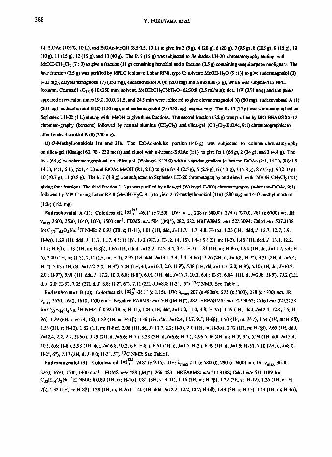
Eudesobovatol A (1); Colorless oil. bi”” D -46.1’ (c 2.50). W: aax 208 (E 58GOO). 274 (E 7200). 281 (E 6700) nm. IR:
Vmax 3600,3530, 1640.1600, 1500 cm-l. FDMS: m/r 504 ([Ml+), 282,222. HRFABMS: m/z 527.3094; Calcd m/r 527.3138
for C33H4404Na. lH NMR: 8 0.93 (3H, s; H-11). 1.01 (lH, ddd, J=11.7. 11.7.4.8; H-la), 1.23 (lH, ddd. J=12.7, 12.7, 3.9;
H-9a), 1.29 (1H. ddd,J=l1.7. 11.7.4.8; H-18). 1.42 (9H, s; H-12, 14. 15), 1.4-1.5 (2H, m; H-2). 1.48 (1H. ddd,J=l3.1. 12.2,
11.7; H-68), 1.53 (lH, m; H-8S), 1.66 (lH, dddd,J=l2.2, 12.2.3.4.3.4 ; H-7). 1.83 (1H. m; H-8a), 1.94 (lH, dd, J=ll.7, 3.4; H-
5). 2.00 (lH, m; H-3). 2.14 (lH, m; H-3). 2.95 (lH, ddd.JEl3.1, 3.4.3.4; H&r), 3.26 (2H. d, J= 6.8; H-7”), 3.31 (2H, d,J=6.4;
H-73.5.03 (IH, dd, J=l7.2.2.0; H-9”), 5.04 (lH, dd, J=lO.3,2.a; H-9’). 5.08 (lH, dd,J=17.1,2.0; H-9’), 5.10 (IH, dd.J=10.3,
2.0 ; H-9”). 5.91 (lH, ddt,J=l7.2, 10.3,6.8; H-8”) 6.01 (lH, ddt,J=l7.1, 10.3.6.4 ; H-8’) 6.84 (IH, d, J=2.0: H-53.7.02 (lH,
d, Jd.0; H-3’). 7.05 (2H, d, J~8.8; H-2”. 6”). 7.11 (2H. dJ=8.8; H-3”. 5”). 13C NMR: See Table 1.
Eudesobovatol B (2); Colorless oil. Ia]: -26.1’ (c 1.15). UV: &,,., 207 (E 48000). 273 (e 5000). 278 (e 4700) nm. IR:
v,,, 352O,1640.1610. 1500 cm-l. Negative FABMS: m/r 503 ([M-H]‘), 282. HRFABMS: m/r 527.3062; Calcd m/r 527.3138
for C33H4404Na. lH NMR: 8 0.92 (3H, s; H-l]), 1.04 (lH, ddd, J=ll.O, 11.0.4.8; H-la), 1.19 (lH, ddd, J=l2.4, 12.4.3.6; H-
9a), 1.29 (6H, s; H-14, 15). 1.29 (IH, m; H-IS), 1.38 (IH, ddd, J=12.4. 11.7,9.5; H-6/3). 1.50 (IH, m; H-7). I.54 (IH, m; H-88).
1.58 (3H, s; H-12). 1.82 (lH, m; H-8a), 2.06 (lH, dd, J=ll.7,2.2: H-5). 210 (IH. m: H-3a), 2.12 (lH, m; H-38). 2.65 (IH, ddd,
J=l2.4.2.2,2.2; H-6a). 3.25 (2H, d, Jz6.6; H-7’), 3.33 (W, d, J=6.6; H-7”). 4.%-5.06 (4H, m; H-9.9”). 5.94 (1H. ddt, J115.4,
10.3,6.6: H-8’). 5.98 (IH, ddt, J=l6.8, 10.2.6.6; H-8”). 6.61 (IH, d,J=1.5; H-3’). 6.99 (IH, d, J=1.5; H-5’). 7.10 (2H,d,J=8.0;
H-2”. 6”). 7.17 (2H, d, J=8.0; H-3”. 5”). l3C NMRz See Table I.
Eudesmagnolol (3); Colorless oil. ia]? -74.8’ (c 9.15). UV: X,,,,,x 211 (E SSOGO), 290 (e 7400) nm. IR: vmax 3610.
3260. 1650, 1500, 1400 cm-*. FDMS: m/z 488 ([Ml+), 266,223. HRFABMS: m/z 511.3188: Calcd m/r 511.3189 for
C33H4403Na. *H NMR: 8 0.80 (lH, m; H-la), 0.81 (3H, s: H-11). 1.16 (lH, m; H-18). 1.22 (3H, s: H-12). 1.26 (IH, m: H-
2p), 1.32 (lH, m; H-8p), 1.38 (IH. m: H-2a). 1.40 (lH, ddd, J=l2.2, 12.2, 10.7; H-q), 1.43 (3H, s: H-15). 1.44 (1H. m: H-3a),

Neurotrophic sesquiteqene-nenlignans 389
1.45 (3H. s; H-14). 1.51 (1H. m; H-3B), 1.59 (IH, dd,J=l2.2.29: H-S), 1.66 (lH, dddd. J=l2.2. 12.2,3.9,2.9; H-7). 1.82 (1H.
ddd.J=l0.7.2.9,2.9; H&r). 2.56 (1H. ddd,J=l2.2.3.9.2.9; H-6a). 3.35 (2H, d.JE6.8; H-T), 3.45(2H, J=6.8; H-7”). 5.01 (lH,
dd,J=lO.3, 19; H-9’). 5.05 (HI, dd. J110.3.1.9; H-9”). 5.10 (lH, dd, J=17.1. 1.9: H-9’), 5.14 (lH, dd. Jx17.1. 1.9; H-9”). 5.97
(111, ddt, J=l7.1. 103.6.8 Hz: H-83, 6.01 (lH, ddt, J=17.1. 10.3.6.8; H-8”). 7.13 (IH. dd, J=8.3.2.0 : H-4’). 7.19 (lH, dd.
J=8.3,2.0 Hz; H-4”). 7.24 (IH, d, Jr8.3; H-3’). 7.26 (1H. d, J=8.3; H-3”), 7.35 (19 d. J=2.0; H-6”). 7.44 (IH, d, J=2.0; H-6’).
t3C NMRz See Table I.
Eudesbonokiol A (4): Colorless oil. io)~ 48.6’ (c 0.45). UW Xmsx 208 (E 67000). 252 (E 32000). 290 (E 13000) om. IR:
vmaX 3670.3550,1640. 1600, 1480 cm-l. FABMS: m/z 511 ([M+Na]+), 266.224. HRFABMS: m/z 511.3164; Calcd m/r
511.3189 for Q3H4403Na. ]HNMRz 60.92 (3H. s; H-II), 1.01 (lH,ddd,J=13.1, 13.1, 2.9: H-la), L25(1H, ddd,J=13.1, 2.9,
2.9; H-l/3), 1.32 (3H, s; H-12). 1.43 (6H, s; H-14. 15). 1.45 (lH,ddd, J=ll.7, 11.7, 11.7; H-6B). 1.53 (lH, m; H-8B), 1.57 (lH,
m; H-3a), 1.66 (lH, dddd, J=11.7, 11.7.3.6, 3.6; H-7). 1.84 (lH,dd, J=11.7,2.1: H-5). 1.86 (1H. m; H-go), 2.02 (IH, m; H-3/3),
2.65 (lH, ddd, 1=11.7,3.6,2.1: H-6a), 3.39 (2H, d, J=6.6; H-7”). 3.55 (1H. dd, J~~15.4.6.6; H-7’) 3.71 (lH, dd, J=15.4,6.6; H-
73,5.06 (2H. dd, J=10.3,2.2; H-9.9”). 5.14 (lH, dd, J=16.9,2.2; H-9’), 5.20 (lH, dd, J=16.9,2.2; H-9”) 6.06 (IH. ddt, Jz16.9.
10.3, .6; H-8”) 6.15 (IH, dddd. J=16.9. 10.3.6.6.6.6; H-8’). 7.13 (lH,dd, J=8.1.2.2; H-4”). 7.26 (lH, d, J&l; H-3”) 7.28
(1H. d, Jz8.1; H-S), 7.41 (IH, d, J=2.2; H-6”). 7.79 (1H. dd. J=8.1.2.2; H-6’). 7.86 (lH, d, J=2.2; H-2’). 1% NMR: SIX Table I.
Eudeshonokiol II (5) ; Colorless oil. [olf -71.7’ (c 1.08). UV: &,,a, 208 (E, 56000) 258 (e 20500) nm. IR: vmax 3580,
3330, 1640, 1600.1480 cm-l. FABMS: m/z 495 ([M+Li]+), 267. HRFABMS: m/r 511.3193; Calcd m/r 511.3189 for
C+4403Na. ‘H NMR (C&j): 6 0.80 (1H. m; H-la), 0.81 (3H, s; H-11). 1.18 (lH, m; H-la), 1.20 (3H, S; H-12). 1.37 (lH,
ddd. Jc12.2, 12.2. 12.2; H-60). 1.42 (3H, S; H-15). 1.45 (3H, s; H-14) 1.50 (lH, ddd. J=12.4, 12.4.3.6; H-3@, 1.66 (lH, dddd,
J=12.2, 12.2,2.5,2.5: H-7), 1.71 (IH, dd, J=l2.2,2.5; H-5), 1.83 (lH, ddd, J=12.4,3.6,3.6; H-3B), 2.69 (lH, ddd, Jz12.2.2.5,
2.5; H-6a). 3.39 (2H. d, J=6.8; H-7”). 3.80 (2H, d, J=6.6; H-7’) 5.04 (IH, dd, J=17.1.2.0; H-9”) 5.07 (lH, dd, J=10.0,2.0; H-
9”). 5.16 (lH, dd, J=lO.O. 1.7; H-9’). 5.30 (lH, dd, J=17.1, 1.7; H-9’). 6.03 (lH, ddt, J=l7.1, 10.0.6.8; H-8”). 6.35 (lH, ddt,
3=17.1, 10.0,6.6; H-8’). 7.11 (lH, dd, J=8.3,2.2; HA”), 7.24 (lH, d, J=8.3; H-3”). 7.29 (lH, d, Jz8.3; H-3’). 7.40 (IH, d, Jz2.2;
H-6”). 7.57 (IH, dd, J=8.3,2.2: H-6’). 7.68 (IH, d, J=2.2; H-2’). 13C NMR: See Table I.
Clovaoemagnolol (6) ; Colorless oil. ir# +21.0’ (C 1.50). Uv: &a, 204 (c 46600). 208 (E 41000), 286 (E 58tX.l) am. IR:
Vmax 3550.3350. 1640, 1500 cm-l. HREIMS: m/r 486.3127: Calcd m/z 486.3134 for C33H4203. 1H NMR (C6D6): S 0.64
(3H. s; H-13). 0.68 (1H. d, J=13.9: H-12a). 0.83 (1H. m; H-7P), 0.84 (3H, s; H-14). 0.94 (3H, S; H-15). 1.02 (IH, dddd, J=11.5,
11.5. 11.u.6.1; H-Go). l.O8(lH,ddd. J=13.6,3.2, 3.2; H-llb). 1.12 (lH, m; H-6P), 1.15 (1H.m; H-7@, 1.24 (lH,dd,J=Il.5,
5.6 ; H-5). 1.38 (1H. dddd, 3=13.6,3.2,3.2,3.2; H-lOa), 1.50 (lH, dd. J=l2.7,8.5: H-3B), 1.56 (lH, d, J=13.9; H-12B), 1.59
(1H. dd. J=12.7,5.8; H-3a). 1.68 (1H. dddd, J=l3.6,13.6, 3.2.3.2: H-lop), 1.77 (lH, dddd, J=13.6, 13.6,3.2,3.2; H-lla), 3.07
(1H. dd, Jz3.2. 3.2; H-9B). 3.19 OH, d, J=6.8: H-7’). 3.24 (lH, d, J=6.6: H-7”). 4.11 (lH, dd, J=8.5, 5.8; H-2@, 4.97 (IH, dd,
J=16.9, 1.2: H-93.4.99 (lH, dd, J=lO.O. 1.2; H-9’). 5.00 (lH, dd, J=lO.O, 1.2; H-9”). 5.02 (1H. dd, Jz16.8, 1.2; H-9”) 5.88 (IH,
ddt. J=16.9, 10.0.6.8; H-8’). 5.94 (lH, ddt, J=l6.8, 10.0,6.6; H-8”). 6.95 (lH, d, J=8.3; H-3’). 7.04 (lH, dd, Jc8.3, 2.2; H-4”)
7.06 (1H. dd, J=8.3,2.% H-4’). 7.17 (lH, d. J=2.4; H-6),7.18 (lH, d, J=2.2, H-6”),7.20 (lH, d, Jz8.3; H-3”). 1% NMR: See
Table I.
Caryolanemagnolol (7) ; Colorless oil. [al: +11.2’ (c 1.85). LJV: &,s, 21 I (E 68000). 285 (e 8700) am. IR: vm,, 3600.
3300, 1670.1640, 1600,148O cm-l. EIMS: m/z 486 ([Ml*), 266. HREIMS: m/r 486.3145; Cakd m/z 486.3134 for C33H42~3.
‘H NMR (CrjUd: S 0.71 (3H. s; H-15). 0.86 (IH. ddd. Jal3.9.4.4.4.4; H-7B). 0.94 (3H, s; H-13). 1.10 (3H, s; H-14) 1.13 (IH,
m: H-7@. 1.20-1.25 (2H. m; H-6), 1.32 (IH. d, J=l2.9; H-12B). 1.46 (lH, dddd. J=l1.4,5.2. 3.6.3.2; H-10/.3), 1.46 (IH, ddd,
J=11.4.3.6 3.6; H-lla), 1.58 (IH, d. J=l2.9: H-12a), 1.65 (lH, dddd. J=llA. 11.4, 3.6.3.2: H-10@, 1.74 (1H, dd, ~=9.8,8.1;

390 Y. FUKUYAMA et al.
H-3@, 1.85 (lH, ddd,J=l1.4, 11.4, 5.2; H-110), 1.92 (lH, ddd,J=ll.Z, 6.8.2.6; H-5). 2.03 (lH, dd, J=9.8,9.8; H-3p). 2.23 (1H.
ddd, J=ll.2,9.8,8.1; H-2). 3.05 (lH, dd, J=3.2,3.2; B9a). 3.17 (ZH, d. J=6.6: H-71, 3.25 (2H, d, J=6.6; H-7”). 4.97 (lH, dd,
J=lO.O, 1.2: H-9).4.99 (1H. dd, J=17.1, 1.2; H-9’). 5.01 (1H. dd, J=17.1, 1.2: H-9”). 5.03 (lH, dd, Jz10.0, 1.2; H-9”). 5.83 (1H.
ddt, J=l7.1. 10.0, 6.6: H-8’). 5.96 (lH, ddt, J=17.1, 10.0.6.6; H-8”). 6.96 (IH, d. Jz8.3; H-3’). 6.98 (lH, dd, Jm8.3, 2.0; H-4’).
7.05 (lH, dd, J=8.3,2.2; Ha”). 7.21 (lH, d, J=2.0; H-6’). 7.23 (1H. d, J=2.2; H-6”), 7.25 (lH, d, Jz8.3; H-3”). 13C NMR: See
Table 1.
2’-0-Methylhonokiol (lla ); Colorless oil. UV: A,,,*x 207 (E 21000), 252 (E 9600), 283 (E 6400) nm. IR: vmllx 3550.
1635, 1600 cm-l. EIMS: m/z 280 ([Ml+), 224. HREIMS: m/r 280.1450; Calcd. m/r 280.1463 for ~19~~00~ 1~ NMR
(CDC13): 6 3.41 (2H, d, J=6.8; H-7’). 3.50 (2H, d, J=6.3: H-7). 3.79 (3H, s, 0CH3), 5.06 (lH, dd, J=10.3,2.0: H-9), 5.10 (1H.
dd, J=l7.1,2.0; H-9), 5.18 (lH, dd. J=9.8,2.0; H-9’). 5.22 (lH, dd, J=17.1.2.0; H-9’), 6.02 (lH, ddt, Jz17.1.9.8.6.8: H-8’). 6.10
(lH.ddt. J=l7.1. 10.3.6.3; H-g), 6.85 (lH, d. J=8.3; H-S), 6.90 (lH, d. J=8.3; H-33, 7.10 (IH, dd, Jz8.3.2.5; H-4’), 7.11 (1H. d.
J =2.5; H-6’). 7.28 (1H. d. J=2.4; H-2). 7.32 (lH, dd, J=8.3 ,2.4; H-6). 13C NMR (CDC13): 6 35.3 (C-7). 39.4 (C-7’). 55.8
(OCH3). 111.4 (C-3). 115.5 (C-4.91, 116.5 (C-9). 124.8 (C-3). 128.0 (C-6), 129.1 (C-6’), 130.4 (C-l), 131.0 (C-41, 131.3 (c-l’),
131.5 (C-2), 132.3 (C-5’). 136.5 (C-8). 137.8 (C-8’). 153.3 (C-4). 154.9 (C-2’).
4-0-Methylhonokiol (lib); Colorless oil. UV: &,,,x 208 (E 25000). 253 (E 7400). 290 (E 4700) nm. IR: vmax 3580, 1640,
16905 cm‘*. EIMS: m/z 280 ([Ml+), 254. lH NMR (CDC13): S 3.35 (2H, d, J=6.8; H-7’). 3.43 (2H, d. J=6.4; H-7). 3.87 (3H, S;
OCH3). 5.97 (lH, ddt,J=17.1, 10.3,6.8: H-8),6.00 (lH, ddt, J=17.1, 10.3.6.4; H-8’). 6.90 (1H. d, Jz8.3; H-3). 6.96 (lH, d, J=
8.3: H-3’). 7.02 (lH, d, J=2.4; H-6). 7.05 (lH, dd, J=8.3,2.4: H-4), 7.23 (lH, d, J=2.4; H-2’). 7.28 (lH, ddJz8.3.2.4; H-6). 13C
NMR (CDC13): 6 34.3 (C-7), 39.4 (C-7’). 55.6 (OCH3), 111.0 (C-5), 115.5 (C-2’. 9’). 115.8 (C-9). 127.9 (C-6). 128.8 (C-6).
129.1 (C-3). 129.7 (C-l), 129.8 (C-l’), 130.2 (C-4’) 130.5 (C-2). 132.2 (C-5’). 136.5 (C-8). 137.8 (C-8’). 150.9 (C-2’). 157.1
(C-4).
Acetylation (Typical procedure). To a solution of 1 (20 mg) in pyridine (0.7 mL) was added acetic anhydride (0.7 mL) and
che mixture was allowed to stand al room temperanne for 48 h. The reaction mixture was diluted with cold water and exracted with
ether. The extracts were washed with water, 1N HCl. sated NaHCO3, and water. After drying over MgS04, solvent was evaporated
in vacua and the residue was chromatographed on silica-gel (n-hexane-EtOAc. 4:l) to give la (14 mg) . la; Colorless oil. IR: &,,, 1740. 1720, 1650, 1608 cm- ‘. ‘H NMR (CDC13): 6 0.91 (3H. s), 1.24 (3H, s), 1.46 (3H. s), 1.98
(3H. s), 2.19 (3H, s), 3.26 (2H, d, J=6.0), 3.35 (2H, d, J=6.0), 5.01-5.07 (4H, m), 5.79-6.01 (ZH, m). 6.50 (lH, d. J=1.8). 6.62
(1H.d. J=1.8), 6.90(2H.d, J=8.5),7.11 (2H,d. J=8.5).
3 (30 mg) was similarly acetylatcd as described above to give diacetate 3a (29 mg) as colorlass oil. ioii -49.4’ (c 0.91) . UV:
&x 234 (E 17500). 274 (E 3900) nm. IR: vmax 1760. 1725, 1635, 1490 cm-l, EIMS: m/z 308,266,223. FDMS: m/z 572
([Ml+), 307, 264. 205. IH NMR: 6 (CDC13) 0.80 (3H. s: H-11). 1.08 (3H, broad s; H-12). 1.41 (3H, s; H-15), 1.46 (3H, s; H-
14). 2.02 (3H, s; H-17). 2.34 (3H, s; H-11”). 3.35 (2H. d. J=6.8; H-T), 3.41 (2H, d, J=6.4; H-7”). 5.04-5.10 (4H. m; H-99”).
5.91 (lH, ddt, J=17.1,9.8,6.4; H-8”). 6.00 (lH, ddt, J=16.6,9.7,6.8; H-8’). 7.27 (lH, d, J=8.3; H-3’). 7.37 (lH, d, J=2.4; H-6’).
7.43 (lH, dd, J=8.3.2.4; H-4’), 7.44 (lH, d, J~7.8; H-3”). 7.52 (lH, d, J=7.8,2.2; H-4”), 7.53 (lH, d, J=2.2; H-6”). t3C NMR
(CDC13): 6 19.0 (C-11). 19.9 (C-2). 21.0 (C-12. AC), 21.5 (C-6). 22.0 (C-8). 22.6 (AC), 23.4 (C-14), 23.8 (C-15). 34.7 (C-10).
38.1 (C-3), 39.5 (C-7’). 39.6 (C-7”). 40.5 (C-l), 44.7 (C-9). 47.1 (C-7). 51.7 (C-5). 84.4 (C-13). 85.2 (C-4). 115.7 (C-9). 115.9
(C-9”). 122.4 (C-3”). 124.1 (C-3’). 128.1 (CA”), 128.2 (C-41. 131.2 (C-6’). 132.1 (C-6”). 133.2 (C-l”), 133.4 (C-l’), 134.3 (C-
5”). 136.9 (C-S), 137.4 (C-8”), 137.6 (C-8’). 146.6 (C-2”). 151.0 (C-2’). 169.4 (CO), 170.5 (CO).
6 (6 mg) was similarly acctylatcd as described above w give 6a (6 mg) as colorless oil. IR: vmax 1760, 1720, 1640. 1600, 1480
cm-l. FABMS: m/r 593 ([M+Na]+). 570 ([Ml+). 308.266, 224,203. lH NMR (CDC13): 6 0.81 (3H, s; H-15). 0.87 (3H. s),
Neurotrophic sesquiterpene-neolignans 391
0.93 (3H, s; H-14). 1.98 (3H. s; H-11”). 2.00 (3H. s; H-17). 3.33 (2H. d,J=6.6; H-7’). 3.37 (2H. d, J=6.8; H-7”). 4.12 (1H. dd,
J=7.3. 5.6; H-2a), 4.49 (lH, m; H-98). 5.03-5.10 (4H.m; H-9,9”). 5.94 (1H. ddt. J=16.9, 10.0,6.8; H-8”). 5.96 (lH, ddt. J=16.8.
10.0.6.6; H-8’). 6.87 (lH, d, J=8.3; H-3’). 7.00 (lH, d. J=2.4; H-6’) 7.02 (IH, d. J=8.3; H-3”). 7.08 (1H. dd, J=8.3,2.+ H-4’).
7.13 (lH, dd, J=8.3.2.2; H-4”). 7.19 (1H. d.J=2.2; H-6”).
Methylation (Typical procedure). Eudesobovatol A (1) (10 mg) was treated with etberial solution of diaxomethane at room
temp. for 36 h. Ether was evaporated and the residue was chromatographcd on silica-gel (CH2ClgBtOAc. 964) to give a mono-
methyl ether lb (5 mg).
lb; Colorless oil. Negative FABMS: m/r 517 ([M-HI-), 297,282. lH NMB (CDCI3): 8 0.92 (3H, s; H-11). 1.21 (3H. s; H-12).
1.23 (3H. s; H-14). 1.25 (3H, s: H-15). 3.24 (2H. d. J=6.4: H-7’) 3.35 (2H, d. J=6.3: H-7”). 3.74 (3H, s: OMe). 6.54 (IH. d.
J=2.0: H-5’). 6.61 (1H. dJ=2.o; H-3’). 6.87 (2H. d,J=8.3: H-2”,6”), 7.11 (2H, d, J=8.3; H-3”J”).
2a; Colorles oil. Negative FABMS: m/r 517 ([M-l]-). 503,297,282. 1~ NMB: 8 (CDCl3) 0.85 (3H, s; H-l I), 0.95 (3H, s; H-
15). 0.98 (3H. S; H-14) 1.32 (3H, S; H-12). 3.27 (2H. d, J=6.8; H-73. 3.33 (2H. d, J=6.8; H-7”). 3.81 (3H, s; OMe), 6.41 (lH,
d, J=2.0; H-5’). 6.52 (1H. d, J=2.0; H-3’). 6.80 (2H. d, J=8.3; H-2”,6”), 7.07 (2H, d, J=8.3; H-3”.5”).
4a; Colorless oil; Negative FABMS: m/z 501 ([M-l)). *H NMB: 8 (CDCl3) 0.94 (3H, s: H-11). 1.23 (3H, s; H-14). 1.25 (3H. s;
H-15). 1.33 (3H, s; H-12). 3.36 (2H. d, J=5.4; H-7”). 3.38 (IH. ddJ=l5.6,6.8; H-7’) 3.48 (IH, dd. J=15.6,6.3: H-7’). 3.77 (3H,
s; OMe), 4.90-5.12 (4H, m; H-9’9”). 5.96 (lH, dddd, J=17.1, 10.5.6.8.6.3; H-8’). 5.97 (lH, ddt, J=17.1, 10.3.5.4; H-8”). 6.90
(1H. d, J=8.3; H-3”). 7.01 (lH, d, J=8.3; H-5”). 7.08 (lH,dd, J=8.3,2.4; HA”), 7.13 (lH, dd, J=8.3,2.4; H-6’). 7.31 (lH, d,
J=2.4; H-6”). 7.40 (1H. d. J=2.4; H-2’).
Sa; Colorless oil. Negative FABMS: m/r 501 ([M-l]-). lH NMR 8 (CDC13) 0.81 (3H, s; H-l I), 1.04 (3H. s; H-12). 1.20 (6H. S;
H-14.15). 3.37 (2H, d, J=6.8; H-7”). 3.42 (2H, d. J=6.3; H-7’). 3.86 (3H, s; OMe), 5.99 (IH. ddt, J=17.1, 10.0.6.8; H-8”). 6.03
(1H. ddt, J=l7.1,10.0.6.3; H-8’). 6.87 (1H. d, J=8.3; H-53.6.92 (1H. d, J=8.3: H-3”). 7.00 (lH, dd, J=8.3,2.4; H-4”). 7.09 (1H.
d, J=2.4; H-6”). 7.28 (IH. d, J=2.2; H-2’). 7.31 (1H. dd, J=8.3.2.2; H-6’).
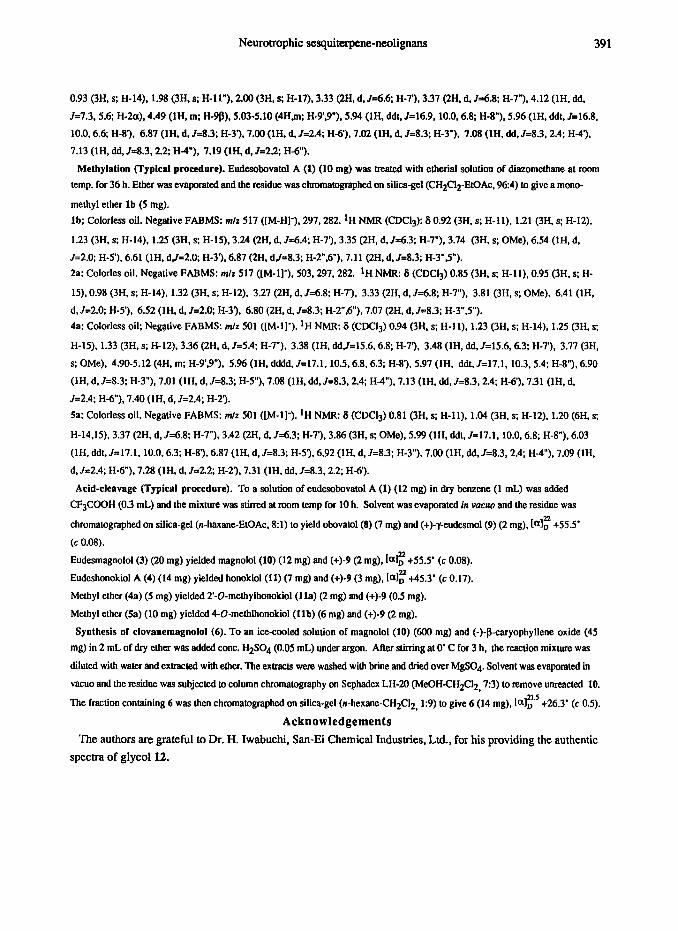
Acid-cleavage (Typical prucedure). To a solution of eudesobovatol A (1) (12 mg) in dry benzene (1 mL) was added
CF$XKIH (0.3 mL) and the mixture was stbred at toom temp for 10 h. Solvent was evaporated in wmw and the residue was
chromatographcd on silica-gel (n-haxane-EtOAc. 81) to yield obovatol(8) (7 mg) and (+)-y-eudesmol(9) (2 mg), (al: +55.5’
(c 0.08).
Eudesmagnolol(3) (20 mg) yielded magnolol(l0) (12 mg) and (+)-9 (2 mg), [a): +55.5’ (c 0.08).
Eudeshonokiol A (4) (14 mg) yielded honokiol(11) (7 me) and (+)-9 (3 mg), [o)f 145.3’ (c 0.17).
Methyl ether (4a) (5 mg) yielded 2’-O-methylhonokiol (11s) (2 mg) and (+)-9 (0.5 mg).
Methyl ether (Sa) (10 mg) yielded 4-0-metblhonokiol (lib) (6 mg) and (+)-9 (2 mg).
Synthesis of clovanemagnolol (6). To an ice-cooled solution of magnolol (10) (600 mg) and (-)-8-caryophyllene oxide (45
mg) in 2 mL of dry ether was added cont. H2SO4 (0.05 mL) under argon. After stirring at 0’ C for 3 h, the reaction mixture was
dihrtcd with water and extracted with ether. The extracts were washed with brine and dried over MgSO4 Solvent was evapomtcd in
vacua and tbe residue was subjected to column chromatography on Sephadex LH-20 (MeOHCH2CI2,7:3) to remove unreactcd 10.
The fraction containing 6 was then chromatograpbed on silica-gel (n-hexane-CH2CC12, 1:9) to give 6 (14 mg), [o)r +26.3’ (c 0.5).
Acknowledgements
The authors are grateful to Dr. H. Iwabuchi, San-Ei Chemical Industries. Ltd., for his providing the authentic
spectra of glycoll2.
392 Y. FuKuYAMA et al.
References
1. Y. Sugii, Yakugaku Zasshi, SO, 183 (1930).
2. M. Fujita, H. Itokawa, and Y. Sashida, Chem. Pharm.Bulf, 20,212 (1972); 3. K. Ito, T. Iida. K. Ichino, M. Tsunezuka, M. Hattori, and T. Nanba, Chem. PhatmBufl., 30, 3347
(1982). 4. M. Fujita, H. Itokawa, and Y. Sashida, Yakugaku Zarshi, 93,415 (1973).
5. Y. Sashida, H. Itokawa, Y. Akita, and M. Fuji&t, Y~ug~~ Zasshi, 96,238 (1976).
6. S. Yahara, T. Nishiyo~, A. Kohda, T. Nohara, and I. Nis~ok~ Chem. Pharm. Bull., 39,2024 (1991). 7. Y.Sugii and H. Shindo, Yogis Zasshi, 50,729 (1930).
8. K. Ito and S. Sakurai, Yakugaku Zasshi, 94,709 (1973).
9. M. Fujita, H. Itokawa, and Y. Sashida, Yakugaku Zasshi, 93,429 (1973). 10. T. Hasegawa, Y. Fukuyama, T. Yamada, and K. Nakagawa, Chemistry Left.,163 (1988); Gem. Phurm.
Buff., 36, 1245 (1988).
II. K. Watanabe, H. Watanabe, Y. Goto, M. Yamaguchi, N. Yamamoto, and K. Hagino, Planta Med., 44,
103 (1983).
12. Y. Fukuyama, Y. Otoshi, M. Kodama, T. Hasegawa, H. Okazaki, and M. Nagasawa, Tetrahedron Left., 30, 5907 (1989).
13. F. J. McQuilIin and J. D. Parrack, J. Chem. Sot., 2973 (1956).
14. Y. Fukuyama, Y. Otoshi, K. N~amu~, M. Kodama, M. Sugawara, and M. Nagasawa, Chemistry Lert,, 295 (1990).
15. F. S. El-Feraly and W. -S. Li, Lloydia, 41,442 (1978); J. K. Nitao, M. G. Nair, D. L. Thorogood, K. S.
Johnson, and J. M. Schriber, Phytochemfstry, 30,2193 (1991).
16. Y. Fukuyama, Y. Otoshi, M. Kodama, T. Hasegawa, and H. Okazaki, Tetrahedron Left., 31,4477 (1990).
17. A. W. Lutz and E. B. Reid, .I. Chem. Sot., 2265 (1954). 18. A. Aebi, D. H. R. Barton, and A. S.Lindsey, j. Chem. Sot., 3124 (1953); F. W. Mckiflop, J. Martin, W.
Parker, J. S. Roberts, and J. R, Stevenson, J. Chem. Sot., (C), 3375 (1971). 19. D. H. R. Barton and P. de Mayo, Qmf. Rev., 2265 (1957); H. Iwabuchi, M. Yoshikura, Y. Ikawa, and
W. Kamisaka, Chem. P&arm. Bull., 35, 1975 (1987). 20. H. Asou, N. Isasaki, S. Hirano, and D. Dahl, Bruin Rese~ch, 332,355 (1985) 21. F. Fonnum, J. ~eurochemist~, 24,407 (1975) 22. Primary neuronal cell culture and measmement of ChAT activity were carried out according to ASOUS’S~~
and Fonnum’s methods,z1 respectively. Detailed nerotrophic property for the active substances will be
reproted in the separate paper.