neurotoxicology and teratology/67531/metadc287047/m2/1/high...neurotoxicology and teratology journal...
TRANSCRIPT
Neurotoxicology and Teratology 34 (2012) 495–504
Contents lists available at SciVerse ScienceDirect
Neurotoxicology and Teratology
j ourna l homepage: www.e lsev ie r .com/ locate /neutera
D-Methionine protects against cisplatin-induced neurotoxicity in cortical networks
Kamakshi V. Gopal a,c,⁎, Calvin Wu a,b,c, Bibesh Shrestha a,c, Kathleen C.M. Campbell d,Ernest J. Moore a,c, Guenter W. Gross b,c
a University of North Texas, Department of Speech & Hearing Sciences, United Statesb University of North Texas, Department of Biological Sciences, United Statesc University of North Texas, Center for Network Neuroscience, United Statesd Southern Illinois University School of Medicine, Department of Surgery, United States
⁎ Corresponding author at: Dept. of Speech & HearinTexas, 1155 Union Circle #311277, Denton, TX 76203‐50565 2481(Voice); fax: +1 940 565 4058.
E-mail address: [email protected] (K.V. Gopal).
0892-0362/$ – see front matter © 2012 Elsevier Inc. Alldoi:10.1016/j.ntt.2012.06.002
a b s t r a c t
a r t i c l e i n f oArticle history:Received 5 December 2011Received in revised form 10 June 2012Accepted 11 June 2012Available online 23 June 2012
Keywords:CisplatinD-MethionineL-MethionineMicroelectrode arrayAuditory cortexIn vitro network
Cisplatin is a platinum-based chemotherapeutic agent widely used for the treatment of various types of can-cer. Patients undergoing cisplatin treatment often suffer from a condition known as “chemobrain”, ototoxic-ity, peripheral neuropathy, weight loss, nausea, vomiting, nephrotoxicity, seizures, hearing loss and tinnitus.D-Methionine (D-Met), a sulfur-containing nucleophilic antioxidant, has been shown to prevent cisplatin-induced side effects in animals without antitumor interference. In this study, we have used an in vitromodel of cortical networks (CNs), enriched in auditory cortex cells; to quantify cisplatin neurotoxicity andthe protective effects of D-Met. Dissociated neurons from auditory cortices of mouse embryos were grownon microelectrode arrays with 64 transparent indium–tin oxide electrodes, which enabled continuous opticaland electrophysiological monitoring of network neurons. Cisplatin at 0.10–0.25 mM induced up to a 200% in-crease in spontaneous spiking activity, while concentrations at or above 0.5 mM caused irreversible loss ofneuronal activity, accompanied by cell death. Pretreatment with D-Met, at a concentration of 1.0 mM,prevented the cisplatin-induced excitation at 0.10–0.25 mM, caused sustained excitation without occurrenceof cell death at 0.5 mM, and delayed cell death at 0.75 mM cisplatin. L-Methionine, the optical isomer, showedlower potency and less efficacy than D-Met, was less protective against 0.1 mM cisplatin, and proved ineffec-tive at a concentration of 0.5 mM cisplatin. Pre-exposure time of D-Met was associated with the protectiveeffects at 0.1 and 0.5 mM cisplatin, with longer pre-exposure times exhibiting better protection. This studyquantifies as a function of concentration and time that D-Met protects central nervous system tissue fromacute cisplatin toxicity.
© 2012 Elsevier Inc. All rights reserved.
1. Introduction
Cisplatin (cis-diamminedichloroplatinum(II)— CDDP) is a platinum‐
based chemotherapeutic agent commonly used in treating various typesof cancer (Kovarík et al., 1972; Stathopoulos, 2010). Cisplatin exerts itscytotoxic effect through the formation of DNA adducts that trigger celldeath by apoptosis (Eastman, 1990; Jamieson and Lippard, 1999).However, its antitumor action is associated with adverse effectssuch as neurotoxicity, ototoxicity, weight loss, nausea, vomiting,and nephrotoxicity (Brock et al., 2012; Lynch and Kil, 2005;Rajeswaran et al., 2008; McWhinney et al., 2009). Neurotoxicity isoften severe, as cisplatin crosses the blood–brain barrier and can ac-cumulate through repeated dosages (Kaasa et al., 1988; Namikawa
g Sciences, University of North10, United States. Tel.: +1 940
rights reserved.
et al., 2000), causing demyelination, axonal shrinkage, neurofibrillaraccumulations, and vacuolar changes in the white matter (Olivi etal., 1993). Cisplatin commonly induces “chemobrain”, a cognitivedecline that occurs in a majority of patients (Whitney et al., 2008).
The central auditory nervous system (CANS), an intricate neuralpathway that is primarily engaged in complex pattern analysis ofacoustic signals, is highly vulnerable to neurotoxicity. Hearing loss in-duced by ototoxicity or neurotoxicity is characterized by irreversiblehearing threshold deficits sometimes associated with tinnitus. Asreviewed by Rybak (2005), hearing loss occurs in 30 to 100% of pa-tients treated with cisplatin. Hearing loss may occur during treatment(Knight et al., 2005, 2007; Orgel et al., 2012) or after discontinuationof chemotherapy (Al-Khatib et al., 2010; Kolinsky et al., 2010). Fur-thermore, patients treated with cisplatin exhibit severe difficulty inword recognition (Einarsson et al., 2011), suggesting a central com-ponent in their hearing loss.
Studies of cisplatin-induced hearing loss have generally focusedon damage to the peripheral auditory system, with morphological
496 K.V. Gopal et al. / Neurotoxicology and Teratology 34 (2012) 495–504
alterations in the organ of Corti of the cochlea, loss of and damage ofcochlear hair cells, damage to the stria vascularis and degeneration ofspiral ganglion neurons (Campbell et al., 1996, 1999; Meech et al.,1998; Hoistad et al., 1998). Cisplatin has been shown to enhancethe formation of reactive oxygen species, and the free radical-induced cell damage is thought to play an important role in cisplatinneurotoxicity (Dehne et al., 2001; Poirrier et al., 2010). Although cis-platin is known to be neurotoxic and is believed to exacerbate the im-pact of the peripheral cisplatin-induced ototoxicity, its effects on thecentral auditory system are not well characterized. Cisplatin hasbeen shown to alter the central conduction time (I–V interval) ofthe auditory brainstem response and prolong cortical-evoked poten-tials from tibial nerve stimulation, suggesting adverse effects on thecentral nervous system (Hansen et al., 1989). Cisplatin induced tinni-tus, a common complaint among sufferers, may also be central in or-igin (Rybak, 2005).
These adverse effects from cisplatin exposure have led to the searchfor preventative treatments (Campbell et al., 1996, 1999; Rybak et al.,2007). Sulfur containing compounds such as sodium thiosulfate (STS)and diethyl-dithiocarbamate (DDTC) were reported to provide goodotoprotection against cisplatin (Otto et al., 1988), but had significantside effects and/or interference with antitumor activity (Jones et al.,1991; Church et al., 1995; Gandara et al., 1995; Muldoon et al., 2000).D-Methionine (D-Met), also a sulfur-containing nucleophilic antioxi-dant, has shown to provide effective nephroprotection against cisplatinwithout interfering with antitumor action (Jones and Basinger, 1989).Cisplatin-induced hearing loss, and subsequent outer hair cell loss andstria vascularis damage, is protected by D-Met (Campbell et al., 1996,1999). In the cochlea, D-Met partially protected cisplatin-induced de-creases in antioxidant enzymes such as superoxide dismutase (SOD),catalase, and glutathione a reductase (GR), and prevented increases inmalondialdehyde levels, a measure of lipid peroxidation (Campbell etal., 2003). Several animal studies have shown that D-Met preventedoxidative-stress induced ototoxicity and nephrotoxicity from cisplatintreatment (Campbell et al., 1996; Reser et al., 1999). Hamstra et al.(2010) evaluated the safety of the MRX-1024 – the bioavailablesuspension of D-Met – in cancer patients. They showed that MRX-1024 decreased the rate and severity of oral mucositis, with no signifi-cant increase in toxicity. D-Met has been regarded as a potentialotoprotective agent, and is in human clinical trials for prevention ofnoise induced hearing loss. Although D-Met crosses the blood brainbarrier, its effects on cisplatin-induced changes in the central auditorynetworks remain unclear (Lauenstein et al., 1987).
The goal of this study was to evaluate the acute effects of cisplatinon central neuronal networks and assess the neuroprotective effectsof D-Met. For that reason, we have used cultured networks (CNs)derived from auditory cortices of mouse embryos growing in vitroon microelectrode arrays (MEAs). Neuronal networks growing onMEAs, unlike brain slices, typically have superior adhesion, stabilityof cell-electrode coupling, and longevity (Gross, 2011; Potter andDeMarse, 2001). These networks exhibit spontaneous activity andare histiotypic (like the parent tissue) in their pharmacological re-sponses (Gopal and Gross, 1996b; Gross and Pancrazio, 2007; Gopaland Gross, 2004; Xia and Gross, 2003). The in vitro environment al-lows precise, reproducible pharmacological manipulations with nohomeostatic interference from other organs. Morphological changesof neurons in the networks can be correlated optically with electro-physiological changes, and network deterioration can be followedsystematically, providing a platform for rapid, quantitative screeningof new chemical and pharmaceutical compounds (Johnstone et al.,2010; Novellino et al., 2011; Wu et al., 2011). Mice have been exten-sively used as the mammalian animal model in physiological andpharmacological experiments. In vitro models allow a drastic reduc-tion in the usage of animals and associated costs. They also showgreat promise for scaling to high throughput (Johnstone et al., 2010;Gross et al., 2006). The hypothesis of this study was that D-Met
is protective against cisplatin-induced acute cytotoxicity as well asfunctional neurotoxicity.
2. Methods
2.1. MEA fabrication and cell culture procedures
In house fabrication and preparation of MEAs, as well as recordingtechniques have been described previously (Gross, 1979; Gross et al.,1985; Gopal and Gross, 1996a; Keefer et al., 2001a). Briefly, indium–
tin oxide sputtered glass plates were photoetched, spin-insulated withmethyltrimethoxysilane, cured, deinsulated at the electrode tips withlaser shots and electrolytically gold-plated to reduce the interfaceimpedance to 1 MΩ at 1.0 kHz. Butane flaming was used to generate3.0 mm diameter hydrophilic adhesion islands for cell growth centeredon the 64-electrode matrix.
The care and use of animals, as well as all procedures involving ani-mals in this studywere approved by and performed in accordance withthe guidelines of the institutional animal care and use committee of theUniversity of North Texas. Mouse embryos were extracted on day E17from ICR mice after CO2 narcosis and cervical dislocation. Tissue fromthe auditory cortex regions was used to provide CNs enriched in audi-tory cortex cells. Auditory cortices (AC) located on the postereolateralsurface and depths of the left temporal cortex were dissected (approxi-mately 2×2×1 mm) from the embryos. The standard culturing processpublished earlier was used in this study (Gopal and Gross, 1996a). TheAC tissue was mechanically and enzymatically dissociated, trituratedand mixed with Dulbecco's Modified Minimal Essential Medium(DMEM), supplemented with horse serum (4%), fetal bovine serum(4%), and 1.0 ml/L B27 (optimized supplement containing vitamins,hormones and other growth factors). The cell suspension was seededat a cellular concentration of 70 K/100 μl onto to the adhesion island(approximately 10 μm2 area), previously treated with poly-D-lysineand laminin, and incubated at 37 °C in 10% CO2 and 90% airatmosphere. The cultures were subjected to a 50% medium changetwice weekly with fresh DMEM supplemented with 6.0% horseserum. The cultures used in this set of experiments on the averagewere 30 days in vitro.
2.2. Drugs and solutions
Drug concentrations were calculated to minimize major bulkvolume changes to the 2.0 ml constant volume experimental bath.D-Met (M9375), L-Met (M9625) and cisplatin (P4393) were obtainedfrom Sigma-Aldrich (St. Louis,MO) in powder form. DMEMand B27wereobtained from GIBCO Products International, Inc. D-Met and L-Met weredissolved in autoclaved water to produce a 200 mM stock solution;cisplatin was dissolved using 1.0 mg/ml in a saline solution (0.9% NaCl2).
2.3. Electrophysiological recordings
MEAs were placed on sterile stainless steel recording chambersmounted on an inverted microscope stage at 37 °C (Gross andSchwalm, 1994). The original medium was then replaced by fresh iso-osmotic DMEM stock medium (without serum). The pH wasmaintained between 7.3 and 7.5 with a continuous 10 mL/min streamof filtered 10% CO2 in air mixed by a gas flow controller (AFC 2600-PRO, Aalborg, Inc.), and confined by a cap with a heated indium–tinoxide (ITO) window to prevent condensation, allowing continuous mi-croscopic observation (Rijal-Oli and Gross, 2008). Medium osmolaritywas maintained at a constant 320 mOsm by infusion of sterile waterwith a syringe pump set at approximately50 μl/h to compensate forevaporation. The neuronal activity was recorded with a two-stage, 64-channel amplifier system (Plexon, Dallas, TX), and digitized simulta-neously at 40 kHz using 64 digital signal processors (DSP). Total systemgain was set to 10,000. Spike identification and separation of multiple
497K.V. Gopal et al. / Neurotoxicology and Teratology 34 (2012) 495–504
spikes recorded by a single electrode were accomplished with a realtime template-matching algorithm (Plexon, Dallas, TX) to providesingle-unit spike rate data. Each DSP could discriminate up to four dif-ferent action potential waveforms or “active” units recorded by a singleelectrode. Multiple unit data were expressed as average network spikeproduction per minute, and monitored in real time. Off-line analysiswas performed using custom programs that allowed for burst patternanalysis based on spike integration with a time constant of 100 ms(Morefield et al., 2000).
Network neurons were monitored using electrophysiological andmorphological recordings. All pharmacological experiments were con-ducted in serum-free medium as serum albumin is known to bind nu-merous chemical compounds (Parviz and Gross, 2006). Criteria for theacceptance of networks after a change to serum-free medium were:(a) a minimum of 30 neurons recorded and discriminated based onwaveshapes, (b) signal to noise ratios of 2:1 or greater, and (c) amean spontaneous network activity exceeding 50 spikes per minute.In addition,minute-to-minute average activityfluctuationwas requiredto be at or below 15%.
The neurotoxicity of cisplatin was quantified, and the potency andefficacy profiles of D-Met were established on par with its enantiomer,the essential amino acid L-Met. Protection was evaluated throughvarying concentration/time pretreatment with D-Met prior to theapplication of cisplatin. The goal of D-Met pretreatment was to effecta minimal change (or no change) in CNs from baseline reference ac-tivity in the presence of cisplatin. The glass carrier plate of the MEAand the transparent indium–tin oxide (ITO) electrode conductorsallowed maximum optical access during recording, and the multisitereadout from a total of 64 electrodes provided continuous action po-tential trains from many individual neurons discriminated on thebasis of waveshapes. Although a 64 electrode MEA records only 5%to 10% of the total number of neurons in the network, the resulting in-formation is sufficient for the generation of reliable population re-sponses that, when normalized and summed or averaged, yieldhighly reproducible electrophysiological and pharmacological data(Gross, 2011).
2.4. Controls
Spontaneously active networks have different baseline (reference)activity. Yet they display highly reproducible pharmacological re-sponses. The simplest approach to tackle the issue of different baselinesis to use the initial stabilized activity (i.e., activity recorded prior toapplication of any compound) of each culture as its own internal control(reference). Compound-induced activity is therewith normalizedand expressed as percent decrease from the internal reference. This
A
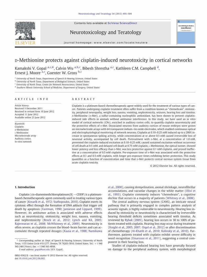
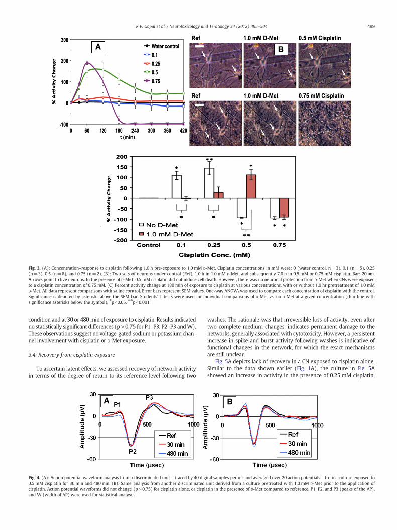
Fig. 1. (A): Concentration–response to cisplatin for 7.0 h at concentrations (mM) of 0 (sali(B): two sets of neurons under control (Ref) and 7.0 h of 0.25 mM or 0.5 mM cisplatin. Bshow obvious morphological changes of neurons (arrows), but 0.5 mM cisplatin induced ce
approach provides reproducible concentration–response curves obtainedfrom different networks and even allows calculation of dissociationconstants (Rijal-Oli and Gross, 2008). To demonstrate long term stabilityin our preparation, we have added data to Figs. 1A and 3A depictingmonitored activity for 7 h in CNs that were exclusively treated withonly the vehicles, i.e., saline (used to dissolve cisplatin) or water (usedto dissolve D- and L-Methionine). If aliquot volumes do not exceed 5% ofthe total volume, no osmolarity effects are noticed and network activitiesremain in a steady plateau state (Gross and Pancrazio, 2007).
To identify the pharmacological up- or down-regulation of neuronalactivity, all electrophysiological data were expressed as percent changefrom reference activity (internal control). Activity variables used weremean spike rate and mean burst rate averaged across all discriminatedunits within the culture, and stability of waveshapes. Bursts are spikeclusters and were identified operationally by digital RC integrationwith rise time constants of approximately 100 ms (Gopal and Gross,2004; Parviz and Gross, 2006).
2.5. Data analyses
The reference activity of neurons remained stable in a serum-freeDMEM stock solution in an environment of pH=7.4, 37±1 °C, with os-molaritymaintained in the range of 300–320 mOsm (Gross et al., 1995;Keefer et al., 2001a, 2001b). The reference activity was recorded for aminimum of 30 min after a medium change from serum containing toserum-free medium. Response variability (after normalization) be-tween different networks was expressed as ± the standard error ofmean (SEM). An activity change of 0% indicated that the networkremained at reference activity after compound addition. Network re-sponses were characterized as excitatory if there was an increase inspike activity compared to reference level, and inhibitory if there wasa decrease in spike activity. A lack of acute recovery or return to refer-ence levels was considered an important indicator of latent, possiblytoxic compound effects. It was always measured after two completemedium changes with an observation interval of 30 min. Therefore, a100% recovery indicated a return to baseline or reference activitywithin30 min. Slower recoveries were not addressed andwill be the subject offuture studies.
Statistical analyses and concentration–response curve fitting usedOrigin software (Origin Lab, Northampton, MA). Differences in spikerates between CNs treated with cisplatin (with or without D-Met)and control (saline) CNs, were statistically analyzed using a one-wayanalysis of variance (ANOVA) followed by Tukey's post-hoc test. Stu-dents' T-tests were used for comparing spike rate data from CNs ex-posed to cisplatin with and without D-Met pretreatment.
B
ne control, n=3), 0.05 (n=3), 0.1 (n=3), 0.25 (n=3), 0.5 (n=5), and 0.75 (n=2).ar: 20 μm. Arrows point to live neurons. Cisplatin concentration of 0.25 mM did notll death (no arrows).
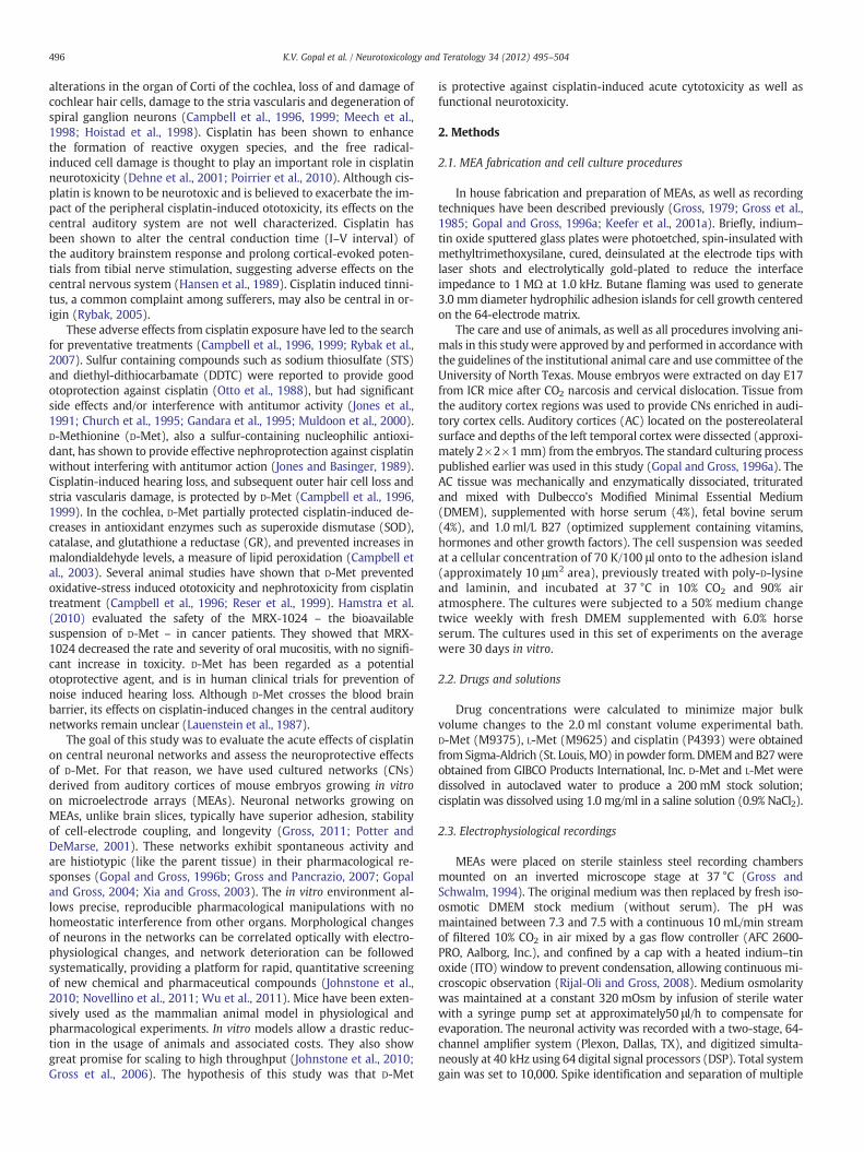
Fig. 2. Concentration–response curve of D-Met (n=4) and L-Met (n=3). The IC50±SEMvalues were 1.03±0.21 mM (Hill slope: 0.94), and 0.64±0.05 mM (Hill slope: 1.54), re-spectively. Note: L-Met has maximal efficacy of only 33% at a maximal concentration of10 mM.
498 K.V. Gopal et al. / Neurotoxicology and Teratology 34 (2012) 495–504
3. Results
3.1. Toxicity of cisplatin
Spontaneously active neurons fromCNswere recorded for an averageexperimental time period of 7.0 h. Cisplatin induced excitationwas char-acterized by an increase in mean spike activity for concentrations rang-ing from 0.05 to 0.25 mM (Fig. 1A). Increasing concentrations withinthis range exhibited steeper slopes of excitation. Network responseswere plotted as percent activity change from a 30-min reference activityperiod (not shown on data graphs).
Cisplatin at 0.05 mM showed a minimal increase in spike activity(5.8 to 11.9%, n=3); while cisplatin at 0.10 mM had an average max-imum increase of 118.1±41.4% after the first hour of exposure, and111.5±32.5% after 7.0 h (n=3) of exposure. Cisplatin at 0.25 mMhad the highest increase of 183.4±31.5% (n=3), with a rapid rise,maximizing at 2.0 h, before gradually leveling off to 85.8±45.2%(n=3). All excitatory phases were sustained for more than 7.0 h. Ex-periments that were monitored for 24 h still showed excitation, level-ing off to 80.3±5.5% (data not shown). At concentrations of 0.5 mMand 0.75 mM, respectively, a brief period of minimal excitation wasobserved, followed by a marked decrease in activity. With cisplatinexposure at 0.5 mM (n=5), 50% and 90% activity losses were seenat 60 and 180 min, respectively. Cisplatin at the 0.75 mM concentra-tion showed a 50% reduction within 60 min, with cessation of activityafter 180 min.
In order to identify morphological changes in neurons, neuronalprocesses and glia cells were visually monitored throughout the ex-periments, and photographs were taken periodically. Fig. 1B showstwo sets of neurons in reference medium (top, left; bottom, left)and after 7.0 h of exposure to cisplatin at 0.25 mM (top panel, right)or 0.5 mM (bottom panel, right). There were no overt changes in neu-ronal morphology (phase bright cells, arrows, ref panels), identifiedafter 7.0 h of exposure to cisplatin at 0.25 mM (top panel, right).Therefore, live neurons can be identified for reference (top and bot-tom panel, left) and cisplatin-treated conditions (top panel, right).However, due to extensive cell death after 7.0 h of exposure to0.5 mM cisplatin, neurons with normal morphology could no longerbe identified (bottom panel, right).
3.2. Concentration response characteristics of D-Met and L-Met exposure
Prior to identifying the potential protective effects of D-Met orL-Met, the pharmacological profiles related to potency and efficacywere characterized. Both isomers of the drugs showed a decrease ofspike activity with an increase in concentration (Fig. 2).
D-Met exhibited maximal efficacy (complete inhibition of neuro-nal spike activity) and potency expressed by the inhibitory concen-tration (IC50) of 1.03±0.21 mM (n=4). In comparison to D-Met,the endogenous isomer L-Met, was found to be less effective with amaximum inhibition of 33.1±0.2%, and an EC50 of 0.64±0.05 mM(n=3). Based on these results, the IC50 value for D-Met (1.0 mM)was chosen to assess its protective effects in subsequent experimentsalong with cisplatin exposure. No morphological changes in the neu-rons were observed with 1.0 mM D-Met application (Fig. 3B, middlepanel) or 1.0 mM L-Met application (data not shown).
3.3. Protection with D-Met
Pretreatment with 1.0 mM D-Met for 1.0 h prior to cisplatin expo-sure had varying effects, depending on the cisplatin concentration(Fig. 3A). In the presence of D-Met, CNs exposed to 0.10 and 0.25 mMcisplatin exhibited substantial attenuation of excitation. The datashow a time-dependent excitation profile for cisplatin at 0.5 mM, withmaximal excitation of 156% compared to reference at approximately120 min. The excitation gradually leveled off to 44% at 7.0 h. With an
application 0.75 mM cisplatin, there was an initial excitatory phase,followed by a shutdown of the network. In comparison to Fig. 1B,which shows extensive cell death when CNs are exposed to 0.5 mMwithout D-Met pretreatment, Fig. 3B (upper panel) depicts the protec-tion provided by D-Met at 0.5 mM cisplatin exposure. However, celldeath and shutdown of the network were unavoidable when thecisplatin concentration was increased to 0.75 mM (Fig. 3A, and lowerpanel of Fig. 3B).
Fig. 3C depicts the results of a one-way ANOVA followed by Tukey'spost-hoc test. Percent change of spike rate in CNs treated with cisplatinfor 180 min (with and without D-Met) was compared to saline controlCNs. The independent variable was the concentration of cisplatin. Com-parisons were made between cisplatin concentrations of 0 (control),0.1 mM, 0.25 mM, 0.5 mMand 0.75 mM. Results showed significant dif-ferences (pb0.05) between control and each of the cisplatin concentra-tions, indicating substantial changes in network activity in the presenceof cisplatin.
In order to identify if pretreatment with D-Met was protective,comparisons were made between CNs exposed to cisplatin with andwithout D-Met pretreatment using Students' T-Tests (also shown inFig. 3C). Results indicated significant differences (pb0.05) betweenCNs exposed to cisplatin alone and CNs exposed to cisplatin in the pres-ence of D-Met. Since the goal of D-Met pretreatmentwas to provide pro-tection against cisplatin toxicity, less change in activity (i.e., activity thatremains close to the zero baseline in Fig. 3C) was considered optimum.Further, if the change in activity was negative as opposed to positive, itwas consideredmore adverse because of the possibility of further dete-rioration of activity leading to network shutdown.
To identify if voltage-gated sodium or potassium channels were in-volved with cisplatin and/or D-Met exposure, we examined AP wave-forms. Fig. 4A depicts representative waveforms from a discriminatedunit with no cisplatin (reference), and 0.5 mM cisplatin exposurefor 30 min and 480 min. Although the activity was close to zero at180 min (Fig. 1A), some units in the networks continued to fire at lowfrequencies, and one of those units was used in the analysis. Fig. 4Bshows a similar sample of waveforms from a network pretreated withD-Met. For quantification, AP amplitudes andAPwidthsweremeasured.P1–P2 and P2–P3 refer to amplitude measures (in μV), and W (width)indicates the time interval (in μs) at the 0 μV baseline between P1 andP3. Mean±SEM amplitude and width measurements from 9 units(3 neurons from 3 different CNs) were analyzed using one-wayANOVA. The independent variablewas the exposure time under cisplat-in. Comparisons were made between waveshapes at the reference
BA
Fig. 3. (A): Concentration–response to cisplatin following 1.0 h pre-exposure to 1.0 mM D-Met. Cisplatin concentrations in mM were: 0 (water control, n=3), 0.1 (n=5), 0.25(n=3), 0.5 (n=8), and 0.75 (n=2). (B): Two sets of neurons under control (Ref), 1.0 h in 1.0 mM D-Met, and subsequently 7.0 h in 0.5 mM or 0.75 mM cisplatin. Bar: 20 μm.Arrows point to live neurons. In the presence of D-Met, 0.5 mM cisplatin did not induce cell death. However, there was no neuronal protection from D-Met when CNs were exposedto a cisplatin concentration of 0.75 mM. (C) Percent activity change at 180 min of exposure to cisplatin at various concentrations, with or without 1.0 hr pretreatment of 1.0 mMD-Met. All data represent comparisons with saline control. Error bars represent SEM values. One-way ANOVA was used to compare each concentration of cisplatin with the control.Significance is denoted by asterisks above the SEM bar. Students' T-tests were used for individual comparisons of D-Met vs. no D-Met at a given concentration (thin-line withsignificance asterisks below the symbol). ⁎pb0.05, ⁎⁎pb0.001.
499K.V. Gopal et al. / Neurotoxicology and Teratology 34 (2012) 495–504
condition and at 30 or 480 min of exposure to cisplatin. Results indicatedno statistically significant differences (p>0.75 for P1–P3, P2–P3 andW).These observations suggest no voltage-gated sodium or potassium chan-nel involvement with cisplatin or D-Met exposure.
3.4. Recovery from cisplatin exposure
To ascertain latent effects, we assessed recovery of network activityin terms of the degree of return to its reference level following two
A
Fig. 4. (A): Action potential waveform analysis from a discriminated unit – traced by 40 dig0.5 mM cisplatin for 30 min and 480 min. (B): Same analysis from another discriminatedcisplatin. Action potential waveforms did not change (p>0.75) for cisplatin alone, or cisplaand W (width of AP) were used for statistical analyses.
washes. The rationale was that irreversible loss of activity, even aftertwo complete medium changes, indicates permanent damage to thenetworks, generally associated with cytotoxicity. However, a persistentincrease in spike and burst activity following washes is indicative offunctional changes in the network, for which the exact mechanismsare still unclear.
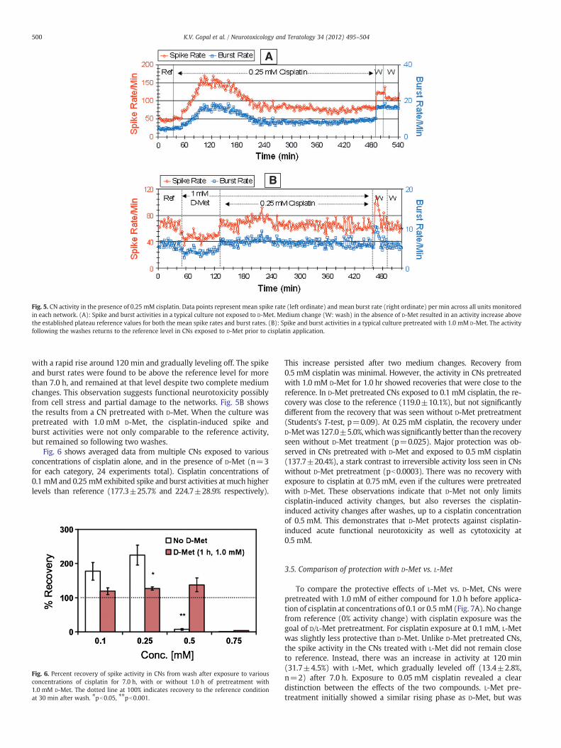
Fig. 5A depicts lack of recovery in a CN exposed to cisplatin alone.Similar to the data shown earlier (Fig. 1A), the culture in Fig. 5Ashowed an increase in activity in the presence of 0.25 mM cisplatin,
B
ital samples per ms and averaged over 20 action potentials – from a culture exposed tounit derived from a culture pretreated with 1.0 mM D-Met prior to the application oftin in the presence of D-Met compared to reference. P1, P2, and P3 (peaks of the AP),
A
B
Fig. 5. CN activity in the presence of 0.25 mM cisplatin. Data points represent mean spike rate (left ordinate) and mean burst rate (right ordinate) per min across all units monitoredin each network. (A): Spike and burst activities in a typical culture not exposed to D-Met. Medium change (W: wash) in the absence of D-Met resulted in an activity increase abovethe established plateau reference values for both the mean spike rates and burst rates. (B): Spike and burst activities in a typical culture pretreated with 1.0 mM D-Met. The activityfollowing the washes returns to the reference level in CNs exposed to D-Met prior to cisplatin application.
500 K.V. Gopal et al. / Neurotoxicology and Teratology 34 (2012) 495–504
with a rapid rise around 120 min and gradually leveling off. The spikeand burst rates were found to be above the reference level for morethan 7.0 h, and remained at that level despite two complete mediumchanges. This observation suggests functional neurotoxicity possiblyfrom cell stress and partial damage to the networks. Fig. 5B showsthe results from a CN pretreated with D-Met. When the culture waspretreated with 1.0 mM D-Met, the cisplatin-induced spike andburst activities were not only comparable to the reference activity,but remained so following two washes.
Fig. 6 shows averaged data from multiple CNs exposed to variousconcentrations of cisplatin alone, and in the presence of D-Met (n=3for each category, 24 experiments total). Cisplatin concentrations of0.1 mMand 0.25 mMexhibited spike and burst activities atmuch higherlevels than reference (177.3±25.7% and 224.7±28.9% respectively).
Fig. 6. Percent recovery of spike activity in CNs from wash after exposure to variousconcentrations of cisplatin for 7.0 h, with or without 1.0 h of pretreatment with1.0 mM D-Met. The dotted line at 100% indicates recovery to the reference conditionat 30 min after wash. ⁎pb0.05, ⁎⁎pb0.001.
This increase persisted after two medium changes. Recovery from0.5 mM cisplatin was minimal. However, the activity in CNs pretreatedwith 1.0 mM D-Met for 1.0 hr showed recoveries that were close to thereference. In D-Met pretreated CNs exposed to 0.1 mM cisplatin, the re-covery was close to the reference (119.0±10.1%), but not significantlydifferent from the recovery that was seen without D-Met pretreatment(Students's T-test, p=0.09). At 0.25 mM cisplatin, the recovery underD-Metwas 127.0±5.0%,whichwas significantly better than the recoveryseen without D-Met treatment (p=0.025). Major protection was ob-served in CNs pretreated with D-Met and exposed to 0.5 mM cisplatin(137.7±20.4%), a stark contrast to irreversible activity loss seen in CNswithout D-Met pretreatment (pb0.0003). There was no recovery withexposure to cisplatin at 0.75 mM, even if the cultures were pretreatedwith D-Met. These observations indicate that D-Met not only limitscisplatin-induced activity changes, but also reverses the cisplatin-induced activity changes after washes, up to a cisplatin concentrationof 0.5 mM. This demonstrates that D-Met protects against cisplatin-induced acute functional neurotoxicity as well as cytotoxicity at0.5 mM.
3.5. Comparison of protection with D-Met vs. L-Met
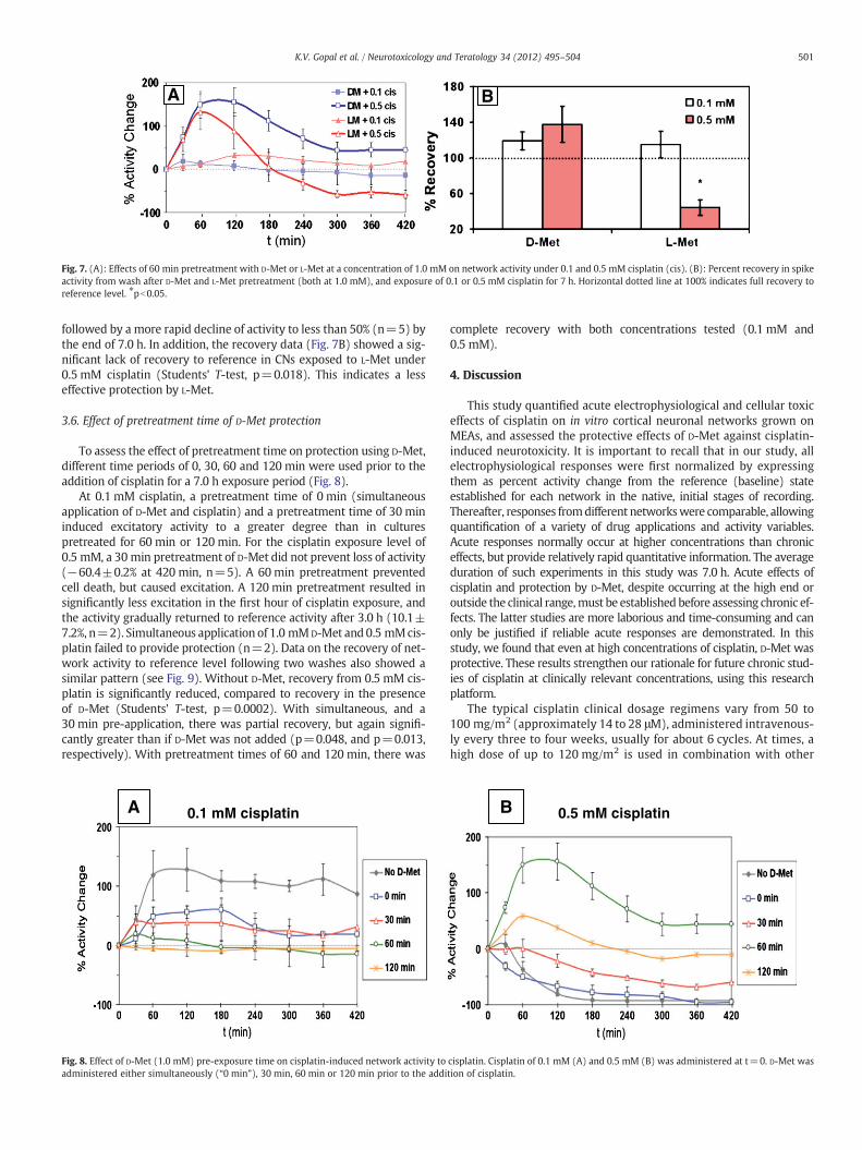
To compare the protective effects of L-Met vs. D-Met, CNs werepretreated with 1.0 mM of either compound for 1.0 h before applica-tion of cisplatin at concentrations of 0.1 or 0.5 mM (Fig. 7A). No changefrom reference (0% activity change) with cisplatin exposure was thegoal of D/L-Met pretreatment. For cisplatin exposure at 0.1 mM, L-Metwas slightly less protective than D-Met. Unlike D-Met pretreated CNs,the spike activity in the CNs treated with L-Met did not remain closeto reference. Instead, there was an increase in activity at 120 min(31.7±4.5%) with L-Met, which gradually leveled off (13.4±2.8%,n=2) after 7.0 h. Exposure to 0.05 mM cisplatin revealed a cleardistinction between the effects of the two compounds. L-Met pre-treatment initially showed a similar rising phase as D-Met, but was
A B
Fig. 7. (A): Effects of 60 min pretreatment with D-Met or L-Met at a concentration of 1.0 mM on network activity under 0.1 and 0.5 mM cisplatin (cis). (B): Percent recovery in spikeactivity from wash after D-Met and L-Met pretreatment (both at 1.0 mM), and exposure of 0.1 or 0.5 mM cisplatin for 7 h. Horizontal dotted line at 100% indicates full recovery toreference level. ⁎pb0.05.
501K.V. Gopal et al. / Neurotoxicology and Teratology 34 (2012) 495–504
followed by a more rapid decline of activity to less than 50% (n=5) bythe end of 7.0 h. In addition, the recovery data (Fig. 7B) showed a sig-nificant lack of recovery to reference in CNs exposed to L-Met under0.5 mM cisplatin (Students' T-test, p=0.018). This indicates a lesseffective protection by L-Met.
3.6. Effect of pretreatment time of D-Met protection
To assess the effect of pretreatment time on protection using D-Met,different time periods of 0, 30, 60 and 120 min were used prior to theaddition of cisplatin for a 7.0 h exposure period (Fig. 8).
At 0.1 mM cisplatin, a pretreatment time of 0 min (simultaneousapplication of D-Met and cisplatin) and a pretreatment time of 30 mininduced excitatory activity to a greater degree than in culturespretreated for 60 min or 120 min. For the cisplatin exposure level of0.5 mM, a 30 min pretreatment of D-Met did not prevent loss of activity(−60.4±0.2% at 420 min, n=5). A 60 min pretreatment preventedcell death, but caused excitation. A 120 min pretreatment resulted insignificantly less excitation in the first hour of cisplatin exposure, andthe activity gradually returned to reference activity after 3.0 h (10.1±7.2%, n=2). Simultaneous application of 1.0 mMD-Met and 0.5 mMcis-platin failed to provide protection (n=2). Data on the recovery of net-work activity to reference level following two washes also showed asimilar pattern (see Fig. 9). Without D-Met, recovery from 0.5 mM cis-platin is significantly reduced, compared to recovery in the presenceof D-Met (Students' T-test, p=0.0002). With simultaneous, and a30 min pre-application, there was partial recovery, but again signifi-cantly greater than if D-Met was not added (p=0.048, and p=0.013,respectively). With pretreatment times of 60 and 120 min, there was
A 0.1 mM cisplatin
Fig. 8. Effect of D-Met (1.0 mM) pre-exposure time on cisplatin-induced network activity toadministered either simultaneously (“0 min”), 30 min, 60 min or 120 min prior to the addi
complete recovery with both concentrations tested (0.1 mM and0.5 mM).
4. Discussion
This study quantified acute electrophysiological and cellular toxiceffects of cisplatin on in vitro cortical neuronal networks grown onMEAs, and assessed the protective effects of D-Met against cisplatin-induced neurotoxicity. It is important to recall that in our study, allelectrophysiological responses were first normalized by expressingthem as percent activity change from the reference (baseline) stateestablished for each network in the native, initial stages of recording.Thereafter, responses fromdifferent networkswere comparable, allowingquantification of a variety of drug applications and activity variables.Acute responses normally occur at higher concentrations than chroniceffects, but provide relatively rapid quantitative information. The averageduration of such experiments in this study was 7.0 h. Acute effects ofcisplatin and protection by D-Met, despite occurring at the high end oroutside the clinical range,must be established before assessing chronic ef-fects. The latter studies are more laborious and time-consuming and canonly be justified if reliable acute responses are demonstrated. In thisstudy, we found that even at high concentrations of cisplatin, D-Met wasprotective. These results strengthen our rationale for future chronic stud-ies of cisplatin at clinically relevant concentrations, using this researchplatform.
The typical cisplatin clinical dosage regimens vary from 50 to100 mg/m2 (approximately 14 to 28 μM), administered intravenous-ly every three to four weeks, usually for about 6 cycles. At times, ahigh dose of up to 120 mg/m2 is used in combination with other
B 0.5 mM cisplatin
cisplatin. Cisplatin of 0.1 mM (A) and 0.5 mM (B) was administered at t=0. D-Met wastion of cisplatin.
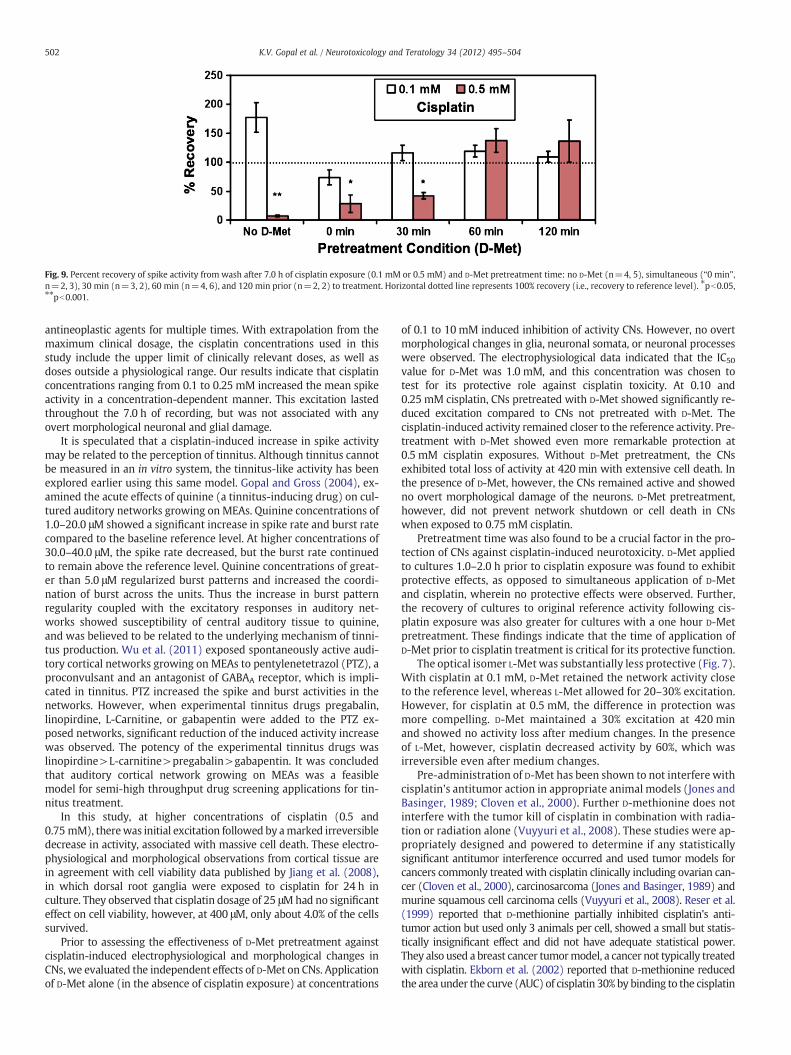
Fig. 9. Percent recovery of spike activity from wash after 7.0 h of cisplatin exposure (0.1 mM or 0.5 mM) and D-Met pretreatment time: no D-Met (n=4, 5), simultaneous (“0 min”,n=2, 3), 30 min (n=3, 2), 60 min (n=4, 6), and 120 min prior (n=2, 2) to treatment. Horizontal dotted line represents 100% recovery (i.e., recovery to reference level). ⁎pb0.05,⁎⁎pb0.001.
502 K.V. Gopal et al. / Neurotoxicology and Teratology 34 (2012) 495–504
antineoplastic agents for multiple times. With extrapolation from themaximum clinical dosage, the cisplatin concentrations used in thisstudy include the upper limit of clinically relevant doses, as well asdoses outside a physiological range. Our results indicate that cisplatinconcentrations ranging from 0.1 to 0.25 mM increased the mean spikeactivity in a concentration-dependent manner. This excitation lastedthroughout the 7.0 h of recording, but was not associated with anyovert morphological neuronal and glial damage.
It is speculated that a cisplatin-induced increase in spike activitymay be related to the perception of tinnitus. Although tinnitus cannotbe measured in an in vitro system, the tinnitus-like activity has beenexplored earlier using this same model. Gopal and Gross (2004), ex-amined the acute effects of quinine (a tinnitus-inducing drug) on cul-tured auditory networks growing on MEAs. Quinine concentrations of1.0–20.0 μM showed a significant increase in spike rate and burst ratecompared to the baseline reference level. At higher concentrations of30.0–40.0 μM, the spike rate decreased, but the burst rate continuedto remain above the reference level. Quinine concentrations of great-er than 5.0 μM regularized burst patterns and increased the coordi-nation of burst across the units. Thus the increase in burst patternregularity coupled with the excitatory responses in auditory net-works showed susceptibility of central auditory tissue to quinine,and was believed to be related to the underlying mechanism of tinni-tus production. Wu et al. (2011) exposed spontaneously active audi-tory cortical networks growing on MEAs to pentylenetetrazol (PTZ), aproconvulsant and an antagonist of GABAA receptor, which is impli-cated in tinnitus. PTZ increased the spike and burst activities in thenetworks. However, when experimental tinnitus drugs pregabalin,linopirdine, L-Carnitine, or gabapentin were added to the PTZ ex-posed networks, significant reduction of the induced activity increasewas observed. The potency of the experimental tinnitus drugs waslinopirdine>L-carnitine>pregabalin>gabapentin. It was concludedthat auditory cortical network growing on MEAs was a feasiblemodel for semi-high throughput drug screening applications for tin-nitus treatment.
In this study, at higher concentrations of cisplatin (0.5 and0.75 mM), therewas initial excitation followed by amarked irreversibledecrease in activity, associated with massive cell death. These electro-physiological and morphological observations from cortical tissue arein agreement with cell viability data published by Jiang et al. (2008),in which dorsal root ganglia were exposed to cisplatin for 24 h inculture. They observed that cisplatin dosage of 25 μMhad no significanteffect on cell viability, however, at 400 μM, only about 4.0% of the cellssurvived.
Prior to assessing the effectiveness of D-Met pretreatment againstcisplatin-induced electrophysiological and morphological changes inCNs, we evaluated the independent effects of D-Met on CNs. Applicationof D-Met alone (in the absence of cisplatin exposure) at concentrations
of 0.1 to 10 mM induced inhibition of activity CNs. However, no overtmorphological changes in glia, neuronal somata, or neuronal processeswere observed. The electrophysiological data indicated that the IC50value for D-Met was 1.0 mM, and this concentration was chosen totest for its protective role against cisplatin toxicity. At 0.10 and0.25 mM cisplatin, CNs pretreated with D-Met showed significantly re-duced excitation compared to CNs not pretreated with D-Met. Thecisplatin-induced activity remained closer to the reference activity. Pre-treatment with D-Met showed even more remarkable protection at0.5 mM cisplatin exposures. Without D-Met pretreatment, the CNsexhibited total loss of activity at 420 min with extensive cell death. Inthe presence of D-Met, however, the CNs remained active and showedno overt morphological damage of the neurons. D-Met pretreatment,however, did not prevent network shutdown or cell death in CNswhen exposed to 0.75 mM cisplatin.
Pretreatment time was also found to be a crucial factor in the pro-tection of CNs against cisplatin-induced neurotoxicity. D-Met appliedto cultures 1.0–2.0 h prior to cisplatin exposure was found to exhibitprotective effects, as opposed to simultaneous application of D-Metand cisplatin, wherein no protective effects were observed. Further,the recovery of cultures to original reference activity following cis-platin exposure was also greater for cultures with a one hour D-Metpretreatment. These findings indicate that the time of application ofD-Met prior to cisplatin treatment is critical for its protective function.
The optical isomer L-Met was substantially less protective (Fig. 7).With cisplatin at 0.1 mM, D-Met retained the network activity closeto the reference level, whereas L-Met allowed for 20–30% excitation.However, for cisplatin at 0.5 mM, the difference in protection wasmore compelling. D-Met maintained a 30% excitation at 420 minand showed no activity loss after medium changes. In the presenceof L-Met, however, cisplatin decreased activity by 60%, which wasirreversible even after medium changes.
Pre-administration of D-Met has been shown to not interfere withcisplatin's antitumor action in appropriate animal models (Jones andBasinger, 1989; Cloven et al., 2000). Further D-methionine does notinterfere with the tumor kill of cisplatin in combination with radia-tion or radiation alone (Vuyyuri et al., 2008). These studies were ap-propriately designed and powered to determine if any statisticallysignificant antitumor interference occurred and used tumor models forcancers commonly treated with cisplatin clinically including ovarian can-cer (Cloven et al., 2000), carcinosarcoma (Jones and Basinger, 1989) andmurine squamous cell carcinoma cells (Vuyyuri et al., 2008). Reser et al.(1999) reported that D-methionine partially inhibited cisplatin's anti-tumor action but used only 3 animals per cell, showed a small but statis-tically insignificant effect and did not have adequate statistical power.They also used a breast cancer tumormodel, a cancer not typically treatedwith cisplatin. Ekborn et al. (2002) reported that D-methionine reducedthe area under the curve (AUC) of cisplatin 30% by binding to the cisplatin
503K.V. Gopal et al. / Neurotoxicology and Teratology 34 (2012) 495–504
and assumed that the binding would reduce the antitumor efficacy of thecisplatin. However Deegan et al. (1994) clearly demonstrated that thecisplatin–methionine complex retains most of its cytotoxic activityagainst tumorswhich is consistentwith the results of the testing in appro-priate tumor models (Jones and Basinger, 1989; Cloven et al., 2000;Vuyyuri et al., 2008). Consequently, D-methionine appears to be an appro-priate candidate for a clinical therapy to reduce the side effects of cisplatintherapy.
D-Met,which can effectively be delivered orally (Campbell et al., 2007)can presumably help circumvent the deleterious effects of cisplatin treat-ment if taken at an optimal period of time. It is possible that the requiredpretreatment time for patients in vivo will be longer than what is ob-served in cell culture. The demonstration of D-Met protection againsthigh concentrations of cisplatin opens the possibility of chronic, low con-centration investigations and also provides a quantitative margin of errorand proof of principle for potential clinical applications.
D-Met has been shown to prevent or ameliorate peripheral cisplatin-induced hearing threshold shifts, outer hair cell loss, loss of hair cellfunction, loss of spiral ganglion cell neurons, and damage to the striavascularis in animals (Campbell et al., 1996, 1999, 2007; Gabaizadehet al., 1997; Wimmer et al., 2004). However, to our knowledge, this in-vestigation is the first evidence of direct cisplatin damage to the corticalneurons, in vitro, and the amelioration of this damage by an over-the-counter supplement — D-Met. If D-Met can provide protection for thecentral nervous system, including the central auditory system, fromcisplatin-induced damage, perhaps the “chemobrain” consequencesand the central component of cisplatin-induced ototoxicity and tinnituscould also be ameliorated. In vitro studies and clinical trials of D-Met toconfirm these in vitro studies will be needed to determine howwell thecortical protection observed in this study translates into the clinicalarena.
Conflict of interest statement
KathleenCampbell, is the sole inventor on thepatents for D-methionineas a protective agent. Her patents are owned by her employer SIUSchool of Medicine.
Kamakshi Gopal: None.Guenter W. Gross: None.Ernest J. Moore: None.Bibesh Shrestha: None.Calvin Wu: None.
Acknowledgments
The authors thank Nga Nguyen for unfailing assistance with cellculture. EJM is supported in part by internal start-up funds fromUNT. The Charles and Josephine Bowen memorial endowment tothe CNNS also supported this research.
References
Al-Khatib T, Cohen N, Carret AS, Daniel S. Cisplatinum ototoxicity in children,long-term follow up. Int J Pediatr Otorhinolaryngol 2010;74(8):913–9. [Aug].
Brock P, Knight K, Freyer D, Campbell K, Steyger PS, Blakley BS, et al. Platinum-inducedototoxicity in children: a consensus review on ototoxicity, otoprotection andmonitoring. J Clin Oncol 2012. [Epub 2012 Apr 30].
Campbell KC, Rybak LP, Meech RP, Hughes L. D-methionine provides excellent protec-tion from cisplatin ototoxicity in the rat. Hear Res 1996;102(1–2):90–8.
Campbell KC, Meech RP, Rybak LP, Hughes LF. D-methionine protects against cisplatindamage to the stria vascularis. Hear Res 1999;138(1–2):13–28.
Campbell KC, Meech RP, Rybak LP, Hughes LF. The effect of D-methionine on cochlearoxidative state with and without cisplatin administration: mechanisms ofotoprotection. J Am Acad Audiol 2003;14(3):144–56.
Campbell KC, Meech RP, Klemens JJ. Prevention of noise- and drug-induced hearingloss with D-methionine. Hear Res 2007;226:92-103.
Church MW, Kaltenbach JA, Blakley BW, Burgio DL. The comparative effects of sodiumthiosulfate, deithyldithiocarbamate, fosfomycin and WR-2712 on amelioratingcisplatin-induced ototoxicity. Hear Res 1995;86(1–2):195–203.
Cloven NG, Re A, McHale MT, Burger RA, DiSala PJ, Rose GS, et al. Evaluation ofD-methionine as a cytoprotectant in cisplatin treatment of an animal modelfor ovarian cancer. Anticancer Res 2000;20:4205–10.
Deegan P, Pratt I, Ryan M. The nephrotoxicity, cytotoxicity and renal handling of acisplatin–methionine complex in male Wistar rats. Toxicology 1994;89:1-14.
Dehne N, Lautermann J, Petrat F, Rauen U, de Groot H. Cisplatin ototoxicity: involvementof iron and enhanced formation of superoxide anion radicals. Toxicol Appl Pharmacol2001;174(1):27–34.
Eastman A. Activation of programmed cell death by anticancer agents: cisplatin as amodel system. Cancer Cells 1990;2:275–80.
Einarsson EJ, Petersen H, Wiebe T, Fransson PA, Magnusson M, Moëll C. Severe difficul-ties with word recognition in noise after platinum chemotherapy in childhood, andimprovements with open-fitting hearing-aids. Int J Audiol 2011;50(10):642–51.[Oct, Epub 2011 Aug 3].
Ekborn A, Laurell G, Johnstrom P, Wallin I, Eksborg S, Ehrsson H. D-Methionine and cis-platin ototoxicity in the guinea pig: D-methionine influences cisplatin pharmacoki-netics. Hear Res 2002;165:53–61.
Gabaizadeh R, Staecker H, Liu W, Kopke R, Malgrange B, Lefebvre PP, et al. Protection ofboth auditory hair cells and auditory neurons from cisplatin-induced damage. ActaOtolaryngol 1997;117:232–8.
Gandara DR, Nahhas WA, Adelson MD, Lichtman SM, Podczaski ES, Yanovich S, et al.Randomized placebo-controlled multicenter evaluation of diethyldithiocarbamatefor chemoprotection against cisplatin-induced toxicities. J Clin Oncol 1995;13(2):490–6. [Feb].
Gopal KV, Gross GW. Auditory cortical neurons in vitro: cell culture and multichannelextracellular recording. Acta Otolaryngol 1996a;116:690–6.
Gopal KV, Gross GW. Auditory cortical neurons in vitro: initial pharmacological studies.Acta Otolaryngol 1996b;116:690–704.
Gopal KV, Gross GW. Unique responses of auditory cortex networks in vitro to low con-centrations of quinine. Hear Res 2004;192(1–2):10–22.
Gross GW. Simultaneous single unit recording in vitro with a photoetched laserdeinsulated gold multi-microelectrode surface. IEEE Trans Biomed Eng 1979;BME-26:273–9.
Gross GW. Multielectrode arrays. Scholarpedia 2011;6(3):5749.Gross GW, Pancrazio JPP. Neuronal network biosensors. In: Knopf GK, Bassi AS, editors. Smart
Biosensor Technology. Taylor and Francis Publishers, CRC Press; 2007. p. 177–201.Gross GW, Schwalm FU. A closed chamber for long-term electrophysiological and mi-
croscopic monitoring of monolayer neuronal networks. J Neurosci Methods1994;52:73–85.
Gross GW, Wen W, Lin J. Transparent indium–tin oxide patterns for extracellular, mul-tisite recording in neuronal cultures. J Neurosci Methods 1985;15:243–52.
Gross GW, Azzazy JME, Wu MC, Rhoades BK. The use of neuronal networks on multi-electrode arrays as biosensors. Biosens Bioelectron 1995;10:553–67.
Gross GW, Rijal-Oli S, Jones V, Hollmuller D, Karg M. High throughput microelectrodearray platforms for quantitative pharmacology and toxicology. Proceedings ofMEA 2006, BIOPRO, Baden-Wuerttemberg; 2006. p. 132–5.
Hamstra DA, Eisbruch A, Naidu MUR, Ramana GV, Sunkara P, Campbell KCM, et al.Pharmacokinetic analysis and phase I study of MRX-1024 in patients treatedwith radiation therapy with or without cisplatinum for head and neck cancer.Clin Cancer Res 2010;16(9):2666–76. [May 1].
Hansen SW, Helweg-Larsen S, Trojaborg W. Long-term neurotoxicity in patients treatedwith cisplatin, vinblastine, and bleomycin for metastatic germ cell cancer. J ClinOncol 1989;7(10):1457–61. [Oct].
Hoistad DL, Ondrey FG, Mutlu C, Schachern PA, Paparella MM, Adams GL. Histopatho-logy of human temporal bone after cis-platinum, radiation, or both. OtolaryngolHead Neck Surg 1998;118(6):825–32.
Jamieson ER, Lippard SJ. Structure, recognition, and processing of cisplatin-DNA ad-ducts. Chem Rev 1999;99:2467–98.
Jiang Y, Guo C, Vasko MR, Kelley MR. Implications of apurinic/apyrimidinic endonucle-ase in reactive oxygen signaling response after cisplatin treatment of dorsal rootganglion neurons. Cancer Res 2008;68(15):6425–34.
Johnstone AFM, Gross GW,Weiss DG, Schroeder O, Gramowski A, Shafer TJ. Micro-electrodearrays: a physiologically-based neurotoxicity testing platform for the 21st century.Neurotoxicology 2010;31:331–50.
Jones MM, Basinger MA. Control of nephrotoxicity in the rat during repeated cis-platinumtreatments. J Appl Toxicol 1989;9(4):229–33.
Jones MM, Basinger MA, Holscher MA. Relative effectiveness of some compounds forthe control of cisplatin-induced nephrotoxicity. Toxicology 1991;68(3):227–47.
Kaasa S, Olsnes BT, Mastekaasa A. Neuropsychological evaluation of patients with inop-erable non-small cell lung cancer treated with combination chemotherapy or ra-diotherapy. Acta Oncol 1988;27(3):241–6.
Keefer EW, Gramowski A, Gross GW. NMDA receptor dependent periodic oscillations incultured spinal cord networks. J Neurophysiol 2001a;86:3030–42.
Keefer EW, Gramowski A, Stenger DA, Pancrazio JJ, Gross GW. Characterization of acute neu-rotoxic effects of trimethylolpropane phosphate via neuronal network biosensors. Bio-sens Bioelectron 2001b;16(7–8):513–25.
Knight KR, Kraemer DF, Neuwelt EA. Ototoxicity in children receiving platinum chemo-therapy: underestimating a commonly occurring toxicity that may influence aca-demic and social development. J Clin Oncol 2005;23(34):8588–96. [Dec 1].
Knight KR, Kraemer DF, Winter C, Neuwelt EA. Early changes in auditory function as aresult of platinum chemotherapy: use of extended high-frequency audiometry andevoked distortion product otoacoustic emissions. Clin Oncol 2007;25(10):1190–5.[Apr 1].
Kolinsky DC, Hayashi SS, Karzon R, Mao J, Hayashi RJ. Late onset hearing loss: a significantcomplication of cancer survivors treated with cisplatin containing chemotherapyregimens. J Pediatr Hematol Oncol 2010;32(2):119–23. [Mar].
504 K.V. Gopal et al. / Neurotoxicology and Teratology 34 (2012) 495–504
Kovarík J, Svec F, Thurzo V. The effect of cis-dichlorodiammineplatinum (II) andacronycin on the proliferation and respiration of HeLa cells in vitro. Neoplasma1972;19(6):569–77.
Lauenstein L, Meyer GJ, Sewing KF, Schober O, Hundeshagen H. Uptake kinetics of 14CL-leucine and 14C L- and 14C D-methionine in rat brain and incorporation into pro-tein. Neurosurg Rev 1987;10(2):147–50.
Lynch ED, Kil J. Compounds for the prevention and treatment of noise-induced hearingloss. Drug Discov Today 2005;10(19):1291–8. [Oct 1].
McWhinney SR, Goldberg RM, McLeod HL. Platinum neurotoxicity pharmacogenetics.Mol Cancer Ther 2009;8(1):10–6.
Meech RP, Campbell KCM, Hughes LP, Rybak LPA. Semiquantitative analysis of theeffects of cisplatin on the rat stria vascularis. Hear Res 1998;124:44–59.
Morefield SI, Keefer EW, Chapman KD, Gross GW. Drug evaluations using neuronal net-works cultured on microelectrode arrays. Biosens Bioelectron 2000;15(7–8):383–96.
Muldoon LL, Pagel MA, Kroll RA, Brummett RE, Doolittle ND, Zuhowski EG. Delayedadministration of sodium thiosulfate in animal models reduces platinum ototoxicitywithout reduction of antitumor activity. Clin Cancer Res 2000;6(1):309–15. [Jan].
Namikawa K, Asakura M, Minami T, Okazaki Y, Kadota E, Hashimoto S. Toxicity ofcisplatin to the central nervous system of male rabbits. Biol Trace Elem Res2000;74(3):223–35.
Novellino A, Scelfo B, Palosaari T, Price A, Sobanski T, Shafer TJ, et al. Development ofmicro-electrode array based tests for neurotoxicity: assessment of interlaboratoryreproducibility with neuroactive chemicals. Front Neuroeng 2011;V4(4):1-14.
Olivi A, Gilbert M, Duncan KL, Corden B, Lenartz D, Brem H. Direct delivery ofplatinum-based antineoplastics to the central nervous system: a toxicity and ultra-structural study. Cancer Chemother Pharmacol 1993;31(6):449–54.
Orgel E, Jain S, Ji L, Pollick L, Si S, Finlay J, et al. Hearing loss among survivors of childhoodbrain tumors treated with an irradiation-sparing approach. Pediatr blood cancer2012;58(6):953–8. http://dx.doi.org/10.1002/pbc.23275. [Epub 2011 Jul 27].
Otto WC, Brown RD, Gage-White L, Kupetz S, Anniko M, Penny JE, et al. Effects of cis-platin and thiosulfate upon auditory brainstem responses of guinea pigs. HearRes 1988;35(1):79–85.
Parviz M, Gross GW. Quantification of zinc toxicity using neuronal networks on micro-electrode arrays. Neurotoxicology 2006;28(3):520–31.
Poirrier AL, Pincemail J, Van Den Ackerveken P, Lefebvre PP, Malgrange B. Oxidativestress in the cochlea: an update. Curr Med Chem 2010;17(30):3591–604.
Potter SM, DeMarse TB. A new approach to neural cell culture for long-term studies.J Neurosci Methods 2001;110:17–24.
Rajeswaran A, Trojan A, Burnand B, Giannelli M. Efficacy and side effects of cisplatin- andcarboplatin-based doublet chemotherapeutic regimens versus non-platinum-baseddoublet chemotherapeutic regimens as first line treatment of metastatic non-smallcell lung carcinoma: a systematic review of randomized controlled trials. Lung Cancer2008;59:1-11.
Reser D, Rho M, Dewan D. L- and D-methionine provide equivalent long term protec-tion against CDDP-induced ototoxicity in vivo, with partial in vitro and in vivo re-tention of antineoplastic activity. Neurotoxicology 1999;20:731–48.
Rijal-Oli S, Gross GW. Determination of dissociation constants using spontaneous neu-ronal network activity recorded with microelectrode arrays in vitro. J NeurosciMethods 2008;173:183–92.
Rybak LP. Neurochemistry of the peripheral and central auditory system after ototoxicdrug exposure: implications for tinnitus. Int Tinnitus J 2005;11(1):23–30.
Rybak LP, Whitworth CA, Mukherjea D, Ramkumar V. Mechanisms of cisplatin-inducedototoxicity and prevention. Hear Res 2007;226(1–2):157–67.
Stathopoulos GP. Liposomal cisplatin: a new cisplatin formulation. Anticancer Drugs2010;21(8):732–6.
Vuyyuri SB, Hamstra DA, Khanna D, Hamilton CA, Marwart SM, Campbell KCM, et al.Evaluation of a D-methionine as a novel oral radiation protector for prevention ofmucositis. Clin Cancer Res 2008;14(7):2161–70.
Whitney KA, Lysaker PH, Steiner AR, Hook JN, Estes DD, Hanna NH. Is “chemobrain” atransient state? A prospective pilot study among persons with non-small celllung cancer. J Support Oncol 2008;6(7):313–21. [Sep-Oct].
Wimmer C, Mees K, Stumpf P, Welsch U, Reichel O, Suckfull M. Round window appli-cation of D-methionine, sodium thiosulfate, brain-derived neurotrophic factor,and fibroblast growth factor-2 in cisplatin-induced ototoxicity. Otol Neurotol2004;25(1):33–40.
Wu C, Gopal KV, Gross GW, Lukas TJ, Moore EJ. An in vitro model for testing drugs totreat tinnitus. Eur J Pharmacol 2011;667:188–94.
Xia Y, Gross GW. Histiotypic electrophysiological responses to cultured neuronal networksto ethanol. Alcohol 2003;30:167–74.